Article

Formation of Microalgal Hunting Nets in Freshwater Microcosm Food Web: Microscopic Evidence

by
Goran Kovačević
1,*,
Daniela Petrinec
2,
Petra Tramontana Ljubičić
3,
Siegfried Reipert
4,
Damir Sirovina
1,
Maria Špoljar
1,
Petra Peharec Štefanić
1 and
Davor Želježić
5
1
Department of Biology, Faculty of Science, University of Zagreb, Horvatovac 102a, HR-10000 Zagreb, Croatia
2
Croatian Institute for Brain Research, School of Medicine, University of Zagreb, Šalata 12, HR-10000 Zagreb, Croatia
3
IV. Gimnazija, Ul. Žarka Dolinara 9, HR-10000 Zagreb, Croatia
4
Cell Imaging and Ultrastructure Research (CIUS), University of Vienna, Djerassiplatz 1, A-1030 Vienna, Austria
5
Unit for Mutagenesis, Institute for Medical Research and Occupational Health, HR-10000 Zagreb, Croatia
*
Author to whom correspondence should be addressed.
Water 2023, 15(19), 3448; https://doi.org/10.3390/w15193448
Submission received: 1 September 2023 / Revised: 27 September 2023 / Accepted: 28 September 2023 / Published: 30 September 2023
(This article belongs to the Special Issue Biodiversity and Functionality of Plankton Communities)

Abstract

:
The microcosm is a laboratory method frequently used in ecological studies related to population and food web interactions and environmental dynamics. It simultaneously brings into interaction different species in the same controlled laboratory experimental area and provides an opportunity for modeling and reconstruction of relationships in the natural biocenoses and ecosystems. We applied that approach to determine and improve our understanding of predator–prey interactions in different freshwater environments. The inhabitants of the microcosms were isolated endosymbiotic microalga Desmodesmus subspicatus (Chlorophyceae) (Chodat) Hegewald et Schmidt (CZ), green hydras, freshwater turbellarians, and large water fleas. Experiments were performed in five replicates, at 25 °C and 13.5 °C, with fed and hungry predators, respectively. Herein, we proposed a mechanism for microalgal hunting net formation in the freshwater microcosm. Ultrastructural visualization of the endosymbiotic microalgae revealed rod-like structures on the cell wall surface, structures that could possibly fit together and interconnect, suggesting the possibility of microalgal hunting net formation. Interspecific cooperation between isolated microalgae and turbellarians resulted in stronger hunting net formation in preying upon water fleas. This study contributes to the diversity of species interactions and shows the producers as a top link, as opposed to what is generally considered as a basic link in the food web, and presents the microalgae as triggers of the dynamics in the freshwater microcosm.

1. Introduction

It is difficult to determine the boundaries of individual ecosystems in nature, but one solution for observations is to isolate a single ecosystem in a container from the rest of the biosphere. Such an isolated system is called a microcosm or microecosystem. It represents a simplified ecosystem under controlled conditions that is used to simulate and predict the behavior and role of organisms within natural ecosystems. In addition to the fact that natural ecosystems are usually large, a number of variable physicochemical factors make it difficult to conduct experiments in natural ecosystems where control of conditions is required. A microcosm approach is being studied to overcome such problems and offers the possibility of modeling and reconstructing relationships in natural biocenoses and ecosystem functioning, with great potential for modern ecological research, which is fundamental due to climate change and anthropogenic influences. This approach provides a mechanistic, rather than merely phenomenological, understanding of environmental processes and leads to theories that enable the development of globally applicable solutions [1,2]. A microcosm method is used for a variety of research purposes, such as to study population dynamics [3,4], food chains and multitrophic interactions [5,6], and competition and predation [7,8,9,10,11]. Such an approach allows the creation of replicable ecosystems that are not feasible in nature. Replicates are conducted in such a way that abiotic conditions can be controlled and materials from the natural ecosystem can be used. Reproducibility and good control of experiments are the main advantages of using microcosms in ecological research [12]. A key feature of a microcosm is the self-organization of the system. Organisms inhabiting similar but geographically distant areas can be placed in the same microcosm, creating their own network of interactions. Another important role of microcosms is that they provide a link between theory and nature itself, where they do not directly reflect nature but can improve understanding of natural processes by simplifying their complexity and enabling the study of natural processes and ecosystems under controlled conditions [13]. In addition to the microcosm approach, there are also mesocosm and macrocosm methods where the size of the experimental ecosystem is different and larger than the microcosm, and the experiment can be conducted outdoors [14,15].
Green hydra (Hydra viridissima Pallas, 1766) (Figure 1a) is a cosmopolitan freshwater invertebrate (Cnidaria, Hydrozoa) that inhabits ponds, lakes, streams and lentic habitats of running waters. Green hydra as a host can contain up to 20 microalgal individuals in 1 gastrodermal myoepithelial cell, each in a structure known as a symbiosome [16]. The exchange of metabolites occurs in both directions. The endosymbiotic microalgae is the stronger symbiotic partner in the symbiosis [17]. The endosymbiotic algae used in this study were isolated from green hydra and were maintained in permanent, stable laboratory cultures. They belong to the Chlorella zagrebiensis group Kovac. & Jelen. (2007) [18]. One of the isolated species is Desmodesmus subspicatus (Chlorophyceae) (Chodat) Hegewald et Schmidt (CZ) (Figure 1b). The cells grow and can occur in coenobial, transitional, or tetrad forms [19,20,21].
Turbellaria belong to the genus Platyhelminthes and are characterized by a dorsoventrally flattened and bilaterally symmetrical body (Figure 1c). The surface of the body is covered by a single-layered ciliated epidermis, which has a covering and protective function. Most Turbellaria have epidermal rhabdoids and rhabdites, structures that provide mucus that has numerous functions, from protecting the body from dehydration, gas exchange, and movement to assisting in the capture and ingestion of prey [22]. Turbellaria glide along on a film of mucus and hunt. As predators or scavengers, they feed on small annelids, larvae of mollusks and insects, and crustaceans.
Daphnia magna (Straus, 1820) (DM) (Branchiopoda) (Figure 1d), a large water flea, is a planktonic freshwater crustacean. Many predators (invertebrates and vertebrates, e.g., fish) feed on large water fleas [23,24,25]. In nature, large water fleas are algivorous, feed on planktonic algae and bacteria [26], are present in many habitats, and are an important link in food chains due to their role as primary consumers of phytoplankton and primary food source for secondary consumers [27,28].
In this study, we established the microcosm communities of small freshwater invertebrates as predators and prey with the aim of investigating the intraspecific and interspecific interactions, competition, and predation that exert strong selection pressure, as well as the influence on the shaping of the ecosystem. The aim of this study was also to investigate the effect and interaction of the isolated microalgal endosymbionts in relation to the present macrozoobenthos constituents to gain insight into the dynamics of freshwater ecosystems with the species involved, including two temperature regimes, with both fed and hungry predators. Also, transmission electron microscopy (TEM), as a powerful tool in describing ultrastructures of endosymbiotic algae, is of ongoing interest [21]. Herein, we aimed to describe ultrastructures and specificities of the isolated endosymbiotic microalga Desmodesmus subspicatus from green hydra. We used two methods, namely chemical fixation and cryofixation, and compared the results. With cryofixation, we expected to better observe the ultrastructures of the isolated microalgae. The results of this study will help highlight the functionality of the phytoplankton component in the system, pointing to the possibility of a twist in the microalgal position in the food web. The results will also provide an understanding of interspecific cooperation in predator–prey systems that include a phytoplankton component.

2. Materials and Methods

2.1. Experimental Organisms

In ex situ microcosm experiments, together with green hydra (Hydra viridissima Pallas, 1766) and an isolated endosymbiotic microalga Desmodesmus subspicatus (Chlorophyceae) (Chodat) Hegewald et Schmidt (CZ), we included two predatory species of freshwater turbellarians, Polycelis felina (Dalyell, 1814) and Dugesia gonocephala (Duges, 1830), as well as algivore large water flea, Daphnia magna (Straus, 1820) (CZ) (Figure 1). The organisms used in this study were obtained from the breeding cultures of the Department of Biology, Faculty of Science, University of Zagreb, while P. felina was isolated from a natural habitat (Gračanski stream, Zagreb, NW Croatia). Green hydras were kept in aerated aquarium water in 2 L glass containers at 21.5 °C and fed twice a week with nauplia of Artemia salina (Linnaeus, 1758). Isolated endosymbiotic microalgae D. subspicatus were cultured in test tubes [29,30] in an air chamber at 24 °C on a sterile deep stock agar. A standardized method of maintaining a culture of isolated algae was used to obtain a constant amount of clonal cultures to conduct the experiments [18]. To prepare the algal suspension, 10 mL of aerated water was measured, to which a quarter of the algal smear length from the test tube was added and homogenized by stirring in the same direction. This suspension was added to another 40 mL of aerated water in crystallizing dishes of 60 mL. Where applicable, other microcosm inhabitants were put into this suspension. P. felina were maintained in aerated water in 1 L glass containers in the refrigerator at 13.5 °C and fed with nauplia of A. salina once a week. D. gonocephala were kept and used as green hydra. For the experiment, fed animals were used directly from the cultures, while for the experiment with hungry animals, individuals were separated into separate glass containers with aerated water and kept in the refrigerator at 13.5 °C for three days prior to the beginning of the experiment. D. magna culture was maintained at 17 to 21 °C in 60 L aquaria with aerated water. The animals were fed once or twice a week with dry yeast, Chlorella sp., and fish food of the smallest granulation, SAK 00. Individuals were used as prey for the experiment directly from the culture. Figure 1a,c,d were obtained using stereomicroscope and digital camera, and Figure 1b was obtained using the light microscope Nikon Eclipse E600 and digital camera Nikon DXM1200.

2.2. Microcosm Setups

Crystallizing dishes with 60 mL contents were used for the microcosm experiments. Each experiment was performed in five replicates at two temperature regimes: 25 °C, with a day/night regime (photoperiod 8 h day/16 h night) and 13.5 °C in the dark, including fed and hungry predators (hydras, planarians). Results were recorded 1 h and 24 h after the start of the experiments (Table 1).
The following interactions between experimental organisms were observed: H. viridissima and isolated endosymbiotic microalga D. subspicatus; P. felina and isolated endosymbiotic D. subspicatus; D. gonocephala and isolated endosymbiotic D. subspicatus; and P. felina and isolated endosymbiotic D. subspicatus and D. magna. Control groups included each individual species and the D. magnaD. subspicatus interaction (Table 2). These organisms are simple and inexpensive to maintain in the laboratory, are mostly widespread inhabitants of freshwater habitats, and are efficiently used in scientific research. To the best of our knowledge, our cultures of isolated microalgae D. subspicatus from green hydra are unique, i.e., the only permanently maintained cultures in the world, and this isolated microalgal species was represented here for the first time as a constituent of a microcosm, i.e., this represents the introduction of the new model organism to microcosm research. The overall experiment was conducted with a lot of combinations of microalgal and invertebrate organisms, but in order to emphasize the observed phenomenon of the net, the shown combinations of organisms in the microcosms were selected.
Experimental dishes (60 mL) were filled with 50 mL of aerated water. Experiments were conducted in a way that 1 or 5 predators (Hydra, Turbellaria) were used in each microcosm, respectively, while a large water flea was added as prey, with 10 individuals per experimental dish. The experimental dishes were placed on trays and exposed to the experimental conditions. An overview of experimental dishes is shown in Figure 2.

2.3. Transmission Electron Microscopy

To perform analysis using TEM, the isolated microalgae samples were embedded in 1.5% agar, and for chemical fixation, the agar pieces containing the algae were fixed with 1% glutaraldehyde in 0.5 M cacodylate buffer (pH 7.2) for 1 h at 4 °C. The samples were then washed twice for 10 min with cold 0.5 M cacodylate buffer and post-fixed for 60 min at 4 °C with 1% osmium tetroxide in the same buffer, followed by a 10 min wash in cold distilled water. Dehydration was accomplished with a series of increasing concentrations of ethanol (50%, 60%, 70%, 80%, 96%), with each concentration acting for 10 min. Finally, the material was soaked overnight in absolute ethanol. The next day, the material was placed in a mixture of absolute ethanol and 100% acetone for 30 min, followed by another 30 min in 100% acetone. Then, the material was placed in a mixture of Spurr’s medium and acetone, first for 30 min in one part Spurr’a/two parts acetone, then for 30 min in one part Spurr’s/one part acetone, and finally for 30 min in two parts Spurr’s/one part acetone. Then, the material was placed in Spurr’s medium for 2 h at 45 °C. Finally, the material was placed in a plastic mold and polymerized in Spurr’s medium at 65 °C for 48 h. Ultrathin sections (50–90 nm) were made using the Leica Ultracut R ultramicrotome. Sections were contrasted with 4% aqueous uranyl acetate for 10 min and then with lead citrate (pH 12.0) for an additional 10 min [31]. The ultrathin sections were analyzed using a transmission electron microscope FEI Morgagni 268D at 70 kV.
For further investigation by TEM, the isolated endosymbiotic algae were cryoimmobilized using high-pressure freezing (HPF) followed by low-temperature dehydration, fixation, and embedding in epoxy resin. Prior to use, carriers type B (3 mm in diameter; 300 µm in depth) for HPF were coated with 1-hexadecene (Merck, Sharp & Dome, Rahway, NJ, USA). Algae in agar were transferred into these carriers and covered with the flat surface of another carrier, type B (Leica Microsystems, Vienna, Austria). To prevent air from being trapped, between the carrier sandwich, 20% BSA was used as filler. Without delay, the mounted sample was inserted in the flat middle plate of a cartridge and frozen at ca. 2000 bar with the high-pressure freezer HPM100 (Leica Microsystems, Austria). Once released automatically in liquid nitrogen, the frozen carrier sandwich had to be separated from the middle plate under liquid nitrogen by using a punching device. Freeze substitution (FS) was performed in an automated freeze substitution system AFS2 (Leica Microsystems, Austria) equipped with an agitation module (Cryomodultech e.U., Vienna, Austria) [32]. Carriers containing the HP-frozen samples were placed onto 1 mL liquid-nitrogen frozen FS medium (1% OsO4 in acetone) in 2 mL cryotubes. Afterward, the tubes were inserted in the tube holders of the agitation module within the cryochamber of the AFS2, which was precooled to −140 °C. FS took place under agitation (15 V) at −85 °C for 44 h. In contrast to previous protocols for algae [33], such a long substitution time was chosen to achieve sufficient dehydration within microbody-like organelles of the freshwater algae. This was followed by a warm-up to room temperature and embedding in epoxy resin. Samples were infiltrated with epoxy resin Agar 100 (Agar Scientific Ltd., Stansted, UK) according to the following schedule: one part resin/two parts acetone for 15 min, one part resin/one part acetone for 30 min, two parts resin/one part acetone for 2 h 30 min. Subsequently, samples were transferred in embedding molds and infiltrated with pure resin overnight. Polymerization of the resin took place in the oven at 65 °C for ca. 36 h. Ultrathin sections (70–90 nm thick) were cut with an ultramicrotome Ultracut S (LEICA Microsystems, Vienna, Austria) by using an oscillating diamond knife, Diatome V7 (Diatome, Nidau, Switzerland), placed on Formvar-coated 200-mesh copper grids, contrasted with 4% neodymium(III)-acetate [34] for 50 min, followed by lead citrate for 8 min, prior to analyses in a TEM ZEISS Libra 120 (ZEISS, Oberkochen, Germany) at 120 kV or, alternatively, in an electronically refurbished ZEISS 900N at 80 kV. Images were acquired by using digital cameras, TRS (4 megapixel) and ImageSp-professional software (Tröndle, Moorenweis, Germany).

3. Results

3.1. Hunting Net Formation

Our results show the formation of microalgal hunting nets in the food web of freshwater microcosms and a step-by-step model proposal for this formation. The results also include a proposal for interspecific cooperation between the isolated microalgae and predatory turbellarians, demonstrating the functionality of this hunting net system.
Controls with a microalgal suspension of isolated endosymbiotic D. subspicatus showed that at 13.5 °C after 1 h, there was a homogeneous condition present, i.e., microalgae were evenly distributed in a microcosm, and it appeared greenish. Moreover, microalgal aggregations, i.e., accumulations of microalgae in the form of smaller clumps/clusters were present at the bottom of the experimental dish. At 25 °C, the condition was homogeneous. After 24 h, microalgal nets were observed macroscopically at both temperatures (Table 3), appearing as green intertwined filaments, showing the ability of the microalgae to form the net only by themselves.
No nets were observed in all other controls. In the control microalgae and D. magna, microalgal aggregations were observed at 13 °C after 1 h and 24 h. Mucus secretion was observed during the experiment: microalgae secreted very little mucus, D. gonocephala slightly more, H. viridissima more, and P. felina the most.
The experimental setup with green hydra and the microalgae showed that microalgal aggregations were present in all microcosm setups. At 13 °C, precipitation of microalgae occurred at the bottom of the experimental dish after 24 h with one and five fed hydras. The presence of the net was confirmed in microcosms with five fed and hungry hydras after 24 h at both temperatures and at the lower temperature with one hungry hydra, showing the ability of microalgal net formation after a period of 24 h only (Table 4).
The microcosm with five fed D. gonocephala and the microalgae at 13 °C demonstrated a homogeneous distribution of microalgae after 1 h. Microcosm with one fed D. gonocephala contained the aggregations and microalgal hunting net after 24 h, but with five fed D. gonocephala, only the aggregations along with the net destruction were present after 24 h at 25 °C. In the experiment with hungry D. gonocephala and microalgae, the aggregations were present in all microcosms set up. The hunting net was confirmed in the presence of 1 D. gonocephala at 13.5 °C after 1 h and 24 h and at 25 °C after 1 h in the presence of 5 D. gonocephala. The ability of hunting net formation was shown after a period of 1 h, and a process of hunting net destruction after 24 h (Table 4).
In all experimental setups with fed P. felina and microalgae, microalgal aggregations were present. At 13.5 °C, microalgal precipitation, aggregations, and a hunting net were observed after 1 h with five P. felina. The net was also observed at a lower temperature after 24 h with one and five P. felina, and net destruction was also present. At 25 °C, the net was present after 1 h with one and five P. felina, and net destruction was observed after 24 h. In the presence of one hungry P. felina, microalgal aggregations and the hunting net were observed after 1 h at 13.5 °C, and the aggregations only were present with five hungry P. felina. At 13.5 °C, both the aggregations and the net were observed after 24 h with one and five hungry P. felina, whereas at 25 °C the aggregations were confirmed in the presence of one hungry P. felina and both the aggregations and the net were observed in the presence of five hungry P. felina. Here, the three steps in the formation of the microalgal hunting net were present (precipitation, aggregations, hunting net), along with the phenomenon of microalgal net destruction after a 24 h period (Table 4).
Experiments with fed P. felina and the microalgae and D. magna showed that at 25 °C after 1 h and 24 h with one P. felina, the hunting net formation was confirmed (Figure 3), and after 24 h with one and five P. felina, microalgal aggregations were present at both temperatures. At 13.5 °C, both microalgal aggregations and hunting nets were present after 1 h with five turbellarians, and the aggregations were observed after 24 h in the presence of one and five P. felina. At 13.5 °C, microalgal aggregations and the hunting nets were present (Table 4) after 1 h and 24 h and after 1 h at 25 °C with one and five hungry P. felina. After 24 h at 25 °C, only the aggregations were present.
Here, the appearance of microalgal filaments on the abdomen of D. magna was observed: at 25 °C after 1 h with five P. felina, at 13.5 °C after 24 h with one P. felina, and at both temperatures with five P. felina. Precipitation of microalgae at the bottom of the experimental dish was present at 13.5 °C after 24 h. Microalgal filaments on the abdomen denoted that some Daphnia managed to escape from microalgal hunting nets, but a part of the net remained attached to their body. Microcosms that included the three species, P. felina, isolated endosymbiotic microalga D. subspicatus, and D. magna, showed that there is a clear connection between the presence of the isolated microalgae and predatory turbellarians in the same microcosm, pointing out the stronger net formation and interspecific cooperation between the microalgae and the turbellarians.

3.2. A Microscopic Evidence

To visualize the ultrastructures of isolated endosymbiotic alga D. subspicatus, chemical fixation and cryofixation of isolated endosymbiotic microalga D. subspicatus were performed. Microscopic images of chemically fixed algal cells using a transmission electron microscope revealed only slightly visible unusual structures on the cell wall of the microalgae (Figure 4).
Cryofixation allowed these unusual structures on the cell wall to be better preserved and clearly visualized. This revealed many small rod-like structures on the microalgal cell wall (Figure 5 and Figure 6), pointing out the possibility of microalgal net formation and the persistence of microalgal filaments on the abdomen of Daphnia. Our results point to the possibility of certain steps (homogenous state, precipitation, aggregations, net formation, net destruction) in the production of the net formed by the microalgae that have the rod-like profile of their cell wall, showing the clear connection between the isolated microalgae and the hunting nets.

4. Discussion

The microcosm method is well suited for setting up scientific theories because it provides a better understanding of ecological processes. While mathematical models rely on assumptions related to biological understanding, the microcosm model can be used to collect data that are already consistent with biological understanding because they incorporate both biological and ecological mechanisms [2]. In this study, well-known organisms belonging to freshwater ecosystems were used: H. viridissima, isolated endosymbiotic microalga D. subspicatus, P. felina, D. gonocephala, and D. magna. Recently, some of these and related species have been used in microcosm research due to the possibility of complete isolation and control of individual systems [11,35,36,37,38,39].
With its constant growth in culture, isolated endosymbiotic microalga D. subspicatus shows characteristic granular and dry growth. These endosymbiotic microalgae isolated from green hydra have a coccoid shape, but the algae start to grow in coenobia and do not separate after division, i.e., the morphological changes from a coccoid to an elliptical coenobial shape occur [19]. Herein, in the presence of D. magna, it is possible that the microalgae, in order to protect themselves, secrete substances that facilitate the formation of algal aggregations that D. magna cannot consume because of its size, i.e., microalgal aggregations could be too large for D. magna to consume them. These microalgal aggregations could present a strong microalgal antipredatory mechanism to avoid predatory attacks of D. magna. Group formation could protect algae from predators if predators are unable to engulf large-sized entities [40]. Wiltshire et al. [41] show that the presence of D. magna individuals induces the production of mucus by the alga Staurastrum, and cells use the produced mucus to form algal clusters that are too large for D. magna individuals to consume. These algal aggregations are thought to be a mechanism by which algae are protected from predatory attack by D. magna. The morphological changes observed in Scenedesmus subspicatus Chodat can be interpreted as an anti-predator strategy that helps increase resistance to predatory attacks by the zooplankton that feeds on it [42]. D. magna causes morphological changes, i.e., the clustering of S. subspicatus algal cells in large coenobia. Even after one day, the share of colonies is elevated in the treated groups. S. subspicatus forms coenobia of 4 to 8 cells and longer and more pointed appendages when exposed to chemical substances released by individuals of D. magna. Either predators or only predator exoproducts promote colony formation [43]. The importance of grazing and protection from grazing in shaping phytoplankton community structure should not be underestimated [44].
During the experiment, we observed the formation of the microalgal nets. It is possible that the microalgae, after aggregation, formed the microalgal nets in order to protect themselves. Given that in control replicates where only a suspension of isolated endosymbiotic microalgae was present, both microalgal aggregations and microalgal net formation occurred, algae are presumably a key factor for the formation of a complex hunting net. Occurrence of the net was also observed in experimental setups when turbellaria and green hydras were present in addition to the suspension of the microalgae, most intensively when P. felina was present in the microcosm. The nets were macroscopically visible as green intertwined filaments. The nets were present in the microcosms after 1 h, but microalgae in microcosms with H. viridissima took longer to form the net, i.e., it was observed after 24 h only. Along with the net, increased mucus secretion by predators was observed, with P. felina secreting the most mucus. Since the microalgae adhered to the bottom of the experimental dish, the increased secretion of mucus could be an adaptation of hydra to facilitate adherence to the bottom of the dish. With the presence of the microalgae, the formation of the hunting net presumably enables even better capture of prey for turbellarians. In the control experiments with microalgae and water fleas, microalgae only formed aggregations and did not manage to form hunting nets and capture the water fleas, but with the presence of turbellarians, this mechanism was effective. Increased mucus secretion during hunting was shown to be an effective technique that allows turbellarians to have better predatory abilities, suggesting that mucus not only protects planarians from predators but also makes them better predators. The mucus secreted from flatworms as a homogenous layer is about 15 µm thick [45].
Phytoplanktonic organisms have evolved a variety of strategies to survive in a variable environment. Induced defense mechanisms in phytoplankton include changes in morphology, biochemistry, and life history traits [46]. The change in the trophic role of microalgae from prey to predator of copepods couples population growth with reduced grazing pressure [47]. Herein, the microalgal net likely emerged as a defense mechanism following microalgal aggregations, but also through interspecific cooperation between microalgae and turbellarians. Microalgal nets were reinforced by mucus secreted by planarians that prey on Daphnia, making them more accessible to turbellarians. Some water fleas managed to escape from the hunting nets, and these individuals had on their abdomen green microalgal filaments that were initially a part of the microalgal hunting net. The appearance of filaments on the abdomen was observed to a greater extent after an exposure time of 1 h when hungry predators were included in the experiment.
We know surprisingly little about the natural environmental conditions that favor the formation of multicellular groups. Adaptations of unicellular organisms to environmental challenges may hold the key to understanding evolutionary pathways to multicellular life [48]. TEM of the microalgal samples provided us the confirmation that the isolated endosymbiotic microalgae may be the base for the microalgal net formation. Ultrathin TEM sections revealed rod-like structures on the cell wall surface of the isolated endosymbiotic microalgae, structures that could potentially fit together and interconnect, indicating the possibility of microalgal net formation. These rod-like profiles could connect the individual microalgal cells and explain the formation of microalgal nets and abdominal microalgal filaments attached to the large water fleas that escaped from the nets. So far, these structures of the isolated endosymbiotic microalgae from green hydra have not been observed/described [21].
Herein, we proposed a step-by-step model for the formation of the microalgal hunting nets: microalgae were basically homogeneously distributed in the microcosm, followed by the precipitation of microalgae at the bottom of the experimental dish. After that, the aggregations of microalgae began to form, finally resulting in the formation of the microalgal net. This formation seemed to be based on the “zip-like” principle between microalgae cells, which had rod-like profiles of their cell wall surface. Together with the mucus produced by the predators, the net became stronger and more effective. Eventually, the destruction of the hunting net occurred. These rod-like profiles could not only explain the formation of the microalgal nets themselves but also reveal the basis of isolated endosymbiotic microalgal species D. subspicatus itself: morphology and color of the culture, formation of coenobia, and transitional forms and tetrads [19,20,21].
Further studies on the functionality of microalgal hunting nets and systems could include (1) using other freshwater microalgal and invertebrate species to investigate their mutual influence and (2) altering the microenvironmental conditions under which the hunting net phenomenon might occur, including other isolated endosymbiotic microalgae and their free-living relatives.

5. Conclusions

Here, in microecosystems, the appearance of mucus and the formation of a complex hunting net occurred. There is a clear connection between the isolated microalgae and the hunting nets, highlighting the potential role of microalgae as a part of the predator chain.
The occurrence of the net in the experiment setup where only endosymbiotic microalgae were present may be a consequence of the microstructure of the endosymbiotic microalgae. In the microcosms with isolated endosymbiotic microalgae and turbellarians, a phenomenon of stronger and more pronounced hunting nets was observed compared to the system with isolated endosymbiotic microalgae only, representing possible interspecific cooperation in which the microalgae facilitate the turbellarians to capture D. magna, and by reducing the number of live D. magna individuals in the system, their predation on microalgae is reduced, i.e., microalgal contribution to the formation of hunting nets allowed microalgae to avoid being eaten by water fleas.
This study contributes to the diversity of species interactions, showing producers as a top link, opposite to what is generally considered as a basic link in the food web, and presents the microalgae as triggers of the dynamics in freshwater microcosm. Further microscopic analyses could provide new insights into the functional diversity of complex formations of hunting nets and food web interactions.

Author Contributions

Conceptualization, G.K., D.P., P.T.L. and M.Š.; methodology, G.K., D.P., P.T.L., S.R., D.S., M.Š. and P.P.Š.; validation, G.K., D.P., P.T.L., S.R. and D.S.; formal analysis, G.K., D.P., P.T.L., S.R., D.S., P.P.Š. and D.Ž.; investigation, G.K., D.P., P.T.L., S.R., and P.P.Š.; resources, G.K., D.P., P.T.L., S.R., M.Š., P.P.Š. and D.Ž.; writing—original draft preparation, G.K., D.P., P.T.L., S.R., D.S., M.Š., P.P.Š. and D.Ž.; writing—review and editing, G.K., D.P., P.T.L., S.R., D.S., M.Š., P.P.Š. and D.Ž.; visualization, G.K., D.P., P.T.L., S.R., D.S., M.Š. and P.P.Š.; supervision, G.K. and D.S.; project administration, G.K.; funding acquisition, G.K. All authors have read and agreed to the published version of the manuscript.

Funding

This research was funded by the University of Zagreb, institutional project number 106-F19-00056/20284116.

Data Availability Statement

The data presented in this study are available in this article.

Acknowledgments

Special thanks to Croatian Microscopy Society for support and cooperation.

Conflicts of Interest

The authors declare no conflict of interest.

Abbreviations

HV (1)Hydra viridissima (Pallas, 1766), 1 individual.
HV (5)H. viridissima (Pallas, 1766), 5 individuals.
PF (1)Polycelis felina (Dalyell, 1814), 1 individual.
PF (5)P. felina (Dalyell, 1814), 5 individuals.
DG (1)Dugesia gonocephala (Duges, 1830), 1 individual.
DG (5)D. gonocephala (Duges, 1830), 5 individuals.
DMDaphnia magna (Straus, 1820).
CZIsolated endosymbiotic microalga Desmodesmus subspicatus (Chlorophyceae) (Chodat)
Hegewald et Schmidt.
TEMTransmission Electron Microscopy.
HPFHigh-Pressure Freezing.
FSFreeze Substitution.

References

  1. Forbes, S.A. The Lake as a Microcosm. Bull. Sci. Assoc. (Peoria IL) 1887, 77–87. [Google Scholar] [CrossRef]
  2. Benton, T.G.; Solan, M.; Travis, M.J.M.; Sait, S.M. Microcosm experiments can inform global ecological problems. Trends Ecol. Evol. 2007, 22, 516–521. [Google Scholar] [CrossRef] [PubMed]
  3. Balčiunas, D.; Lawler, S.P. Effects of basal resources, predation, and alternative prey in microcosm food chains. Ecology 1995, 76, 1327–1336. [Google Scholar] [CrossRef]
  4. Holyoak, M.; Lawler, S.P. Persistence of an extinction-prone predator-prey interaction through metapopulation dynamics. Ecology 1996, 77, 1867–1879. [Google Scholar] [CrossRef]
  5. Lawler, S.P.; Morin, P.J. Food web architecture and population dynamics in laboratory microcosms of protists. Am. Nat. 1993, 141, 675–686. [Google Scholar] [CrossRef] [PubMed]
  6. Diehl, S.; Feißel, M. Effects of enrichment on three-level food chains with omnivory. Am. Nat. 2000, 155, 200–218. [Google Scholar] [CrossRef]
  7. Tilman, D. Resource competition between planktonic algae: An experimental and theoretical approach. Ecology 1977, 58, 338–348. [Google Scholar] [CrossRef]
  8. Bonsall, M.B.; Hassell, M.P. Apparent competition structures ecological assemblages. Nature 1997, 388, 371–373. [Google Scholar] [CrossRef]
  9. Morin, P.J. Productivity, intra-guild predation, and population dynamics in experimental food webs. Ecology 1999, 80, 752–760. [Google Scholar] [CrossRef]
  10. Petchey, O.L. Prey diversity, prey composition, and predator population dynamics in experimental microcosms. J. Anim. Ecol. 2000, 69, 874–882. Available online: https://www.jstor.org/stable/2647408 (accessed on 30 August 2023.). [CrossRef]
  11. Walsh, E.J.; Salazar, M.; Remirez, J.; Moldes, O.; Wallace, R.L. Predation by invertebrate predators on the colonial rotifer Sinantherina socialis. Invert. Biol. 2006, 125, 325–335. [Google Scholar] [CrossRef]
  12. Beyers, R.J. The Microcosm Approach to Ecosystem Biology. Am. Biol. Teach. 1964, 26, 491–498. [Google Scholar] [CrossRef]
  13. Vidican, R.; Sandor, A.V. Microcosm experiments as a tool in soil ecology studies. Bull. UASVM Agric. 2015, 72, 319–320. [Google Scholar] [CrossRef] [PubMed]
  14. Jeppesen, E.M.; Søndergaard, M.; Lauridsen, T.L.; Davidson, T.A.; Liu, Z.; Mazzeo, N.; Trochine, C.; Özkan, K.; Jensen, H.S.; Trolle, D.; et al. Biomanipulation as a restoration tool to combat eutrophication: Recent advances and future challenges. Adv. Ecol. Res. 2012, 47, 411–488. [Google Scholar]
  15. Landkildehus, F.; Søndergaard, M.; Beklioglu, M.; Adrian, R.; Angeler, D.G.; Hejzlar, J.; Papastergiadou, E.; Zingel, P.; Çakiroğlu, A.I.; Scharfenberger, U.; et al. Climate change effects on shallow lakes: Design and preliminary results of a cross-European climate gradient mesocosm experiment. Est. J. Ecol. 2014, 63, 71–89. [Google Scholar] [CrossRef]
  16. Douglas, A.E. Symbiotic Interactions; Oxford University Press Inc.: Oxford, UK; New York, NY, USA, 1994. [Google Scholar]
  17. Kovačević, G.; Kalafatić, M.; Jelenčić, B.; Franjević, D. Endosymbiotic alga as the stronger evolutionary partner in green hydra symbiosis. J. Endocyt. Cell Res. 2010, 20, 13–15. [Google Scholar]
  18. Kovačević, G.; Franjević, D.; Jelenčić, B.; Kalafatić, M. Isolation and Cultivation Endosymbiotic Algae from Green Hydra and Phylogenetic Analysis of 18S rDNA Sequences. Folia Biol. 2010, 58, 135–143. [Google Scholar] [CrossRef]
  19. Kovačević, G.; Radić, S.; Jelenčić, B.; Kalafatić, M.; Poslović, H.; Pevalek-Kozlina, B. Morphological features and isoenzyme characterization of endosymbiotic algae from green hydra. Plant. Syst. Evol. 2010, 284, 33–39. [Google Scholar] [CrossRef]
  20. Ivšić, M.; Kovačević, G. Evaluation of algae farming using the Chlorella bioassay. Croat. J. Fish. 2018, 7, 99–106. [Google Scholar] [CrossRef]
  21. Kević, N.; Radić Brkanac, S.; Vincek, N.; Peharec Štefanić, P.; Faraguna, F.; Kovačević, G.; Kalafatić, M.; Franjević, D. Endosymbiotic green algae in European Hydra strains show quantitative difference on morphological and isoenzyme level. Symbiosis 2019, 77, 161–175. [Google Scholar] [CrossRef]
  22. Habdija, I.; Primc Habdija, B.; Radanović, I.; Špoljar, M.; Matoničkin Kepčija, R.; Vujčić Karlo, S.; Miliša, M.; Ostojić, A.; Sertić Perić, M. Protista-Protozoa i Metazoa-Invertebrata. Strukture i funkcije; Alfa d.d.: Zagreb, Croatia, 2011. [Google Scholar]
  23. Pestana, J.L.T.; Baird, D.J.; Soares, A.M.V.M. Predator threat assessment in Daphnia magna: The role of kairomones versus conspecific alarm cues. Mar. Freshw. Res. 2013, 64, 679–686. [Google Scholar] [CrossRef]
  24. Stoks, R.; Govaert, L.; Pauwels, K.; Jansen, B.; De Meester, L. Resurrecting complexity: The interplay of plasticity and rapid evolution in the multiple trait response to strong changes in predation pressure in the water flea Daphnia magna. Ecol. Lett. 2016, 19, 180–190. [Google Scholar] [CrossRef] [PubMed]
  25. Ekvall, M.T.; Sha, Y.; Palmér, T.; Bianco, G.; Bäckman, J.; Åström, K.; Hansson, L.A. Behavioural responses to co-occurring threats of predation and ultraviolet radiation in Daphnia. Freshw. Biol. 2020, 65, 1509–1517. [Google Scholar] [CrossRef]
  26. Ebert, D. Introduction to Daphnia Biology. In Ecology, Epidemiology, and Evolution of Parasitism in Daphnia [Internet]; Bethesda (MD): National Library of Medicine (US), National Center for Biotechnology Information; 2005; Available online: https://www.ncbi.nlm.nih.gov/books/NBK2039/ (accessed on 30 August 2023.).
  27. Abe, T.; Saito, H.; Niikura, Y.; Shigeoka, T.; Nakano, Y. Embryonic development assay with Daphnia magna: Application to toxicology of aniline derivates. Chemosphere 2001, 45, 487–495. [Google Scholar] [CrossRef] [PubMed]
  28. Miner, B.E.; De Meester, L.; Pfrender, M.E.; Lampert, W.; Hairston, N.G. Linking genes to communities and ecosystems: Daphnia as an ecogenomic model. Proc. Biol. Sci. 2012, 279, 1873–1882. [Google Scholar] [CrossRef]
  29. Pratt, R. Studies on Chlorella vulgaris IV. Am. J. Bot. 1941, 28, 492–497. [Google Scholar] [CrossRef]
  30. Horvatić, J.; Vidaković-Cifrek, Ž.; Regula, I. Trophic Level, Bioproduction and Toxicity of the Water of Lake Sakadaš (Nature Park Kopački Rit, Croatia). Limnol. Rep. 2000, 33, 89–94. [Google Scholar]
  31. Reynolds, E.S. The use of lead citrate at high pH as an electronopaque stain in electron microscopy. J. Cell. Biol. 1963, 17, 208–212. [Google Scholar] [CrossRef]
  32. Reipert, S.; Goldammer, H.; Richardson, C.; Goldberg, M.W.; Hawkins, T.J.; Hollergschwandtner, E.; Kaufmann, W.A.; Antreich, S.; Stierhof, Y.D. Agitation Modules: Flexible Means to Accelerate Automated Freeze Substitution. J. Histochem. Cytochem. 2018, 66, 903–921. [Google Scholar] [CrossRef]
  33. Goldammer, H.; Hollergschwandtner, E.; Elisabeth, N.H.; Frade, P.R.; Reipert, S. Automatized freeze substitution of algae accelerated by a novel agitation module. Protist 2016, 167, 369–376. [Google Scholar] [CrossRef]
  34. Kuipers, J.; Giepmans, B.N.G. Neodymium as an alternative contrast for uranium in electron microscopy. Histochem. Cell Biol. 2020, 153, 271–277. [Google Scholar] [CrossRef] [PubMed]
  35. Majdi, N.; Kreuzinger-Janik, B.; Traunspurger, W. Effects of flatworm predators on sediment communities and ecosystem functions: A microcosm approach. Hydrobiologia 2016, 776, 193–207. [Google Scholar] [CrossRef]
  36. Petrinec, D.; Kovačević, G.; Tramontana, P.; Peharec-Štefanić, P.; Špoljar, M. Visualisation of hunting nets formed by algae: A perfect hunting mechanism? In Proceedings of the 19th International Microscopy Congress, Sydney, Australia, 9–14 September 2018.
  37. Hevrøy, T.H.; Golz, A.L.; Hansen, E.L.; Xie, L.; Bradshaw, C. Radiation effects and ecological processes in a freshwater microcosm. J. Environ. Radioact. 2019, 203, 71–83. [Google Scholar] [CrossRef] [PubMed]
  38. Kovačević, G.; Petrinec, D.; Tramontana, P.; Špoljar, M. Hydra vs. turbellarians: Who is the strongest constituent in a given micro-(eco)system?—preliminary observations. In Proceedings of the 3rd Symposium of Freshwater Biology, Zagreb, Croatia, 15 February 2019. [Google Scholar]
  39. Ptatscheck, C.; Brüchner-Hüttemann, H.; Kreuzinger-Janik, B.; Weber, S.; Traunspurger, W. Are meiofauna a standard meal for macroinvertebrates and juvenile fish? Hydrobiologia 2020, 847, 2755–2778. [Google Scholar] [CrossRef]
  40. Kapsetaki, S.E.; Fisher, R.M.; West, S.A. Predation and the formation of multicellular groups in algae. Evol. Ecol. Res. 2016, 17, 651–669. [Google Scholar]
  41. Wiltshire, K.; Boersma, M.; Meyer, B. Grazer-induced changes in the desmid Staurastrum. Hydrobiologia 2003, 491, 255–260. [Google Scholar] [CrossRef]
  42. Lürling, M.; van Donk, E. Morphological changes in Scenedesmus induced by infochemicals released in situ from zooplankton grazers. Limnol. Oceanogr. 1997, 42, 783–788. [Google Scholar] [CrossRef]
  43. Fisher, R.M.; Bell, T.; West, S.A. Multicellular group formation in response to predators in the alga Chlorella vulgaris. J. Evol. Biol. 2016, 29, 551–559. [Google Scholar] [CrossRef]
  44. Lürling, M. Grazing resistance in phytoplankton. Hydrobiologia 2021, 848, 237–249. [Google Scholar] [CrossRef]
  45. Wilden, B.; Majdi, N.; Kuhlicke, U.; Neu, T.R.; Traunspurger, W. Flatworm mucus as the base of a food web. BMC Ecology 2019, 19, 15. [Google Scholar] [CrossRef]
  46. Van Donk, E.; Ianora, A.; Vos, M. Induced defences in marine and freshwater phytoplankton: A review. Hydrobiologia 2011, 668, 3–19. [Google Scholar] [CrossRef]
  47. Berge, T.; Poulsen, L.K.; Moldrup, M.; Daugbjerg, N.; Hansen, P.J. Marine microalgae attack and feed on metazoans. ISME J. 2012, 6, 1926–1936. [Google Scholar] [CrossRef] [PubMed]
  48. Cornwallis, C.K.; Svensson-Coelho, M.; Lindh, M.; Li, Q.; Stábile, F.; Hansson, L.A.; Rengefors, K. Single-cell adaptations shape evolutionary transitions to multicellularity in green algae. Nat. Ecol. Evol. 2023, 7, 889–902. [Google Scholar] [CrossRef] [PubMed]
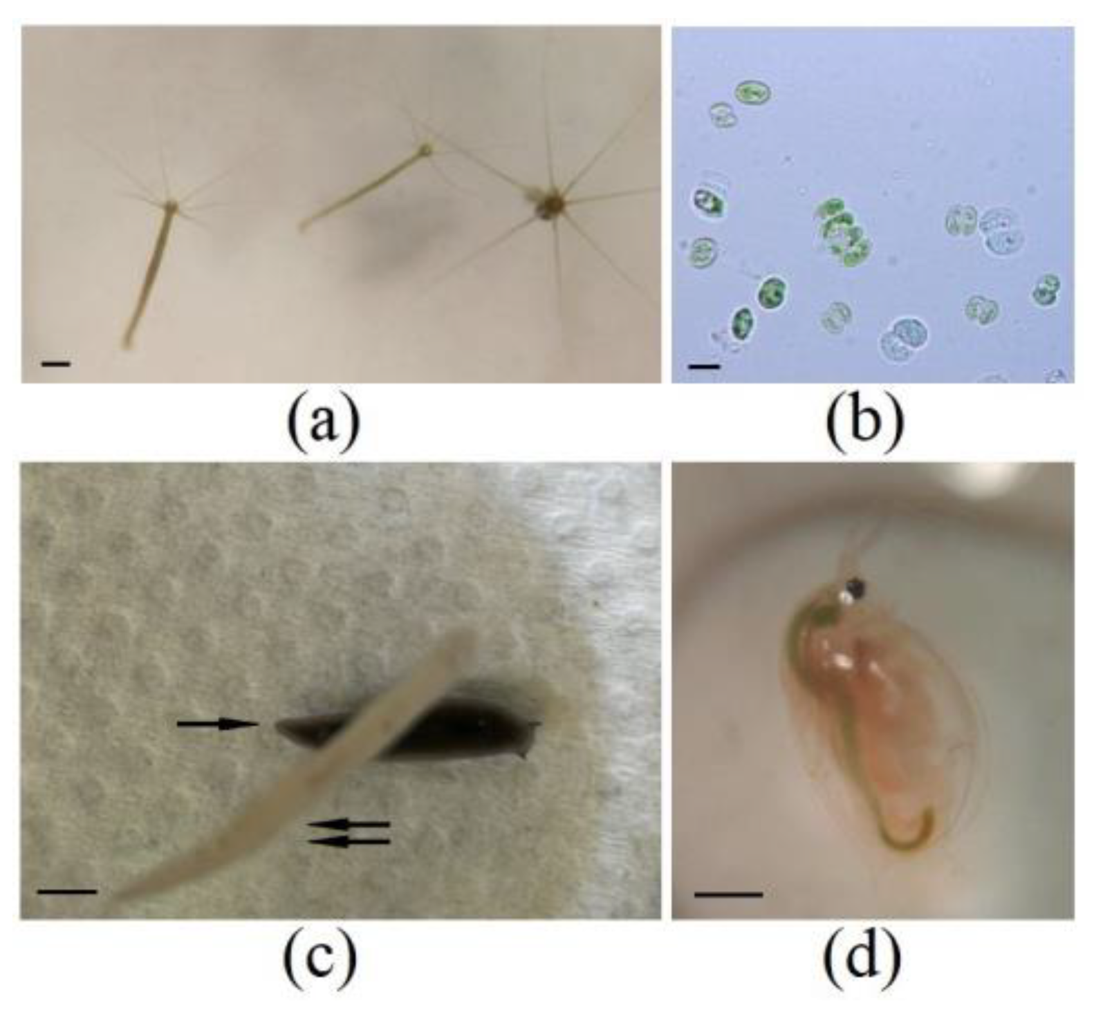
Figure 1. (a) H. viridissima. Scale bar 1 mm; (b) isolated endosymbiotic microalga D. subspicatus. Scale bar 5 µm; (c) P. felina (arrow), D. gonocephala (2 arrows). Scale bar 1 mm; (d) D. magna. Scale bar 1 mm.
Water 15 03448 g001
Figure 2. An overview of experimental dishes.
Water 15 03448 g002
Figure 3. (a) Microcosm including D. magna as prey, 1 fed P. felina as predator, and a suspension of isolated endosymbiotic microalga D. subspicatus at 25 °C after 1 h. Hunting net at the bottom of experimental dish (framed by a hexagon) in which water fleas were trapped (circled); (b) hunting net highlighted by digital processing.
Water 15 03448 g003
Figure 4. Isolated endosymbiotic microalga D. subspicatus, transmission electron microscopy (TEM), chemical fixation. An unusual form on the cell wall (arrow) and interconnection of the cells (2 arrows). Scale bar 250 nm.
Water 15 03448 g004
Figure 5. Isolated endosymbiotic microalga D. subspicatus, TEM, high-pressure freezing/freeze substitution (HPF/FS). Rod-like profile of the cell wall (arrow) and proposed “zip-like“ principle for interconnection of the cells (2 arrows). Scale bar 250 nm.
Water 15 03448 g005
Figure 6. Isolated endosymbiotic microalga D. subspicatus, TEM, HPF/FS. Rod-like profile (arrow) and interconnection of the cells (2 arrows). Scale bar 250 nm.
Water 15 03448 g006
Table 1. Experimental conditions of set up microcosms.
Temperature
Conditions
Light ConditionsNutritional Status of
Turbellarians and Hydras
Result—Recorded Hours (after Setting up the Experiments)
13.5 °C25 °CPhotoperiod of 8 h day/16 h nightdarkhungryfed124
Table 2. Ex situ microcosms setups: two temperature regimes (13.5 °C and 25 °C) with fed and hungry predators (hydras and turbellarians; 1 or 5), D. magna as prey (10 individuals), and a suspension of isolated endosymbiotic microalga Desmodesmus subspicatus (Chlorophyceae) (Chodat) Hegewald et Schmidt (CZ).
Hydra + Microalgae MicrocosmTurbellarians + Microalgae MicrocosmTurbellarians + Microalgae + D. magna Microcosm
Hydra viridissima + Desmodesmus subspicatusPolycelis felina + D. subspicatusDugesia gonocephala + D. subspicatusP. felina + D. subspicatus+ Daphnia magna
controls
H. viridissimaD. subspicatusP. felinaD. magnaD. subspicatus + D. magna
Table 3. Microalgal net formation in experimental dishes with microalgae after 1 h and 24 h of exposure at 13.5 °C and 25 °C.
Formation/Arrangement of Microalgae1 h24 h
13.5 °C25 °C13.5 °C25 °C
Homogeneous-+--
Aggregations----
Homogeneous with aggregations+---
Net formation--++
Table 4. Microalgal net occurrence in experimental setups under experimental conditions and with isolated endosymbiotic microalga D. subspicatus. Hydra viridissima (Pallas, 1766), 1 individual (HV (1)); Polycelis felina (Dalyell, 1814), 1 individual (PF (1)); P. felina (Dalyell, 1814), 5 individuals (PF (5)); Dugesia gonocephala (Duges, 1830), 1 individual (DG (1)); D. gonocephala (Duges, 1830), 5 individuals (DG (5)); Daphnia magna (Straus, 1820) (DM).
Experimental ConditionExperimental OrganismsNet Formation
1 h24 h
Fed predators
(13.5 °C)
HV (1) + CZ--
HV (5) + CZ-+
Hungry predators
(13.5 °C)
HV (1) + CZ-+
HV (5) + CZ-+
Fed predators
(25 °C)
HV (1) + CZ--
HV (5) + CZ-+
Hungry predators
(25 °C)
HV (1) + CZ--
HV (5) + CZ-+
Fed predators
(13.5 °C)
PF (1) + CZ-+
PF (5) + CZ++
Hungry predators
(13.5 °C)
PF (1) + CZ++
PF (5) + CZ-+
Fed predators
(25 °C)
PF (1) + CZ++ *
PF (5) + CZ++ *
Hungry predators
(25 °C)
PF (1) + CZ--
PF (5) + CZ-+
Fed predators
(13.5 °C)
DG (1) + CZ-+
DG (5) + CZ-+ *
Hungry predators
(13.5 °C)
DG (1) + CZ++
DG (5) + CZ--
Fed predators
(25 °C)
DG (1) + CZ-+ *
DG (5) + CZ-+ *
Hungry predators
(25 °C)
DG (1) + CZ--
DG (5) + CZ+-
Fed predators
(13.5 °C)
PF (1) + CZ + DM (10)--
PF (5) + CZ + DM (10)+-
Hungry predators
(13.5 °C)
PF (1) + CZ + DM (10)++
PF (5) + CZ + DM (10)++
Fed predators
(25 °C)
PF (1) + CZ + DM (10)++
PF (5) + CZ + DM (10)--
Hungry predators
(25 °C)
PF (1) + CZ + DM (10)+-
PF (5) + CZ + DM (10)+-
Note(s): * Net residues only.
Disclaimer/Publisher’s Note: The statements, opinions and data contained in all publications are solely those of the individual author(s) and contributor(s) and not of MDPI and/or the editor(s). MDPI and/or the editor(s) disclaim responsibility for any injury to people or property resulting from any ideas, methods, instructions or products referred to in the content.