Article

Facet-Dependent Adsorption of Phosphate on Hematite Nanoparticles: Role of Singly Coordinated Hydroxyl

1
Information Materials and Intelligent Sensing Laboratory of Anhui Province, Institutes of Physical Science and Information Technology, Anhui University, Hefei 230601, China
2
School of Resources and Environmental Engineering, Anhui University, Hefei 230601, China
*
Author to whom correspondence should be addressed.
Water 2023, 15(23), 4070; https://doi.org/10.3390/w15234070
Submission received: 22 October 2023 / Revised: 16 November 2023 / Accepted: 22 November 2023 / Published: 23 November 2023
(This article belongs to the Section Water Quality and Contamination)

Abstract

:
Phosphorus is an essential nutrient for plant and animal growth, while excessive phosphorus discharges can cause eutrophication of water bodies, leading to deterioration of water quality and posing serious risks to biodiversity and human health. Hematite is abundant on the Earth’s surface and plays a key role in phosphorus cycle. In particular, hematite nanoparticles may adsorb organic or inorganic phosphorus, consequently affecting phosphorus fate and effects. Yet, how the intrinsic properties of hematite (e.g., crystal facet) affect its adsorption process of phosphorus remains unclear. Here, three hematite nanoparticles with different exposed crystal facets were controllably synthesized to investigate their adsorption of phosphate under different pH conditions. The results revealed that the efficiency of hematite for adsorbing the phosphate depends on the crystal facets of hematite in the order of {104} > {110} > {001}. The phosphate adsorption on the hematite surface involves inner-sphere complexation between the phosphate and surface hydroxyl groups of hematite. The facet-dependent adsorption affinity of phosphate to hematite is mainly determined by the content of singly coordinated hydroxyl groups of hematite. These findings are useful to evaluate the potential environmental risks of iron oxide nanoparticles as a medium to the biochemical cycle of P element.

Graphical Abstract

1. Introduction

Phosphorus is an essential nutrient in natural ecosystems and is widely used in human life, industrial and agricultural production processes [1,2]. However, excessive phosphorus discharges are responsible for eutrophication of water bodies, leading to deterioration of water quality and posing serious risks to biodiversity and human health [3,4]. Therefore, it is particularly important to understand the processes of phosphorus enrichment, transport and transformation in water bodies. Phosphorus can interact with naturally occurring minerals in the water body or sediments in a variety of ways, thereby affecting the form of phosphorus present, its bioavailability, and its environmental effects [5,6]. The adsorption of phosphorus by minerals reduces the concentration of phosphorus in water and acts as a “sink” for dissolved phosphorus in the water body [7,8]. Therefore, minerals in the aqueous environment have a very important influence on phosphorus enrichment and transport.
As abundant metal oxide in nature, iron oxide minerals play a crucial role in various biogeochemical processes, ecosystem functions, as well as industrial and agricultural production due to their diverse morphology and abundant valence states [9,10,11]. Early studies have indicated that it is difficult to find a process or environment in nature where iron oxide is not involved [12]. Noteworthy, iron oxide nanoparticles (NPs) are widely found in atmosphere, soil, oceans, groundwater, surface water, sediments and even in living organisms [13,14]. Due to their large specific surface area and excellent physicochemical properties, iron oxide NPs are strong scavengers of various substances [15]. In particular, iron oxide NPs can affect the transport, fate and transformation pathways of pollutants and nutrients in ecosystems through surface adsorption of environmental inorganic or organic ligands, thus causing a series of environmental effects [12,16]. For example, previous studies had demonstrated the ability of iron oxides to adsorb dissolved organic matter, which enhanced their adsorption of antibiotics and ultimately altered the bioavailability and toxicity of antibiotics to organisms [17]. Therefore, it is essential to explore the interactions between iron oxide NPs and coexisting substances (e.g., phosphate) in the surrounding environment.
Iron oxides play a crucial role in the biogeochemical cycling of elemental phosphorus [18,19]. In aqueous environments, adsorption of iron oxides to environmental substances occurs mainly on mineral surfaces with interfacial interactions including surface complexation, hydrogen bonding or electrostatic interactions [20,21]. It is worth noting that iron oxides in the environment expose different crystal facets due to different growth orientations [22]. Accordingly, iron oxide surfaces have different surface atomic arrangements and coordination states due to different exposed crystal facets [23], which affects their adsorption of environmental substances and poses unknown environmental risks [24]. Currently, there are few studies on the effect of exposed crystal facets on the adsorption of phosphate by iron oxides [25,26], especially the adsorption of phosphate by hematite (α-Fe2O3) on different exposed crystal surfaces has rarely been reported.
Hematite is extremely widespread in the environment due to its stable nature, wide distribution and excellent heat resistance [27]. Hematite may expose {001}, {110}, {100}, {101}, {104}, {012}, {018} and {113} crystal facets in the natural environment, which inevitably influence the fate, transport, transformation and bioavailability of environmental pollutants [28]. For example, the exposed crystal facets could significantly affect the ability of hematite to catalyze the hydrolysis of phosphate esters [29,30]. The exposed facets also influenced the photocatalytic or photo-Fenton degradation of pollutants by controlling the state of atomic arrangement on the hematite surface [31]. Therefore, it is extremely important to study the effect of exposed crystal facet of hematite nanoparticles on their adsorption of phosphate. In particular, the mechanisms of phosphorus control by intrinsic properties of minerals in eutrophic waters need to be further revealed.
The aim of this study was to examine the effect of exposed crystal facet on phosphate adsorption by hematite. Firstly, three hematite NPs with mainly exposed {001}, {110} and {104} crystal facets, respectively, were controllably synthesized by hydrothermal method in the laboratory. The crystalline phase, morphology, sizes, specific surface area and chemical composition of the three NPs were characterized to determine the properties of the synthesized nanoparticles. In addition, the effect of the three NPs on phosphate adsorption under environmentally relevant pH conditions were compared by the results of adsorption isotherms fitting. Finally, the facet-dependent adsorption mechanism of phosphate by hematite were further investigated through the change of functional groups on the surface of hematite before and after adsorption as well as the surface charge and hydroxyl group content of the three NPs. These results provide a basis for the better understanding and prediction of mineral-mediated phosphorus cycling processes in aqueous environments.

2. Materials and Methods

2.1. Chemicals

Ferric chloride hexahydrate (FeCl3·6H2O), sodium acetate (CH3COONa), ammonium chloride (NH4Cl), potassium dihydrogen phosphate (KH2PO4) and sodium hydroxide (NaOH) were purchased from Aladdin Reagent Co., Ltd. (Shanghai, China). Ethanol absolute (C2H5OH) and hydrochloric acid (HCl) were obtained from Tianjin Kemiou Chemical Reagent Co., Ltd. (Tianjin, China). The ultrapure water was used across the entire experiment. All chemical reagents were analytical grade without further purification.

2.2. Material Preparation

The three NPs with different crystal facets in this study were prepared according to the previously reported hydrothermal/solvothermal method [32,33,34]. Hematite with {001} crystal facet was synthesized as follows: 1.09 g of ferric chloride hexahydrate was added to a 100 mL polytetrafluoroethylene vessel containing 40 mL of ethanol and 2.8 mL of ultrapure water under magnetic stirring. Then, 3.2 g of sodium acetate was added to the above solution. The vessel was sealed and kept in an oven at 180 °C for 12 h. After the reaction was completed and cooled to room temperature, the resulting precipitate was washed with ultrapure water and anhydrous ethanol and dried in an oven at 60 °C overnight. The sample was denoted as H_001. Hematite with {110} crystal facet was synthesized as follows: 1.35 g of ferric chloride hexahydrate was dissolved in a solution containing 77 mL of ammonium chloride at a concentration of 0.16 mol L−1 under magnetic stirring. The reactor was sealed and kept in an oven at 120 °C for 12 h. After cooling to room temperature, the precipitate was washed with ultrapure water and absolute ethanol and then dried in an oven at 60 °C. The powder was subsequently milled and calcined in a muffle furnace at 500 °C for 2 h. The sample was labeled H_110. Hematite with {104} crystal facet was synthesized as follows: 0.27 g of ferric chloride hexahydrate was dissolved into a 50 mL vessel containing 30 mL of absolute ethanol. Then, 5 mL of ammonia was added to the above mixed solution and stirred for 30 min. Finally, the sealed reactor is placed in an oven at 180 °C for 24 h, washed by centrifugation and dried at 60 °C. The sample was labeled H_104.

2.3. Characterization

The crystalline phase of the NPs was identified by X-ray diffraction (XRD, X‘Pert Pro MPD, Panalytical, Almelo, Holland). The morphology and sizes were measured using scanning electron microscopy (SEM, GeminiSEM 300, ZEISS, Oberkochen, Germany) and transmission electron microscopy (TEM, Tecnai F20, FEI, Hillsboro, OR, USA). The specific surface area (SABET) of the NPs was measured by nitrogen adsorption on a Brunauer–Emmett–Teller (BET) instrument (Autosorb iQ, Quantachrome, Boynton Beach, FL, USA). The chemical composition of NPs was analyzed by X-ray photoelectron spectroscopy (XPS, PHI-5000 Versaprobe III, ULVAC-PHI, Kanagawa, Japan). The surface functional group changes in three hematite NPs before and after phosphate adsorption were investigated by Fourier transform infrared spectra (FTIR, Invenio R, Bruker, Karlsruhe, Germany). The zeta potential of the NPs in aqueous suspension was determined using an Analyzer (NanoPlus, Micromeritics, Norcross, GA, USA). The adsorption capacities (ΔE) of phosphate on different crystal facets of hematite were estimated using density functional theory (DFT) calculations (see details in the Supporting Information (SI)).

2.4. Adsorption Experiments of Phosphate

An amount of 20 mg of hematite NPs was added to several brown vials containing 20 mL of phosphate solutions of different mass concentrations. The pH values of 6.0, 7.0 and 8.0 were selected to represent the environmental relevant pH conditions. 0.1 mol L−1 of NaOH and HCl were used to adjust the pH of the reaction system. The vials were then sealed and equilibrated in a thermostatic shaking incubator (25 °C) for 60 h. A quantity of reaction solution was taken in each vial using a syringe and the hematite was filtered out using a 0.22 μm filter. Phosphorus content was determined by the previously reported molybdenum blue photometric method [35] and measured at 880 nm with a UV-Vis spectrophotometer (UV-1900i, Shimadzu, Kyoto, Japan). All adsorption experiments were set up with three parallel samples.

2.5. Hematite NPs Surface Hydroxyl Density Titration

Surface hydroxyl density titration of three hematite NPs were performed using previously reported methods [36]. In detail, a certain amount of hematite nanoparticles was added to a series of glass vials containing different concentrations of NaOH solution (ranging from 10 mmol to 100 mmol). The vials were shaken on a thermostatic shaker at 25 °C for 4 h and the solution was filtered through a 0.45 μm membrane. Titrate the above transparent filtrate with standard HNO3 solution to measure the remaining NaOH concentration. The above process is a surface acid–base saturation reaction. When the surface acidic hydroxyl groups of hematite NPs react with NaOH, its hydroxyl density can be quantified by the amount of NaOH consumed. According to the theory of charge balance, the surface of a material should contain an equal number of acidic and basic hydroxyl groups. Thus, the sum density of hydroxyl groups on the surface of hematite NPs is twice as high as that of NaOH-consumed hydroxyl groups.

2.6. Estimation of the Densities of Singly Coordinated Hydroxyl Group

The primary exposed crystal facets of the three hematite NPs were identified based on SEM, TEM and high-resolution TEM results, and then geometrical models of the hematite NPs with different morphologies were constructed (inset of Figure 1). Based on the dimensional and geometrical modeling of the three hematite NPs, the exposure ratio of the primary crystal facet of each was calculated (Table 1, see details in the Supporting Information (SI)). Moreover, the densities of singly coordinated hydroxyl (–FeOH) groups on different crystal facets of hematite were obtained from the previous literature [37,38]. Finally, the density of total –FeOH sites on the hematite surface was calculated based on the proportion of different exposed facets of the three hematite NPs and the density of –FeOH on their different crystal facets.

2.7. Data Analysis

Adsorption experimental data were fitted using the Freundlich model equation:
Q e = K F C e n
where Qe (mg m−2) and Ce (mg L−1) are the equilibrium concentrations of phosphate in adsorbent and solution after SABET normalization, respectively; KF (mg1−n Ln m−2) is the Freundlich affinity coefficient after SABET normalization; n is the Freundlich nonlinearity index. The correlation coefficient R2 was used to assess the fit of the adsorption data to the equation. Langmuir model equation information is provided in the SI.
The Pearson correlation coefficient (r) was calculated to determine the relationship between the three NP properties and the Freundlich affinity coefficients. The Pearson correlation coefficient depends on the degree of covariance can be any value between −1 and 1. Positive and negative values of r indicate positive and negative correlations between datasets, respectively. Analysis of Pearson’s correlation was performed using IBM SPSS statistics 26 software.

3. Results and Discussion

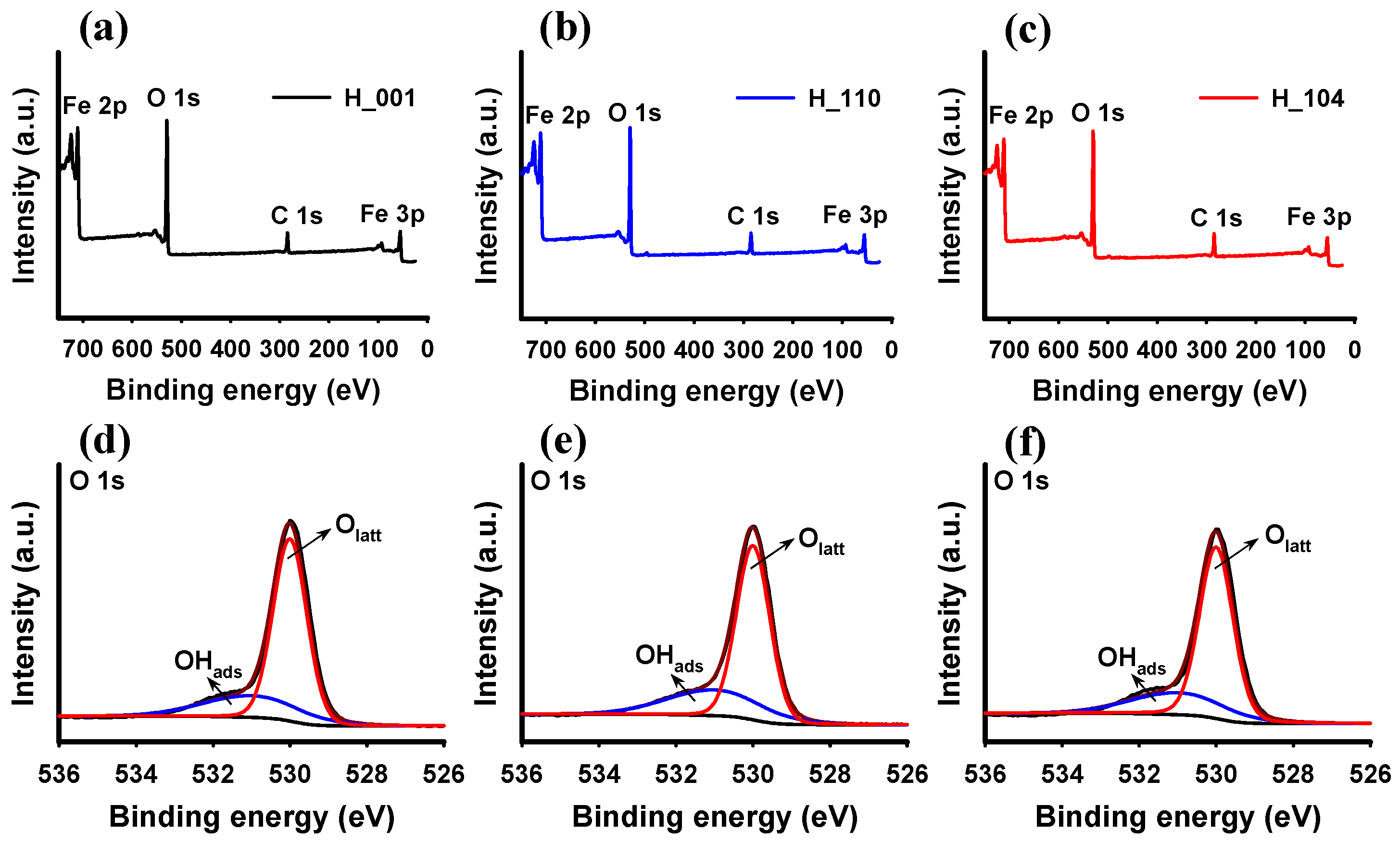
3.1. Characteristics of Hematite NPs

The characterization results of the three hematite NPs were shown in Figure 1, and the proportion of exposed crystal facets and the main physicochemical properties were presented in Table 1. The XRD patterns (Figure 1a,e,i) of the three NPs displayed the respective diffraction peaks at 24.1°, 33.1°, 35.6°, 40.8°, 49.4°, 54.0°, 62.4° and 63.9° corresponding to the (012), (104), (110), (113), (024), (116), (214) and (300) crystal planes of hematite phase (JCPDS No. 33-0664), respectively. The shape of their respective diffraction peaks was sharp, indicating that the three NPs had good crystallinity. Notably, the intensity ratios between the (104) and (110) diffraction peaks of XRD for the three hematite NPs were different, suggesting that the three hematite NPs may have different exposed facets [39]. According to SEM and TEM images, the H_001 NP consisted of hexahedral nanosheets with a thickness of about 15 nm (Figure 1b,c). The high-resolution TEM images (Figure 1d) and the fast Fourier transform (FFT) patterns (inset of Figure 1d) showed three sets of 0.25 nm diffraction fringes corresponding to the (110), (−210) and (−120) planes of hematite, suggesting that the main exposed facet of H_001 NP is the {001} crystal facet. According to the literature [29] and the geometric configuration in inset 1c, the H_001 NP was composed of an upper and lower {001} crystal plane and six equivalent {102} crystal planes around it. Figure 1f,g showed that the morphology of the H_110 NP was nanorods with a length of about 200 nm. The HRTEM image (Figure 1h) exhibited three sets of 0.25 nm diffraction fringes corresponding to the (110), (−120) and (−210) planes of hematite, while the corresponding FFT pattern (inset of Figure 1h) showed diffraction spots of the {110}, {−120} and {−210} facets. Combining the previous study [32] and the geometrical configuration in inset 1g revealed that H_110 was composed of {110}, {001}, {210} and {120} crystal facets. However, the morphology of the H_104 NP was a rhombohedral hexahedron with an edge length of about 400 nm (Figure 1j,k). The HRTEM image (Figure 1l) displayed two sets of 0.27 nm diffraction streaks corresponding to the (1 0 4) and (0–14) planes of hematite, while the diffraction spots of the corresponding FFT patterns (inset of Figure 1l) were attributed to (110), (0–14) and (104) facets along the [−441] direction. From the existing study [40] and the geometric configuration in inset 1k, it was evident that that H_104 NP was composed of six equivalent {104} crystal planes. Moreover, the SABET of the three hematite NPs were 20.9 m2 g−1, 24.2 m2 g−1 and 4.3 m2 g−1, respectively (Table 1). All three NPs exhibited a wide range of pore distribution (Figure S1), while the total pore volume of H_104 NPs was significantly smaller than that of H_001 and H_110 NPs, which explained the smaller SABET value of H_104 NPs.
The surface chemical states of three different facet hematite NPs were analyzed by XPS. Figure 2a–c showed the full spectrum with peaks at binding energies of 711.0 eV, 530.0 eV, 284.6 eV and 55.4 eV positions corresponding to Fe 2p, O 1s, C 1s and Fe 3p orbitals [41], respectively. This result suggested that the surface of the hematite NP contains only Fe, O and C elements. The C element was generally thought to come from substrate compounds in the XPS instrument itself or from air interference [42]. The O 1s high-resolution spectra of the three NPs were divided into two peaks at 530.0 eV and 531.0 eV (Figure 2d–f), where the peak at 530.0 eV was attributed to lattice oxygens (Olatt), while the peak at 531.0 eV was considered to be the surface adsorbed hydroxyl oxygens (OHads) [43]. From the above analysis, it can be seen that there was no significant difference in the surface elemental states of the three NPs, in which the surface hydroxyl content was only slightly different. Thus, these results demonstrated that the synthesized three NPs are suitable for the study of crystal facet effects.

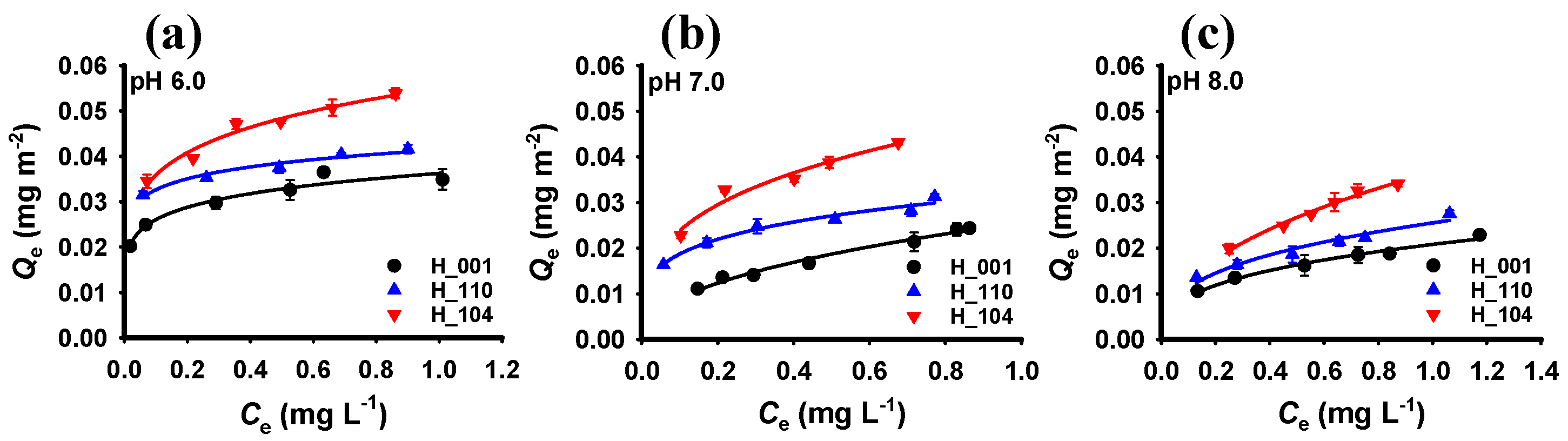
3.2. Adsorption of Phosphate by Hematite with Different Crystal Facets

Adsorption experiments were used to examine the adsorption efficiency of hematite NPs with different crystal facets towards phosphate. The adsorption data of phosphate to the three hematite NPs at different pH conditions were shown in Figure S2. The H_110 NPs, which has a larger SABET, exhibited greater adsorption capacity for phosphates. To exclude the effect of the SABET of NPs, we normalized the adsorption data by the SABET. The Freundlich and Langmuir adsorption isotherms are two commonly used models for describing the adsorption of substances on solid surfaces. Therefore, the Freundlich (Figure 3) and Langmuir (Figure S3) model were used to fit the normalized phosphate adsorption data. The results of the fitted Freundlich and Langmuir model parameters were shown in Table 2 and Table S1, respectively. Comparison of the fitting results of the two models showed that the adsorption of phosphate by the three NPs was more consistent with the Freundlich model, and the correlation coefficients R2 were greater than 0.95 for all three NPs. Phosphate adsorption onto hematite of different crystal facets exhibited different degrees of nonlinearity with little difference.
As shown in the results of Figure 3 and Table 2, the Freundlich affinity coefficients of the three NPs for phosphate increased by 1.7-fold, 1.6-fold and 1.5-fold for H_001, H_110, and H_104, respectively, with the decrease in pH, indicating that the adsorption capacity of all the three NPs increased with decreasing pH, which was also consistent with the results of the previous studies [44]. It has been demonstrated that when the pH of the adsorption system was gradually lower than the isoelectric point (7.5–7.6, Table 1) of the NP surface, the hematite surface became positively charged [45]. Therefore, more phosphate ions were enriched on the surface of the NP by electrostatic interaction at low pH, which was also more favorable for complexation [46].
It is noteworthy that the adsorption capacities of the three NPs for phosphate after normalizing the specific surface area showed significant differences with the following order: H_104 > H_110 > H_001. For example, the adsorption of phosphate by H_104 was higher than that of H_110 and higher than that of H_001 for different initial concentrations of phosphate at pH 7, and the Freundlich affinity coefficient increased from 0.026 mg1−n Ln m−2 for H_001 to 0.032 mg1−n Ln m−2 for H_110 and to 0.048 mg1−n Ln m−2 for H_104. These results suggested that the adsorption of phosphate by hematite was dependent on the exposed crystal facets [38,47].

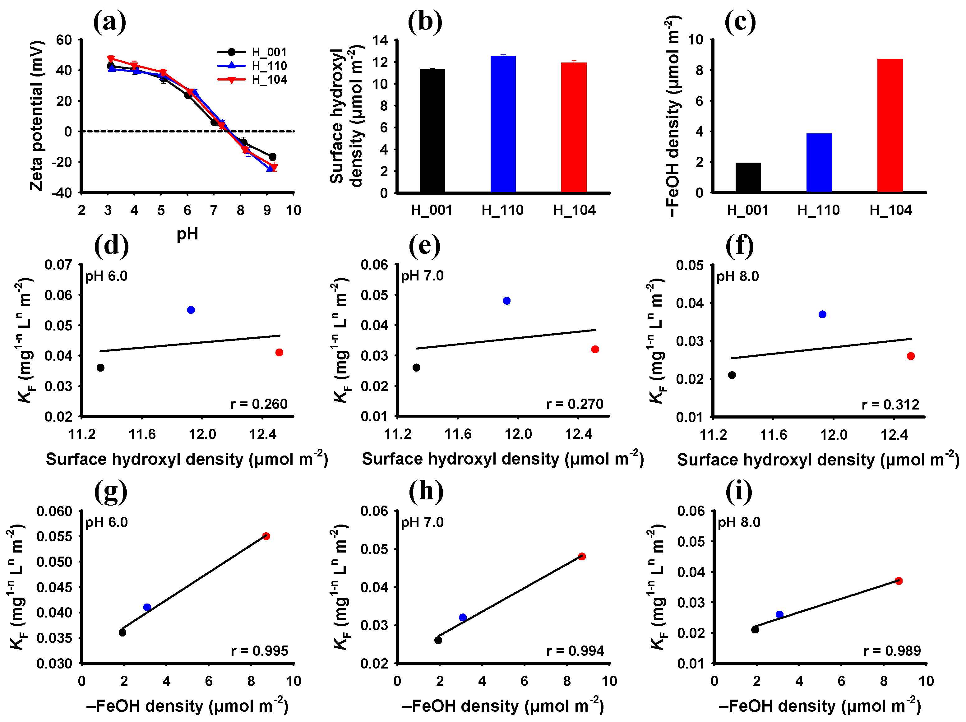
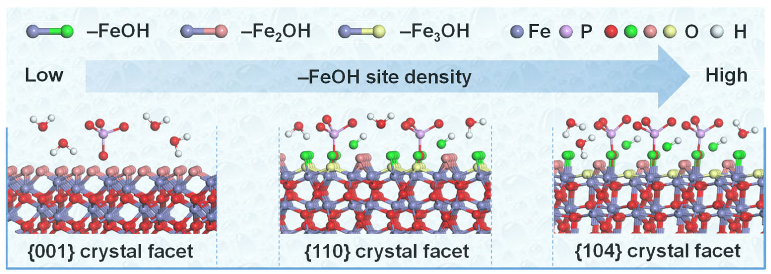
3.3. Facet-Dependent Adsorption Mechanism of Hematite to Phosphate

Previous studies proposed that the mechanisms of phosphate adsorption by iron oxides are mainly hydrogen bonding, electrostatic and ligand exchange interactions [26,48]. To further investigate the crystal facets-determined adsorption mechanism, the surface functional group compositions of the three hematite NPs and their changes before and after phosphate adsorption were characterized by FTIR spectroscopy. As shown in Figure 4, the characteristic peaks at about 537 cm−1, 558 cm−1, 632 cm−1 and 645 cm−1 in the FTIR spectra of hematite before and after the adsorption of phosphate corresponded to the stretching vibration of Fe-O functional group [49]. In addition, the peak at 875 cm−1 was attributed to Fe-OH vibration of the surface hydroxyl groups on hematite [48]. Meanwhile, the positions of the Fe-OH characteristic peaks in the three hematite NPs barely changed before and after the adsorption of phosphate, indicating that the hydrogen bonding between the hydroxyl groups on the surface of hematite and phosphate was negligible. In particular, the FTIR spectra (Figure 4) of hematite after adsorption of phosphate showed new characteristic peaks at 1106 and 1299 cm−1 compared to those before adsorption, which were usually attributed to the P-O stretching mode and P=O vibration mode of phosphate compounds (Figure S4), respectively [50,51,52]. These results further suggested that the phosphates were strongly interacting with the hematite NPs through electrostatic and ligand exchange interactions. The zeta potential usually represents the surface charge of the NP in solution, and the results in Figure 5a showed that the zeta potentials of the three NPs in aqueous suspensions were also not significantly different, suggesting that electrostatic interactions were not responsible for the differences in the adsorption of phosphate by the three NPs. Furthermore, the XPS survey spectra (Figure S5) of three hematite nanoparticles after adsorption of phosphate showed a P 2p peak at binding energy of 133 eV [53], while the Fe-OH (OHads) peak in the O 1s high-resolution spectra was weakened (Table S2). Therefore, the above results indicated that the main factor leading to the different adsorption of phosphate by the three NPs was the inner-sphere complexation formed by ligand exchange.
In order to gain a deeper understanding of the facet-dependent mechanism of phosphate adsorption by inner-sphere complexation formed through ligand exchange, the surface state of hematite in the aqueous environment needed to be clarified. It is well known that when metal oxides are present in aqueous solution, the unsaturated Fe atoms on their surfaces can dissociate the adsorbed H2O molecules to form surface hydroxyl groups [54]. Moreover, the ligand exchange process is mainly molecular or ionic replacement of hydroxyl functional groups on the surface of metal oxides [55]. Thus, the content and differences in surface hydroxyls may contribute to differences in phosphate adsorption by hematite NPs [56]. The hydroxyl titration results (Figure 5b) showed that the surface hydroxyl content of H_001, H_110 and H_104 NPs were 11.33 μmol m−2, 12.51 μmol m−2 and 11.93 μmol m−2, respectively, in the order of H_110 > H_104 > H_001, which was also consistent with the XPS results (Figure 2 and Table 1). However, the surface hydroxyl content of the three hematite NPs showed poor correlation with their adsorption capacity for phosphate, as shown by the Pearson correlation coefficients (r) of 0.260, 0.270 and 0.312 (Figure 5d–f). Hence, the hydroxyl content of the hematite surface may not be the main factor contributing to the differences in phosphate adsorption.
According to the position of the coordinating iron atoms on the surface of the iron oxides, the surface hydroxyl functional groups can be classified into single coordinating hydroxyl (–FeOH), double coordinating hydroxyl (–Fe2OH), and triple coordinating hydroxyl (–Fe3OH) functional groups [24,40]. Due to the differences in atomic arrangement on the different crystal facets of hematite, the corresponding surface hydroxyl species and densities are different [38]. For example, the –FeOH, –Fe2OH and –Fe3OH hydroxyl functional groups are present on the {102} crystal facet of hematite with densities of 5.1 sites nm−2, 5.1 sites nm−2 and 5.9 sites nm−2, respectively [37]. Previous studies have shown that inorganic ions or organic compounds preferentially undergoes ligand exchange with –FeOH groups on the surface of iron oxides [18,38,57,58,59]. Especially, Ding et al. proposed that –FeOH groups were the primary adsorption centers for ligand exchange between phosphate and iron oxides using in situ attenuated total reflectance (ATR)-FTIR spectra characterization, while the contributions of –Fe2OH and –Fe3OH groups in this process were negligible [56]. Therefore, we hypothesize that the content of –FeOH groups on the surface of hematite is the main factor influencing its facet-dependent adsorption of phosphate. To verify this speculation, the –FeOH functional group content of the three hematite surfaces were calculated as shown in Figure 5c. For three different crystal facets of hematite, the –FeOH site densities of the {110} and {104} crystal facets are reported to be 5.0 sites nm−2 and 5.3 sites nm−2, respectively, while the {001} crystal facet of hematite has only –Fe2OH sites [37]. Thus, the –FeOH site of the H_001 NP is located on its contained {102} crystal facet [29]. Combining the proportions of exposed crystal facets for each of the H_001, H_110 and H_104 NPs (Table 1), the –FeOH site densities of the three NPs were calculated to be 1.93 μmol m−2, 3.09 μmol m−2 and 8.71 μmol m−2 (Figure 5c), respectively [38]. Furthermore, the –FeOH site densities on the surface of the three NPs were analyzed by Pearson correlation with their Freundlich affinity coefficients for phosphate adsorption. The results showed that the Pearson correlation coefficients (r) were 0.995, 0.994 and 0.989 for pH 6, 7 and 8 conditions, respectively, indicating that the Freundlich affinity coefficients showed a significant positive correlation with single coordinating hydroxyl density (Figure 5g–i). Moreover, DFT calculations further analyzed the adsorption capacity of phosphate on the three crystal facets after replacement of –FeOH. The results (Figure S6) showed that the adsorption energy of phosphate on the {104} facet (−3.96 eV) is greater than that on the {110} (−3.36 eV) and {102} (−3.11 eV, H_001 NPs primary adsorption plane) facets, which is also consistent with the above results. Consequently, the main explanation for the differences in phosphate adsorption by hematite was the preferential ligand exchange of phosphate with –FeOH sites on the hematite surface (Figure 6), which was related to the facet-dependent density of –FeOH sites on the hematite surface.

4. Conclusions

In this work, the adsorption capacities of three hematite NPs with different exposed facets for phosphate were examined. The findings demonstrated that the adsorption of phosphate by hematite was face-dependent, with the adsorption capacity in the order of H_104 > H_110 > H_001. The results of XPS analysis, FTIR spectra, zeta potential and hydroxyl titration further revealed the facet-dependent adsorption mechanism of phosphate by hematite. Although pH can affect the adsorption capacity of hematite for phosphate through electrostatic interactions, the facet-dependent adsorption was mainly determined by the ligand exchange between phosphate and hematite. Different crystal exposed facets have different contents and densities of corresponding single coordinated hydroxyl sites due to the different atomic arrangements, which determine the adsorption capacity of hematite for phosphate. Therefore, the influence of the intrinsic properties of metal oxide particles (e.g., crystal facets) should be considered in the promotion of nano-agricultural technology and the management of eutrophication in water bodies.

Supplementary Materials

The following supporting information can be downloaded at: https://www.mdpi.com/article/10.3390/w15234070/s1, Text S1: Calculation of percentage the predominant facet; Text S2: Data analysis; Text S3: Density functional theory (DFT) computational details, Table S1: Model parameters of the Langmuir model for phosphate adsorption on different faceted hematite nanoparticles; Table S2: Surface chemistry properties of the different-faceted hematite nanoparticles after adsorption of phosphate determined by XPS; Figure S1: The pore size distribution curves of the different-faceted hematite nanoparticles; Figure S2: Adsorption isotherms of phosphate on different-faceted hematite nanoparticles under pH 6.0 (a), pH 7.0 (b) and pH 8.0 (c) conditions; Figure S3: The surface area-normalized adsorption isotherms of phosphate on different-faceted hematite nanoparticles under pH 6.0 (a), pH 7.0 (b) and pH 8.0 (c) conditions. Experimental data were fitted to the Langmuir model (Equation (S7)); Figure S4: FTIR spectra of the potassium dihydrogen phosphate; Figure S5: XPS survey spectra and high-resolution O 1s spectra of the H_001 (a, d), H_110 (b, e) and H_104 (c, f) nanoparticles after adsorption of phosphate, respectively; Figure S6: Optimized surface complex geometries of phosphate on {102} (a), {110} (b) and {104} (c) facets of hematite. Scheme S1. Schematic structure of H_001 nanoparticle. Scheme S2. Schematic structure of H_110 nanoparticle. Scheme S3. Schematic structure of H_104 nanoparticle.

Author Contributions

Conceptualization, T.L.; methodology, T.L. and F.S.; software, F.S., Y.J. and Z.D.; validation, T.L., Y.J. and Z.D.; formal analysis, T.L. and F.S.; investigation, F.S., Y.J. and Z.D.; resources, T.L. and Y.J.; data curation, T.L. and F.S.; writing—original draft preparation, T.L., F.S. and Y.J.; writing—review and editing, T.L.; visualization, T.L., F.S., Y.J. and Z.D.; supervision, T.L.; project administration, T.L.; funding acquisition, T.L. All authors have read and agreed to the published version of the manuscript.

Funding

This research was funded by the National Natural Science Foundation of China, grant number 22106003.

Data Availability Statement

The data presented in this study are available on request from the corresponding author. The data are not publicly available due to the laboratory’s sharing policy.

Acknowledgments

We are grateful to the potential reviewers for their valuable comments and suggestions. We are also very grateful to Anhui University for providing startup funding support to T.L.

Conflicts of Interest

The authors declare no conflict of interest.

References

  1. Schindler, D.W.; Carpenter, S.R.; Chapra, S.C.; Hecky, R.E.; Orihel, D.M. Reducing phosphorus to curb lake eutrophication is a success. Environ. Sci. Technol. 2016, 50, 8923–8929. [Google Scholar] [CrossRef] [PubMed]
  2. Jung, J.; Choi, M.; Police, A.K.R.; Lee, J.; Bae, S. Removal of phosphorus by ferric ion-rich solutions prepared using various Fe(III)-containing minerals. Water 2022, 14, 3765. [Google Scholar] [CrossRef]
  3. Wilfert, P.; Kumar, P.S.; Korving, L.; Witkamp, G.J.; van Loosdrecht, M.C.M. The relevance of phosphorus and iron chemistry to the recovery of phosphorus from wastewater: A review. Environ. Sci. Technol. 2015, 49, 9400–9414. [Google Scholar] [CrossRef] [PubMed]
  4. Liu, L.; He, N.N.; Borham, A.; Zhang, S.W.; Xie, R.Q.; Zhao, C.; Hu, J.W.; Wang, J.J. The effect of iron-modified biochar on phosphorus adsorption and the prospect of synergistic adsorption between biochar and iron-oxidizing bacteria: A review. Water 2023, 15, 3315. [Google Scholar] [CrossRef]
  5. Wang, R.G.; Zhao, X.; Wang, T.C.; Guo, Z.Z.; Hu, Z.; Zhang, J.; Wu, S.B.; Wu, H.M. Can we use mine waste as substrate in constructed wetlands to intensify nutrient removal? A critical assessment of key removal mechanisms and long-term environmental risks. Water Res. 2022, 210, 118009. [Google Scholar] [CrossRef]
  6. Han, C.N.; Dai, Y.; Sun, N.N.; Wu, H.; Tang, Y.; Dai, T.H. Algae bloom and decomposition changes the phosphorus cycle pattern in taihu lake. Water 2022, 14, 3607. [Google Scholar] [CrossRef]
  7. Fahmi, A.; Hairani, A.; Alwi, M.; Nurzakiah, S. Fe-P pools as phosphorus source for rice in acid sulfate soils. Chilean J. Agric. Res. 2023, 83, 626–634. [Google Scholar] [CrossRef]
  8. Lin, J.W.; Li, Y.; Zhan, Y.H.; Wu, X.G. Combined amendment and capping of sediment with ferrihydrite and magnetite to control internal phosphorus release. Water Res. 2023, 235, 119899. [Google Scholar] [CrossRef]
  9. Hochella, M.F.; Mogk, D.W.; Ranville, J.; Allen, I.C.; Luther, G.W.; Marr, L.C.; McGrail, B.P.; Murayama, M.; Qafoku, N.P.; Rosso, K.M.; et al. Natural, incidental, and engineered nanomaterials and their impacts on the Earth system. Science 2019, 363, 1414. [Google Scholar] [CrossRef] [PubMed]
  10. Tao, Z.X.; Zhou, Q.X.; Zheng, T.; Mo, F.; Ouyang, S.H. Iron oxide nanoparticles in the soil environment: Adsorption, transformation, and environmental risk. J. Hazard. Mater. 2023, 459, 132107. [Google Scholar] [CrossRef]
  11. Tartaj, P.; Morales, M.P.; Gonzalez-Carreño, T.; Veintemillas-Verdaguer, S.; Serna, C.J. The iron oxides strike back: From biomedical applications to energy storage devices and photoelectrochemical water splitting. Adv. Mater. 2011, 23, 5243–5249. [Google Scholar] [CrossRef]
  12. Navrotsky, A.; Mazeina, L.; Majzlan, J. Size-driven structural and thermodynamic complexity in iron oxides. Science 2008, 319, 1635–1638. [Google Scholar] [CrossRef]
  13. Lv, Y.; Liu, J.; Zhu, R.L.; Zhu, J.X.; Chen, Q.Z.; Liang, X.L.; He, H.P. Photoreductive dissolution of iron (hydr)oxides and its geochemical significance. ACS Earth Space Chem. 2022, 6, 811–829. [Google Scholar] [CrossRef]
  14. Hochella, M.F.; Lower, S.K.; Maurice, P.A.; Penn, R.L.; Sahai, N.; Sparks, D.L.; Twining, B.S. Nanominerals, mineral nanoparticles, and Earth systems. Science 2008, 319, 1631–1635. [Google Scholar] [CrossRef] [PubMed]
  15. Di Iorio, E.; Circelli, L.; Angelico, R.; Torrent, J.; Tan, W.F.; Colombo, C. Environmental implications of interaction between humic substances and iron oxide nanoparticles: A review. Chemosphere 2022, 303, 135172. [Google Scholar] [CrossRef]
  16. Huang, X.P.; Hou, X.J.; Zhang, X.; Rosso, K.M.; Zhang, L.Z. Facet-dependent contaminant removal properties of hematite nanocrystals and their environmental implications. Environ. Sci. Nano 2018, 5, 1790–1806. [Google Scholar] [CrossRef]
  17. Bai, L.L.; Cao, C.C.; Wang, C.L.; Wang, C.H.; Zhang, H.; Jiang, H.L. Roles of phytoplankton- and macrophyte-derived dissolved organic matter in sulfamethazine adsorption on goethite. Environ. Pollut. 2017, 230, 87–95. [Google Scholar] [CrossRef] [PubMed]
  18. Kim, J.; Li, W.; Philips, B.L.; Grey, C.P. Phosphate adsorption on the iron oxyhydroxides goethite (α-FeOOH), akaganeite (β-FeOOH), and lepidocrocite (γ-FeOOH): A 31P NMR study. Energy Environ. Sci. 2011, 4, 4298–4305. [Google Scholar] [CrossRef]
  19. Siwek, H.; Bartkowiak, A.; Wlodarczyk, M. Adsorption of phosphates from aqueous solutions on alginate/goethite hydrogel composite. Water 2019, 11, 633. [Google Scholar] [CrossRef]
  20. Yang, Y.L.; Wang, S.R.; Xu, Y.S.; Zheng, B.H.; Liu, J.Y. Molecular-scale study of aspartate adsorption on goethite and competition with phosphate. Environ. Sci. Technol. 2016, 50, 2938–2945. [Google Scholar] [CrossRef]
  21. Wang, Z.Y.; Zhang, L.; Zhao, J.; Xing, B.S. Environmental processes and toxicity of metallic nanoparticles in aquatic systems as affected by natural organic matter. Environ. Sci. Nano 2016, 3, 240–255. [Google Scholar] [CrossRef]
  22. Parkinson, G.S. Iron oxide surfaces. Surf. Sci. Rep. 2016, 71, 272–365. [Google Scholar] [CrossRef]
  23. Huang, X.P.; Hou, X.J.; Jia, F.L.; Song, F.H.; Zhao, J.C.; Zhang, L.Z. Ascorbate-promoted surface iron cycle for efficient heterogeneous fenton alachlor degradation with hematite nanocrystals. ACS Appl. Mater. Interfaces 2017, 9, 8751–8758. [Google Scholar] [CrossRef] [PubMed]
  24. Vindedahl, A.M.; Strehlau, J.H.; Arnold, W.A.; Penn, R.L. Organic matter and iron oxide nanoparticles: Aggregation, interactions, and reactivity. Environ. Sci. Nano 2016, 3, 494–505. [Google Scholar] [CrossRef]
  25. Wang, M.L.; Kong, B.; Zhang, Y.Y.; Shan, C.; Pan, B.C. Facet-dependent phosphate adsorptive reactivity by lanthanum hydroxides of different crystal structure: Role of surface hydroxyl groups. Appl. Surf. Sci. 2021, 538, 147910. [Google Scholar] [CrossRef]
  26. Ge, X.F.; Wang, L.J.; Zhang, W.J. Molecular-scale determination of facet- and adsorbent-dependent phosphate adsorption by metal-based adsorbents. Environ. Sci. Nano 2022, 9, 3372–3384. [Google Scholar] [CrossRef]
  27. Stirner, T.; Scholz, D.; Sun, J.Z. Ab initio simulation of structure and surface energy of low-index surfaces of stoichiometric α-Fe2O3. Surf. Sci. 2018, 671, 11–16. [Google Scholar] [CrossRef]
  28. Cornell, R.M.; Schwertmann, U. The Iron Oxides: Structure, Properties, Reactions, Occurrences and Uses; John Wiley & Sons: Hoboken, NJ, USA, 2003. [Google Scholar]
  29. Li, T.; Zhong, W.; Jing, C.Y.; Li, X.G.; Zhang, T.; Jiang, C.J.; Chen, W. Enhanced hydrolysis of p-nitrophenyl phosphate by iron (hydr)oxide nanoparticles: Roles of exposed facets. Environ. Sci. Technol. 2020, 54, 8658–8667. [Google Scholar] [CrossRef] [PubMed]
  30. Jin, X.; Wu, D.D.; Liu, C.; Huang, S.H.; Zhou, Z.Y.; Wu, H.; Chen, X.R.; Huang, M.Y.; Zhou, S.D.; Gu, C. Facet effect of hematite on the hydrolysis of phthalate esters under ambient humidity conditions. Nat. Commun. 2022, 13, 6125. [Google Scholar] [CrossRef]
  31. Asif, A.H.; Rafique, N.; Hirani, R.A.K.; Wu, H.; Shi, L.; Zhang, S.; Wang, S.B.; Yin, Y.; Saunders, M.; Sun, H.Q. Morphology/facet-dependent photo-Fenton-like degradation of pharmaceuticals and personal care products over hematite nanocrystals. Chem. Eng. J. 2022, 432, 134429. [Google Scholar] [CrossRef]
  32. Huang, X.P.; Hou, X.J.; Zhao, J.C.; Zhang, L.Z. Hematite facet confined ferrous ions as high efficient Fenton catalysts to degrade organic contaminants by lowering H2O2 decomposition energetic span. Appl. Catal. B 2016, 181, 127–137. [Google Scholar] [CrossRef]
  33. Wang, W.T.; Zhang, W.J.; Fan, Y.K.; Qu, C.C.; Ren, W.Y.; Huang, X.P.; Hong, M.; Liu, F.; Yin, H. Facet-dependent adsorption of aluminum(iii) on hematite nanocrystals and the influence on mineral transformation. Environ. Sci. Nano 2022, 9, 2073–2085. [Google Scholar] [CrossRef]
  34. Lin, M.; Tan, H.R.; Tan, J.P.Y.; Bai, S.Q. Understanding the growth mechanism of α-Fe2O3 nanoparticles through a controlled shape transformation. J. Phys. Chem. C 2013, 117, 11242–11250. [Google Scholar] [CrossRef]
  35. Prato, J.G.; Millán, F.C.; González, L.C.; Ríos, A.C.; López, E.; Ríos, I.; Navas, S.; Márquez, A.; Carrero, J.C.; Díaz, J.I. Adsorption of phosphate and nitrate ions on oxidic substrates prepared with a variable-charge lithological material. Water 2022, 14, 2454. [Google Scholar] [CrossRef]
  36. Tamura, H.; Tanaka, A.; Mita, K.Y.; Furuichi, R. Furuichi Surface hydroxyl site densities on metal oxides as a measure for the ion-exchange capacity. J. Colloid Interface Sci. 1999, 209, 225–231. [Google Scholar] [CrossRef] [PubMed]
  37. Barrón, V.; Torrent, J. Surface hydroxyl configuration of various crystal faces of hematite and goethite. J. Colloid Interface Sci. 1996, 177, 407–410. [Google Scholar] [CrossRef]
  38. Lv, J.T.; Miao, Y.X.; Huang, Z.Q.; Han, R.X.; Zhang, S.Z. Facet-mediated adsorption and molecular fractionation of humic substances on hematite surfaces. Environ. Sci. Technol. 2018, 52, 11660–11669. [Google Scholar] [CrossRef]
  39. Li, X.G.; Li, T.; Zhang, T.; Gu, C.; Zheng, S.R.; Zhang, H.J.; Chen, W. Nano-TiO2-catalyzed dehydrochlorination of 1,1,2,2-tetrachloroethane: Roles of crystalline phase and exposed facets. Environ. Sci. Technol. 2018, 52, 4031–4039. [Google Scholar] [CrossRef] [PubMed]
  40. Chan, J.Y.T.; Ang, S.Y.; Ye, E.Y.; Sullivan, M.; Zhang, J.; Lin, M. Heterogeneous photo-Fenton reaction on hematite (α-Fe2O3) {104}, {113} and {001} surface facets. Phys. Chem. Chem. Phys. 2015, 17, 25333–25341. [Google Scholar] [CrossRef]
  41. Rashid, N.M.A.; Haw, C.; Chiu, W.; Khanis, N.H.; Rohaizad, A.; Khiew, P.; Rahman, S.A. Structural- and optical-properties analysis of single crystalline hematite (α-Fe2O3) nanocubes prepared by one-pot hydrothermal approach. CrystEngComm 2016, 18, 4720–4732. [Google Scholar] [CrossRef]
  42. Yu, H.G.; Liu, R.; Wang, X.F.; Wang, P.; Yu, J.G. Enhanced visible-light photocatalytic activity of Bi2WO6 nanoparticles by Ag2O cocatalyst. Appl. Catal. B 2012, 111, 326–333. [Google Scholar] [CrossRef]
  43. Huang, X.P.; Hou, X.J.; Song, F.H.; Zhao, J.C.; Zhang, L.Z. Facet-dependent Cr(VI) adsorption of hematite nanocrystals. Environ. Sci. Technol. 2016, 50, 1964–1972. [Google Scholar] [CrossRef] [PubMed]
  44. Luengo, C.; Brigante, M.; Antelo, J.; Avena, M. Kinetics of phosphate adsorption on goethite: Comparing batch adsorption and ATR-IR measurements. J. Colloid Interface Sci. 2006, 300, 511–518. [Google Scholar] [CrossRef] [PubMed]
  45. Baldwin, D.S.; Beattie, J.K.; Coleman, L.M.; Jones, D.R. Phosphate ester hydrolysis facilitated by mineral phases. Environ. Sci. Technol. 1995, 29, 1706–1709. [Google Scholar] [CrossRef] [PubMed]
  46. Yan, W.; Jing, C.Y. Molecular insights into glyphosate adsorption to goethite gained from ATR-FTIR, two-dimensional correlation spectroscopy, and DFT study. Environ. Sci. Technol. 2018, 52, 1946–1953. [Google Scholar] [CrossRef]
  47. Li, X.C.; Reich, T.; Kersten, M.; Jing, C.Y. Low-molecular-weight organic acid complexation affects antimony(III) adsorption by granular ferric hydroxide. Environ. Sci. Technol. 2019, 53, 5221–5229. [Google Scholar] [CrossRef]
  48. Wang, H.; Zhu, J.; Fu, Q.L.; Hong, C.; Hu, H.Q.; Violante, A. Phosphate adsorption on uncoated and humic acid-coated iron oxides. J. Soils Sediments 2016, 16, 1911–1920. [Google Scholar] [CrossRef]
  49. Chen, M.N.; Xie, Z.M.; Yang, Y.; Gao, B.; Wang, J. Effects of calcium on arsenate adsorption and arsenate/iron bioreduction of ferrihydrite in stimulated groundwater. Int. J. Environ. Res. Public Health 2022, 19, 3465. [Google Scholar] [CrossRef] [PubMed]
  50. Wang, Z.; Mahmood, A.; Xie, X.F.; Wang, X.; Qiu, H.X.; Sun, J. Surface adsorption configurations of H3PO4 modified TiO2 and its influence on the photodegradation intermediates of gaseous o-xylene. Chem. Eng. J. 2020, 393, 124723. [Google Scholar] [CrossRef]
  51. Saito, S.; Hamai, R.; Shiwaku, Y.; Hasegawa, T.; Sakai, S.; Tsuchiya, K.; Sai, Y.; Iwama, R.; Amizuka, N.; Takahashi, T.; et al. Involvement of distant octacalcium phosphate scaffolds in enhancing early differentiation of osteocytes during bone regeneration. Acta Biomater. 2021, 129, 309–322. [Google Scholar] [CrossRef] [PubMed]
  52. Park, K.H.; Song, H.J.; Park, Y.J. Microwave treatment of calcium phosphate/titanium dioxide composite to improve protein adsorption. Materials 2022, 15, 4773. [Google Scholar] [CrossRef]
  53. Mäkie, P.; Persson, P.; Österlund, L. Adsorption of trimethyl phosphate and triethyl phosphate on dry and water pre-covered hematite, maghemite, and goethite nanoparticles. J. Colloid Interface Sci. 2013, 392, 349–358. [Google Scholar] [CrossRef] [PubMed]
  54. Mu, R.T.; Zhao, Z.J.; Dohnálek, Z.; Gong, J.L. Structural motifs of water on metal oxide surfaces. Chem. Soc. Rev. 2017, 46, 1785–1806. [Google Scholar] [CrossRef] [PubMed]
  55. Waiman, C.V.; Avena, M.J.; Regazzoni, A.E.; Zanini, G.P. A real time in situ ATR-FTIR spectroscopic study of glyphosate desorption from goethite as induced by phosphate adsorption: Effect of surface coverage. J. Colloid Interface Sci. 2013, 394, 485–489. [Google Scholar] [CrossRef]
  56. Ding, X.B.; Song, X.W.; Boily, J.F. Identification of fluoride and phosphate binding sites at FeOOH surfaces. J. Phys. Chem. C 2012, 116, 21939–21947. [Google Scholar] [CrossRef]
  57. Hiemstra, T.; Riemsdijk, W.H.V. A surface structural approach to ion adsorption: The charge distribution (CD) model. J. Colloid Interface Sci. 1996, 179, 488–508. [Google Scholar] [CrossRef]
  58. Boily, J.F.; Persson, P.; Sjöberg, S. Benzenecarboxylate surface complexation at the goethite (α-FeOOH)/water interface: III. The influence of particle surface area and the significance of modeling parameters. J. Colloid Interface Sci. 2000, 227, 132–140. [Google Scholar] [CrossRef] [PubMed]
  59. Song, X.W.; Boily, J.F. Structural controls on OH site availability and reactivity at iron oxyhydroxide particle surfaces. Phys. Chem. Chem. Phys. 2012, 14, 2579–2586. [Google Scholar] [CrossRef] [PubMed]
Figure 1. XRD spectra, SEM, TEM and HRTEM images of H_001 (ad), H_110 (eh) and H_104 (il) nanoparticles, respectively. Insets in the TEM and HRTEM images are the geometric models and the corresponding FFT patterns of hematite nanoparticles, respectively.
Water 15 04070 g001
Figure 2. XPS survey spectra and high-resolution O 1s spectra of the H_001 (a,d), H_110 (b,e) and H_104 (c,f) nanoparticles, respectively.
Water 15 04070 g002
Figure 3. The surface area-normalized adsorption isotherms of phosphate on different-faceted hematite nanoparticles under pH 6.0 (a), pH 7.0 (b) and pH 8.0 (c) conditions. Experimental data were fitted to the Freundlich model (Equation (1)).
Water 15 04070 g003
Figure 4. FTIR spectra of H_001 (a), H_110 (b) and H_104 (c) nanoparticles before and after phosphate adsorption.
Water 15 04070 g004
Figure 5. The zeta potential (a), surface hydroxyl densities (b) and –FeOH site densities (c) of the different-faceted hematite nanoparticles. Correlations between the Freundlich affinity coefficient (KF) and the surface hydroxyl densities (df) or the –FeOH site densities (gh) on hematite surface at pH 6.0 (d,g), 7.0 (e,h) and 8.0 (f,i). The black, blue and red dots in (d-i) represent the H_001, H_110 and H_104 NPs, respectively.
Water 15 04070 g005
Figure 6. Proposed facet-dependent adsorption mechanism of hematite to phosphate.
Water 15 04070 g006
Table 1. Selected physicochemical properties of the different-faceted hematite nanoparticles.
SampleSABET 1
(m2 g−1)
Proportion of Exposed Facets 2Surface Fe 3
(at %)
Surface O 3
(at %)
Fe/Olatt
Ratio 3
Isoelectric Point
OlattOHads
H_00120.977.2%
{001}
22.8%
{102}
--33.250.216.60.667.5
H_11024.237.2%
{110}
55.6%
{001}
3.6%
{120}
3.6%
{210}
32.448.119.50.677.6
H_1044.399.0%
{104}
---31.251.417.40.617.5
Notes: 1 Measured using the BET method. 2 Calculated by SEM and TEM images. 3 Analyzed by XPS spectra.
Table 2. Model parameters of the Freundlich model for phosphate adsorption on different-faceted hematite nanoparticles.
SamplepH 6.0pH 7.0pH 8.0
KF
(mg1−n Ln m−2)
nR2 *KF
(mg1−n Ln m−2)
nR2 *KF
(mg1−n Ln m−2)
nR2 *
H_0010.0360.1440.9520.0260.4540.9820.0210.3500.981
H_1100.0410.1040.9610.0320.2300.9700.0260.3510.951
H_1040.0550.1850.9710.0480.3080.9590.0370.4560.987
Note: * The correlation coefficient of the Freundlich model fit.
Disclaimer/Publisher’s Note: The statements, opinions and data contained in all publications are solely those of the individual author(s) and contributor(s) and not of MDPI and/or the editor(s). MDPI and/or the editor(s) disclaim responsibility for any injury to people or property resulting from any ideas, methods, instructions or products referred to in the content.

