Article

Ecotoxicological Effects of Sodium Metasilicate on Two Hydra Species, Hydra viridissima Pallas, 1766 and Hydra oligactis Pallas, 1766

1
Department of Biology, Faculty of Science, University of Zagreb, Horvatovac 102A, 10000 Zagreb, Croatia
2
Graduate School, University of Nova Gorica, Vipavska 13, 5000 Nova Gorica, Slovenia
*
Author to whom correspondence should be addressed.
Water 2023, 15(24), 4228; https://doi.org/10.3390/w15244228
Submission received: 27 October 2023 / Revised: 1 December 2023 / Accepted: 6 December 2023 / Published: 8 December 2023

Abstract

:
Sodium metasilicate (SM) is a synthetic hazardous water-soluble salt used in industry as an active ingredient in household cleaning products. The impact of SM on the aquatic environment has been discussed worldwide, but its toxicity has not been well documented and researched. Studies have only been performed on a handful of aquatic organisms: algae, plants, blackworms, water fleas, dipteran larvae, and two fish species. Hydra is a simple freshwater cnidarian with diploblastic organisation, where all the cells are in permanent contact with the surrounding aqueous media, and represents a sensitive model organism for environmental toxicity assessments. This research aimed to determine and compare the effect of SM on the morphology, excitability, and behaviour of green and brown hydra and endosymbiotic microalgae as a microbiome of green hydra. The hydras were treated with four sublethal SM concentrations (0.050, 0.365, 0.380, and 0.390 g/L) for 72 h. Standard preparations were made for the cyto-histological analysis of green hydra, and damaged cellular layers and mesoglea and a changed distribution of microalgae were recorded. The SM caused muted responses to mechanical stimuli and damage to the tentacles in both hydra species. The changes were more pronounced in brown hydra, while green hydra showed better adaptability to unfavourable environmental conditions.

1. Introduction

The mass production and use of chemicals, and the human dependence on them, causes the continuous enormous release of numerous hazardous substances into the natural environment [1], especially aquatic ecosystems [2]. Soluble silicates are a class of synthetic compounds produced in large amounts worldwide. Chemically, these are inorganic salts of silicon dioxide and alkali metals [3]. Their application includes different branches of industry, from construction to the cosmetic industry and the production of paper, detergents, and glue. Besides that, they are used in the water treatment process to remove toxic metals and for fire protection [3,4]. Sodium metasilicate (Na2SiO3 × nH2O) is a synthetic solid substance in the form of water-soluble inorganic salt that may easily spread in water systems through laundry detergent products [5,6,7]. According to the European Chemicals Agency (ECHA) and Regulation (EC) No. 1907/2006—REACH of the European Parliament and of the Council, SM belongs to hazardous components with PNEC values (Predicted No-Effect Concentration) for freshwater and marine water below 7.5 mg/L and 1 mg/L, respectively. Conservative and predicted concentrations (PNEC values) at which SM would have no toxic effect are crucial for further EU regulations, which implies the inclusion of the results of ecotoxicological research on different groups of organisms, which, to date, have been highly insufficient regarding dangerous, water-soluble chemicals, such as SM. Spills of SM can have a damaging effect on the environment as a very corrosive strong base (pH 12.7 at a concentration of 1%). Therefore, it is the only silicate classified as corrosive in Annex 1 of the European Dangerous Substances Directive 67/548/EC [8,9,10,11]. Alkaline earth and heavy metal ions are precipitated as metasilicates from SM solutions. When heated or acidified, solutions of SM are hydrolysed to free sodium ions and silicic acid [12].
According to [6], using soluble silicates in household cleaning products has a negligible effect on aquatic ecosystems. However, the primary hazard of commercially used soluble silicates in aquatic environments is their moderate to strong alkalinity, which could cause a negative physical effect on aquatic animals. Additionally, previous research shows the inhibitory effect of SM on some enzymes, such as urease and invertase, while there was no effect on the activity of other enzymes, like pepsin, trypsin, lipase, catalase, and cholinesterase [4]. The toxicity of SM to aquatic organisms is not well documented, and the existing studies include research on algae and aquatic plants, blackworms, water fleas, dipteran larvae (chironomid, muscids, and scatophagids), and fish [13,14,15,16]. In laboratory conditions, the effects of sodium silicate solutions on aquatic invertebrates were investigated on the water flea Daphnia magna Straus, 1820. The reported 48 h EC50 values for the studied crustacean species ranged from 0.28 to 0.57 mg/L of SM (US EPA, Ecotox database—Aquatic Toxicity Data) and 216 to 1700 mg/L of sodium silicate [13,14,15,16]. The zebrafish Danio rerio (Hamilton, 1822) was exposed to SM (anhydrous) by Richterich and Müehlberg [13,14,15,16]. The 96 h LC50 was determined to be 210 mg/L. The toxicity of SM to the mosquito fish Gambusia affinis (Baird et Girard, 1853) was examined by [17], where the 96 h LC50 was determined to be 2320 mg/L.
Biomonitoring and choosing and using sensitive biological indicators represent key points for ecosystem health assessment during the last decade. One type of biomonitoring is surveillance before and after a toxic substance enters the water. It involves using bioindicators, indicator species, or indicator communities [18]. A limiting factor is the prevalent use of mortality as the most common indicator of toxicity. Hydras can be extremely good indicators of environmental changes and are widely used in ecotoxicological research [19,20,21,22,23]. Moreover, hydra is a suitable test organism in ecotoxicological research due to numerous reasons [19,21,22,24,25,26,27,28,29]. It is widely distributed, with well-known morphology, physiology, and ecology [21,23,30,31]. In laboratory conditions, it is relatively easy to maintain, reproduces quickly, and produces genetically identical individuals. Since the life processes of this sessile organism take place by diffusion, changes in the surrounding medium can be easily monitored for hydra [19,22,26,29]. It has been established that hydra can be a good experimental object for determining lethal and sublethal doses of toxicants and in explaining the effect of pollutants on freshwater ecosystems [21,23]. Hydra has a great ability to regenerate, so the degree of damage and the action of the investigated xenobiotic can be monitored [19,22,26]. Extensive research has been carried out on hydras, in which the effects of various xenobiotics, such as pesticides, heavy metals, cytostatics, and antibiotics, have been monitored regarding behaviour, mortality, cytological and histological structure, locomotion, regeneration, physiology, asexual reproduction, and genotoxicity [20,22,29,32]. Hydra is a ubiquitous inhabitant of freshwater ecosystems. Given that it is part of the lower level of the food web of stagnant freshwater, changes in the hydra population could have significant effects on the rest of the freshwater community. Hydra plays the role of both predator and prey within the ecosystem. It feeds on various zooplankton species, while it can become prey to flatworms. As such, it has ecological importance in structuring the plankton community of freshwater ecosystems and represents an important indicator species in ecotoxicology [33]. However, more subtle changes in the external hydra morphology, behaviour, and structure of the endosymbiotic hydra biome are rarely considered [34,35]. These kinds of subtle changes include disorders in growth and changes in the reproduction and behaviour of the organisms [36,37]. All of these parameters may have an important effect on the relationships between organisms and the stability of ecosystems.
Aquatic ecosystems require special attention regarding ecotoxicological research and environmental protection. The existing methods and framework for hazard assessment in the aquatic environment comply with the current EU regulations [8,9,10,11], and the test organisms that have been used range from plants to plenty of unicellular and small tiny multicellular organisms, such as Hydra spp. [33]. Chemical substances from the soil, atmosphere, sewage, and industrial wastewater often end up in rivers, lakes, and seas [38]. Hydra is a widespread inhabitant of freshwater ecosystems around the world. It takes up the position at the bottom of aquatic food networks. The role of a hydra inside an ecosystem can be both predator and prey [39]. It feeds on different zooplankton, while it represents food for platyhelminths. As such, it is important in structuring the freshwater planktonic community and is a crucial bioindicator. The disappearance of hydra populations from the environment could influence the stability of entire networks of aquatic organisms [33,38]. Hydra is an invertebrate from the phylum Cnidaria that lives on the bottom of still or slow-flowing freshwater [21]; it is sedentary and attaches to rocks, leaves, and twigs on the bottom of aquatic ecosystems. The body of the hydra is cylindrical. Its size varies from 2 mm to 5 mm and is divided into three regions. The apical part of the body consists of a hypostome with a mouth opening and tentacles. Below the apical is the gastral region, a part of which is the budding region. With the basal part of the body, hydra attaches itself to different surfaces. Between the two layers, there is a noncellular layer: the mesoglea. The green hydra Hydra viridissima Pallas, 1766 shows less susceptibility to the harmful effects of some toxicants, such as iron, than the brown hydra Hydra oligactis Pallas, 1766 [40].
Holobionts and hologenomes are units of biological organization, organised as complex assemblages of organisms, their microbial symbionts, and their genomes. The simple architecture of the hydra body, the limited number of different bacterial symbionts, and the experimental accessibility of both host tissue and colonizing microbes have made the hydra an excellent model organism for studying host–microbe interactions [41,42,43].
Hydra’s ectodermal epithelial layer is tightly colonized by a stable multispecies bacterial community [44], which is species-specific and mirrors the phylogenetic relationship of their host (phylosymbiosis) [45,46]. Recent research [47] demonstrated that in the freshwater green polyp H. viridissima species, the photosynthetic Chlorella symbiont plays an important role in stabilizing the host bacterial community.
Since the molecular mechanisms controlling these tripartite complex interactions (Hydra—photobiont, Chlorella—resident microbiota) are still unknown, reductionistic model systems, such as hydra, are used for a functional understanding of these mechanisms [48]. The hydra model system has also contributed to the discovery of the role of interactions between symbiotic bacteria in colonization resistance [49]. It seems that microbial metabolites stimulate hydra’s immune response, body contractions [50], and responsive behaviour and affect the differentiation capacity of stem cells [48,51].
According to all the facts mentioned above, we can summarise that hydra is a more than suitable model organism for ecotoxicological studies since (1) it is a widespread inhabitant of freshwater ecosystems around the world; (2) its polyps propagate asexually by budding, so it can be easily raised in laboratory conditions; (3) it has diploblastic organisation and is composed of an outer ectoderm, inner endoderm, and noncellular mesoglea [52], and all the cells are in constant contact with the aqueous environment, which facilitates the permeation of toxic substances into the animal media; (4) the study population is often from the same strain, so individual variations are minimal; and (5) it can be used as a holobiont and hologenome for studying host–microbe interactions. This research aimed to determine the effects of sublethal concentrations of SM on green and brown hydra by keeping track of morphological and behavioural changes. Furthermore, considering the endosymbiotic microalgae in green hydra as a function of the microbiome, we aimed to see whether symbiotic green hydra better adapts to unfavourable environmental conditions than free-living brown hydra. Since SM is a compound with wide application in industry, using hydra as a sensitive cosmopolitan aquatic organism makes this experiment extremely important and provides us with information on the influence that metasilicate may have on aquatic ecosystems worldwide.

2. Materials and Methods

2.1. Test Organisms

The experiments were carried out using two different species of hydra: green hydra and brown hydra. The individuals of brown hydra were collected in a stream (Maksimir) in Zagreb, Croatia, while the green hydras used in this experiment were taken from the permanent laboratory cultivation at the Faculty of Science, Department of Biology, Zagreb, Croatia. The data from the former analysis on the chemical composition and characteristics of the aquarium water for hydras used in the experiments are given in [53]. Prior to the experiment, the brown hydras were maintained at 10 °C in a refrigerator in a glass bowl filled with aerated water. The animals were fed twice a week with freshly hatched Artemia salina (Linnaeus, 1758) nauplia. Budless hydras of similar size and morphology were selected for the experiment.

2.2. Toxicity Tests

Toxicity tests were performed with one control and four experimental groups for each species. Each group of green hydras consisted of 10 individuals, while each group of brown hydras consisted of 5 individuals. The experiment was performed in triplicate in the experiment with green hydras and in duplicate in the experiment with brown hydras. The concentrations of SM used for the treatment of green and brown hydras in the experimental groups were as follows: 0.050, 0.365, 0.380, and 0.390 g/L. Small differences in the concentrations are targeted to help establish threshold values for mortality. Therefore, the differences in the values were adjusted to the third decimal number because the mortality suddenly appeared after the SM concentration was increased. This claim is supported by the mortality data in the Supplementary Materials (Table S5). The experiment lasted for three days (72 h), during which the following changes were observed by using a stereomicroscope: mortality, morphological structure, and response to mechanical stimuli with a preparation needle. We also analysed the green hydra with its holobiotic endosymbiont (histopathological changes in the green hydra and the reaction of endosymbiotic microalgae (microbiome) from the hydra’s gastrodermal myoepithelial cells). The micrographs of the green and brown hydra were taken using a Dino-Lite Digital Microscope Camera (Figures S1 and S2 in Supplementary Materials).

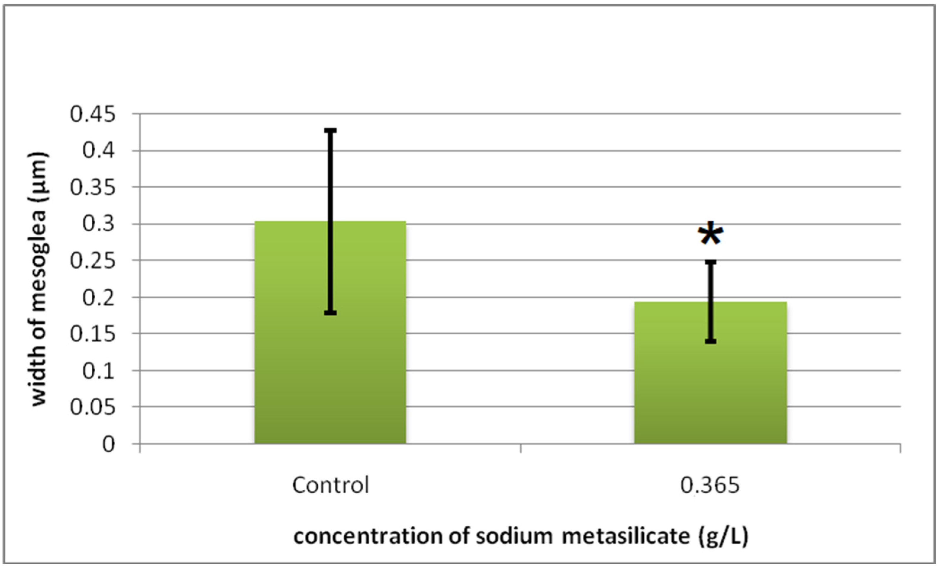
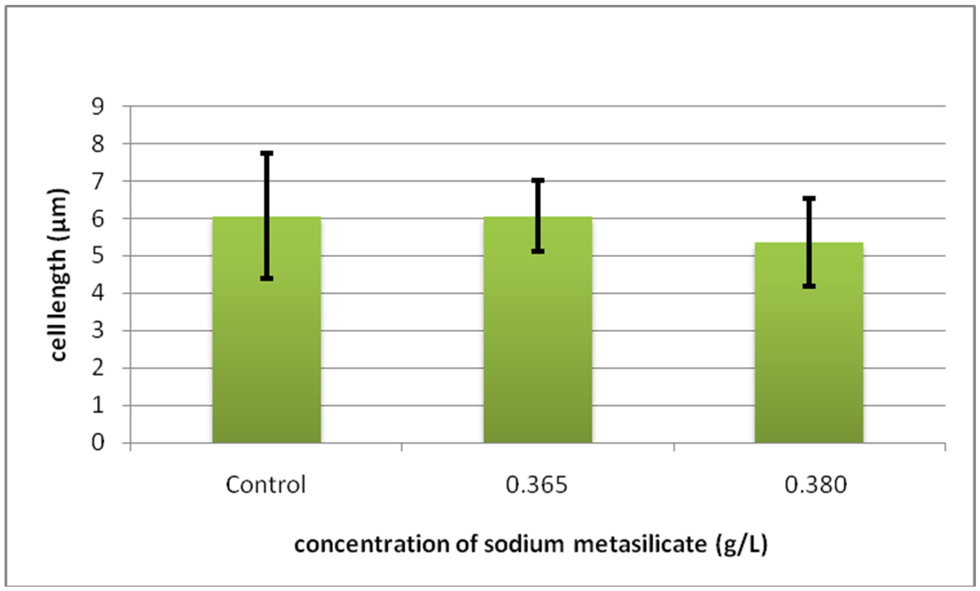
2.3. Histomorphometric and Cytometric Analysis

Histological analysis was conducted on individuals of green hydra treated with 0.365, 0.380, and 0.390 g/L SM collected on the second day of the experiment. Whole animals were fixed in Bouin’s solution (composition: 15 mL saturated water solution of picric acid, 5 mL 40% formalin, 1 mL glacial acetic acid) for 24 h. Afterwards, the animals were dehydrated through a graded ethanol series (70%, 80%, 96%, and 100%), exposed to chloroform, and embedded in Paraplast embedding media (Sherwood Medical, Norfolk, NE, USA). The Paraplast blocks were sectioned on a rotating microtome (Shandon Finesse 325, Thermo Fisher Scientific, Waltham, MA, USA) at 7 μm, and the slides were stained with hematoxylin–eosine (HE) and toluidine blue (TB) (dissolved in acetate buffer, pH = 5.6) and viewed through a Nikon Eclipse E600 light microscope equipped with an AxioCam ERc5s digital camera and ZEN 2 lite software (Carl Zeiss Microscopy GmbH, Jena, Germany) at 1000× magnification.
Histomorphometric measurements were conducted on permanent histological slides from the second day of the experiment using computer software for morphometric analysis, Ellise version 2081 (ViDiTo, 2009). The morphometric parameters included the length of the ectodermal myoepithelial cells and the length and width of the gastrodermal myoepithelial cells on the sample of 30 cells, and the thickness of the mesoglea was measured on 30 randomly chosen places, in each experimental and control group. For the morphometry of the endosymbiotic algae, hydras from all the groups were macerated, and the algae were directly measured in a fresh sample. Cytometric analysis was performed on the sample of 100 algal cells from each experimental and control group, where the diameter and area of the algal cells were measured.

2.4. Data Analysis

Statistical analysis was performed using STATISTICA 12.0 (StatSoft, Inc., Tulsa, OK, USA). The summary statistics were evaluated to express the value range (mean ± SD) and measure the variability within the datasets (the width of the mesoglea, the length of the myoepithelial ectodermal cells, the length of the gastrodermal myoepithelial cells, the width of the gastrodermal myoepithelial cells, the cell diameter of the endosymbiotic algae, and the cell area of the endosymbiotic algae). The normality of the data was tested using Shapiro–Wilk’s W test. The homogeneity of variance for each variable was tested using Levene’s test. The possible difference in the mean values of the selected variables between two groups was assessed using a t-test, while in the case of three or more groups, one-way ANOVA followed by a Newman-Keuls post hoc comparison test was employed. Prior to the tests, all the data were normalised by log transformation. The statistical significance was set to p < 0.05.

3. Results

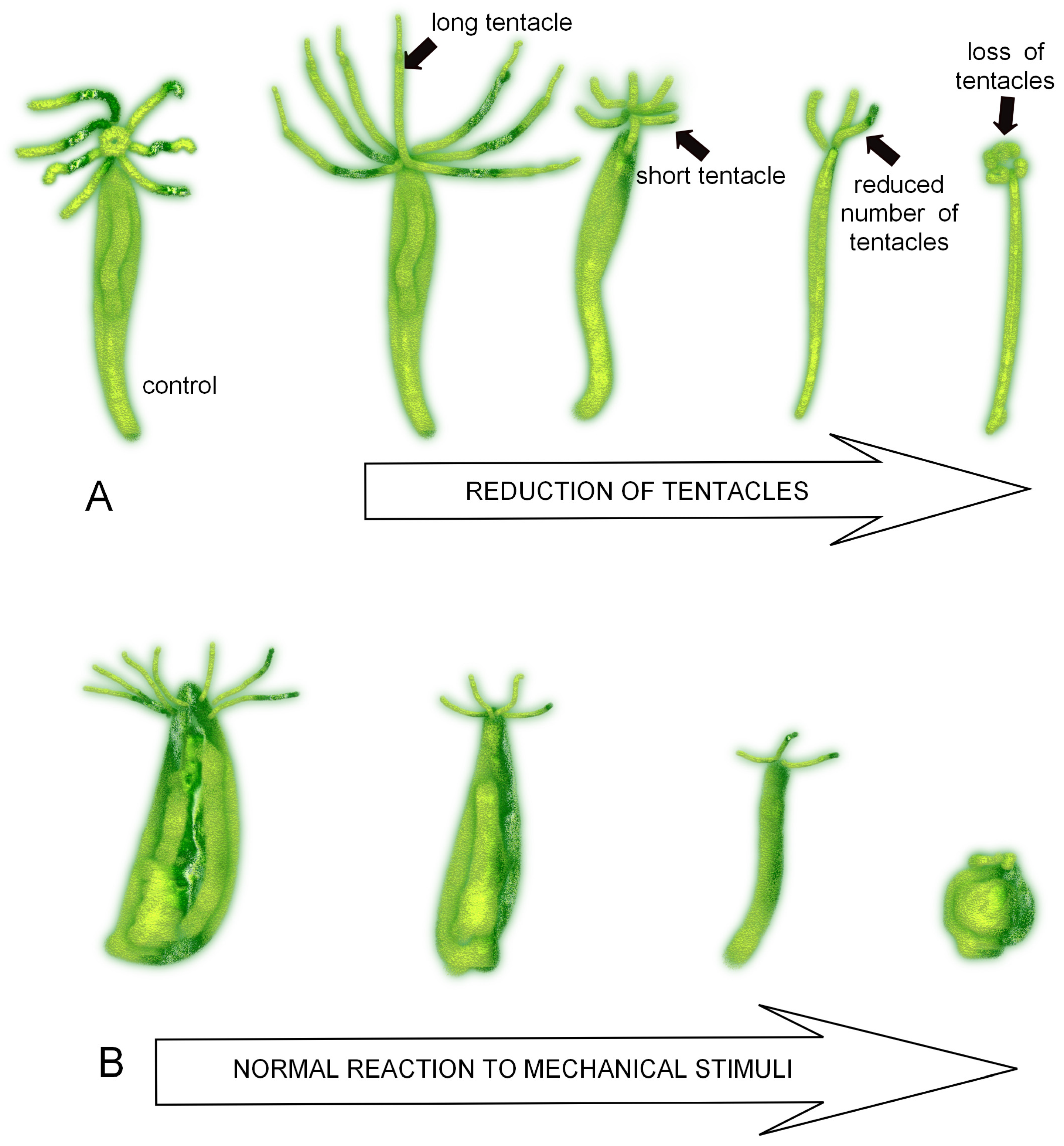
3.1. Morphological and Behavioural Changes in Green and Brown Hydra Caused by SM

A variable percentage of individuals of both hydra species (from 0 to 70% of green hydra and from 0 to 100% of brown hydra) showed morphological and behavioural changes over an exposure period of three days with increasing SM concentration (≥0.365 g/L). The morphological changes included damage to the tentacles (length, number, and loss of tentacles). The behavioural changes included measuring the percentage of the hydra’s reaction to mechanical stimuli (normal excitability—fast contractions/relaxations; lower excitability—slower contractions/relaxations; low excitability—slow contractions/relaxations; no excitability—very low contractions/relaxations or no reaction) (Figure 1A). Exposing the green hydra to a higher SM concentration (≥0.365 g/L) caused overall lower morphological and behavioural changes in comparison to the brown hydra (from 0 to 70% of green hydra and from 0 to 60% of brown hydra showed morphological changes, while behavioural changes were observed in 0 to 66% of green hydra and 0 to 100% of brown hydra). Shortening of the tentacles was observed on exposure at higher concentrations. A reduction in the number of tentacles increased in the brown hydra with increased SM concentration. A loss of tentacles was observed towards the end of the test period on the third day (Figure 1A). Decreasing excitability with increasing SM concentration was observed in the brown hydra (up to 100% of the individuals) (Figure 1B; detailed results are presented in Supplementary Materials, Tables S1 and S2).

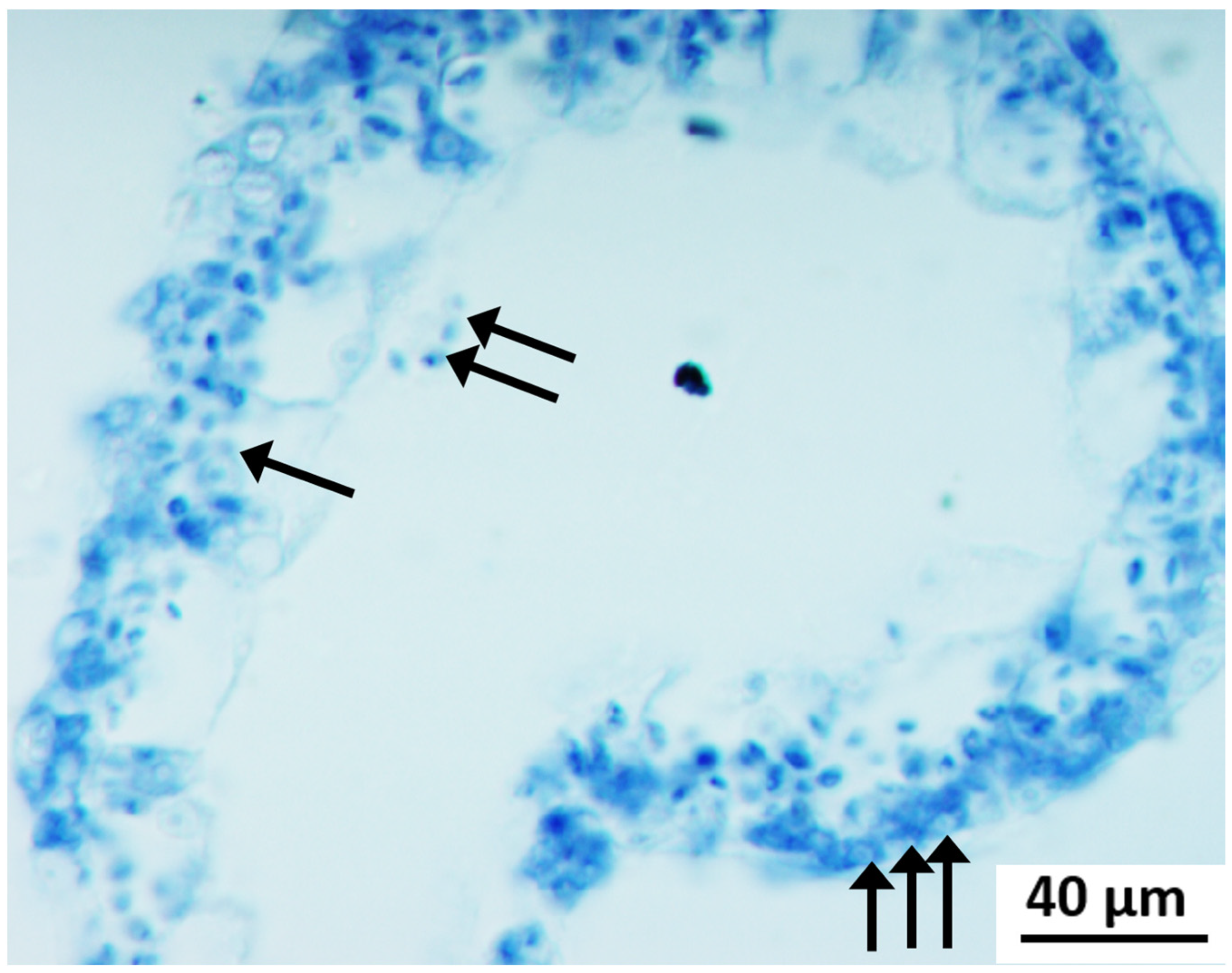
Cyto-Histological Changes in Green Hydra Caused by SM

Individuals from the control group showed no damage to the ectoderm and gastroderm with all the cell types present, and the mesoglea was uninterrupted (Figure 2A). Algae were regularly located in the column-like formations in the basal part of the gastrodermal myoepithelial cells (Figure 2B). The individuals of the green hydra treated with 0.365 g/L water solution of SM showed places of ectoderm interruptions. The mesoglea was visible and well-defined. It was wrinkled in the places of ectoderm damage. The gastroderm was not damaged (Figure 3A,B). The individuals of the green hydra treated with 0.380 g/L water solution of SM showed hardly visible cellular layers. The ectoderm was reduced, damaged, and interrupted. The mesoglea was not visible. The gastroderm was in a state of disintegration. The gastrodermal cells were hard to distinguish. Algae were dispersed around the gastrodermal cells. A relatively low number of algae were also present in the gastrovascular cavity (Figure 4). Due to severe damage, measuring the mesoglea’s width or the ectodermal myoepithelial cells’ length was impossible. The individuals of the green hydra treated with 0.390 g/L water solution of SM showed low resolution of the cellular layers. The mesoglea could be detected only in some places. The gastrovascular cavity was reduced, and the microalgae of the hydra’s microbiome were dispersed around the body. Morphometry could not be performed due to the high level of damage (Figure 5).
The mesoglea width ranged from 0.18 to 0.66 µm (0.30 ± 0.12 µm) in the control group and decreased significantly (p < 0.001) following treatment with 0.365 g/L of SM and ranged from 0.09 to 0.31 µm (0.19 ± 0.05 µm) (Figure 6). The length of the ectodermal myoepithelial cells in the control group ranged from 1.76 to 5.46 µm (3.62 ± 0.84 µm). The treatment with 0.365 g/L of SM resulted in a significant decrease in the values (p < 0.001), which ranged from 0.70 to 2.56 µm (1.47 ± 0.42 µm) (Figure 7). The length of the gastrodermal myoepithelial cells in the control group ranged from 4.41 to 11.45 µm (6.06 ± 1.68 µm). There was no change in the mean value following treatment with 0.365 g/L of SM (6.06 ± 0.94 µm) (Figure 8). A further increase in the SM concentration to 0.380 g/L resulted in a slight decrease in the values, which ranged from 3.92 to 7.00 µm (5.36 ± 1.16 µm). In the control samples, the width of the gastrodermal myoepithelial cells ranged from 1.89 to 5.29 µm (3.22 ± 1.03 µm). The treatment with 0.365 g/L of SM significantly increased the values (p < 0.001), which ranged from 3.26 to 9.69 µm (6.38 ± 1.82 µm). Furthermore, an increase in the concentration of SM to 0.380 g/L resulted in a slight decrease in the values compared to the previous concentration. However, those values were significantly higher compared to the control group (p < 0.001) and ranged from 3.17 to 8.46 µm (5.36 ± 1.33 µm) (Figure 9).

3.2. Cytological Changes in Endosymbiotic Algae

In the control group, the cell diameter ranged from 1.45 to 3.39 µm (2.44 ± 0.37 µm) (Figure 10). A slight decrease was observed following treatment with 0.050 g/L water solution of SM, and the values ranged from 1.63 to 3.00 µm (2.42 ± 0.31 µm). An increase in the concentration of SM to 0.365 g/L resulted in a slight increase in the values, which ranged from 1.89 to 3.44 µm (2.45 ± 0.33 µm). By increasing the concentration of SM to 0.380 g/L, the values decreased significantly compared to both the control (p < 0.001) and the groups treated with 0.05 g/L (p < 0.001), 0.365 g/L (p), and 0.390 g/L (<0.001). The values ranged from 1.50 to 3.04 µm (2.03 ± 0.33 µm). The highest treatment concentration (0.390 g/L) resulted in the highest cell diameter, which ranged from 1.72 to 3.48 µm (2.49 ± 0.32 µm).
The area of the algal cells in the control group ranged from 2.37 to 5.94 µm2 (4.17 ± 0.83 µm2) and the treatment with 0.050 g/L of SM resulted in a significant decrease in the mean value of the cell area (p < 0.001). The values ranged from 1.43 to 5.25 µm2 (3.21 ± 0.85 µm2). Following the treatment with 0.365 g/L of SM, the mean value slightly increased to 3.77 ± 0.75 µm2 (2.19 to 5.72 µm2). This mean value was still significantly lower than the control (p < 0.05) and significantly higher than the samples treated with 0.050 g/L (p < 0.001). A further increase of the SM to 0.380 g/L resulted in a significant decrease in the mean value compared to both the control (p < 0.001) and the groups treated with 0.050 g/L (p < 0.001), 0.365 g/L (p < 0.001), and 0.390 g/L (p < 0.001). The values ranged from 1.47 to 4.03 µm2 (2.46 ± 0.49 µm2). By increasing the concentration of SM to 0.390 g/L, the mean value increased compared to all the other treatment groups, with a mean value and standard deviation of 3.85 ± 0.84 µm2 (range 2.04 to 5.71 µm2). This mean value was still significantly lower compared to the control (p < 0.05) but significantly higher compared to the groups treated with 0.050 g/L (p < 0.001) and 0.380 g/L (p < 0.001) (Figure 11).

4. Discussion

This study presents the detrimental effects of SM on two different hydra species: frequently used simple freshwater model organisms that can absorb the tested chemical in the medium from both outside (through the ectoderm) and inside (through the gastroderm, which surrounds the gastrovascular cavity with a continuous flow of water). Thus, hydra is a simple and efficient in vivo test system that can demonstrate the toxicological effects at the organism and cellular levels. Researchers commonly use morphological markers when using hydra as an environmental assessment tool for ecotoxicology in freshwater. However, when histological biomarkers are added to complement the morphological ones, these new biomarkers may allow a more sensitive assessment of the impacts [54,55,56]. Previous studies have shown that SM can cause eye, skin, and respiratory system irritations in rats, dogs, and pigs [4].
The natural environment is affected by various environmental pollutants that cause unfavourable health changes and damage to aquatic and terrestrial ecosystems. Although releasing these chemicals into the environment raises concerns, the number of scientific studies that investigate the behaviour and effects of synthetic chemicals is not proportional to the rate of their development and production. While most of the research focuses on the effects of these chemicals on human health, more recent studies have explored the toxicological effects on other organisms and the ecosystem [57]. However, natural ecosystems are complex systems; therefore, these efforts are still insufficient to understand the long-term individual or collective impact these chemicals can have on the environment [58]. Severe morphological changes were observed in the present study following exposure to increasing sublethal concentrations of SM. Short-term exposure to the concentrations used in this experiment showed damaging effects at higher concentrations. Some insecticides can cause various morphological and cyto-histological changes in green hydras. Experiments concerning the effects of insecticides (Dimiline WP 25, Gamacide 20, and Primore WP 20) recorded toxicological effects, including changes in the body shape and length of the tentacles as well as damage to the tentacles and hypostome region [25]. Some compounds like Gamacide 20 can have strong effects and cause serious damage to whole regions of the hydra’s body [59]. When hydras were treated with antibiotic chloramphenicol, heavy lesions were recorded on the tentacles and were positively correlated with higher concentrations of the tested chemicals [22], similar to the reported SM effects in the present paper. Thus, higher concentrations caused a complete loss of the tentacles, while lower concentrations caused the shortening and loss of tentacles. Since tentacles are composed of a layer of thin myoepithelial cells, substances like SM can easily intrude into them and damage them [22]. Similar research conducted so far has shown differences in sensitivity between different species of hydra. Thus, green hydra seems to be more tolerant against some compounds, while brown hydra shows greater tolerance against others [34]. Additionally, some hydras are more sensitive to toxic chemicals because of their algicidal and anti-zoochlorellae activities when microalgae are present as endosymbiotic microbiome organisms. Hence, metal toxicity varies between symbiotic and aposymbiotic hydra species: symbiotic hydra species may tolerate a lower metal concentration (e.g., Cu), with the endosymbiotic algae being able to sequester Cu at lower concentrations, which reduces the degree of toxicity and effects on the polyps [34,60].
The contraction and relaxation of the body in hydras are well-known defence mechanisms in unfavourable environmental conditions [40]. The brown hydras exhibited greater changes in body contraction than the green hydras. The increased body relaxation of the control group of brown hydras may have been due to their greater susceptibility to high temperatures and/or oxygen deficiency inside the Petri dishes they were kept in [61]. If this was in fact the case, the relaxation of the body could be a way of increasing the oxygen diffusion inside the body. Increased body contraction occurred first at higher concentrations of SM. On the third day, it was evident in all the concentrations. The increased contraction could be a mechanism that hydra uses to prevent or reduce the entry of harmful substances into its body [19]. The reaction to mechanical stimuli was rapid in the green and brown hydra control groups. A small number of brown hydras in the control group showed a slower reaction, which could indicate that brown hydras are generally less adaptable to the environment’s changing conditions than green hydras [39,62]. With an increase in the concentration of SM, the hydras showed lower excitability. This effect was more substantial in the brown hydras than in the green hydras. This may confirm the hypothesis that the green hydra exhibits greater adaptability because of its endosymbionts and the overall notion of the importance of symbiotic relationships for evolution [62].
As the concentration of 0.050 g/L SM did not cause mortality, the cyto-histological analysis of the green hydra comprised the three highest concentrations of SM, and the changes observed in the green hydra included damage to the two cellular layers, the ectoderm and the gastroderm, and the mid-layer mesoglea. SM caused the thinning of the ectoderm and its breakage in some places. In experiments with the antibiotic chloramphenicol, similar changes were observed in both layers, with the addition of an increased amount of mucous surrounding the body, which represents a defence mechanism against toxicant entry into the body or a detoxification mechanism [22]. Histological preparations have shown that SM damages the ectoderm and mesoglea first. The collagen in hydra is located in the mesoglea, so it is possible that prolonged exposure to SM would induce changes in its composition. For further research, we would suggest using 0.050 and 0.365 g/L concentrations of SM to determine the effects of prolonged exposure on the mesoglea. With an increase in the concentration, damage also starts to appear in the gastroderm, causing the expansion of its influence on the algae. The first noticeable influence is a change in the algal distribution inside the gastrodermal cells. In the control group, the green hydra’s algae were orderly located in the basal part of the cells, near the mesoglea. At higher concentrations of SM, namely 0.380 g/L, the algae were scattered around the gastrodermal cells and also the gastrovascular cavity. A similar effect was noticed in experiments conducted with chloramphenicol and pirimicarb [22,26]. As the final effect, endosymbiotic algae were dispersed around the body at the highest concentrations of SM. SM treatment has a lower influence on the cell diameter than the cell area. Still, changes in the structure of the algal cells were not observed, which may be the reason for the greater tolerance exhibited by the green hydra compared to the brown hydra.
The results of the present study show that the use of hydra as a key model organism in toxicity testing, despite numerous limiting factors and difficulties [63], represents a possible fine key step in future protocols for testing the toxicity of new chemicals in industry, as it provides fast and progressive results. As an aquatic invertebrate with a rapid exchange of generations in a short period, a multicellular (and endosymbiotic and holobiotic) organism with a simple body plan and numerous easily recognised cell structures [39,64], and the great role played by its stem cells in regeneration [19], we confirmed hydra as an excellent model organism. We propose hydra for the inexpensive and rapid in vivo and in vitro monitoring of the impact of devastating environmental pollutants. On one hand, its extreme sensitivity to minute concentrations of various types of toxic substances (from heavy metals, pharmaceuticals, and nanomaterials to microplastics) [34,53,60,65], and, on the other hand, its high survival rate even when interstitial stem cells are eliminated [66,67], makes hydra an efficient multifunctional biosensor for numerous aquatic ecosystems.

5. Conclusions

The wide application of SM in the cosmetic industry and in the production of detergents and the particular sensitivity of aquatic ecosystems make it essential to know its effects on freshwater communities of organisms. In this quest, two species of the genus Hydra represent a good and popular model organism, considering their easy-to-observe morphological and behavioural changes and their worldwide presence.
In the experiment with sublethal concentrations of SM, its toxic effect on both green and brown hydra was observed. The effects on the brown hydra were stronger than on the green hydra. An endosymbiotic relationship may be the reason for the greater tolerance of the green hydra. It could be concluded that natural populations of green hydras would be more resistant and adaptable to exposure to this toxicant than natural populations of brown hydras.
The higher the concentrations, the more considerable changes were observed, which means that exposure to higher concentrations in the natural environment would possibly cause a reduction in the population density. Conducting similar, longer-lasting experiments on hydras would be useful for following the recovery process in detail.
The hydra has proven to be a significant alternative organism for preliminary toxicity screening, avoiding the use of sensitive and rare vertebrate species. In seeking to minimize the unnecessary use of animals in experiments by finding ideal alternative methodologies and protocols for toxicity testing, the hydra has proven to be a very powerful model organism for toxicity testing at the organism, cell, and microbiome levels. We are living in an era of rapid global economic growth and there is an increasing accumulation of toxic chemicals in aquatic environments; therefore, it is essential to connect new ecotoxicological records and methodological principles in the biomonitoring of SM as a dangerous toxic chemical contaminant. Reducing hazardous chemicals, such as SM, and understanding their harmful effects on aquatic organisms and their microbiomes should be a priority worldwide for human and environmental risk assessment.

Supplementary Materials

The following supporting information can be downloaded at https://www.mdpi.com/article/10.3390/w15244228/s1, Figure S1. Habitus of green hydra in control condition; Figure S2. Habitus of brown hydra in control condition; Table S1: Morphological changes of tentacles in green hydra (% of analysed individuals) during the treatment with sodium metasilicate (SM); Table S2: Morphological changes of tentacles in brown hydra (% of analysed individuals) during the treatment with sodium metasilicate (SM); Table S3: Response to mechanical stimuli in green hydra (% of analysed individuals) during the treatment with sodium metasilicate (SM); Table S4: Response to mechanical stimuli in brown hydra (% of analysed individuals) during the treatment with sodium metasilicate (SM); Table S5: Mortality of green and brown hydra.

Author Contributions

Conceptualization, G.K. and S.G.; methodology, G.K. and S.G.; software, S.G.; validation, G.K., R.G. and S.G.; formal analysis, G.K. and S.G.; investigation, G.K. and S.G.; resources, G.K., S.G. and R.G.; data curation, G.K., S.G. and R.G.; writing, G.K., S.G. and R.G.; writing—review and editing, G.K., R.G. and S.G.; visualization, G.K., S.G. and R.G. All authors have read and agreed to the published version of the manuscript.

Funding

This research received no external funding.

Data Availability Statement

Data are contained within the article.

Acknowledgments

We wish to thank Ana Šimičev, mag. biol. exp. for her help with the experimental work.

Conflicts of Interest

The authors declare no conflict of interest.

References

  1. Naidu, R.; Biswas, B.; Willett, I.R.; Cribb, J.; Singh, B.K.; Nathanail, C.P.; Coulon, F.; Semple, K.T.; Jones, K.C.; Barclay, A.; et al. Chemical pollution: A growing peril and potential catastrophic risk to humanity. Environ. Int. 2021, 156, 106616. [Google Scholar] [CrossRef] [PubMed]
  2. Wharfe, J. Historical perspective and overview. In Environmental Toxicity Testing; Thompson, K.C., Wadhia, K., Loibner, A.P., Eds.; Blackwell Publishers Ltd.: Oxford, UK, 2005. [Google Scholar]
  3. Fawer, M.; Concannon, M.; Rieber, W. Life cycle inventories for production of sodium silicates. Int. J. Life Cycle Assess. 1999, 4, 207–212. [Google Scholar] [CrossRef]
  4. Elmore, A.R. Final Report on Safety Assessment of Potassium Silicate, Sodium Metasilicate, and Sodium Silicate. Int. J. Toxicol. 2005, 24, 103–117. [Google Scholar] [PubMed]
  5. Wills, J.H. A short history of the manufacture of soluble silicates in the United States. ACS Symp. Ser. 1982, 194, 3–15. [Google Scholar]
  6. HERA. Soluble Silicates (CAS No.: 1344-09-8, 6834-92-0, 10213-79-3, 13517-24-3, 1312-76-1): Human & Environmental Risk Assessment on Ingredients of European Household Cleaning Products; Draft for Public Comment; HERA Substance Team: Wendelsheim, Germany, 2005; pp. 1–64. [Google Scholar]
  7. Baehr, C.H.; Koehl, W. Soluble Silicates–Highly Versatile and Safe. SÖFW-J.–Int. J. Appl. Sci. 2007, 133, 88–94. [Google Scholar]
  8. EC 648/2004. Regulation (EC) No 648/2004 of the European Parliament and of the Council of 31 March 2004 on detergents. Off. J. L 2004, 104, 1–35. Available online: https://eur-lex.europa.eu/legal-content/EN/ALL/?uri=CELEX%3A32004R0648 (accessed on 17 May 2020).
  9. EC 907/2006. Commission Regulation (EC) No 907/2006 of 20 June 2006 amending Regulation (EC) No 648/2004 of the European Parliament and of the 10. Council on detergents, in order to adapt Annexes III and VII thereto. Off. J. L 2006, 168, 5–10. Available online: https://eur-lex.europa.eu/legal-content/EN/ALL/?uri=CELEX%3A32006R0907 (accessed on 17 May 2020).
  10. EC 1272/2008. Regulation (EC) No1272/2008 of the European Parliament and of the Council of 16 December 2008 on Classification, Labelling and Packaging of Substances and Mixtures, Amending and Repealing Directives 67/548/EEC and 1999/45/EC, and Amending Regulation (EC) No 1907/2006. Off. J. L 2008, 353, 1–1355. Available online: https://eur-lex.europa.eu/legal-content/EN/TXT/?uri=celex%3A32008R1272 (accessed on 17 May 2020).
  11. EC 1336/2008. Regulation (EC) No 1336/2008 of the European Parliament and of the Council of 16 December 2008 Amending Regulation (EC) No 648/2004 in order to adapt it to Regulation (EC) No 1272/2008 on Classification, Labelling and Packaging of Substances and Mixtures. Off.J. L 2008, 354, 60–61. Available online: https://eur-lex.europa.eu/legal-content/EN/TXT/?uri=uriserv%3AOJ.L_.2008.354.01.0060.01.ENG (accessed on 17 May 2020).
  12. Haneke, K.E. Sodium Metasilicate, Anhydrous [6834-92-0], sodium metasilicate pentahydrate [10213-79-3], and sodium metasilicate nonahydrate [13517-24-3]. In Review of toxicological literature; Prepared by Integrated Laboratory Systems, Inc., for National Institute of Environmental Health Sciences: Research Triangle Park, NC, USA, 2002. [Google Scholar]
  13. Richterich, K.; Mühlberg, B. Silicic Acid, Potassium Salt. Daphnia Magna, Acute Toxicity; Final Report, R-0100925; Henkel KGaA: Dusseldorf, Germany, 2001. [Google Scholar]
  14. Richterich, K.; Mühlberg, B. Silicic Acid, Potassium Salt. Fish, Acute Toxicity; Final Report, R-0100924; Henkel KGaA: Dusseldorf, Germany, 2001. [Google Scholar]
  15. Richterich, K.; Mühlberg, B. Silicic Acid (H2SiO3), Disodium Salt. Acute Bacterial Toxicity (Pseudomonas Oxygen Consumption Inhibition Test, DIN 38412-27); Final Report, R-0100923; Henkel KGaA: Dusseldorf, Germany, 2001. [Google Scholar]
  16. Richterich, K.; Mühlberg, B. Silicic Acid (H2SiO3), Disodium Salt. Fish, Acute Toxicity; Final Report, R-0100922; Henkel KGaA: Dusseldorf, Germany, 2001. [Google Scholar]
  17. Wallen, I.E.; Greer, W.C.; Lasater, R. Toxicity to “Gambusia Affinis” of Certain Pure Chemicals in Turbid Waters. Sew. Ind. Wastes 1957, 29, 695–711. [Google Scholar]
  18. Holt, E.A.; Miller, S.W. Bioindicators: Using organisms to measure environmental impacts. Nat. Educ. Knowl. 2011, 2, 1–10. [Google Scholar]
  19. Kalafatić, M. Regeneration and asexual reproduction of Hydra oligactis treated with different pesticides. Biologia 1997, 52, 475–480. [Google Scholar]
  20. Kalafatić, M.; Kopjar, N. Response of Green Hydra to the Treatment with Different Pesticides under Laboratory Conditions. Zeitsch. Angewan. Zool. 1994, 2, 213–223. [Google Scholar]
  21. Beach, M.J.; Pascoe, D. The role of Hydra vulgaris (Pallas) in assessing the toxicity of freshwater pollutants. Wat. Res. 1998, 32, 101–106. [Google Scholar] [CrossRef]
  22. Kovačević, G.; Kalafatić, M.; Ljubešić, N.; Šunjić, H. The effect of chloramphenicol on the symbiosis between alga and hydra. Biologia 2021, 56, 605–610. [Google Scholar]
  23. Arkhipchuk, W.; Blaise, C.; Malinovskaya, M.V. Use of hydra for chronic toxicity assessment of waters intended for human consumption. Environ. Pollut. 2005, 142, 200–211. [Google Scholar] [CrossRef] [PubMed]
  24. Kalafatić, M.; Žnidarić, D.; Lui, A.; Wrischer, M. Effects of insecticides (Dimiline WP 25, Torak EC 24 and Gamacide 20) on hydra (Hydra vulgaris Pallas). Int. J. Dev. Biol. 1991, 35, 335–340. [Google Scholar] [PubMed]
  25. Kalafatić, M.; Žnidarić, D.; Kopjar, N. The effect of insecticides upon green hydra. Period. Biol. 1994, 1, 129–130. [Google Scholar]
  26. Kalafatić, M.; Kopjar, N. Response of green hydra to pirimicarb. Biologia 1995, 50, 289–292. [Google Scholar]
  27. Kessler, E.; Kauer, G.; Rahat, M. Excretion of Sugars by Chlorella Species Capable and Incapable of Symbiosis with Hydra viridis. Bot. Acta. 1991, 104, 58–63. [Google Scholar] [CrossRef]
  28. Müller-Parker, G.; Pardy, R.L. Response of green hydra to feeding and starvation at four irradiances. Biol. Bull. 1987, 172, 46–60. [Google Scholar] [CrossRef]
  29. Žnidarić, D.; Kalafatić, M.; Kopjar, N. The survival of Hydra oligactis Pallas in unpleasant conditions. Zeitsch Angewan Zool. 1995, 2, 157–163. [Google Scholar]
  30. Burnett, A.L. Biology of Hydra; Academic Press: New York, NY, USA; London, UK, 1973. [Google Scholar]
  31. Habetha, M.; Anton-Erksleben, F.; Neumann, K.; Bosch, T.C.G. The Hydra viridis/Chlorella symbiosis. Growth and sexual differentiation in polyps without symbionts. Zoology 2003, 106, 101–108. [Google Scholar] [CrossRef] [PubMed]
  32. Fradkin, M.; Kakis, H.; Campbell, R. Effects of irradiation on hydra. Elimination of interstitial cells from viable hydra. Radiat. Res. 1978, 76, 187–197. [Google Scholar] [CrossRef] [PubMed]
  33. Quinn, B.; Gagne, F.; Blaise, C. Hydra, a model system for environmental studies. Int. J. Dev. Biol. 2012, 56, 613–625. [Google Scholar] [CrossRef] [PubMed]
  34. Karntanut, W.; Pascoe, D. The toxicity of copper, cadmium and zinc to four different Hydra (Cnidaria: Hydrozoa). Chemosphere 2002, 47, 1059–1064. [Google Scholar] [CrossRef] [PubMed]
  35. Bosch, T.C.G. What Hydra Has to Say About the Role and Origin of Symbiotic Interactions. Biol. Bull. 2012, 223, 78–84. [Google Scholar] [CrossRef] [PubMed]
  36. Bell, G.; Wolfe, L.M. Sexual and asexual reproduction in a natural population of Hydra pseudoligactis. Can. J. Zool. 1984, 63, 851–856. [Google Scholar] [CrossRef]
  37. Campbell, R.D. Transmission of symbiotic algae through sexual reproduction in Hydra: Movement of algae into the oocyte. J. Tissue Cell 1990, 22, 137–147. [Google Scholar] [CrossRef] [PubMed]
  38. Bhat, S. Ecotoxicology and impact on biodiversity. J. Pharmacogn. Phytochem. 2013, 2, 1–19. [Google Scholar]
  39. Slobodkin, L.B. The connection between single species and ecosystems. In Water Quality & Stress Indicators in Marine and Freshwater Systems: Linking Levels of Organization; Sutcliffe, D.W., Ed.; Freshwater Biological Association, FBA Special Publications: Cumbria, UK, 1994; Volume 4, pp. 75–87. [Google Scholar]
  40. Kovačević, G.; Gregorović, G.; Matijević, A.; Kalafatić, M. Toxic effects of iron on green and brown hydra. Curr. Sci. 2015, 110, 502–504. [Google Scholar]
  41. Klimovich, A.V.; Bosch, T.C.G. Rethinking the Role of the Nervous System: Lessons from the Hydra Holobiont. Bioessays 2018, 40, e1800060. [Google Scholar] [CrossRef] [PubMed]
  42. McFall-Ngai, M.; Bosch, T.C.G. Animal development in the microbial world: The power of experimental model systems. Curr. Top. Dev. Biol. 2021, 141, 371–397. [Google Scholar] [PubMed]
  43. Lousada, M.B.; Lachnit, T.; Edelkamp, J.; Paus, R.; Bosch, T.C.G. Hydra and the Hair Follicle—An Unconventional Comparative Biology Approach to Exploring the Human Holobiont. BioEssays 2022, 44, e2100233. [Google Scholar] [CrossRef] [PubMed]
  44. Franzenburg, S.; Fraune, S.; Altrock, P.M.; Künzel, S.; Baines, J.F.; Traulsen, A.; Bosch, T.C.G. Bacterial colonization of Hydra hatchlings follows a robust temporal pattern. ISME J. 2013, 7, 781–790. [Google Scholar] [CrossRef] [PubMed]
  45. Brucker, R.M.; Bordenstein, S.R. The hologenomic basis of speciation: Gut bacteria cause hybrid lethality in the genus Nasonia. Science 2013, 341, 667–669. [Google Scholar] [CrossRef] [PubMed]
  46. Fraune, S.; Bosch, T.C.G. Long-term maintenance of species-specific bacterial microbiota in the basal metazoan Hydra. Proc. Natl. Acad. Sci. USA 2007, 104, 13146–13151. [Google Scholar] [CrossRef] [PubMed]
  47. Bathia, J.; Schröder, K.; Fraune, S.; Lachnit, T.; Rosenstiel, P.; Bosch, T.C.G. Symbiotic algae of Hydra viridissima play a key role in maintaining homeostatic bacteria colonization. Front. Microbiol. 2022, 13, 869666. [Google Scholar] [CrossRef]
  48. Bosch, T.C.G. Beyond Lynn Margulis’ green hydra. Symbiosis 2022, 87, 11–17. [Google Scholar] [CrossRef]
  49. Fraune, S.; Anton-Erxleben, F.; Augustin, R.; Franzenburg, S.; Knop, M.; Schröder, K.; Willoweit-Ohl, D.; Bosch, T.C.G. Bacteria–bacteria interactions within the microbiota of the ancestral metazoan Hydra contribute to fungal resistance. ISME J. 2015, 9, 1543–1556. [Google Scholar] [CrossRef]
  50. Murillo-Rincon, A.P.; Klimovich, A.; Pemöller, E.; Taubenheim, J.; Mortzfeld, B.; Augustin, R.; Bosch, T.C.G. Spontaneous body contractions are modulated by the microbiome of Hydra. Sci. Rep. 2017, 7, 15937. [Google Scholar] [CrossRef] [PubMed]
  51. Taubenheim, J.; Willoweit-Ohl, D.; Knop, M.; Franzenburg, S.; He, J.; Bosch, T.C.; Fraune, S. Bacteria- and temperature-regulated peptides modulate β-catenin signaling in Hydra. Proc. Natl. Acad. Sci. USA 2020, 117, 35, 21459–21468. [Google Scholar] [CrossRef] [PubMed]
  52. Glauber, K.M.; Dana, C.E.; Steele, R.E. Hydra. Curr. Biol. 2010, 22, R964–R965. [Google Scholar] [CrossRef] [PubMed]
  53. Kovačević, G.; Želježić, D.; Horvatin, K.; Kalafatić, M. Morphological features and comet assay of green and brown hydra treated with aluminium. Symbiosis 2007, 44, 145–152. [Google Scholar]
  54. Ambrosone, A.; Mattera, L.; Marchesano, V.; Quarta, A.; Susha, A.S.; Tino, A.; Rogach, A.L.; Tortiglione, C. Mechanisms underlying toxicity induced by CdTe quantum dots determined in an invertebrate model organism. Biomaterials 2012, 33, 1991–2000. [Google Scholar] [CrossRef] [PubMed]
  55. Murugadas, A.; Zeeshan, M.; Thamaraiselvi, K.; Ghaskadbi, S.; Akbarsha, M.A. Hydra as a model organism to decipher the toxic effects of copper oxide nanorod: Eco-toxicogenomics approach. Sci. Rep. 2016, 6, 29663. [Google Scholar] [CrossRef] [PubMed]
  56. Cera, A.; Cesarini, G.; Spani, F.; Scalici, M. Hydra vulgaris assay as environmental assessment tool for ecotoxicology in freshwaters: A review. Mar. Freshw. Res. 2020, 72, 745–753. [Google Scholar] [CrossRef]
  57. Boxall, A.B.A. The environmental side effects of medication. EMBO Rep. 2004, 5, 1110–1116. [Google Scholar] [CrossRef]
  58. Bernhardt, E.S.; Rosi, E.J.; Gessner, M.O. Synthetic chemicals as agents of global change. Front. Ecol. Environ. 2017, 15, 84–90. [Google Scholar] [CrossRef]
  59. Kopjar, N.; Kalafatić, M.; Žnidarić, D. Combined effect of Gamacide 20 and UV irradiation on green hydra (Hydra viridissima Pallas). Biologia 1994, 49, 371–375. [Google Scholar]
  60. Vimalkumar, K.; Sangeetha, S.; Felix, L.; Kay, P.; Pugazhendhi, A. A systematic review on toxicity assessment of persistent emerging pollutants (EPs) and associated microplastics (MPs) in the environment using the Hydra animal model. Comp. Biochem. Physiol. Part C Toxicol. Pharmacol. 2022, 256, 109320. [Google Scholar] [CrossRef]
  61. Kovačević, G.; Matulić, A. Effects of Querzetin on the Green Hydra (Hydra viridissima Pallas). Int. J. Biol. 2013, 5, 57–63. [Google Scholar] [CrossRef]
  62. Kovačević, G.; Kalafatić, M.; Ljubešić, N. Effects of Norflurazon on Green and Brown Hydra. Folia Biol. 2009, 57, 91–96. [Google Scholar] [CrossRef]
  63. Patwardhan, V.; Ghaskadbi, S. Invertebrate alternatives for toxicity testing: Hydra stakes its claim. Altex Proc. 2013, 2, 69–76. [Google Scholar]
  64. Bosch, T.C.; Miller, D.J. The Hydra Holobiont: A Tale of Several Symbiotic Lineages. In The Holobiont Imperative: Perspectives from Early Emerging Animals; Bosch, T.C., Miller, D.J., Eds.; Springer: Vienna, Austria, 2016; pp. 79–97. [Google Scholar]
  65. Tortiglione, C. An ancient model organism to test in vivo novel functional nanocrystals. In Biomedical Engineering—From Theory to Applications; Fazel, R., Ed.; InTechOpen: Rijeka, Croatia, 2011; pp. 225–252. [Google Scholar]
  66. Bode, H.R. The interstitial cell lineage of hydra: A stem cell system that arose early in the evolution. J. Cell Sci. 1996, 109, 1155–1164. [Google Scholar] [CrossRef]
  67. Buzgariu, W.; Haddad, H.A.; Tomczyk, S.; Wenger, Y.; Galliot, B. Multi-functionality and plasticity characterise epithelial cells in Hydra. Tissue Barriers 2015, 3, e1068908. [Google Scholar] [CrossRef] [PubMed]
Figure 1. Scheme of quantitative and repeatable identification of morphological and behavioural changes in hydra in the experiment: (A) Reduction of hydra’s tentacles: long tentacles—prolongation of tentacles; short tentacles—shortening of tentacles; reduced number of tentacles—at least one tentacle is missing; loss of tentacles—reduction to the very base or no tentacles. Detectable within 24 h and on. (B) Hydra’s reaction to mechanical stimuli: normal excitability—immediate fast contractions/relaxations of the body; lower excitability—slower contractions/relaxations compared to control, within 4 s; low excitability—slow contractions/relaxations, mostly not reaching the extent of contraction compared to control, within 5 s; no excitability—very low and slow partial contractions/relaxations, hardly noticeable, or no reaction at all.
Water 15 04228 g001
Figure 2. Regular cellular layers (ectoderm and gastroderm) and mesoglea in the control group of green hydra on the second day of the experiment. (A) Three arrows—ectoderm, four arrows—gastroderm. (B) One arrow—mesoglea, two arrows—regular distribution of endosymbiotic algae inside gastrodermal myoepithelial cells. Hematoxylin–eosine (HE).
Water 15 04228 g002
Figure 3. (A) Cellular layers (ectoderm and gastroderm). (B) Mesoglea in green hydra treated with 0.365 g/L SM, the second day of the experiment. One arrow—damage of ectoderm, two arrows—low number of algae inside gastrodermal myoepithelial cells, and three arrows—continuous mesoglea (HE).
Water 15 04228 g003
Figure 4. Cellular layers (ectoderm and gastroderm) and mesoglea in green hydra treated with 0.380 g/L SM, the second day of the experiment. One arrow—endosymbiotic algae dispersed in gastrodermal myoepithelial cells, two arrows—endosymbiotic algae inside the gastrovascular cavity, and three arrows—reduced ectoderm (TB).
Water 15 04228 g004
Figure 5. Green hydra treated with 0.390 g/L SM, the second day of the experiment. Cellular layers (ectoderm and gastroderm) and mesoglea are reduced and hardly distinguishable (HE).
Water 15 04228 g005
Figure 6. Width of mesoglea (µm) (mean ± SD) in untreated (control group) green hydra and green hydra treated with 0.365 g/L water solution of SM, the second day of the experiment (* statistically significant difference, p < 0.05).
Water 15 04228 g006
Figure 7. Length of myoepithelial ectodermal cells (µm) (mean ± SD) in untreated (control group) green hydra and green hydra treated with 0.365 g/L water solution of SM, the second day of the experiment (* statistically significant difference, p < 0.05).
Water 15 04228 g007
Figure 8. Length of gastrodermal myoepithelial cells (µm) (mean ± SD) in untreated (control group) green hydra and green hydras treated with 0.365 and 0.380 g/L water solution of SM, second day of the experiment.
Water 15 04228 g008
Figure 9. Width of gastrodermal myoepithelial cells (µm) (mean ± SD) in untreated (control group) green hydra and green hydras treated with 0.365 and 0.380 g/L water solution of SM, second day of the experiment (* statistically significant difference between concentrations 0.365/0.380 g/L and control, p < 0.05).
Water 15 04228 g009
Figure 10. Cell diameter (µm) (mean ± SD) of endosymbiotic algae in green hydra, untreated (control) group, and groups treated with different concentrations of water solutions of SM, the second day of the experiment (* statistically significant difference between concentrations 0.380 g/L and all the other groups, p < 0.05).
Water 15 04228 g010
Figure 11. Cell area (µm2) (mean ± SD) of endosymbiotic algae in green hydra, untreated (control) group, and groups treated with different concentrations of water solutions of SM, second day of the experiment (* statistically significant difference between concentrations 0.050/0.365 g/L and a control; between concentration 0.380 g/L and all the other groups; between concentration 0.390 g/L, concentrations 0.050/0.380 g/L, and control, p < 0.05).
Water 15 04228 g011
Disclaimer/Publisher’s Note: The statements, opinions and data contained in all publications are solely those of the individual author(s) and contributor(s) and not of MDPI and/or the editor(s). MDPI and/or the editor(s) disclaim responsibility for any injury to people or property resulting from any ideas, methods, instructions or products referred to in the content.

