Abstract
To enhance the removal of COD (Chemical Oxygen Demand) by microalgae, this study constructed a novel microalgae–microalgae microbial fuel cell system (AA-MFC). It investigated the coupling relationship between the COD treatment efficiency at the anode and the production of high-value microalgal products at the cathode, as well as explored the effects of different initial inoculum densities and light–dark cycles. The experiment first measured the operational performance of the newly constructed AA-MFC in open-circuit and closed-circuit modes, demonstrating that this novel AA-MFC could start up rapidly within 32 h and operate stably. The results showed that the AA-MFC enhanced the removal of COD and the growth of microalgae biomass at the anode while maintaining stable power generation. When the initial inoculation density of the anode was 1.2 × 10⁸ cell/cm2 and the light–dark cycle time was 18:6 h, the AA-MFC had the most obvious promoting effect on the COD removal of the anode. Compared with normal culture conditions, the COD removal rate increased by 26.0% to 96.1%. These results indicate that the AA-MFC can not only effectively remove pollutants, but also promote the accumulation of high-value microalgae biomass.
1. Introduction
The microbial fuel cell (MFC) is an innovative wastewater treatment technology that utilizes the metabolic activity of microorganisms to convert chemical energy in wastewater into electrical energy. This technology has the potential to provide clean electricity while treating wastewater and has important prospects for development. Jiang et al. [1] used an MFC to treat organic matter in sludge, and the results showed that the removal rate of the total Chemical Oxygen Demand (COD) in sludge under a closed-loop condition was 8% higher than that under open-circuit control. Numerous studies have shown that MFCs can enhance the treatment of pollutants such as petroleum hydrocarbons and phenols [2].
In a conventional MFCS, microorganisms that are electrochemically active at the anode thrive under anaerobic conditions, where they electrochemically oxidize organic pollutants, producing electrons and protons. These electrons and protons are then transferred to the cathode through an external conductor and a proton exchange membrane. In the cathode, they perform an aerobic reaction with the electron acceptor, O2, completing the closed loop [3].
In contrast to the electrochemically active anaerobic microorganisms documented in the literature [4], some microalgae are electrochemically active aerobic microorganisms. Microbial fuel cells usually undergo two processes of inoculation and start-up before achieving stable operation. The start-up cycle of microbial fuel cells is often relatively long, generally taking about 1~2 months. The start-up speed of microbial fuel cells has an important influence on their practical applications [5].
In addition, in conventional microbial fuel cells, the air used as an oxygen source for the cathode permeates through the proton exchange membrane to the anode, potentially compromising the power generation performance of the anode’s anaerobic microorganisms. This may be detrimental to the actual operation and application of the reactor [6]. Microbial fuel cells are often difficult to scale up due to their inoperability and slow start-up, which is why most microbial fuel cell research remains stuck at the laboratory scale [7].
Microalgae are a kind of aerobic microorganism with electrochemical activity. Microalgae such as Chlorella and Spirulina have been shown to be electrochemically active [8]. In addition, microalgae have been widely utilized in various types of wastewater treatment and have the potential to produce economically valuable substances, such as lipids and proteins, that can transform wastewater into valuable resources [8]. Although microalgae can effectively remove nitrogen and phosphorus from wastewater, their COD removal performance is often less than ideal [9]. Using electrochemically active microalgae to construct MFCs for wastewater treatment can enhance COD removal by microalgae and reduce the complexity of reactor operation [10].
While the application of MFCs in wastewater treatment has seen rapid development due to widespread attention from researchers, there have been relatively few reports on the combination of microalgae-based water treatment technology with MFCs. Subhash et al. [11] constructed a single-chamber microalgal fuel cell, which achieved a COD removal rate of 72.6% after seven days of operation. The maximum power density generated with an external load of 200 Ω was 3.55 μW/m2, but the experiment experienced voltage reversal issues. This issue arose because oxygen, an electron acceptor that significantly reduces current, and the oxygen produced by anode algae photosynthesis can harm the MFC. Xu et al. [12] introduced sodium bisulfite as a deoxygenating agent in the anodic microalgal MFC they constructed, resulting in a significant increase in MFC current. However, as sodium bisulfite rapidly oxidizes, its deoxygenating effect is limited and unsustainable in the MFC system, causing stagnation in the research of microalgal anodic MFCs.
In another category of wastewater treatment research involving microalgae-based cathodic MFCs, microalgae are used to produce oxygen at the cathode as an electron acceptor, replacing mechanical aeration. Kakarla et al. [13] constructed a microalgae cathode microbial fuel cell using Chlamydomonas reinhardtii, and found that the use of algal aeration could increase the concentration of dissolved oxygen in the cathode chamber from 0 to 15.7 mg/L, indicating that microalgae could continuously supply oxygen in the cathode, replacing energy-intensive mechanical aeration. At the same time, microalgae harvested from the cathode can be used for biofuel production, thereby reducing operating costs.
Using microalgae as an electron donor and electron acceptor, a novel algae–algae-type microbial fuel cell (AA-MFC) with fast start-up and stable power generation was constructed for the first time in this study, which improved the COD removal efficiency while harvesting high-value microalgal biomass. We also investigated the effects of different initial inoculum densities and different light–dark cycles on AA-MFC wastewater treatment and microalgal biomass.
2. Materials and Methods
2.1. Algal Cultivation
By reviewing the relevant literature [14,15] on microalgae wastewater treatment and microalgae by-product production, we selected five strains of microalgae with high oil content commonly used in wastewater treatment for experiments. These selected microalgae strains include the following: Parachlorella kessleri (P. kessleri), Spirulina platensis (FACHB-439), Chlorella sp. (FACHB-9), Tetradesmus obliquus (FACHB-416), and Desmodesmus armatus (A1). P. kessleri and Desmodesmus armatus were cultured in our laboratory, and other microalgae strains were obtained from the Chinese Academy of Sciences Institute of Hydrobiology.
The preservation and cultivation of these microalgal strains were carried out as follows.
Preparation of Culture Media
For P. kessleri, Chlorella sp., Tetradesmus obliquus, and Desmodesmus armatus, BG11 medium was used. For Spirulina platensis, Zarrouk medium was employed. All culture media were sterilized at 121 °C for 20 min. The microalgae strains were cultured at 25 °C, under a light intensity of 80 μmol·m−2·s−1, with a 12 h light and 12 h dark cycle in a plant growth chamber.
2.2. Artificial Wastewater Formulation
In this experiment, artificial wastewater was prepared to simulate real wastewater conditions. The composition of the artificial wastewater was designed to mimic the pollutant concentrations found in the influent water quality of the Li Miao Town Central Wastewater Treatment Plant in Nanzhang County, Xiangyang City. The influent water quality of Li Miao Town Central Wastewater Treatment Plant is representative of municipal wastewater. The components of the artificial wastewater were as follows: 425 mg/L C6H12O6, 42.5 mg/L NH4(SO4)2, 11 mg/L KH2PO4, 18 mg/L K2HPO4, 0.35 mg/L CaCl2·2H2O, 0.35 mg/L MgSO4·7H2O, 0.35 mg/L FeCl3, 0.22 mg/L ZnSO4·7H2O, 0.08 mg/L CuSO4·5H2O, 0.05 mg/L CoCl2·2H2O, 1.86 mg/L MnCl2·4H2O, 2.86 mg/L H3BO3.
These components were used to prepare the artificial wastewater used in the experiments.
2.3. AA-MFC Reactor Configuration
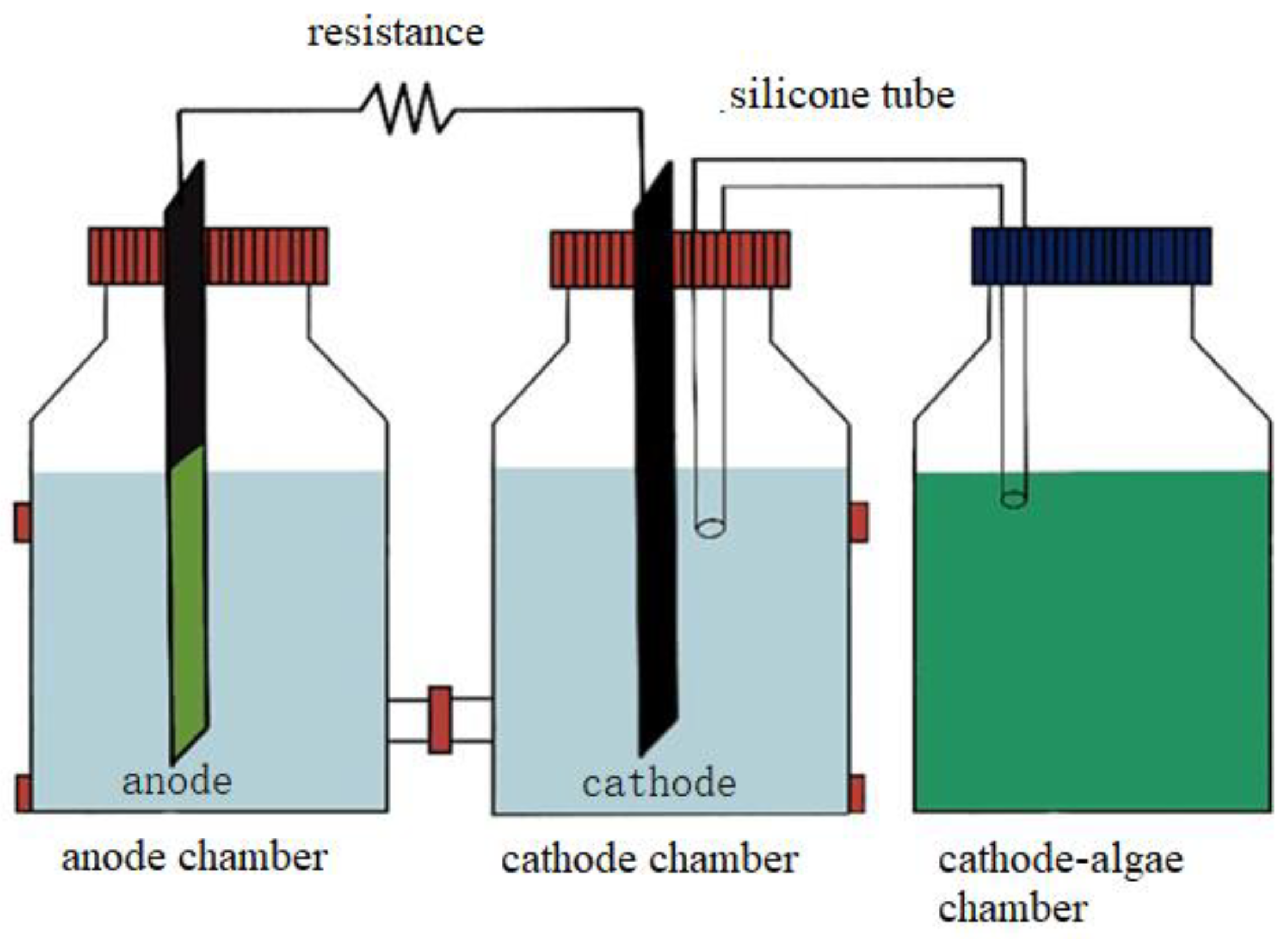
An H-type double-chamber MFC was constructed using organic glass, with each single chamber having a diameter of 7 cm. Both chambers had equal volumes of 285 mL, separated by a proton exchange membrane (Nafion 117) and silicone gasket. The connection between the two chambers was secured using clamps. Carbon felt electrodes (3.5 cm × 9 cm, thickness 3 mm) were placed at 6.6 cm on either side of the proton exchange membrane. The anode and cathode were connected to the external circuit via wires, and a 1000 Ω load was connected between them. Silicone gaskets were used to ensure complete sealing at the bottleneck where it contacts the Teflon stopper, maintaining a sterile environment to the best extent possible. Each chamber had a sampling port and a venting port. The venting port of the anode was sealed with a 0.22-micron organic filter to maintain sterility and prevent external gas exchange. The venting port of the cathode was connected to the sealed blue bottle via silicone tubing, supplying oxygen to the cathode compartment, which was separated from the cathode. The configuration of the microbial fuel cell is shown in Figure 1.

Figure 1.
Configuration of AA-MFC.
2.4. Biofilm Cultivation
Carbon felt electrodes were placed into glass bottles containing P. kessleri to cultivate a normal biofilm. This normal biofilm was then subjected to a constant potential polarization of 0.2 V using an electrochemical workstation to obtain electrochemical biofilm. The attachment density of algae on the biofilm was 1.2 × 10⁸ cell/cm2. The microalgae biofilm was cultured at 25 °C, under a light intensity of 80 μmol·m−2·s−1.
2.5. Electrochemical Analysis and Calculations
Voltage data were collected every 10 s using a data acquisition card (MPS10010) connected to a computer. Polarization curves were obtained by adjusting the external resistor box to various loads of 1000, 2000, 3000, 4000, 5000, 6000, 7000, 8000, 9000, and 9999 Ω. The internal resistance and electromotive force of the cell were calculated using Ohm’s law, where U is the cell voltage, E is the cell electromotive force, I is the current, and r is the cell internal resistance. Cyclic voltammetry measurements were conducted under open-circuit conditions using a three-electrode system, with a saturated calomel electrode as the reference electrode. The linear cyclic voltammetry scan ranged from −0.8 V to 0.8 V (vs. SCE) with a scan rate of 5 mV/s. The electrochemical workstation used in this study is the Koster150M (Koster, Wuhan, China).
2.6. Parameter Measurements
COD (Chemical Oxygen Demand) content was determined using the potassium dichromate method refer to China national standard GB11914-89 [16]. Algae cell density was measured by measuring the absorbance of the microalgal species at 540 nm. A curve function correlating absorbance with dry weight and cell density was constructed to convert absorbance to cell numbers.
Dissolved oxygen in the cathode microalgal chamber was measured using a dissolved oxygen meter. The model of the dissolved oxygen meter is multi from the Hach Company (New York, NY, USA).
2.7. Scanning Electron Microscope
The surface morphology of the biofilm electrodes was examined. Carbon felt electrodes were carefully removed with sterile forceps, cut into 0.5 × 0.5 cm pieces, soaked in 2.5% glutaraldehyde, and placed in a refrigerator at 4 °C for 12 h. After fixation, they were washed three times with a 0.2 M phosphate buffer solution at pH 7.4. Subsequently, the samples were dehydrated using a series of ethanol concentrations (30%, 50%, 75%, 90%, 95%, and 100%). The dried samples were gold-coated for observation.
2.8. Three-Dimensional Fluorescence Spectroscopy of Microalgal Extracellular Polymers
For three-dimensional fluorescence analysis of extracellular polymers, carbon felt electrodes were gently removed with clean forceps and added to 10 mL of 0.9% NaCl solution. Then, 10 mL of 0.9% NaCl solution at 70 °C was added, followed by two minutes of ultrasonication and vortexing. Afterward, the carbon felt was washed with 5 mL of NaCl solution. The microalgal suspension was centrifuged at 5000× g for 15 min, and the supernatant was filtered using a 0.45 µm water series filter. Blank spectra were obtained using ultrapure water. Fluorescence measurements were carried out using a fluorescence spectrophotometer with an excitation wavelength range of 220 nm to 450 nm and an emission wavelength range of 250 nm to 700 nm, with 5 nm increments. The scan speed was set to 1200 nm/min [17].
2.9. Experimental Methods
2.9.1. Effects of Different Operating Modes on AA-MFC Performance
Under sterile conditions, carbon felt electrodes enriched with P. kessleri biofilm (initial inoculum density of 0.3 g/L) were placed in the anode chamber, and a suspension of Spirulina platensis (initial inoculum of 0.2 g/L) was placed in the cathode chamber to create the AA-MFC. One group was operated in an open-circuit mode (control group), meaning the connection between the cathode and anode of the AA-MFC was disconnected, preventing electron transfer from the anode to the cathode. This group can be considered a normal microalgal biofilm wastewater treatment system. The other group was operated in a closed-circuit mode (experimental group), with the cathode and anode of the AA-MFC connected by wires, allowing electron transfer from the anode to the cathode, forming a closed loop. The AA-MFCs were cultured for 72 h under conditions of 25 °C, a light intensity of 80 μmol·m−2·s−1, and a 24:0 h light–dark cycle. Samples were taken at the end of the experiment to compare the performance of the two modes in terms of electricity generation, pollutant degradation, and biomass production.
2.9.2. Effects of Different Initial Inoculation Densities on the Anode of AA-MFC
Different initial inoculum densities (0.4 × 10⁸ cell/cm2, 0.8 × 10⁸ cell/cm2, 1.2 × 10⁸ cell/cm2) of P. kessleri were introduced to the anode of the AA-MFC under both closed-circuit and open-circuit operating modes. In the open-circuit conditions, the cathode microalgal chamber can be regarded as a normally closed culture system, so the data from the three different initial inoculum densities in the cathode microalgal chamber were averaged as a control group.
2.9.3. Effects of Different Light–Dark Cycles on the Anode of the AA-MFC
The performance of the AA-MFC under different light–dark cycles (24:0 h, 18:6 h, 12:12 h) for the anode was evaluated in both closed-circuit and open-circuit operating modes. Data from the cathode control group were handled in the same manner as described above.
2.9.4. AA-MFC Electron Transfer Mechanism
This experiment aimed to investigate the electron transfer mechanism of P. kessleri. Suspended P. kessleri cells with the same density (1.2 × 10⁸ cell/cm2) and cultivation time were placed in the AA-MFC system and operated in the closed-circuit mode to assess their electron transfer mechanism.
3. Results and Discussion
3.1. Selection of Anode and Cathode Microalgae Species
To ensure efficient anode and cathode reactions, the experiment selected microalgal species based on their specific traits. Algal species demonstrating strong electricity generation capabilities were chosen for the anode, while those with strong oxygen production capabilities were selected for the cathode. A stable potassium ferricyanide solution was used as the cathode electrolyte, and experiments were conducted using the chosen five microalgal species to create microalgal cathode-type microbial fuel cells. The open-circuit voltage produced by the cells was measured to assess their electricity generation capabilities. No external load was connected during the measurement of open-circuit voltage. The open-circuit voltage reflects the theoretical maximum voltage that the microbial fuel cell can achieve. After running the microbial fuel cells constructed from the five microalgal species for 72 h, the voltage stabilized. Therefore, the voltage values of the final 12 h were used, and the average was calculated to obtain the open-circuit voltage of the microbial fuel cells, as shown in Figure 2.
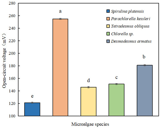
Figure 2.
Comparison of open-circuit voltage of different algae species.
Among these five microalgae species, P. kessleri exhibited the highest open-circuit voltage, reaching 255 mV. The next three microalgal species were Chlorella sp., Desmodesmus armatus, and Tetradesmus obliquus, with corresponding open-circuit voltages of 181 mV, 151 mV, and 146 mV, respectively. Spirulina platensis produced the lowest open-circuit voltage of 121 mV. P. kessleri, with an open-circuit voltage significantly higher than the other microalgal species, is the best performer in terms of electricity generation. Its open-circuit voltage was approximately 2.1 times higher than the lowest open-circuit voltage. In summary, P. kessleri is the microalgae species with the best electricity generation performance, making it suitable for placement at the MFC anode for electricity generation.
In microbial fuel cells, the functionality of electron generation and wastewater treatment is undertaken by the anode. However, the rate at which the cathode receives electrons from the anode is a critical step in limiting the power generation capacity of microbial fuel cells. Therefore, it is crucial to ensure that the cathode has an ample supply of oxidants. To guarantee that the algae in the cathode provide sufficient oxygen as an electron acceptor, the rates of oxygen accumulation for five algal strains were measured and compared.
As shown in Figure 3, among these five microalgal strains, the highest rate of dissolved oxygen accumulation was observed in Spirulina platensis, reaching 1.32 ± 0.09 mg·L−1·h−1. The subsequent three algal strains in descending order of oxygen accumulation rate were Desmodesmus armatus (0.63 ± 0.11 mg·L−1·h−1), Tetradesmus obliquus (0.53 ± 0.06 mg·L−1·h−1), and P. kessleri (0.46 ± 0.06 mg·L−1·h−1). The minimum dissolved oxygen accumulation rate was observed in Chlorella sp., at 0.43 ± 0.06 mg·L−1·h−1. The dissolved oxygen accumulation rate of Spirulina platensis was significantly higher than that of the other algal strains, being approximately three times higher than the lowest rate.
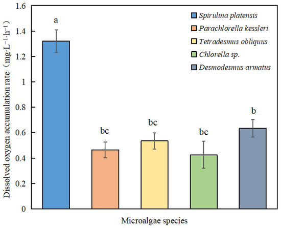
Figure 3.
Comparison of dissolved oxygen accumulation rates of different algae species.
Dissolved oxygen refers to the content of dissolved molecular oxygen in water, expressed in milligrams of oxygen per liter of water. The normal dissolved oxygen content in uncontaminated surface water is in the range of 5 to 10 mg/L [18]. In water bodies where algae grow, the oxygen produced by algae photosynthesis keeps the dissolved oxygen in a state of long-term oversaturation, continuously escaping to the atmosphere. Liu et al. [19] found that, during the cultivation of microalgae in an airlift photobioreactor, the dissolved oxygen could reach as high as 17.91 mg/L. Experimental results by Chi et al. [20] indicated that microalgae cultured in wastewater could release a large amount of oxygen, raising the dissolved oxygen concentration in water to over 20 mg/L. In this study, Spirulina platensis demonstrated excellent oxygen-supply efficiency, making it the optimal choice among the cathodic algal strains.
3.2. Effects of Different Operating Modes on AA-MFC Performance
In this section, we assess the impact of different operating modes on the performance of the algae–algae microbial fuel cell. Specifically, carbon felt electrodes enriched with P. kessleri biofilm, which had been acclimated for growth, and artificial municipal wastewater were introduced into the anode chamber of the AA-MFC. In the cathode microalgae chamber, Spirulina platensis was added to construct the algae–algae microbial fuel cell.
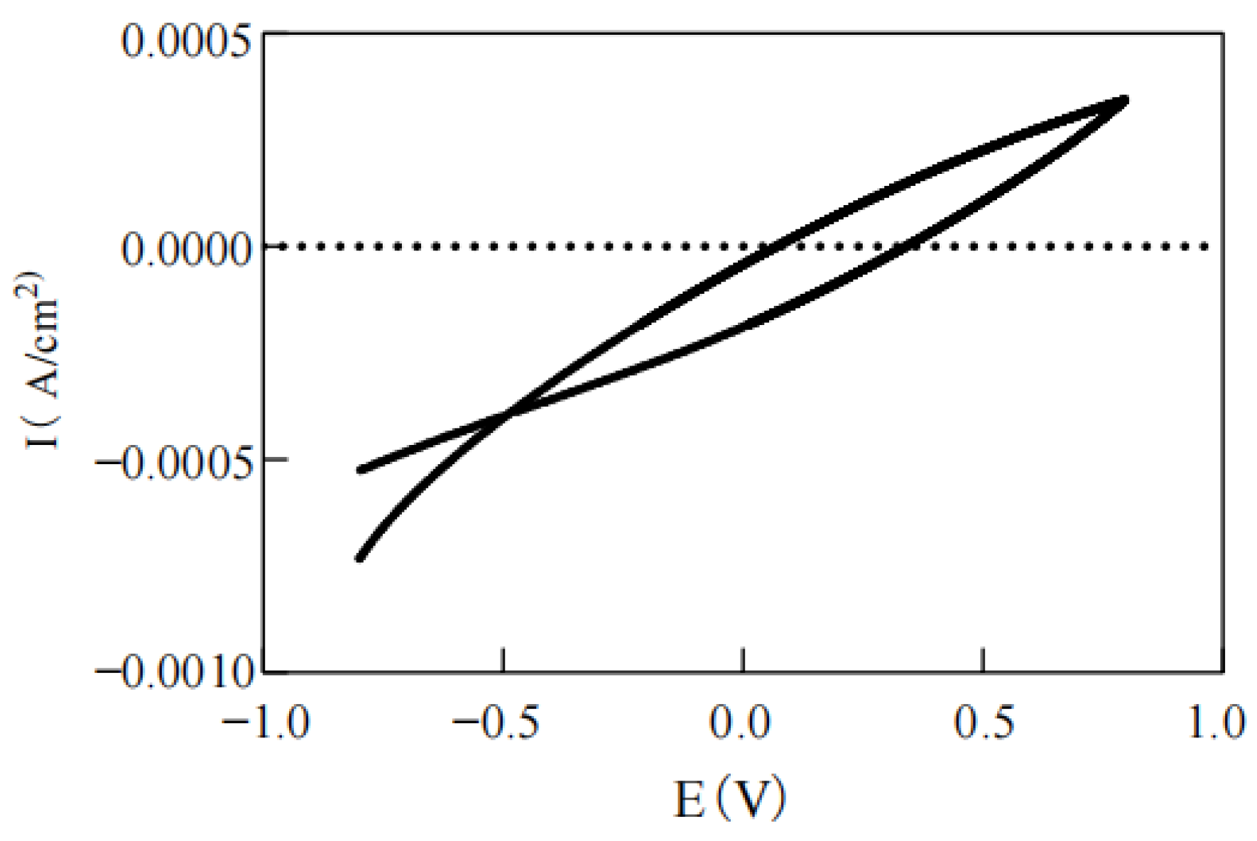
As depicted in Figure 4, in the closed-circuit operating mode, the AA-MFC initially exhibited a current of around 5 μA, which rapidly declined for a certain duration before stabilizing at approximately 2 μA. This behavior was consistent with the findings in Xu et al.’s [12] study, where a Parachlorella MFC demonstrated a gradual reduction in current to stabilize at 5–10 μA. The linear cyclic voltammetry curve, as shown in Figure 5, indicates that the AA-MFC built in this study generated a maximum oxidation–reduction current of 0.0003 A without apparent oxidation–reduction peaks, which aligns with results observed in the research of Subhash [11].
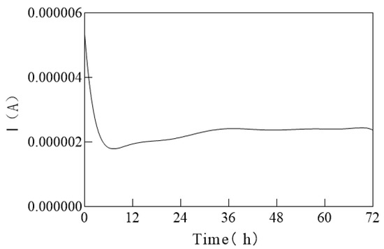
Figure 4.
Algae–algae microbial fuel cell current variation with time.
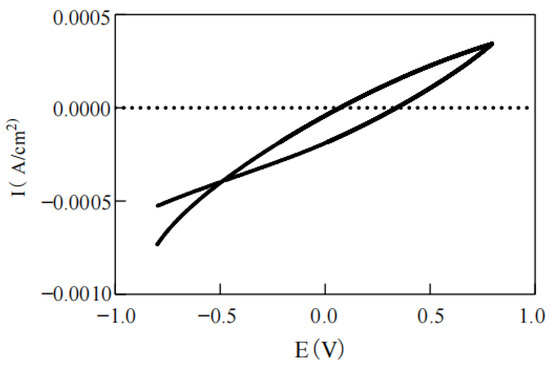
Figure 5.
Linear cyclic voltammetry.
Microbial fuel cells often need to undergo two processes of inoculation and start-up before formal operation. The process from the inoculation of electroactive microorganisms to stable electricity generation is called the start-up process of microbial fuel cells. The start-up time of microbial fuel cells primarily depends on the formation of electrochemically active microbial biofilms on the anode. The growth of electrochemically active biofilms is crucial, and the higher the biomass on the electrode surface within the same period, the faster the start-up of microbial fuel cells. As shown in Figure 4, after a 24 h pre-cultivation of biofilms, an AA-MFC can achieve stable electricity production within 8 h, with a smooth power generation curve, indicating a start-up time of only 32 h. Lu et al. [5] found that pre-attachment of microorganisms to electrode materials can shorten start-up times. In this study, we directly utilized microalgae biofilms attached to the electrodes, enabling rapid and stable current generation, and overcoming the traditional issue of slow start-up in bacterial MFCs, thus facilitating subsequent applications.
According to Table 1, whether operating under closed-circuit or open-circuit conditions, the COD removal efficiency of the AA-MFC in the anode exceeded 90%. However, the COD removal efficiency under closed-circuit conditions was approximately 5% higher than that under open-circuit conditions, suggesting that the closed-circuit operation mode (forming a complete microbial fuel cell system) enhanced the removal efficiency of COD by microalgae biofilms.

Table 1.
Performance of AA-MFC in different operating modes.
After 72 h of operation, the biomass yield of the anode under closed-circuit conditions increased by 12.5% compared to open-circuit conditions, indicating that closed-circuit conditions favor the growth and biomass accumulation of microalgae in the AA-MFC anode chamber.
The biomass growth of microalgae in wastewater is closely related to their efficiency in COD removal. In this study, the AA-MFC system may enhance the removal of COD by promoting the growth of microalgae. The COD removal efficiency of this system far exceeded that of MFC systems reported previously, where Parachlorella, Diatom, or other microalgae were used as the anode [21]. Research has shown that bacteria with electrochemical activity are more easily enriched and grow better under closed-circuit operation, and the high COD removal rate may be related to the higher microbial metabolic activity in a closed-circuit environment [2].
Table 1 also reveals that after 72 h of operation in the cathode chamber, the biomass increment of Spirulina platensis under closed-circuit conditions was 1.6 times that under open-circuit conditions. Additionally, the accumulation of dissolved oxygen in the cathode microalgal chamber under open-circuit conditions was approximately 40% higher than that under closed-circuit conditions.
Weissman et al. [22] demonstrated that in closed cultivation systems, the accumulation of oxygen produced by microalgae photosynthesis can lead to excessively high dissolved oxygen levels, inhibiting microalgae photosynthesis, and even damaging microalgal cells. However, this study found that the accumulation of cathode oxygen was significantly lower in the closed-circuit mode of AA-MFC than in the open-circuit mode. This mitigated the growth stress caused by excessive accumulation of dissolved oxygen in the closed system, thus promoting the growth and biomass accumulation of Spirulina. In Yang’s study [6] on a microalgae cathode MFC, the cathode potential was positively correlated with dissolved oxygen concentration, and dissolved oxygen was one of the main influencing factors of cathode potential. However, too much cathode-dissolved oxygen can penetrate into the anode and negatively affect the anode potential.
The formation of cathodic microalgae biofilms is conducive to the absorption of electrons produced by the anode, but an increase in biofilm thickness may affect the oxygen transfer efficiency [19]. In this study, the cathode was divided into two chambers, and the oversaturated dissolved oxygen in the cathode microalgal chamber was released to the cathode, providing the cathode chamber with an appropriate electron acceptor. Moreover, in comparison to traditional microalgae cathodes, in this study, microalgae did not grow directly on the cathode electrode to form a biofilm. This avoided the impact of increased biofilm thickness on oxygen transfer rates, facilitating the long-term stable operation of the system.
3.3. Effect of Different Initial Inoculum Density in Anode on Performance of AA-MFC
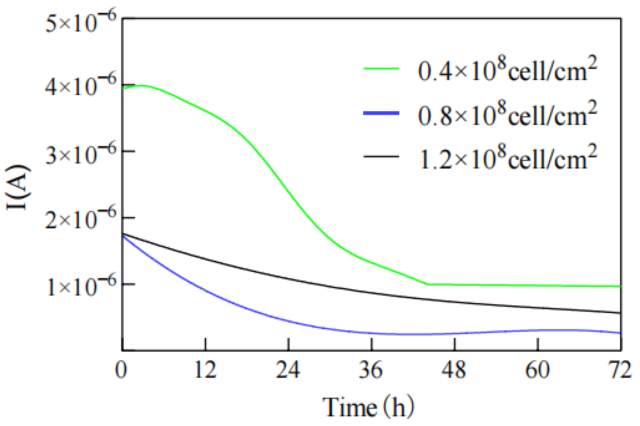
As shown in Figure 6, in all three groups with different initial inoculum densities, the output current initially exhibited a slow decrease followed by a stable trend. The experimental group with the lowest initial inoculum density of 0.4 × 10⁸ cell/cm2 achieved the highest stable output current, whereas the group with an initial inoculum density of 0.8 × 10⁸ cell/cm2 obtained the lowest stable output current.
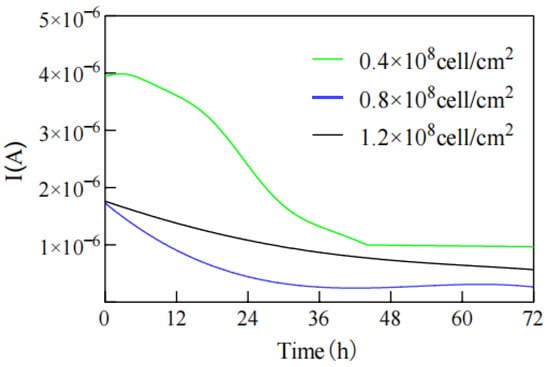
Figure 6.
Current changes with different initial inoculations in anode.
Xu et al. [12] also studied the relationship between microalgal cell density and current and found that higher microalgal cell densities resulted in lower currents. This differed slightly from the findings of this study. In theory, more microalgal cells attached to the anode electrode should provide more electron donors for electricity generation, leading to higher current production. However, the algae on the anode perform photosynthesis, producing oxygen as an electron acceptor, which can reduce their electricity-generating capacity. When the negative impact of increased dissolved oxygen content on the MFC exceeded the positive contribution of biomass to electricity generation, the output current decreased. At this stage, it was true that higher microalgal cell density resulted in lower electricity generation. However, when the total number of microalgal cells increased to a certain threshold, the increase in biomass led to an increase in the thickness of the biofilm. Based on an estimation of 10 μM of electrode surface area occupied by each microalgal cell in this experiment, a single layer of the biofilm accumulated 105~107 microalgal cells per cm2 of electrode surface [23]. After the biofilm reached a certain thickness, the inner layer of microalgae directly in contact with the electrode was covered by the outer layer of microalgae. This reduced the light intensity received and, consequently, the oxygen produced through photosynthesis. The negative impact of dissolved oxygen on electricity generation also diminished. At this point, an increase in microalgal cell density led to an increase in output current. Therefore, in this study, there was a situation where an increase in microalgal cell density at the anode initially resulted in a decrease in output current, followed by an increase.
By measuring the COD removal rate of the AA-MFC anode at 72 h with different initial inoculum densities (Figure 7), it was observed that both closed-circuit and open-circuit modes of the AA-MFC exhibited an increase in COD removal rate with higher initial inoculum densities. Additionally, under all initial inoculum densities, the closed-circuit mode had a promoting effect on COD removal. The greatest enhancement of the COD removal rate in the closed-circuit mode was observed at an initial inoculum density of 0.4 × 10⁸ cell/cm2, which was 17.51% higher than in the open-circuit mode. At an initial inoculum density of 0.8 × 10⁸ cell/cm2, the closed-circuit mode showed the weakest promoting effect on COD removal, only 5.81% higher than the open-circuit mode. These results suggest that the enhancement of COD removal in an AA-MFC is consistent with the magnitude of its output current, with higher current output leading to greater gains in COD removal.
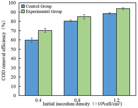
Figure 7.
COD removal efficiency of the AA-MFC at different initial inoculum densities in the anode.
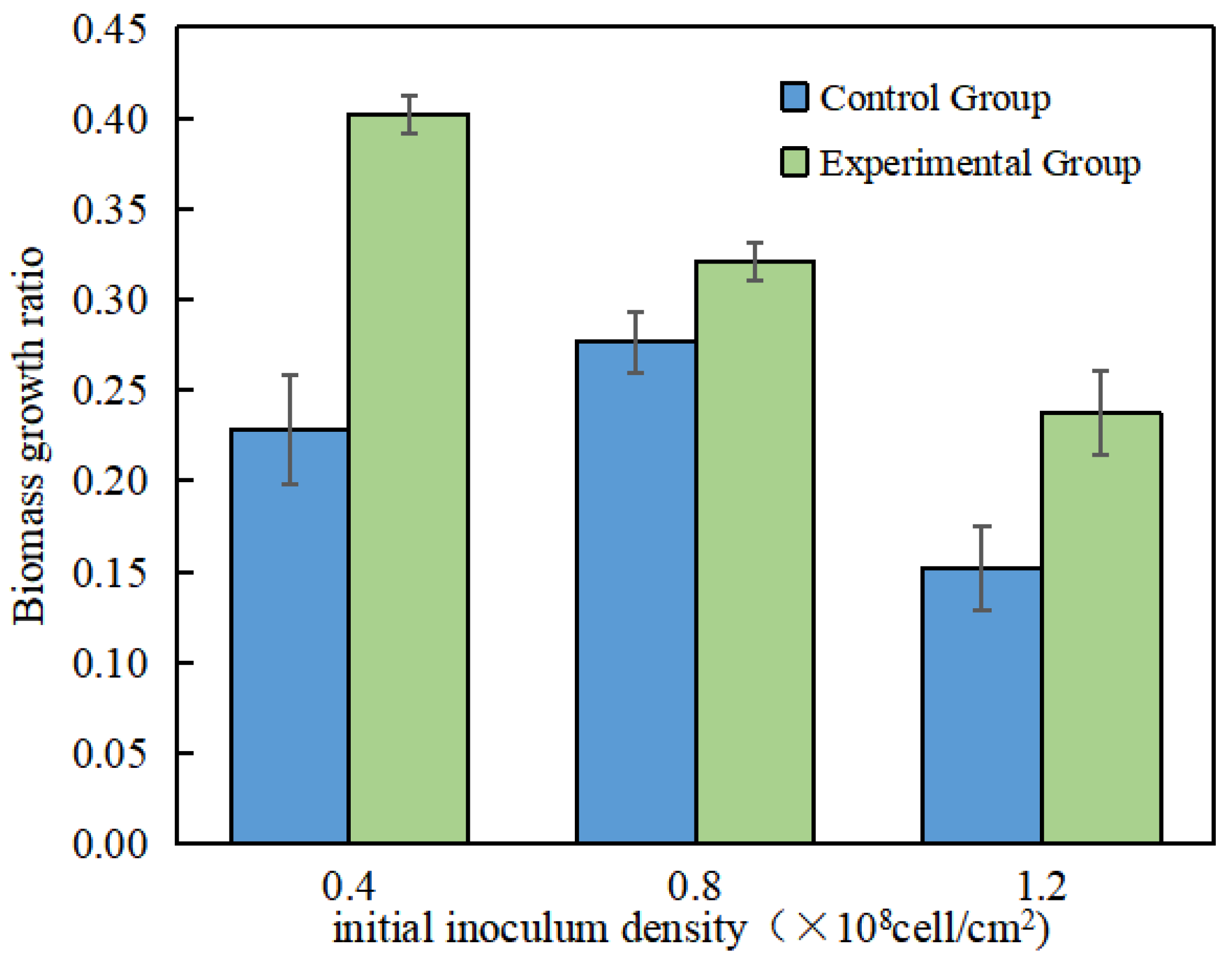
In the process of COD removal by algae, it is often accompanied by the growth of their biomass. As shown in Figure 8, the AA-MFC running in the closed-circuit mode at various initial inoculum densities had a promoting effect on biomass growth. When the initial inoculum density was 0.4 × 10⁸ cell/cm2, the closed-circuit mode had the strongest promoting effect on biomass growth, with an increase of 76.4% compared to the open-circuit mode. However, when the initial inoculum density was 0.8 × 10⁸ cell/cm2, the closed-circuit mode had the weakest promoting effect on the growth of P. kessleri, increasing by only 16.1% compared to the open-circuit mode. This result also indicates that the promoting effect of a closed-circuit AA-MFC on anode biomass growth is consistent with the magnitude of its output current, with higher current output leading to a stronger promoting effect on P. kessleri growth at the anode.

Figure 8.
Biomass growth ratio of the AA-MFC at different initial inoculum densities in the anode.
Combining the results from both operating modes and the three different initial inoculum densities, a fitting analysis was performed on the data for the anode COD removal rate and anode biomass growth factor. The experimental results indicate that there was a positive correlation between the anode biomass growth factor and COD removal rate in the AA-MFC (y = 111.9x + 50.129, R2 = 0.9557).
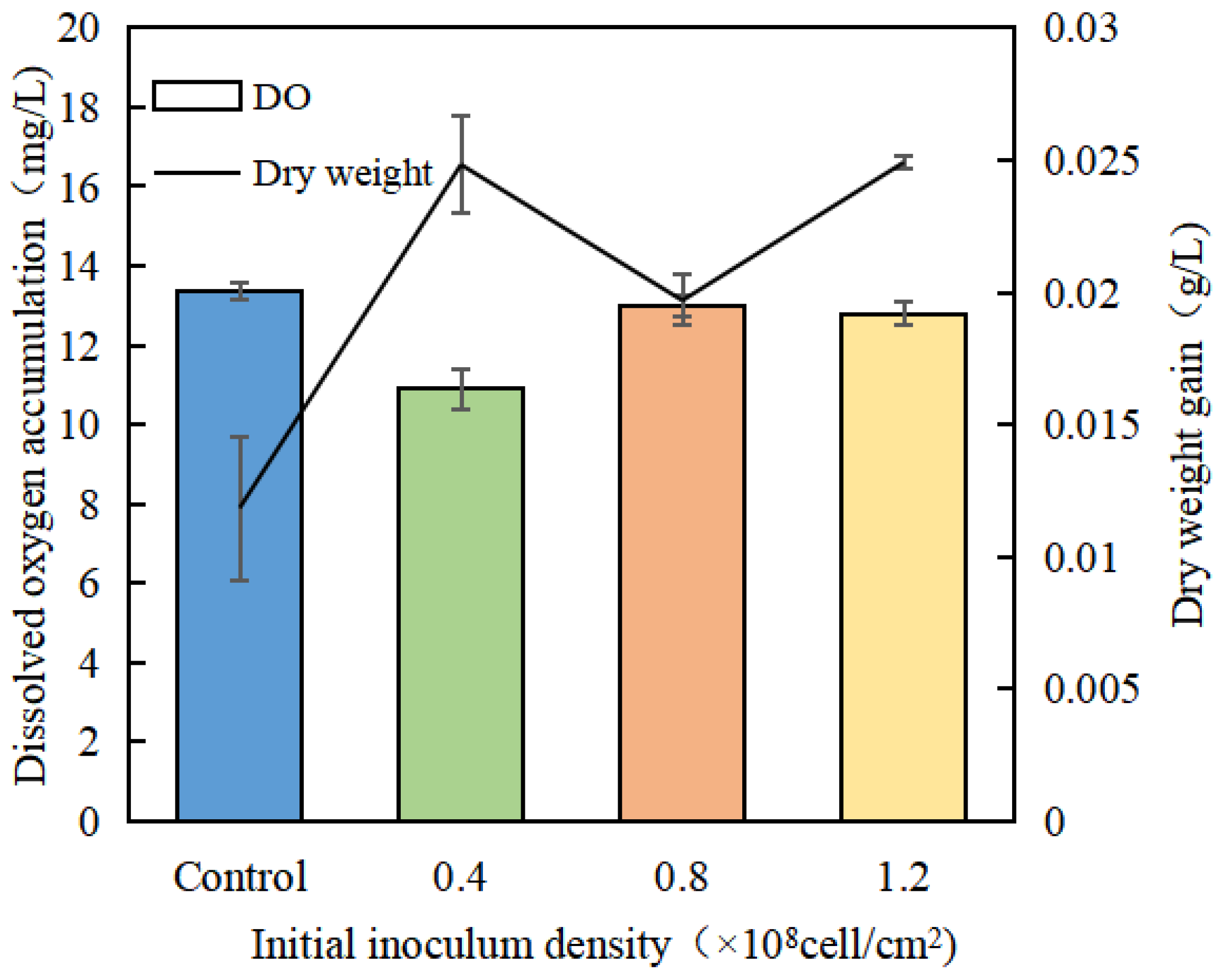
As shown in Figure 9, under different initial inoculum densities of P. kessleri at the anode, the growth and dissolved oxygen accumulation of Spirulina platensis in the AA-MFC cathode varied. After 72 h of growth, all three groups of AA-MFC cathode chambers showed faster growth of Spirulina compared to a normal closed-culture system. This suggests that the AA-MFC system promotes the growth of algae in the cathode compared to normal closed-culture systems. Simultaneously, all three groups of AA-MFC systems alleviated the issue of dissolved oxygen accumulation observed in the closed-culture system to varying degrees.
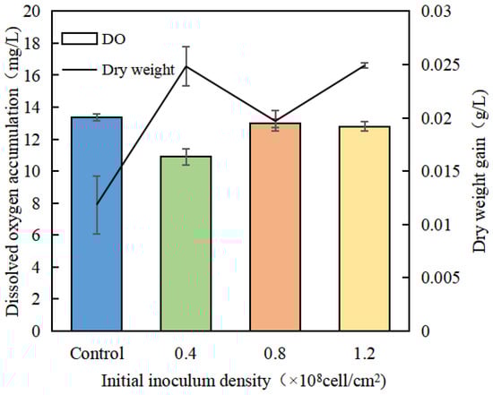
Figure 9.
Changes in dissolved oxygen and dry weight in the cathode chamber with algae at different initial inoculum densities in the anode.
A comprehensive analysis of the results from both operating modes and the three different initial inoculum densities for the anode was performed to fit the data for the cathode Spirulina platensis dry weight increase and dissolved oxygen accumulation. The experimental results indicate a negative correlation between dissolved oxygen accumulation and cathode Spirulina platensis dry weight increase (y = −0.0037x + 0.0669). This suggests that dissolved oxygen is the primary factor influencing cathode biomass growth.
In the case of the 0.4 × 10⁸ cell/cm2 initial inoculum group, P. kessleri attached thinly and evenly to the anode electrode. While photosynthesis was vigorous, the lower total biomass resulted in relatively less oxygen production, which had a minor negative impact on the MFC system. Correspondingly, this group exhibited the best electrical output and the fastest growth of anode P. kessleri biomass, resulting in a more significant enhancement of COD removal. The higher electrical current generated by this group consumed more oxygen at the cathode, reducing dissolved oxygen accumulation in the cathode chamber. This alleviated the inhibitory effect on the growth of Spirulina platensis in the cathode, promoting the growth of cathode biomass.
For the 1.2 × 10⁸ cell/cm2 initial inoculum group, the increased growth of P. kessleri biomass on the anode led to higher electron transfer resistance. Therefore, this group generated a lower current compared to the 0.4 × 10⁸ cell/cm2 group. However, due to the lesser negative effect of dissolved oxygen compared to the 0.8 × 10⁸ cell/cm2 group, the current was higher than the 0.8 × 10⁸ cell/cm2 group. Additionally, the higher initial biomass in this group allowed for the best COD removal rate.
3.4. Effect of Different Light–Dark Cycles on AA-MFC Performance in Anode
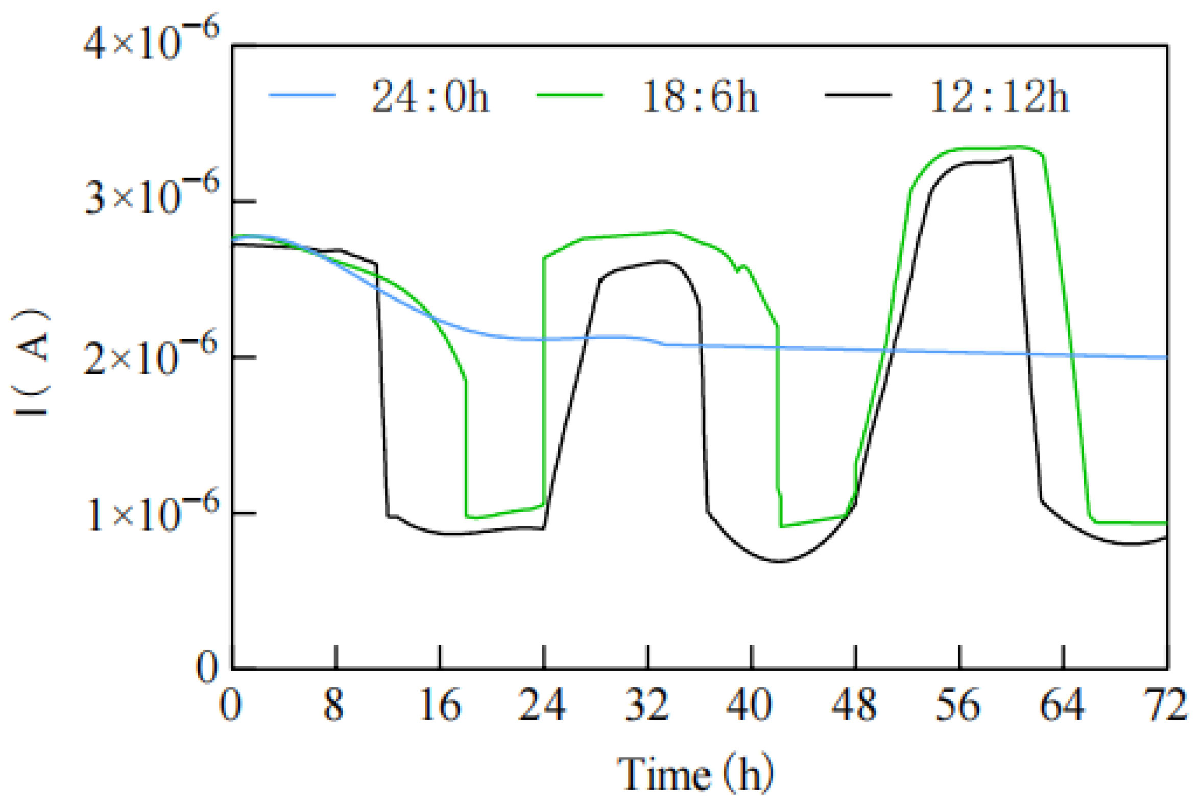
In Figure 10, when the light–dark cycle was set at 24:0 h, the AA-MFC exhibited an initial increase in current, followed by a gradual decrease and stabilization. However, for light–dark cycles of 18:6 h and 12:12 h, the current in the AA-MFC increased during the light period and decreased during the dark period, with a rapid response to light. Even during the dark period, the AA-MFC still generated a relatively small current, possibly attributed to microalgae releasing electrons extracellularly through respiratory processes, in line with the findings of Lam et al. [24]. When the light–dark cycles were set at 18:6 h and 12:12 h, the current generated during each light period after a dark period surpassed the current generated in the previous light period. This phenomenon might be due to the consumption of oxygen by MFC anode microalgae, specifically P. kessleri, during the dark period through respiratory processes, negatively affecting the electron acceptor for anodic electricity generation.
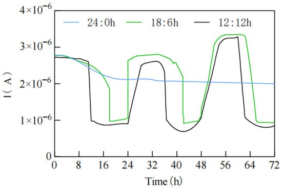
Figure 10.
Current changes in AA-MFC anode at different light and dark periods.
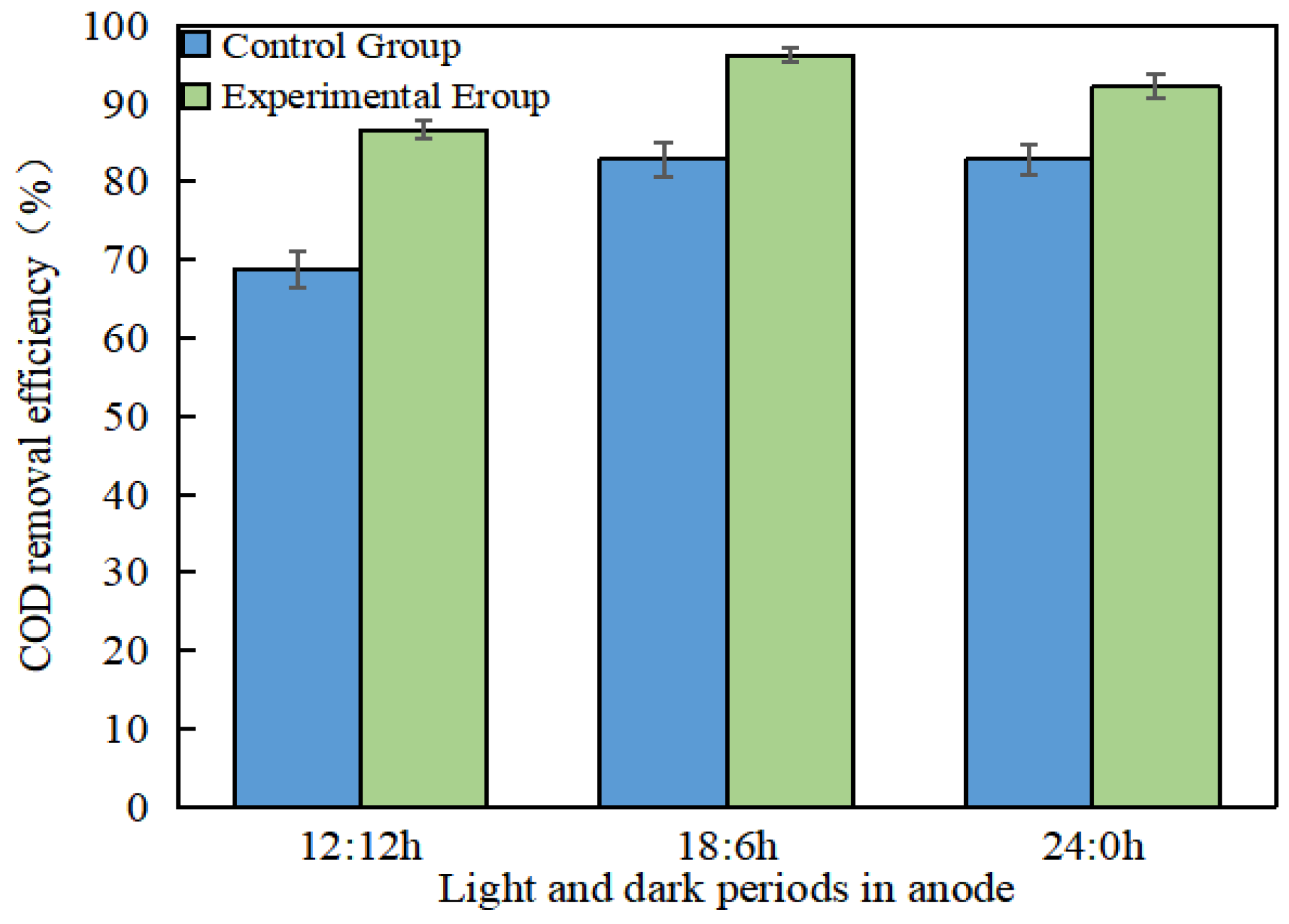
Figure 11 shows the COD removal rates of the AA-MFC under different light–dark cycles in open-circuit and closed-circuit operation modes. The results indicate that closed-circuit operation enhances COD removal. Among the different light–dark cycle conditions, the 12:12 h cycle exhibited the highest enhancement, with a COD removal rate increase of 26.04%. The 18:6 h cycle was followed by a 15.99% enhancement, while the 24:0 h cycle had the weakest enhancement effect, with only a 13.13% increase in COD removal.
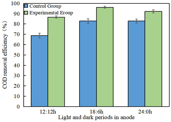
Figure 11.
The COD removal rate of the AA-MFC at different light and dark cycles in the anode.
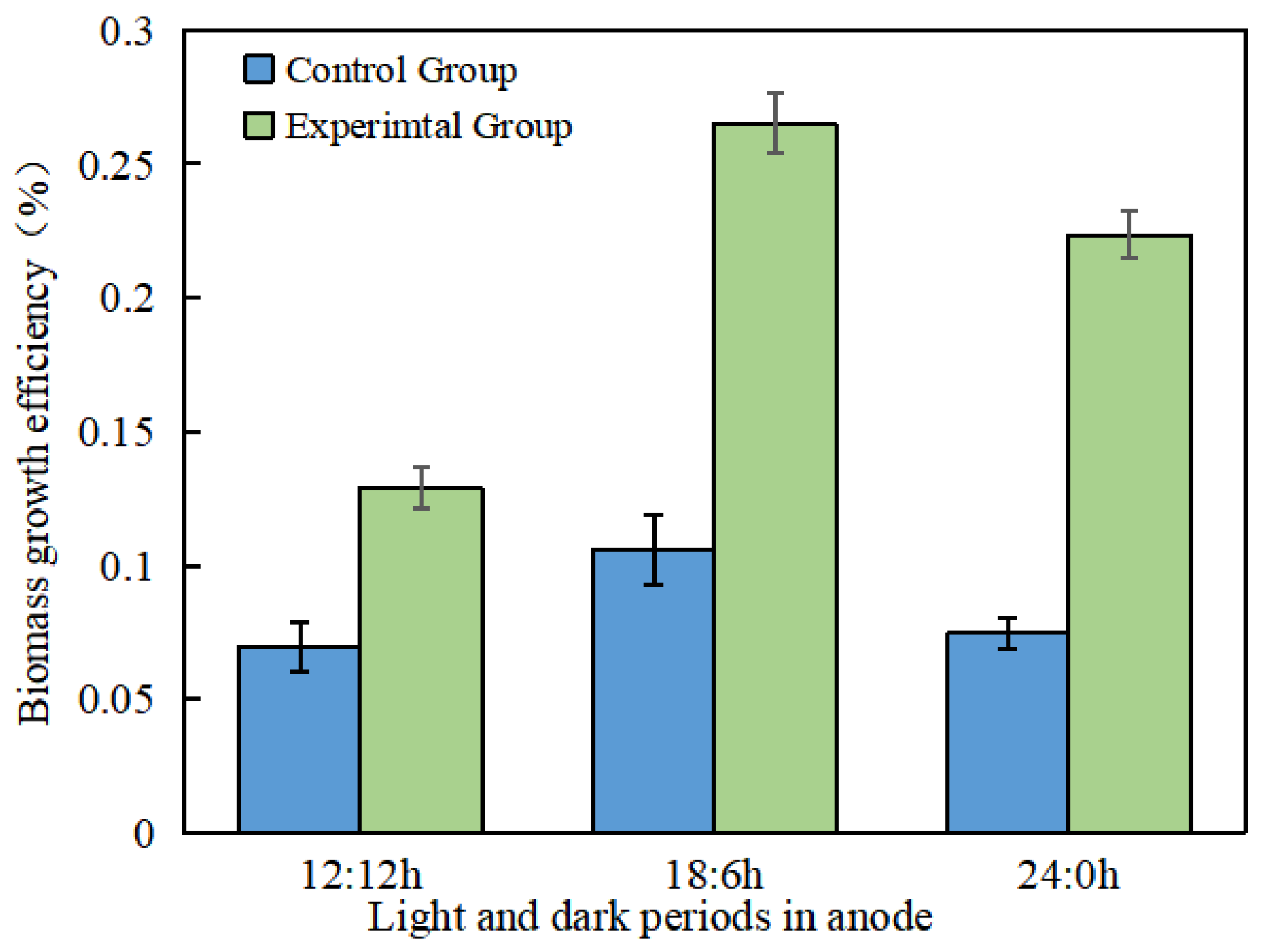
The promotion of closed-circuit operation on anode biomass growth under various light–dark cycles for the AA-MFC is shown in Figure 12. The results reveal that the anode biomass growth was fastest under the 18:6 h light–dark cycle, while it was the slowest under the 24:0 h cycle. Furthermore, under the 18:6 h light–dark cycle, the promotion of biomass growth in the anode was the strongest when compared to open-circuit operation, whereas the 12:12 h cycle group exhibited the weakest promotion effect.
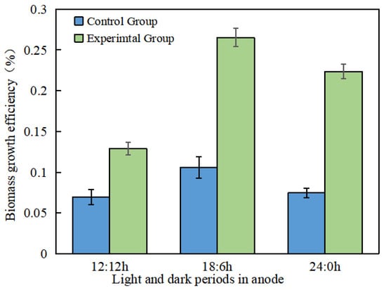
Figure 12.
Biomass growth ratio of AA-MFC at different light and dark cycles in the anode.
The fitting results for the COD removal efficiency and biomass growth ratio under three different light–dark cycles for the AA-MFC in the anode also indicate that there was a positive correlation between the biomass growth ratio and AA-MFC COD removal efficiency (y = 100.98x + 70.3, R2 = 0.7465).
Under the 18:6 h light–dark cycle, the anode biomass growth was the highest. The 18:6 h group exhibited faster biomass growth, which led to a quicker response to light and higher current production. Corresponding to the biomass growth levels, the COD removal efficiency was the highest under the 18:6 h light–dark cycle. The 12:12 h light–dark cycle significantly enhanced the COD removal effect. While the 12:12 h light–dark cycle effectively promoted COD removal, the best COD treatment performance was achieved under the 18:6 h light–dark cycle.
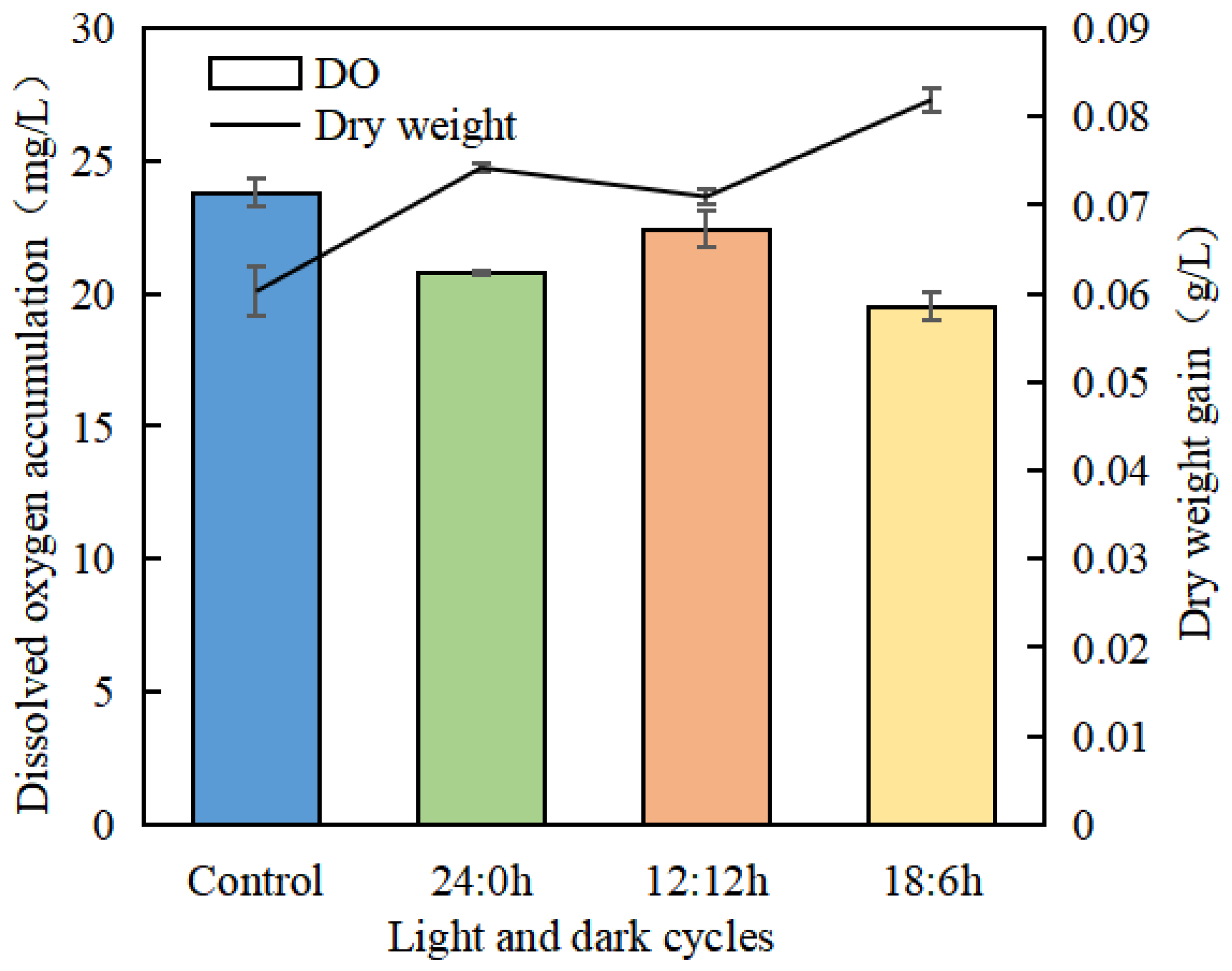
As shown in Figure 13, after 72 h of growth, the growth of Spirulina platensis in the cathode chamber of the three AA-MFC groups is faster than that in the control group of the ordinary closed-circuit culture system. Among the AA-MFC groups, the cathode chamber Spirulina platensis exhibited the best growth under the 18:6 h light–dark cycle. The ordinary closed-circuit culture system has a higher accumulation of dissolved oxygen compared to the three experimental groups under closed-circuit conditions, with the least dissolved oxygen accumulation observed under the 18:6 h light–dark cycle.
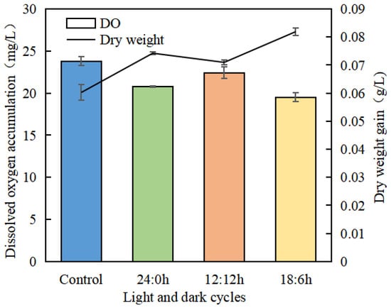
Figure 13.
Changes in dissolved oxygen and dry weight in the cathode chamber with algae at different light and dark cycles in the anode.
By combining the experimental results of two operating modes and three different light–dark cycles for the anode, the data for Spirulina platensis dry weight increase and dissolved oxygen accumulation in the cathode were fitted. The experimental results further validate that there was a negative correlation between dissolved oxygen accumulation and Spirulina platensis dry weight increase (y = −0.0047x + 0.1731, R2 = 0.9493), indicating that dissolved oxygen was indeed the primary factor influencing cathode biomass growth.
To further investigate the coupling relationship between the efficiency of wastewater treatment at the anode and the production of high-value microalgal products at the cathode in the AA-MFC, the data for the anode COD removal rate and the ratio of cathode dissolved oxygen accumulation to biomass growth were fitted based on different initial inoculum densities, light–dark cycles, and closed-circuit vs. open-circuit operating modes. The experimental results show that there was a positive correlation between anode COD removal efficiency and the ratio of cathode dissolved oxygen accumulation to biomass growth (y = 13.426x + 1.4035, R2 = 0.7763). The higher the COD removal rate, the greater the ratio of dissolved oxygen accumulation to dry weight increase. The closed-circuit operation mode provided a greater benefit for COD removal and faster biomass growth. The coupling model between the cathode and anode revealed the relationship between COD removal and dissolved oxygen accumulation and dry weight increase, providing data support for cost-effective wastewater treatment and economic microalgae production.
3.5. Mechanism of Enhanced COD Treatment through Electron Transfer in AA-MFC
Microalgae played a crucial role in the microbial fuel cell by generating an output current in the anode. The electrons produced by microalgae traveled through the electrode and conductive wires to the cathode, where they were received. This process effectively promoted the removal of COD. However, the mechanism by which electrons generated by microalgae are transferred from the cells to the electrode is not yet well understood.
The mechanism of how electrons produced by microalgae are transferred from the cells to the electrode in AA-MFCs is not fully understood. There are four main extracellular electron transport mechanisms on an anode’s electrically active biofilm: (1) a direct contact mechanism, (2) a nanowire mechanism, (3) an electron shuttle mechanism (using exogenous or endogenous media), and (4) an electrotaxis mechanism (using flagella). The presence of nanowires in cyanobacteria Synnechocystis sp. PCC 6803 suggests that microalgae may transfer electrons directly [25].
According to the direction of electron transfer between electrically active microorganisms and carriers, electrically active biofilms can be divided into anode electrically active biofilms and cathode electrically active biofilms. The anode electrically active biofilms can output electrons to the electrode, while the cathode electrically active biofilms can receive electrons from the electrode. In this study, the microalgae biofilm that produced electricity was an anode electrically active biofilm. There are two effective ways to cultivate electrically active biofilms: (1) enrichment and domestication in bioelectrochemical systems, such as microbial fuel cells, microbial electrolysis cells, and microbial desalination fuel cells; (2) using electrochemical workstations and techniques such as constant potential, potential step, constant current, etc., to culture electrochemically active biofilms using the working electrode of the electrochemical workstation as a carrier for EABs.
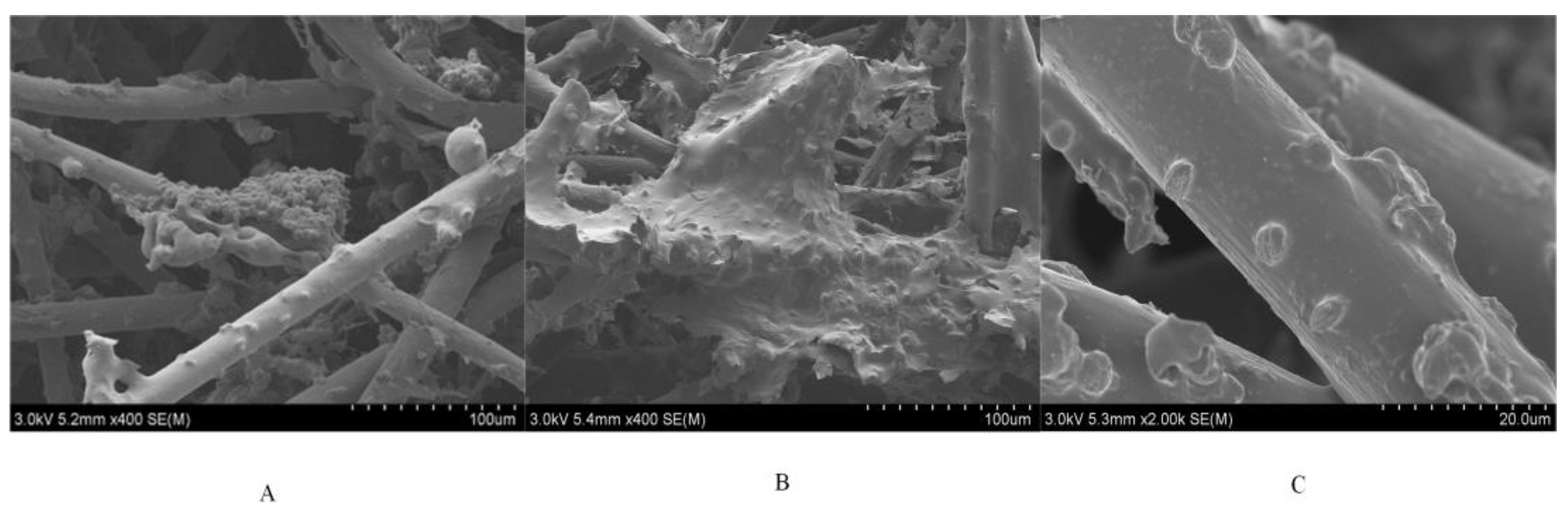
This study compared the P. kessleri biofilm obtained from constant potential cultivation with normal P. kessleri biofilm under the same cultivation time. The structure and morphology of the normal biofilm (Figure 14A) and the electrochemical biofilm (Figure 14B) on the carbon felt surface were observed using scanning electron microscopy. As shown in Figure 14C, the P. kessleri cells are the class of P. kessleri used in the experiment. The electrochemical biofilm and the normal biofilm were attached to the 3D scaffold of carbon felt. The carbon felt consisted of numerous coiled carbon fibers, with each carbon fiber having a larger diameter than the class of P. kessleri. This provided a favorable environment for the growth and attachment of the P. kessleri. The electrochemical biofilm covered the surface of the carbon felt uniformly in a sheet-like structure, while the normal biofilm was attached more sporadically to the carbon fibers. In the image with a scale of 100 μm, it can be seen that more cells were attached to the carbon felt electrode in the electrochemical biofilm group, and the attachment density of the electrochemical biofilm was greater than that of the normal biofilm. In the image with a scale of 20 μm (Figure 14C), it can be observed that there were no nanowires or flagella between the P. kessleri on the electric biofilm and the carbon felt electrode. Since no additional electron mediators were added in this study, the way electrons transferred from the P. kessleri cell membrane to the electrode surface was likely the direct contact mechanism.

Figure 14.
SEM image. (A) Common biofilm (100 μm); (B) electrochemically active biofilm (100 μm); (C) electrochemically active biofilm (20 μm).
In this study, it was observed that the MFCs containing suspended microalgae, classified as a distinct group, generated significantly lower and less stable electric currents (1 × 10⁻⁷ A) in comparison to MFCs with microalgae forming a biofilm (2 × 10⁻⁶ A). This observation aligns with findings from Liu’s research on bacterial microbial fuel cells [26], thereby affirming that direct physical contact between microalgae cells and the electrode surface serves as one of the primary mechanisms for electron transfer in MFCs. Moreover, experiments involving different initial inoculum densities of the biofilm-forming microalgae on the anode revealed a decreasing trend in electric current output as the biofilm thickness increased. When the biofilm thickness reached a certain threshold, it impeded the direct substrate oxidation by algae cells in contact with the electrode, resulting in a decline in the electric current. The experiments employing diverse light–dark cycles further provided evidence of the rapid response of the current to light variations, reinforcing the mechanism of direct electron transfer by the microalgae on the anode.
Additionally, in this study, we employed a fluorescence spectrophotometer to analyze extracellular polymeric substances extracted from both the normal biofilm and the electrochemical biofilm of P. kessleri, which had grown on the carbon felt electrode after 72 h, as depicted in Figure 15.
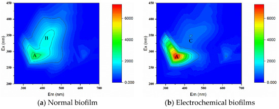
Figure 15.
Three-dimensional fluorescence spectra of common biofilms and electrochemical biofilms.
The results indicate distinct differences in the three-dimensional fluorescence spectra of extracellular polymeric substances between electrochemical biofilm and normal biofilm. This disparity underscores structural variations in EPS composition arising from the stimulation of extracellular electron transfer by the same species of microalgae forming different biofilms. The contrast in three-dimensional fluorescence spectra of the EPS between normal biofilm and electrochemically active biofilm reveals the impact of extracellular electron transfer by P. kessleri on changes in EPS components.
Previous studies have indicated that Peak A was associated with tryptophan-containing proteins [27], while Peaks B and C represent humic acid substances generated from organic matter degradation [28]. This suggests that tryptophan-containing proteins and humic acid-like substances are the main constituents of extracellular polymeric substances in electrochemical biofilm and normal biofilm. The corresponding fluorescence intensity (Table 2) in Figure 15 reveals that the fluorescence intensity of tryptophan-containing proteins in the EPS from the electrochemical biofilm was enhanced by 111% compared to the EPS from the control group’s normal biofilm. This enhancement may be attributed to increased metabolic activity due to enhanced photosynthesis and respiration, resulting in changes in metabolic products. Liu et al. [29] found that electrical stimulation can boost bacterial metabolism, leading to increased protein secretion that regulates physiological activities. Zhang et al. [30] also reported that the use of closed-circuit electron flow can construct a highly reducing environment around individual bacterial cells, improving the electron donor-to-acceptor ratio, and enhancing nitrate reduction to ammonium efficiency. The effective enrichment of electroactive bacteria involved in nitrate reduction to ammonium on the biofilm electrode in the reactor indicated enhanced nitrate reduction to ammonium under closed-circuit electron flow conditions. In this study, closed-circuit electron flow effectively enriched power-generating P. kessleri on the electrode, leading to changes in metabolic products, including increased protein secretion. This aligns with one of the mechanisms reported for microbial fuel cells in the enhancement of pollutant degradation.
Table 2.
3D fluorescence intensity.
There is speculation that the metabolic mode of electrochemical biofilms is more efficient than normal biofilms [31]. In the electrochemical biofilm of microalgae-based microbial fuel cells, metabolic products increase significantly, indicating that the metabolic mode of microalgae-based microbial fuel cells is more efficient than normal biofilm treatment systems. Previous studies have suggested that the anodic electrons in microalgae-based microbial fuel cells may originate from the photosynthetic PS II phase [32]. Lumistra’s [33] research also shows that microalgae-based microbial fuel cells exhibit a continuous increase in open-circuit potential under illumination, while they exhibit a linear decrease under dark conditions. This phenomenon suggests that photosynthesis is the source of electrons in microalgae-based microbial fuel cells. In the experiments on the impact of different light–dark cycles on microalgae-based microbial fuel cells with algae–electrode interactions, the current increased during the light period and decreased during the dark period, consistent with Lumistra’s observations.
Furthermore, photosynthesis is crucial for microalgae growth using wastewater as a culture medium while removing nutrients from the water. Light saturation effects result in much lower actual light utilization efficiency than theoretical values. The electron transfer from photosystem II is limited downstream in microalgae. Subhash’s study found that the electrons generated by photosystem II in microalgae can be transferred externally [10]. In this study, P. kessleri in the microbial fuel cell system can transfer some of the photosynthesis-generated electrons to the exterior, broadening the photosynthetic electron transfer pathway.
Microalgae-based microbial fuel cells may enhance photosynthesis efficiency by improving the photosynthetic electron transfer pathway’s efficiency, which promotes microalgae growth and enhances COD removal efficiency. This aligns with the results of the experiments in this study concerning different initial inoculum densities and light–dark cycles, where a stronger microalgal electricity generation capacity led to faster growth of anodic biomass and higher COD removal efficiency. A more substantial electricity generation capacity in the anodic microalgae led to higher dissolved oxygen content, reducing the stress of dissolved oxygen on cathodic microalgae growth, and consequently, better cathodic microalgal growth.
4. Conclusions
To enhance the removal of COD from synthetic wastewater using microbial fuel cell technology and mitigate the damage to microalgal cells in closed-system photobioreactors due to excess dissolved oxygen, this study constructed a novel alga–alga-type MFC. Building upon the existing knowledge of microalgae wastewater treatment and MFC technology and understanding the principles of MFC electricity generation and microalgae wastewater treatment, this research aimed to demonstrate the feasibility of COD removal enhancement using a microalgal-assisted MFC and to increase the COD removal efficiency while harvesting high-value microalgal biomass by optimizing the factors influencing system performance.
The study selected suitable algae species for placement on the anode and cathode and used them to construct a new AA-MFC that started up more rapidly and operated more stably than traditional MFCs. By comparing their performance under open- and closed-circuit conditions, it was confirmed that the anodic microalgae in the closed system had more potential for sewage COD removal and cathodic microalgal biomass growth. The impacts of different initial inoculum densities on the performance of the AA-MFC were investigated, showing that the three initial inoculum densities could promote the removal of anode COD and the growth of cathode biomass. The best electricity generation capacity was observed at an initial inoculum density of 0.4 × 10⁸ cell/cm2, with the most significant enhancement of COD degradation. An initial inoculum density of 1.2 × 10⁸ cell/cm2 resulted in the highest anodic COD removal rate and the most significant yield of cathodic biomass.
The study further investigated the influence of different light–dark cycles on AA-MFC performance. The results showed that the AA-MFC performed well under all three light–dark cycle conditions, which is conducive to anodic COD removal and cathodic biomass growth. The 12:12 h light–dark cycle exhibited the most significant promotion of anodic COD removal, and the 18:6 h light–dark cycle resulted in the highest COD removal rate and the most production of cathodic microalgal biomass. The mechanism of COD removal enhanced by the AA-MFC was also discussed, which showed that the direct electron transfer mechanism is the primary mechanism for microalgal electricity generation. By improving the efficiency of the photosynthetic electron transfer pathway, the AA-MFC enhanced photosynthesis efficiency, promoting microalgal growth and, hence, strengthening COD removal.
Future research could focus on optimizing and innovating the configurations of existing photobioreactors, applying the AA-MFC to actual wastewater treatment, studying its practical applications, and further investigating the mechanisms of the AA-MFC’s enhancement of pollutant removal. In addition, further research into the electron transport mechanisms of microalgae in MFC systems and the role of photosynthesis in AA-MFC power generation and pollutant enhancement could provide valuable insights for future research.
Author Contributions
Y.Z.: Formal analysis, Investigation, Methodology, Validation, Writing—original draft, Writing—review and editing. H.M.: Conceptualization, Writing—review and editing, Funding acquisition. K.C.: Writing—review and editing. W.Q.: Writing—review and editing, Funding acquisition, Supervision. All authors have read and agreed to the published version of the manuscript.
Funding
This research was supported by the National Natural Science Foundation of China under Grants 32170383 and 52179065, as well as the Ministry of Education’s Chunhui Program Collaborative Research Project (Grant No. 202201970).
Data Availability Statement
Data will be made available on request.
Conflicts of Interest
The authors declare no conflicts of interest.
References
- Jiang, J.; Zhao, Q.; Wei, L.; Wang, K.; Lee, D.J. Degradation and characteristic changes of organic matter in sewage sludge using microbial fuel cell with ultrasound pretreatment. Bioresour. Technol. 2011, 102, 272–277. [Google Scholar] [CrossRef] [PubMed]
- Xiang, Y.B.; Yang, Y.G.; Sun, G.P.; Xu, M.Y. Enhanced contaminant degradations in microbial fuel cells and the potential mechanisms: A review. Microbiol. China 2014, 41, 344–351. [Google Scholar]
- Logan, B.E.; Hamelers, B.; Rozendal, R.; Schröder, U.; Keller, J.; Freguia, S.; Aelterman, P.; Verstraete, W.; Rabaey, K. Microbial fuel cells: Methodology and technology. Environ. Sci. Technol. 2006, 40, 5181–5192. [Google Scholar] [CrossRef] [PubMed]
- Tang, J.H.; Liu, Y.; Zhou, S.G.; Yuan, Y. Electrochemically active biofilms: Formation, characterization and application. Chin. J. Appl. Environ. Biol. 2014, 20, 1096–1103. [Google Scholar]
- Lu, H.; Yu, Y.; Xi, H.; Zhou, Y.; Wang, C. A quick start method for microbial fuel cells. Chemosphere 2020, 259, 127323. [Google Scholar] [CrossRef]
- Yang, Z. Process and Performance of Treatment of Anaerobically Digested Effluent from Kitchen Waste Using an Algae-Assisted Microbial Fuel Cell. Ph.D. Thesis, Shandong University, Jinan, China, 2020. [Google Scholar]
- Kumar, G.; Bakonyi, P.; Zhen, G.; Sivagurunathan, P.; Kook, L.; Kim, S.H.; Tóth, G.; Nemestóthy, N.; Bélafi-Bakó, K. Microbial electrochemical systems for sustainable biohydrogen production: Surveying the experiences from a start-up viewpoint. Renew. Sustain. Energy Rev. 2017, 70, 589–597. [Google Scholar] [CrossRef]
- Wu, X.Y.; Zhou, C.X.; Zhi, Y.F.; Chen, L.Y. Progress of microbial fuel cell with microalgae. Environ. Sci. Technol. 2012, 35, 82–86. [Google Scholar]
- Li, Y.L.; Zhao, G.Q.; Wu, S.; Wang, W.Q.; Hua, W.; Cheng, Y.L.; Bai, J. Cultivation of microalgae based on wastewater and utilization of microalgae. Mod. Chem. Ind. 2021, 41, 48–51. [Google Scholar]
- Maity, J.P.; Hou, C.P.; Majumder, D.; Bundschuh, J.; Kulp, T.R.; Chen, C.Y.; Chuang, L.T.; Chen, C.N.N.; Jean, J.S.; Yang, T.C.; et al. The production of biofuel and bioelectricity associated with wastewater treatment by green algae. Energy 2014, 78, 94–103. [Google Scholar] [CrossRef]
- Subhash, G.V.; Chandra, R.; Mohan, S.V. Microalgae mediated bio-electrocatalytic fuel cell facilitates bioelectricity generation through oxygenic photomixotrophic mechanism. Bioresour. Technol. 2013, 136, 644–653. [Google Scholar] [CrossRef]
- Xu, C.; Poon, K.; Choi, M.M.; Wang, R. Using live algae at the anode of a microbial fuel cell to generate electricity. Environ. Sci. Pollut. Res. 2015, 22, 15621–15635. [Google Scholar] [CrossRef] [PubMed]
- Kakarla, R.; Min, B. Photoautotrophic microalgae Scenedesmus obliquus attached on a cathode as oxygen producers for microbial fuel cell (MFC) operation. Int. J. Hydrogen Energy 2014, 39, 10275–10283. [Google Scholar] [CrossRef]
- Diniz, G.S.; Silva, A.F.; Araújo, O.Q.F.; Chaloub, R.M. The potential of microalgal biomass production for biotechnological purposes using wastewater resources. J. Appl. Phycol. 2017, 29, 821–832. [Google Scholar] [CrossRef]
- Mat Aron, N.S.; Khoo, K.S.; Chew, K.W.; Show, P.L.; Chen, W.H.; Nguyen, T.H.P. Sustainability of the four generations of biofuels—A review. Int. J. Energy Res. 2020, 44, 9266–9282. [Google Scholar] [CrossRef]
- GB11914-89; Standard determination method of COD GB11914-89. Standardization Administration of China: Beijing, China, 1989.
- Pei, H.; Yang, Z.; Nie, C.; Hou, Q.; Zhang, L.; Wang, Y.; Zhang, S. Using a tubular photosynthetic microbial fuel cell to treat anaerobically digested effluent from kitchen waste: Mechanisms of organics and ammonium removal. Bioresour. Technol. 2018, 256, 11–16. [Google Scholar] [CrossRef]
- Zhang, P.L.; Sun, C.J. The Influence of Algae Growing on pH and DO in Surface Water. Environ. Monit. China 2004, 20, 2. [Google Scholar]
- Liu, W.N.; Wu, Y.; Xu, Z.; Sun, J.M. The change of dissolved oxygen and pH of the marine microalgae cultivation in air-lift photobioreactor. Fish. Moderniz. 2008, 35, 6–10. [Google Scholar]
- Chi, K. Influencing Factors of Microalgal Culturing in Helotism Wastewater Treatment System. Master’s Thesis, Harbin Institute of Technology, Harbin, China, 2010. [Google Scholar]
- Fan, G.D.; Lin, R.J.; Su, Z.Y.; Lin, X.Y.; Xu, R.X.; Chen, W. Construction of microbial fuel cells using algae. Chem. Ind. Eng. Prog. 2016, 35, 3847–3853. [Google Scholar]
- Weissman, J.C.; Goebel, R.P.; Benemann, J.R. Photobioreactor design: Mixing, carbon utilization, and oxygen accumulation. Biotechnol. Bioeng. 1988, 31, 336–344. [Google Scholar] [CrossRef]
- Zhou, C.R.; Feng, Q.; Zhang, K.J.; Luo, L.W.; Zhang, Y.; Guo, W.; Luo, M.M. Research Progress in Cultivation Devices for Microalgae Biofilms. Appl. Chem. Ind. 2020, 49, 7. [Google Scholar]
- Lam, K.B.; Chiao, M.; Lin, L. A micro photosynthetic electrochemical cell. In Proceedings of the Sixteenth Annual International Conference on Micro Electro Mechanical Systems, MEMS-03 Kyoto, IEEE, Kyoto, Japan, 19–23 January 2003. [Google Scholar]
- Gorby, Y.A.; Yanina, S.; Mclean, J.S.; Rosso, K.M.; Moyles, D.; Dohnalkova, A.; Beveridge, T.J.; Chang, I.S.; Kim, B.H.; Kim, K.S. Electrically conductive bacterial nanowires produced by Shewanella oneidensis strain MR-1 and other microorganisms. Proc. Natl. Acad. Sci. USA 2006, 103, 11358–11363. [Google Scholar] [CrossRef] [PubMed]
- Liu, H.; Ramnarayanan, R.; Logan, B.E. Production of electricity during wastewater treatment using a single chamber microbial fuel cell. Environ. Sci. Technol. 2004, 38, 2281–2285. [Google Scholar] [CrossRef]
- Reynolds, D.M.; Ahmad, S.R. Rapid and direct determination of wastewater BOD values using a fluorescence technique. Water Res. 1997, 31, 2012–2018. [Google Scholar] [CrossRef]
- Parlanti, E.; Wörz, K.; Geoffroy, L.; Lamotte, M. Dissolved organic matter fluorescence spectroscopy as a tool to estimate biological activity in a coastal zone submitted to anthropogenic inputs. Org. Geochem. 2000, 31, 1765–1781. [Google Scholar] [CrossRef]
- Liu, H.; Tong, S.; Chen, N.; Liu, Y.; Feng, C.; Hu, Q. Effect of electro-stimulation on activity of heterotrophic denitrifying bacteria and denitrification performance. Bioresour. Technol. 2015, 196, 123–128. [Google Scholar] [CrossRef]
- Zhang, H.R.; Zhu, C.; Guo, Z.R.; Liu, C.L.; Zhu, G.B. Performance and functional bacterial community analysis for highly efficient nitrate dissimilatory reduction to ammonium induced by electron flow with closed circuit. Acta Sci. Circumst. 2023, 43, 217–227. [Google Scholar]
- Lovley, D.R.; Nevin, K.P. A shift in the current: New applications and concepts for microbe-electrode electron exchange. Curr. Opin. Biotech. 2011, 22, 441–448. [Google Scholar] [CrossRef]
- Cevik, E.; Tombuloglu, H.; Anıl, I.; Senel, M.; Sabit, H.; Abdulazeez, S.; Borgio, J.F.; Barghouthi, M. Direct electricity production from Microalgae Choricystis sp. and investigation of the boron to enhance the electrogenic activity. Int. J. Hydrogen Energy 2020, 45, 11330–11340. [Google Scholar] [CrossRef]
- Luimstra, V.M.; Kennedy, S.; Güttler, J.; Wood, S.A.; Williams, D.E.; Packer, M.A. A cost-effective microbial fuel cell to detect and select for photosynthetic electrogenic activity in algae and cyanobacteria. J. Appl. Phycol. 2014, 26, 15–23. [Google Scholar] [CrossRef]


Disclaimer/Publisher’s Note: The statements, opinions and data contained in all publications are solely those of the individual author(s) and contributor(s) and not of MDPI and/or the editor(s). MDPI and/or the editor(s) disclaim responsibility for any injury to people or property resulting from any ideas, methods, instructions or products referred to in the content. |
© 2024 by the authors. Licensee MDPI, Basel, Switzerland. This article is an open access article distributed under the terms and conditions of the Creative Commons Attribution (CC BY) license (https://creativecommons.org/licenses/by/4.0/).