Article

Investigation on Lanthanum Modified Kaolinite for Control of Cyanobacterial Growth and Microcystin Production

1
School of Life and Environmental Sciences, Hangzhou Normal University, Hangzhou 311121, China
2
Key Laboratory of Ecological Prewarning, Protection and Restoration of Bohai Sea, Ministry of Natural Resources, Qingdao 266033, China
3
State Key Laboratory for Mineral Deposits Research, School of Earth Sciences and Engineering, Nanjing University, Nanjing 210023, China
4
College of Civil Engineering and Architecture, Zhejiang University, Hangzhou 310058, China
*
Author to whom correspondence should be addressed.
Water 2025, 17(3), 428; https://doi.org/10.3390/w17030428
Submission received: 23 December 2024 / Revised: 27 January 2025 / Accepted: 1 February 2025 / Published: 4 February 2025

Abstract

:
Eutrophication and its resultant cyanobacterial blooms are a severe environmental issue in global water bodies, and phosphate is regarded as one of the primary triggers. In this study, the in situ-synthesized heated kaolinite lanthanum hydroxide composite (HKL-LH) was used to treat cyanobacterial blooms through phosphate removal. A typical cyanobacteria species—Microcystis aeruginosa—was selected as the target organism. HKL-LH efficiently removed phosphate in the solution with the inoculation of M. aeruginosa over the course of one day. A good performance of HKL-LH on control cyanobacterial blooms with initial cell densities ranging from 104 cells mL−1 to 105 cells mL−1 was observed. Although the genetic expression relating to photosynthesis and cell division was upregulated under the stress of phosphorus deficiency, M. aeruginosa growth was significantly inhibited, i.e., the inhibition rate of up to 98% was achieved by 0.1g L−1 of HKL-LH. In addition to cell growth, the photosynthetic activity and viability of M. aeruginosa cells were decreased by HKL-LH. Furthermore, the production of associated toxins (microcystins) and algal organic matters were effectively inhibited, which can reduce the ecological risk and challenges that follow water treatment. In this study, it is shown that HKL-LH has excellent application potential in the mitigation of cyanobacterial blooms in eutrophic water.

Graphical Abstract

1. Introduction

Eutrophication in various water bodies is a critical environmental problem worldwide, leading to the frequent occurrence of cyanobacterial (blue–green algal) blooms [1]. These blooms can negatively impact aquatic environments [2], inducing the depletion of dissolved oxygen, death of fish and invertebrates, and reduction in biodiversity [3]. Microcystis aeruginosa (M. aeruginosa), one of the dominant bloom-forming species of cyanobacteria [4], can produce harmful metabolites including microcystins (MCs) which are potent liver toxins [5]. In addition, the algal organic matters (AOMs) produced via cyanobacteria may interfere with coagulation/flocculation, block filters/membranes, and act as the precursors of disinfection by-products (DBPs) during drinking water treatments [6,7,8,9]. Although various methods have been proposed for controlling M. aeruginosa blooms, they continue to occur in many countries such as Brazil, China, and the United States, causing huge economic loss [10,11,12]. Therefore, there is a significant need for approaches to develop efficient and low-cost techniques, which can inhibit cyanobacterial blooms and simultaneously alleviate the risk of secondary metabolites.
Reducing external nutrient loading is considered as a fundamental solution for suppressing cyanobacterial blooms [13,14]. However, it seems impractical to restrict nutrient inputs (i.e., phosphorus) in many countries or areas. Phosphorus usually exists in the form of phosphate in water [15,16]. Hence, using adsorption to remove phosphate is regarded as one of the most effective methods to mitigate eutrophication [17,18]. Nevertheless, some traditional phosphate adsorbents (e.g., aluminum-based salts) may lead to pH reduction and release of aluminum, negatively impacting aquatic ecosystems [19,20,21,22]. Although biodegradable materials (e.g., chitosan) are generally environmentally friendly, they have shown poor adsorption capability and have been ineffective in controlling M. aeruginosa blooms [23,24]. Thus, it is important to seek alternative methods to effectively restrict phosphorus for M. aeruginosa control.
Kaolinite is regarded as an abundant, economical, and eco-friendly natural material [25,26,27]. Its plentiful surface hydroxyl groups and negative charge make it suitable for the preparation of advanced composites [28,29]. Because of an inherently high affinity for phosphate, lanthanum (La) is known as a promising candidate for phosphate removal [30,31]. As documented in a recent study, an in situ-synthesized heated kaolinite–lanthanum hydroxide complex (HKL-LH) could remove ~99% of phosphate in a pure phosphate solution with concentrations of up to 20 mg L−1, showing its potential as an effective adsorbent [32]. However, the presence of cyanobacteria and their metabolites may affect the efficiency of adsorbents for phosphate removal [33,34]. Moreover, phosphorus availability has been considered the principal nutrient that limits cyanobacterial biomass [35,36]; even a low phosphate concentration (0.02 mg L−1) is adequate for stimulating cyanobacterial growth [37]. Thus, it is currently unclear whether HKL-LH can effectively remove phosphate in a solution inoculated with the cells of M. aeruginosa and inhibit their growth. Additionally, the possible effects of HKL-LH on MCs and AOMs produced by M. aeruginosa remain unknown and require further research.
Consequently, the aims of this study were as follows: (1) to determine the phosphate removal capability of HKL-LH in M. aeruginosa samples, (2) to access the effects of HKL-LH on M. aeruginosa growth and viability (photosynthetic activity and cellular integrity), and (3) evaluate the production of MCs and AOMs in M. aeruginosa treated by HKL-LH.

2. Materials and Methods

2.1. Materials and Reagents

M. aeruginosa (strain FACHB-905) was purchased from the Institute of Hydrobiology, Chinese Academy of Science. The strain was observed to produce MCs, primarily MC-LR [38]. It was cultivated in the sterilized BG-11 media with a modified phosphate concentration (0.6 mg L−1), according to a eutrophication evaluation standard in natural waters [39]. NaOH and HCl were used to adjust the pH of BG-11 media to 7.5 ± 0.1. The strain was cultured under a cool fluorescent light flux (25 μmol photos m−2 s−1) with a 12 h: 12 h light/dark cycle at a constant temperature of 25 ± 1 °C. All solutions were prepared with ultra-pure water purified by a Milli-Q water purification system (Synergy, Merck Millipore, Darmstadt, Germany). All the chemicals utilized in this study were analytical grade. The preparation of heated kaolinite (HKL) and HKL-LH was detailed in a previous study [32].

2.2. Treatment of M. aeruginosa by HKL-LH

To investigate the effects of HKL-LH on M. aeruginosa, 0.023, 0.1, and 0.2 g L−1 of HKL-LH were applied in M. aeruginosa samples, and HKL (0.2 g L−1) was selected for comparison. Different eutrophication conditions were simulated as background water in this study, using initial phosphate concentrations of 0.6, 1.2, and 3.0 mg L−1, respectively. The samples of M. aeruginosa were taken at specific intervals for cell counting (0, 6, 9, 12, 15, 18, 21, 24, 30, 36, 48, and 60 days) and determination of phosphate concentrations (0, 1, 2, 3, 6, 9, 12, 15, 18, 21, 24, and 30 days). Fv/Fm and cell integrity of cyanobacterial samples were measured every 6 days. Samples were also collected to determine the amounts of MCs and dissolved organic carbon (DOC) on days 0, 6, 12, 24, and 36. In addition, the M. aeruginosa cells were obtained for real-time quantitative PCR (qPCR) testing after treatment for 12 days.

2.3. Analytical Methods

2.3.1. Kinetic Model

To describe the phosphate removal by HKL-LH in M. aeruginosa samples quantitatively, the second-order kinetic (Equation (1)) was applied for fitting the data [40], as follows:
C t = C c / 1 + C c k t
where Cc (mg L−1) means the phosphate concentration in the control at a time t; Ct (mg L−1) means the phosphate concentration in M. aeruginosa treated by HKL-LH at a time t; and k (L mg−1 d−1) means the second-order kinetic constant of phosphate removal. The reduced phosphate value during the first three days in the control was taken into account in order to obtain a better fit.

2.3.2. Growth Assessment of M. aeruginosa

To determine the cell density of M. aeruginosa, 2 mL of each sample was treated with Lugol’s iodine and examined under a microscope (ECLIPSE E100, Nikon, Tokyo, Japan) with a total 100× magnification [41]. The growth inhibition rate of M. aeruginosa samples was calculated by Equation (2), as follows:
i n h i b i t i o n   r a t e % = N 0 N t / N 0 × 100
where N0 presents the M. aeruginosa cell density in the control at a time t and Nt presents the M. aeruginosa cell density by HKL-LH treatment at a time t.

2.3.3. Fv/Fm and Cell Integrity Analysis

The chlorophyll fluorescence parameter Fv/Fm is a measure of the maximum quantum efficiency of photosystem II, which is used as a typical indicator of the photosynthetic activity in cyanobacteria [42,43]. The Fv/Fm values were quantified by a Phyto-PAM chlorophyll fluorometer (Walz, Effeltrich, Germany), after a modified dark incubation period of 15 min [44].
The cell integrity of M. aeruginosa cells was measured by a flow cytometer (CytoFLEX, Beckman Coulter, Brea, CA, USA) equipped with a laser emitting at 488 nm wavelength. SYTOX Green nucleic acid stain (Invitrogen, Waltham, MA, USA) was used to identify the intact and damaged M. aeruginosa cells [41]. The detailed operation procedures were referred to a previous study [45].

2.3.4. Determination of Cyanobacterial Cell Morphology

M. aeruginosa suspensions were centrifuged at 5000 rpm for 10 min and the supernatant was subsequently removed. The cells were immobilized with 2.5% glutaraldehyde for 8 h and then washed with phosphate buffer solution (PBS, 0.1 M, pH = 7.0) for three times. The cells were dehydrated in a graded ethanol series (30% (v/v), 50%, 70%, 80%, 90%, 95%, and 100%) for 15 min at each step. The processed samples were finally sprayed with gold and analyzed by a scanning electron microscope (SEM, Sigma 500, Zeiss, Oberkochen, Germany).

2.3.5. MC-LR Analysis

For the determination of MC-LR production, the sampled M. aeruginosa underwent three freeze–thaw cycles and then filtered through 0.45 μm and 0.22 μm glass fiber filters (Xingya, Shanghai, China) [45]. High-performance liquid chromatography (Symmetry C18, Waters, Milford, MA, USA) equipped with a C18 reverse phase column (4.6 × 150 mm) was used for the analysis of MC-LR concentrations [46]. The mobile phases were methanol and PBS (pH = 3.0) (56%/44%, v/v) at a flow rate of 1.0 mL min−1.

2.3.6. Measurement of Phosphate and AOMs

The concentrations of phosphate were measured by the molybdenum blue method using a UV–Visible spectrophotometer (Evolution 300, Thermo Scientific, Waltham, MA, USA) [47]. AOMs (characterized by DOC content) include extracellular organic matter (EOM) and intracellular organic matter (IOM) [48]. M. aeruginosa samples were filtered with 0.45 μm glass fiber filter. The supernatant was used for EOM determination, and the cells on the membrane were suspended in ultrapure water and underwent three freeze-thaw cycles and was then filtered for IOM analysis. The concentrations of DOC in EOM and IOM were analyzed by a total organic carbon (TOC) analyzer (TOC–V CPH, Shimadzu, Kyoto, Japan).

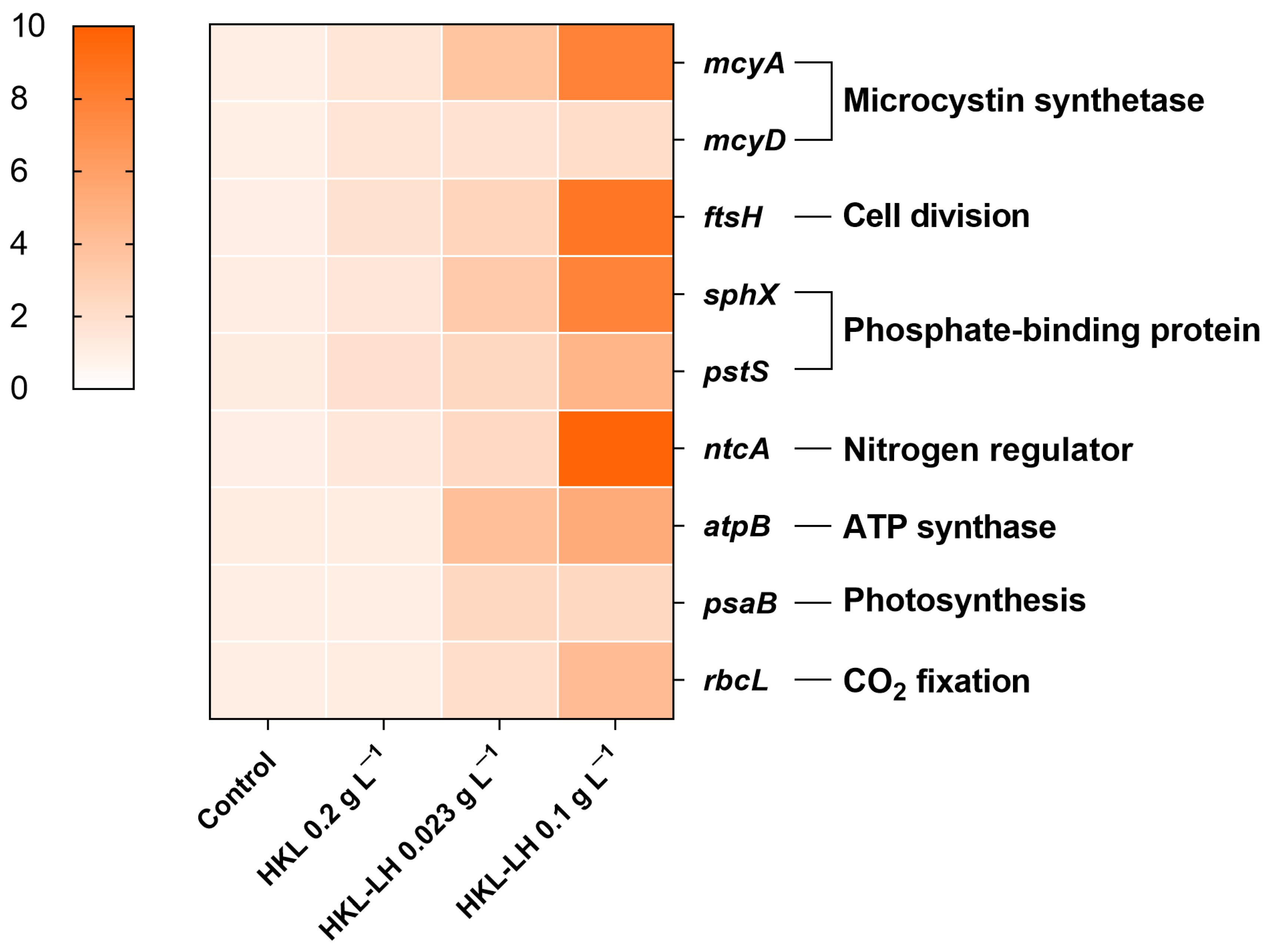
2.3.7. Gene Expression Test

The M. aeruginosa cells were collected by centrifugation (5000 rpm, 5 min) at 4 °C, then washed with PBS (pH = 7.2) three times. After immediately being frozen in liquid nitrogen, the cells were stored at −80 °C until sequencing. To explore the effects of phosphate restriction on the physiological and biochemical functions of M. aeruginosa, mcyA, mcyD, ftsH, sphX, pstS, ntcA, atpB, psaB, and rbcL were chosen as the target genes, and quantified by qPCR. Primer sequences and functions for the housekeeping gene (16S rRNA) and target genes are listed in Table S1 (Supplementary Materials). The relative expression was calculated by the 2−ΔΔCt method [49].

2.4. Statistical Analysis

A one-way ANOVA test was performed on Prism 8.0 (GraphPad Software, San Diego, CA, USA) to evaluate statistically significant differences between treatment and control groups. The figures and heatmap presented were created by Prism 8.0. All the experiments were conducted in triplicates, and the error bar represents mean ± standard deviation.

3. Results and Discussion

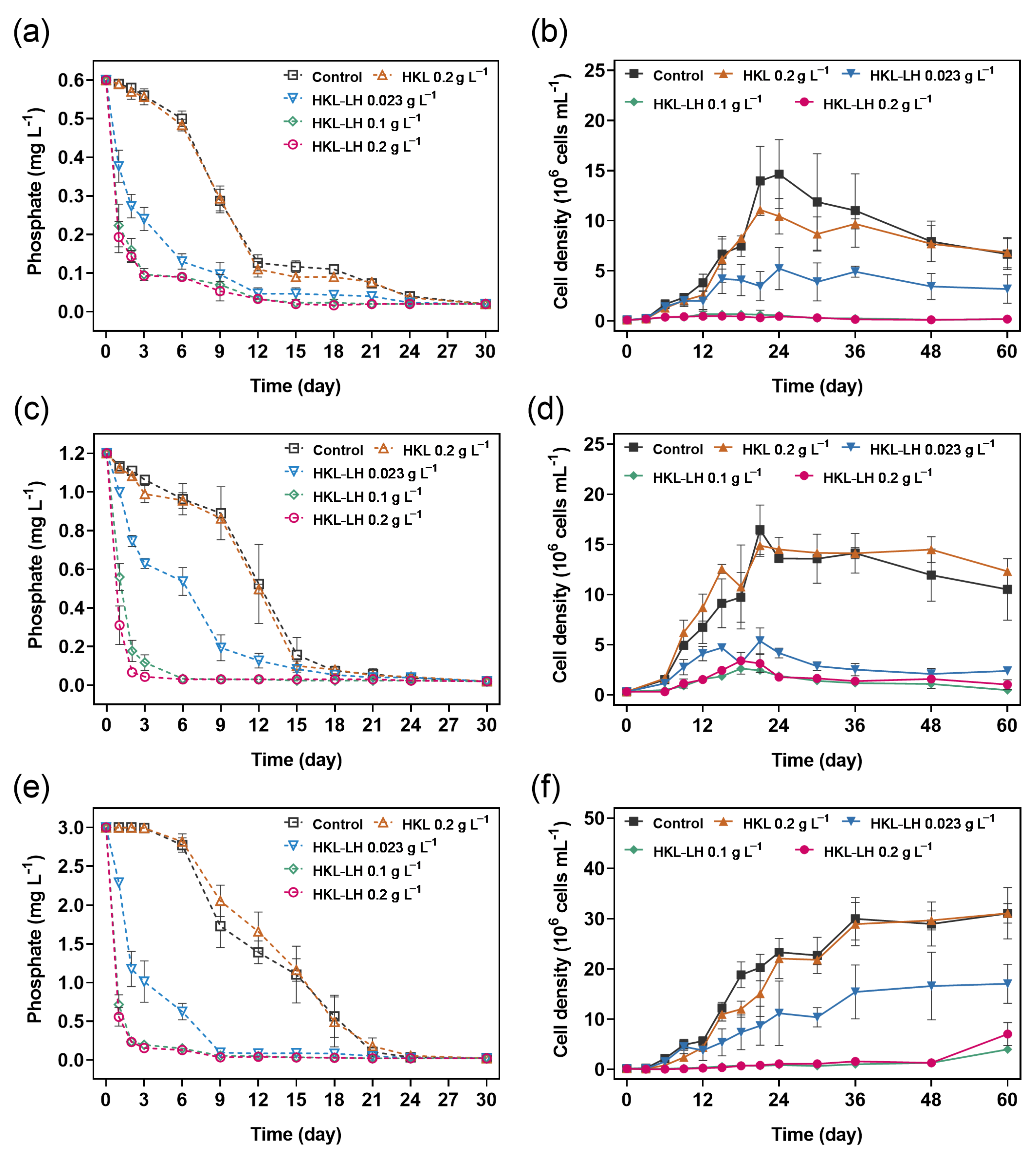
3.1. Phosphate Removal by HKL-LH in M. aeruginosa Samples

The changes in phosphate concentration in M. aeruginosa samples were investigated during HKL-LH treatment (Figure 1a,c,e). There were no significant differences found in the phosphate concentrations between the HKL-treated and control samples (p > 0.05). However, HKL-LH efficiently removed phosphate; i.e., reductions of 84–94% in phosphate concentration were achieved with 0.1 g L−1 HKL-LH treatment for 3 days. The results were comparable to those in a study by Zheng et al. [32], documenting 91% of phosphate was removed by 1.0 g L−1 HKL-LH in pure phosphate solution after 3 days. Previous studies have suggested that anions (e.g., CO32−, SO42−, Cl, and NO3−), cations (e.g., Ca2+ and Mg2+), and humic substances in the background water may influence the performance of La-based adsorbents [50,51,52]. However, in this study, it was shown that these factors scarcely abated the effectiveness of HKL-LH in phosphate removal, indicating HKL-LH is suitable for application in much more complicated water environments.
The capacity of phosphate removal by HKL-LH in M. aeruginosa samples was promoted with its increasing dosage (Table 1). For instance, the constant values (k) for phosphate removal were 0.86, 2.68, and 2.91 for HKL-LH dosages of 0.023, 0.1, and 0.2 g L−1, respectively, where an initial phosphate concentration of 0.6 mg−1 was applied. Generally, the k value in HKL-LH treatment was increased with reduced initial phosphate concentration. For instance, the k values were 2.68, 2.38, and 1.67, respectively, in the samples treated by 0.1 g L−1 HKL-LH, with 0.6, 1.2, and 3.0 mg L−1 phosphate initially. It indicates that the phosphate removal capacity is positively correlated with HKL-LH dosages while it is negatively associated with initial phosphate concentrations. Similar findings were reported on other La-containing adsorbents, such as hydrated lanthanum oxide-modified diatomite composites [53].

3.2. The Impact of HKL-LH on the Growth of M. aeruginosa

The M. aeruginosa cell densities of the control samples increased to peak values of 1.5 × 10⁷ and 1.6 × 10⁷ cells mL−1 on days 24 and 21, with initial phosphate concentrations of 0.6 and 1.2 mg L−1, respectively (Figure 1b,d). For the control samples added with 3.0 mg L−1 phosphate initially, higher cell density (3.0 × 10⁷ cells mL−1) was achieved and maintained after 36 days (Figure 1f). The growth dynamic of M. aeruginosa by HKL treatment was similar to that of the control, with equivalent initial phosphate concentration. However, HKL-LH effectively inhibited the growth of M. aeruginosa (Figure 1b,d,f). The inhibition degree of cell growth increased as HKL-LH dosages increasing from 0.023 g L−1 to 0.1 g L−1, while no significant differences (p > 0.05) were observed at HKL-LH dosages > 0.1 g L−1. For instance, with the initial phosphate concentration of 0.6 mg L−1, the inhibition rates of M. aeruginosa were 48%, 98%, and 97% after 0.023, 0.1, and 0.2 g L−1 HKL-LH treatments for 24 days, respectively (Figure 1b and Figure S1, Supplementary Materials). As a limiting factor, phosphorus plays a key role in the growth of cyanobacteria [54]. Thus, the growth of M. aeruginosa was inhibited with phosphate deficiency caused by HKL-LH (Figure 1a,c,e). It agrees with previous studies showing that phosphate adsorbents (e.g., lanthanum peroxide-loaded sepiolite nanocomposite and zero-valent iron-modified biochar) inhibited the metabolism of M. aeruginosa by preventing nutrient intake [55,56]. Notably, commercial phosphate fixatives such as Phoslock® (a lanthanum-embedded bentonite) only showed less than 15% decrease in the growth rate of M. aeruginosa at the concentration of 0.1 g L−1, while the cell growth was effectively inhibited by HKL-LH at the same concentration [57].
The effects of HKL-LH on the growth of M. aeruginosa with different initial cell densities were also investigated, initially with 0.6 mg L−1 phosphate (Figure 2). Generally, the inhibition rate of M. aeruginosa decreased with increasing initial cell densities. For instance, the inhibition rate of M. aeruginosa (initial cell density = 2.8 × 10⁴ cells mL−1) was 97% by 0.1 g L−1 HKL-LH treatment (Figure 2a), while it was 75% at a higher initial cell density of 6.3 × 10⁵ cells mL−1 (Figure 2c). As reported, cyanobacterial blooms with cell densities of 104 cells mL−1 are already visible at the water surface [58]. Therefore, HKL-LH is generally efficient to treat cyanobacterial blooms (with initial cell densities of 104−105 cells mL−1). However, to achieve optimization, it would be recommended to apply HKL-LH at their early bloom stage with a relatively lower biomass (≤104 cells mL−1).

3.3. Impacts of HKL-LH on the Viability of M. aeruginosa

The Fv/Fm value of M. aeruginosa in the control gradually increased to 0.46 during the first 12 days and then began to decline (Figure 3a). The photosynthetic activity in the HKL-treated samples showed similar trends to those observed in control. The Fv/Fm value of samples treated by 0.023 g L−1 HKL-LH was similar to that of control during the initial 18 days, but it became lower than the value in the control thereafter. The Fv/Fm value of M. aeruginosa by 0.1, and 0.2 g L−1 HKL-LH treatment was 74.4% and 78.9% lower than the control samples on day 36, respectively, indicating the photosynthetic system was seriously damaged [59]. It may be attributed to the efficient removal of phosphate by HKL-LH (Figure 1a), which led to the reduction in electron transport rate and thus inhibition of photosynthetic performance in M. aeruginosa cells [60,61]. In addition, according to the SEM images, HKL-LH particles may decrease cell integrity, enhance the cell aggregation, and then block the light that can illuminate M. aeruginosa cells, thereby suppressing the photosynthetic activity (Figure S2, Supplementary Materials).
More than 98% of the M. aeruginosa cells maintained membrane integrity in the control, 0.2 g L−1 HKL, and 0.023 g L−1 HKL-LH treatments over the course of 36 days (Figure 3b). A slight decrease in the proportion of intact cells could be observed after 0.1 and 0.2 g L−1 HKL-LH treatments for 36 days. Phosphorus is one of the important elements in the synthesis of phospholipids in cell membranes [62]. The membranes of M. aeruginosa cells are more likely to rupture when they are unable to take up sufficient phosphate over a long period [63]. SEM images also showed that the membranes of some cells in the groups treated by HKL-LH were wrinkled and weakened (Figure S2, Supplementary Materials). Similar results were reported that magnetite/lanthanum hydroxide composite caused the membrane rupture and morphological deformation of M. aeruginosa cells [64]. Therefore, HKL-LH can not only reduce the phosphate concentration and inhibit the growth of M. aeruginosa (Figure 1) but may also inactivate the cells through phosphate deficiency.

3.4. Changes in MCs and AOMs by HKL-LH Treatment

Accompanied by the increasing cyanobacterial biomass, the amount of MCs in the control gradually increased from 2.8 μg L−1 (day 0) to 366.0 μg L−1 (day 36) (Figure 4). The amount of MCs in the HKL-treated samples were similar to that of control, while they were significantly decreased after exposure to HKL-LH (p < 0.01). The concentrations of MCs were 186.1, 56.1, and 55.2 μg L−1, with 0.023, 0.1, and 0.2 g L−1 HKL-LH treatments for 36 days, respectively. Although the MC production was reduced by HKL-LH, the expression of mcyA (MC biosynthesis gene) was significantly upregulated (Figure 5). This may be attributed to a self-protective mechanism of M. aeruginosa cells in response to phosphate stress [65,66]. Thus, the reduced MCs may be owing to the decrease in M. aeruginosa biomass (Figure 1b). Previous studies have also shown that the production of MCs is positively correlated with the amounts of biomass [67,68]. Additionally, the amounts of MCs in cyanobacteria can be affected by the inhibition of photosynthetic activity, which decreases the availability of synthetic substrates and the energy required for MC synthesis (Figure 3a) [69,70].
The concentration of total DOC in the control reached 49.8 mg L−1 on day 24 and then decreased to 35.5 mg L−1 on day 36, which was consistent with the tendency in cell density of M. aeruginosa (Figure 6). The amounts of total DOC in the M. aeruginosa samples treated with HKL were similar to the control (p > 0.05). Conversely, HKL-LH significantly decreased the total DOC production in M. aeruginosa cells (p < 0.01), with decreases of 54.0%, 70.0%, and 74.2% by 0.023, 0.1, and 0.2 g L−1 dosages on day 36, respectively, compared to the control. The extracellular DOC concentration in the control increased from 4.3 mg L−1 (day 0) to 10.7 mg L−1 (day 36), probably due to release of intracellular substances from decaying cells. The extracellular DOC of M. aeruginosa were only 3.9 and 4.2 mg L−1 after 0.1 and 0.2 g L−1 HKL-LH treatments for 36 days, respectively. These lower concentrations of extracellular DOC may be attributed to the significant reduction in total DOC in M. aeruginosa with decreased biomass (Figure 1b). The accumulation of extracellular DOC is considered a precursor of DBPs, which can pose a significant challenge to water treatment plants due to their risks to human health and negative impacts on treatment processes [71,72]. Therefore, a reduction in extracellular DOC by HKL-LH treatment can effectively mitigate the associated risks.

4. Conclusions

In this study, the effects of HKL-LH on M. aeruginosa treatment were investigated. HKL-LH showed an outstanding phosphate removal performance in M. aeruginosa samples, thereby significantly inhibiting the growth of M. aeruginosa cells with various initial cell densities. Although the related genes were upregulated in response to the stress caused by HKL-LH, the photosynthetic activity and vitality of M. aeruginosa was significantly suppressed. In addition, HKL-LH efficiently reduced the production of MCs and AOMs in M. aeruginosa samples, which can lighten the burden on subsequent water treatment processes. Therefore, the implementation of HKL-LH may be practically applicable for water authorities to control cyanobacterial blooms in various water bodies, including recreational waters, aquaculture ponds, and urban lakes. In this study, it was shown that HKL-LH could effectively control cyanobacteria in media with different phosphate concentrations, while its application in other environments with more complex water matrices remains to be further explored.

Supplementary Materials

The following supporting information can be downloaded at: https://www.mdpi.com/article/10.3390/w17030428/s1, Table S1: Sequences of primer pairs used in qPCR. Figure S1: The inhibition rate of M. aeruginosa samples by HKL-LH treatments, with initial phosphate concentration: (a) 0.6 mg L−1, (b) 1.2 mg L−1, and (c) 3.0 mg L−1. The initial cell density was approximately 1.0 × 10⁵ cells mL−1. Figure S2: SEM images of M. aeruginosa samples after (a, b) HKL and (c, d) HKL-LH treatments for 24 days [73,74,75,76,77,78,79,80].

Author Contributions

Conceptualization, J.F. and X.L.; methodology, J.F.; validation, K.Z.; formal analysis, Y.M.; investigation, S.Z.; resources, J.F. and K.Z.; data curation, Y.M. and S.Z.; writing—original draft preparation, Y.M.; writing—review and editing, X.L. and J.F.; visualization, Y.M.; supervision, X.L. and J.F.; project administration, J.F. All authors have read and agreed to the published version of the manuscript.

Funding

This study was supported by the National Natural Science Foundation of China (41730316 and 51708490) and the Scientific Research Foundation for Scholars of Hangzhou Normal University (2021QDL059).

Data Availability Statement

The data are available from the corresponding author upon request.

Conflicts of Interest

The authors declare no conflict of interest.

References

  1. Paerl, H.W.; Huisman, J. Climate. Blooms like it hot. Science 2008, 320, 57–58. [Google Scholar] [CrossRef] [PubMed]
  2. Huisman, J.; Codd, G.A.; Paerl, H.W.; Ibelings, B.W.; Verspagen, J.M.H.; Visser, P.M. Cyanobacterial blooms. Nat. Rev. Microbiol. 2018, 16, 471–483. [Google Scholar] [CrossRef]
  3. Facey, J.A.; Michie, L.E.; King, J.J.; Hitchcock, J.N.; Apte, S.C.; Mitrovic, S.M. Severe cyanobacterial blooms in an Australian lake; causes and factors controlling succession patterns. Harmful. Algae 2022, 117, 102284. [Google Scholar] [CrossRef] [PubMed]
  4. Cook, K.V.; Li, C.; Cai, H.; Krumholz, L.R.; Hambright, K.D.; Paerl, H.W.; Steffen, M.M.; Wilson, A.E.; Burford, M.A.; Grossart, H.-P.; et al. The global Microcystis interactome. Limnol. Oceanogr. 2020, 65, S194–S207. [Google Scholar] [CrossRef]
  5. Codd, G.A.; Morrison, L.F.; Metcalf, J.S. Cyanobacterial toxins: Risk management for health protection. Toxicol. Appl. Pharmacol. 2005, 203, 264–272. [Google Scholar] [CrossRef]
  6. Gonzalez-Torres, A.; Pivokonsky, M.; Henderson, R.K. The impact of cell morphology and algal organic matter on algal floc properties. Water Res. 2019, 163, 114887. [Google Scholar] [CrossRef] [PubMed]
  7. Li, L.; Wang, Z.; Rietveld, L.C.; Gao, N.; Hu, J.; Yin, D.; Yu, S. Comparison of the effects of extracellular and intracellular organic matter extracted from Microcystis aeruginosa on ultrafiltration membrane fouling: Dynamics and mechanisms. Environ. Sci. Technol. 2014, 48, 14549–14557. [Google Scholar] [CrossRef]
  8. Tomlinson, A.; Drikas, M.; Brookes, J.D. The role of phytoplankton as pre-cursors for disinfection by-product formation upon chlorination. Water Res. 2016, 102, 229–240. [Google Scholar] [CrossRef]
  9. Ihsanullah, I.; Khan, M.T.; Hossain, M.F.; Bilal, M.; Ali Shah, I. Eco-Friendly Solutions to Emerging Contaminants: Unveiling the Potential of Bioremediation in Tackling Microplastic Pollution in Water. Adv. Sustain. Syst. 2024, 8, 2400172. [Google Scholar] [CrossRef]
  10. Aguilera, A.; Almanza, V.; Haakonsson, S.; Palacio, H.; Benitez Rodas, G.A.; Barros, M.U.G.; Capelo-Neto, J.; Urrutia, R.; Aubriot, L.; Bonilla, S. Cyanobacterial bloom monitoring and assessment in Latin America. Harmful. Algae 2023, 125, 102429. [Google Scholar] [CrossRef]
  11. Ho, J.C.; Michalak, A.M.; Pahlevan, N. Widespread global increase in intense lake phytoplankton blooms since the 1980s. Nature 2019, 574, 667–670. [Google Scholar] [CrossRef] [PubMed]
  12. Huo, D.; Gan, N.; Geng, R.; Cao, Q.; Song, L.; Yu, G.; Li, R. Cyanobacterial blooms in China: Diversity, distribution, and cyanotoxins. Harmful. Algae 2021, 109, 102106. [Google Scholar] [CrossRef]
  13. Conley, D.J.; Paerl, H.W.; Howarth, R.W.; Boesch, D.F.; Seitzinger, S.P.; Havens, K.E.; Lancelot, C.; Likens, G.E. Ecology. Controlling eutrophication: Nitrogen and phosphorus. Science 2009, 323, 1014–1015. [Google Scholar] [CrossRef] [PubMed]
  14. Xie, L.Q.; Xie, P.; Tang, H.J. Enhancement of dissolved phosphorus release from sediment to lake water by Microcystis blooms--an enclosure experiment in a hyper-eutrophic, subtropical Chinese lake. Environ. Pollut. 2003, 122, 391–399. [Google Scholar] [CrossRef]
  15. Correll, D.L. The Role of Phosphorus in the Eutrophication of Receiving Waters: A Review. J. Environ. Qual. 1998, 27, 261–266. [Google Scholar] [CrossRef]
  16. Nollet, L.M.L.; Gelder, L.S.P.D. (Eds.) Handbook of Water Analysis, 2nd ed.; CRC Press: Boca Raton, FL, USA, 2007. [Google Scholar] [CrossRef]
  17. Loganathan, P.; Vigneswaran, S.; Kandasamy, J.; Bolan, N.S. Removal and Recovery of Phosphate from Water Using Sorption. Crit. Rev. Environ. Sci. Technol. 2014, 44, 847–907. [Google Scholar] [CrossRef]
  18. Wang, Z.; Xia, D.; Cui, S.; Yu, W.; Wang, B.; Liu, H. A high-capacity nanocellulose aerogel uniformly immobilized with a high loading of nano-La(OH)3 for phosphate removal. Chem. Eng. J. 2022, 433, 134439. [Google Scholar] [CrossRef]
  19. D’Haese, P.C.; Douglas, G.; Verhulst, A.; Neven, E.; Behets, G.J.; Vervaet, B.A.; Finsterle, K.; Lürling, M.; Spears, B. Human health risk associated with the management of phosphorus in freshwaters using lanthanum and aluminium. Chemosphere 2019, 220, 286–299. [Google Scholar] [CrossRef]
  20. Ma, C.; Hu, W.; Pei, H.; Xu, H.; Pei, R. Enhancing integrated removal of Microcystis aeruginosa and adsorption of microcystins using chitosan-aluminum chloride combined coagulants: Effect of chemical dosing orders and coagulation mechanisms. Colloids Surf. A Physicochem. Eng. Asp. 2016, 490, 258–267. [Google Scholar] [CrossRef]
  21. Reitzel, K.; Andersen, F.Ø.; Egemose, S.; Jensen, H.S. Phosphate adsorption by lanthanum modified bentonite clay in fresh and brackish water. Water Res. 2013, 47, 2787–2796. [Google Scholar] [CrossRef] [PubMed]
  22. Yin, H.; Ren, C.; Li, W. Introducing hydrate aluminum into porous thermally-treated calcium-rich attapulgite to enhance its phosphorus sorption capacity for sediment internal loading management. Chem. Eng. J. 2018, 348, 704–712. [Google Scholar] [CrossRef]
  23. Eltaweil, A.S.; Omer, A.M.; El-Aqapa, H.G.; Gaber, N.M.; Attia, N.F.; El-Subruiti, G.M.; Mohy-Eldin, M.S.; Abd El-Monaem, E.M. Chitosan based adsorbents for the removal of phosphate and nitrate: A critical review. Carbohydr. Polym. 2021, 274, 118671. [Google Scholar] [CrossRef] [PubMed]
  24. Lürling, M.; Noyma, N.P.; de Magalhães, L.; Miranda, M.; Mucci, M.; van Oosterhout, F.; Huszar, V.L.M.; Marinho, M.M. Critical assessment of chitosan as coagulant to remove cyanobacteria. Harmful. Algae 2017, 66, 1–12. [Google Scholar] [CrossRef]
  25. Awad, M.E.; López-Galindo, A.; Setti, M.; El-Rahmany, M.M.; Iborra, C.V. Kaolinite in pharmaceutics and biomedicine. Int. J. Pharm. 2017, 533, 34–48. [Google Scholar] [CrossRef]
  26. Detellier, C. Functional Kaolinite. Chem. Rec. 2018, 18, 868–877. [Google Scholar] [CrossRef]
  27. Johnson, E.B.G.; Arshad, S.E. Hydrothermally synthesized zeolites based on kaolinite: A review. Appl. Clay Sci. 2014, 97–98, 215–221. [Google Scholar] [CrossRef]
  28. Dill, H.G. Kaolin: Soil, rock and ore: From the mineral to the magmatic, sedimentary and metamorphic environments. Earth Sci. Rev. 2016, 161, 16–129. [Google Scholar] [CrossRef]
  29. García, K.I.; Quezada, G.R.; Arumí, J.L.; Toledo, P.G. Phosphate aggregation, diffusion, and adsorption on kaolinite in saline solutions by molecular dynamics simulation. Appl. Clay Sci. 2023, 233, 106844. [Google Scholar] [CrossRef]
  30. Xie, J.; Wang, Z.; Lu, S.; Wu, D.; Zhang, Z.; Kong, H. Removal and recovery of phosphate from water by lanthanum hydroxide materials. Chem. Eng. J. 2014, 254, 163–170. [Google Scholar] [CrossRef]
  31. Yang, Z.; Hou, J.; Pan, Z.; Wu, M.; Zhang, M.; Yin, X.; Wu, J.; Miao, L.; Liu, Q. A novel La(OH)3 decorated co-graft tannin and polyethyleneimine co-coating magnetic adsorbent for effective and selective phosphate removal from natural water and real wastewater. J. Clean. Prod. 2022, 369, 133345. [Google Scholar] [CrossRef]
  32. Zheng, S.; Fan, J.; Lu, X. Heated kaolinite-La(III) hydroxide complex for effective removal of phosphate from eutrophic water. Appl. Clay Sci. 2023, 231, 106729. [Google Scholar] [CrossRef]
  33. Lürling, M.; Faassen, E.J. Controlling toxic cyanobacteria: Effects of dredging and phosphorus-binding clay on cyanobacteria and microcystins. Water Res. 2012, 46, 1447–1459. [Google Scholar] [CrossRef]
  34. Peschek, G.A.; Obinger, C.; Renger, G. (Eds.) Bioenergetic Processes of Cyanobacteria; Springer: Dordrecht, The Netherlands, 2011. [Google Scholar] [CrossRef]
  35. Briddon, C.L.; Szekeres, E.; Hegedüs, A.; Nicoară, M.; Chiriac, C.; Stockenreiter, M.; Drugă, B. The combined impact of low temperatures and shifting phosphorus availability on the competitive ability of cyanobacteria. Sci. Rep. 2022, 12, 16409. [Google Scholar] [CrossRef] [PubMed]
  36. Paerl, H.W.; Otten, T.G. Harmful cyanobacterial blooms: Causes, consequences, and controls. Microb. Ecol. 2013, 65, 995–1010. [Google Scholar] [CrossRef] [PubMed]
  37. Bacelo, H.; Pintor, A.M.A.; Santos, S.C.R.; Boaventura, R.A.R.; Botelho, C.M.S. Performance and prospects of different adsorbents for phosphorus uptake and recovery from water. Chem. Eng. J. 2020, 381, 122566. [Google Scholar] [CrossRef]
  38. Sun, F.; Pei, H.-Y.; Hu, W.-R.; Ma, C.-X. The lysis of Microcystis aeruginosa in AlCl3 coagulation and sedimentation processes. Chem. Eng. J. 2012, 193–194, 196–202. [Google Scholar] [CrossRef]
  39. Du, H.; Chen, Z.; Mao, G.; Chen, L.; Crittenden, J.; Li, R.Y.M.; Chai, L. Evaluation of eutrophication in freshwater lakes: A new non-equilibrium statistical approach. Ecol. Indic 2019, 102, 686–692. [Google Scholar] [CrossRef]
  40. de Farias Silva, C.E.; de Oliveira Cerqueira, R.B.; de Lima Neto, C.F.; de Andrade, F.P.; de Oliveira Carvalho, F.; Tonholo, J. Developing a kinetic model to describe wastewater treatment by microalgae based on simultaneous carbon, nitrogen and phosphorous removal. J. Environ. Chem. Eng. 2020, 8, 103792. [Google Scholar] [CrossRef]
  41. Fan, J.; Ho, L.; Hobson, P.; Brookes, J. Evaluating the effectiveness of copper sulphate, chlorine, potassium permanganate, hydrogen peroxide and ozone on cyanobacterial cell integrity. Water Res. 2013, 47, 5153–5164. [Google Scholar] [CrossRef]
  42. Macedo, R.S.; Lombardi, A.T.; Omachi, C.Y.; Rörig, L.R. Effects of the herbicide bentazon on growth and photosystem II maximum quantum yield of the marine diatom Skeletonema costatum. Toxicol. Vitr. 2008, 22, 716–722. [Google Scholar] [CrossRef]
  43. Wu, H.; Wei, G.; Tan, X.; Li, L.; Li, M. Species-dependent variation in sensitivity of Microcystis species to copper sulfate: Implication in algal toxicity of copper and controls of blooms. Sci. Rep. 2017, 7, 40393. [Google Scholar] [CrossRef] [PubMed]
  44. Campbell, D.; Hurry, V.; Clarke, A.K.; Gustafsson, P.; Öquist, G. Chlorophyll Fluorescence Analysis of Cyanobacterial Photosynthesis and Acclimation. Microbiol. Mol. Biol. Rev. 1998, 62, 667–683. [Google Scholar] [CrossRef] [PubMed]
  45. Lin, S.; Yu, X.; Fang, J.; Fan, J. Influences of the micropollutant erythromycin on cyanobacteria treatment with potassium permanganate. Water Res. 2020, 177, 115786. [Google Scholar] [CrossRef]
  46. Nicholson, B.C.; Rositano, J.; Burch, M.D. Destruction of cyanobacterial peptide hepatotoxins by chlorine and chloramine. Water Res. 1994, 28, 1297–1303. [Google Scholar] [CrossRef]
  47. Knochen, M.; Rodríguez-Silva, J.C.; Silva-Silva, J. Exploitation of reaction mechanisms for sensitivity enhancement in the determination of phosphorus by sequential injection analysis. Talanta 2020, 209, 120589. [Google Scholar] [CrossRef]
  48. Li, L.; Gao, N.; Deng, Y.; Yao, J.; Zhang, K. Characterization of intracellular & extracellular algae organic matters (AOM) of Microcystic aeruginosa and formation of AOM-associated disinfection byproducts and odor & taste compounds. Water Res. 2012, 46, 1233–1240. [Google Scholar] [CrossRef]
  49. Livak, K.J.; Schmittgen, T.D. Analysis of Relative Gene Expression Data Using Real-Time Quantitative PCR and the 2−ΔΔCT Method. Methods 2001, 25, 402–408. [Google Scholar] [CrossRef] [PubMed]
  50. He, Q.; Zhao, H.; Teng, Z.; Wang, Y.; Li, M.; Hoffmann, M.R. Phosphate removal and recovery by lanthanum-based adsorbents: A review for current advances. Chemosphere 2022, 303, 134987. [Google Scholar] [CrossRef]
  51. Li, J.; Li, B.; Yu, W.; Huang, H.; Han, J.-C.; Huang, Y.; Wu, X.; Young, B.; Wang, G. Lanthanum-based adsorbents for phosphate reutilization: Interference factors, adsorbent regeneration, and research gaps. Sustain. Horiz. 2022, 1, 100011. [Google Scholar] [CrossRef]
  52. de Lucena-Silva, D.; Molozzi, J.; Severiano, J.D.S.; Becker, V.; de Lucena Barbosa, J.E. Removal efficiency of phosphorus, cyanobacteria and cyanotoxins by the “flock & sink” mitigation technique in semi-arid eutrophic waters. Water Res. 2019, 159, 262–273. [Google Scholar] [CrossRef]
  53. Wu, Y.; Li, X.; Yang, Q.; Wang, D.; Xu, Q.; Yao, F.; Chen, F.; Tao, Z.; Huang, X. Hydrated lanthanum oxide-modified diatomite as highly efficient adsorbent for low-concentration phosphate removal from secondary effluents. J. Environ. Manag. 2019, 231, 370–379. [Google Scholar] [CrossRef]
  54. Correll, D.L. Phosphorus: A rate limiting nutrient in surface waters. Poult. Sci. 1999, 78, 674–682. [Google Scholar] [CrossRef]
  55. Ren, L.; Li, Y.; Wang, K.; Ding, K.; Sha, M.; Cao, Y.; Kong, F.; Wang, S. Recovery of phosphorus from eutrophic water using nano zero-valent iron-modified biochar and its utilization. Chemosphere 2021, 284, 131391. [Google Scholar] [CrossRef] [PubMed]
  56. Shan, S.; Chen, Z.; Yuen Koh, K.; Cui, F.; Paul Chen, J. Development and application of lanthanum peroxide loaded sepiolite nanocomposites for simultaneous removal of phosphate and inhibition of cyanobacteria growth. J. Colloid Interface Sci. 2022, 624, 691–703. [Google Scholar] [CrossRef]
  57. Kang, L.; Mucci, M.; Lürling, M. Compounds to mitigate cyanobacterial blooms affect growth and toxicity of Microcystis aeruginosa. Harmful. Algae 2022, 118, 102311. [Google Scholar] [CrossRef]
  58. Graham, J.L.; Loftin, K.A.; Ziegler, A.C.; Meyer, M.T. Guidelines for Design and Sampling for Cyanobacterial Toxin and Taste-and-Odor Studies in Lakes and Reservoirs; Scientific Investigations Report No. 2008–5038; USGS: Reston, VA, USA, 2008. [Google Scholar] [CrossRef]
  59. Sipka, G.B.; Magyar, M.; Mezzetti, A.; Akhtar, P.; Zhu, Q.; Xiao, Y.; Han, G.; Santabarbara, S.; Shen, J.-R.; Lambrev, P.H.; et al. Light-adapted charge-separated state of photosystem II: Structural and functional dynamics of the closed reaction center. Plant Cell 2021, 33, 1286–1302. [Google Scholar] [CrossRef] [PubMed]
  60. Peng, X.; Lin, Q.; Liu, B.; Huang, S.; Yan, W.; Zhang, L.; Ge, F.; Zhang, Y.; Wu, Z. Effect of submerged plant coverage on phytoplankton community dynamics and photosynthetic activity in situ. J. Environ. Manag. 2022, 301, 113822. [Google Scholar] [CrossRef] [PubMed]
  61. Wu, Z.; Zeng, B.; Li, R.; Song, L. Physiological regulation of Cylindrospermopsis raciborskii (Nostocales, Cyanobacteria) in response to inorganic phosphorus limitation. Harmful. Algae 2012, 15, 53–58. [Google Scholar] [CrossRef]
  62. Butusov, M.; Jernelöv, A. Phosphorus in the Organic Life: Cells, Tissues, Organisms. In Phosphorus: An Element That Could Have Been Called Lucifer; Butusov, M., Jernelöv, A., Eds.; Springer: New York, NY, USA, 2013; pp. 13–17. [Google Scholar] [CrossRef]
  63. Zhang, C.; Chen, G.; Wang, Y.; Guo, C.; Zhou, J. Physiological and molecular responses of Prorocentrum donghaiense to dissolved inorganic phosphorus limitation. Mar. Pollut. Bull. 2018, 129, 562–572. [Google Scholar] [CrossRef]
  64. Song, Q.; Huang, S.; Xu, L.; Li, Q.; Luo, X.; Zheng, Z. Response of Magnetite/Lanthanum hydroxide composite on cyanobacterial bloom. Chemosphere 2021, 275, 130017. [Google Scholar] [CrossRef]
  65. Chen, Q.; Wang, M.; Zhang, J.; Shi, W.; Mynett, A.E.; Yan, H.; Hu, L. Physiological effects of nitrate, ammonium, and urea on the growth and microcystins contamination of Microcystis aeruginosa: Implication for nitrogen mitigation. Water Res. 2019, 163, 114890. [Google Scholar] [CrossRef] [PubMed]
  66. Christiansen, G.; Fastner, J.; Erhard, M.; Börner, T.; Dittmann, E. Microcystin biosynthesis in planktothrix: Genes, evolution, and manipulation. J. Bacteriol. 2003, 185, 564–572. [Google Scholar] [CrossRef] [PubMed]
  67. MacKeigan, P.W.; Zastepa, A.; Taranu, Z.E.; Westrick, J.A.; Liang, A.; Pick, F.R.; Beisner, B.E.; Gregory-Eaves, I. Microcystin concentrations and congener composition in relation to environmental variables across 440 north-temperate and boreal lakes. Sci. Total Environ. 2023, 884, 163811. [Google Scholar] [CrossRef]
  68. Xin, R.; Yu, X.; Fan, J. Physiological, biochemical and transcriptional responses of cyanobacteria to environmentally relevant concentrations of a typical antibiotic-roxithromycin. Sci. Total Environ. 2022, 814, 152703. [Google Scholar] [CrossRef] [PubMed]
  69. Dai, R.; Li, Z.; Yan, F.; An, L.; Du, W.; Li, X. Evaluation of changes in M. aeruginosa growth and microcystin production under phosphorus starvation via transcriptomic surveys. Sci. Total Environ. 2023, 893, 164848. [Google Scholar] [CrossRef]
  70. Rantala, A.; Fewer, D.P.; Hisbergues, M.; Rouhiainen, L.; Vaitomaa, J.; Börner, T.; Sivonen, K. Phylogenetic evidence for the early evolution of microcystin synthesis. Proc. Natl. Acad. Sci. USA 2004, 101, 568–573. [Google Scholar] [CrossRef]
  71. Wang, X.; Qian, Y.; Chen, Y.; Liu, F.; An, D.; Yang, G.; Dai, R. Application of fluorescence spectra and molecular weight analysis in the identification of algal organic matter-based disinfection by-product precursors. Sci. Total Environ. 2023, 882, 163589. [Google Scholar] [CrossRef] [PubMed]
  72. Zhou, S.; Shao, Y.; Gao, N.; Deng, Y.; Li, L.; Deng, J.; Tan, C. Characterization of algal organic matters of Microcystis aeruginosa: Biodegradability, DBP formation and membrane fouling potential. Water Res. 2014, 52, 199–207. [Google Scholar] [CrossRef] [PubMed]
  73. Harke, M.J.; Berry, D.L.; Ammerman, J.W.; Gobler, C.J. Molecular response of the bloom-forming cyanobacterium, Microcystis aeruginosa, to phosphorus limitation. Microb. Ecol. 2012, 63, 188–198. [Google Scholar] [CrossRef]
  74. Kaebernick, M.; Neilan, B.A.; Börner, T.; Dittmann, E. Light and the transcriptional response of the microcystin biosynthesis gene cluster. Appl. Environ. Microbiol. 2000, 66, 3387–3392. [Google Scholar] [CrossRef] [PubMed]
  75. Li, D.; Kang, X.; Chu, L.; Wang, Y.; Song, X.; Zhao, X.; Cao, X. Algicidal mechanism of Raoultella ornithinolytica against Microcystis aeruginosa: Antioxidant response, photosynthetic system damage and microcystin degradation. Environ. Pollut. 2021, 287, 117644. [Google Scholar] [CrossRef] [PubMed]
  76. Lu, Y.; Wang, J.; Yu, Y.; Shi, L.; Kong, F. Changes in the physiology and gene expression of Microcystis aeruginosa under EGCG stress. Chemosphere 2014, 117, 164–169. [Google Scholar] [CrossRef] [PubMed]
  77. Muro-Pastor, M.I.; Florencio, F.J. Regulation of ammonium assimilation in cyanobacteria. Plant Physiol. Biochem. 2003, 41, 595–603. [Google Scholar] [CrossRef]
  78. Pimentel, J.S.M.; Giani, A. Microcystin production and regulation under nutrient stress conditions in toxic microcystis strains. Appl. Environ. Microbiol. 2014, 80, 5836–5843. [Google Scholar] [CrossRef]
  79. Qian, H.; Yu, S.; Sun, Z.; Xie, X.; Liu, W.; Fu, Z. Effects of copper sulfate, hydrogen peroxide and N-phenyl-2-naphthylamine on oxidative stress and the expression of genes involved photosynthesis and microcystin disposition in Microcystis aeruginosa. Aquat. Toxicol. 2010, 99, 405–412. [Google Scholar] [CrossRef] [PubMed]
  80. Rinta-Kanto, J.M.; Ouellette, A.J.A.; Boyer, G.L.; Twiss, M.R.; Bridgeman, T.B.; Wilhelm, S.W. Quantification of toxic Microcystis spp. during the 2003 and 2004 blooms in western Lake Erie using quantitative real-time PCR. Environ. Sci. Technol. 2005, 39, 4198–4205. [Google Scholar] [CrossRef]
Figure 1. The changes in phosphate concentrations and the growth of M. aeruginosa by HKL-LH treatments, with different initial phosphate concentrations in the background water: (a,b) 0.6 mg L−1, (c,d) 1.2 mg L−1, and (e,f) 3.0 mg L−1. The initial cell density was approximately 1.0 × 10⁵ cells mL−1.
Water 17 00428 g001
Figure 2. The inhibition rate of M. aeruginosa samples by 0.023–0.2 g L−1 HKL-LH treatments, with initial cell densities: (a) 2.8 × 10⁴ cells mL−1, (b) 9.7 × 10⁴ cells mL−1, and (c) 6.3 × 10⁵ cells mL−1. The initial phosphate concentration was 0.6 mg L−1.
Water 17 00428 g002
Figure 3. The effects of HKL and HKL-LH on (a) photosynthetic efficiency and (b) cell integrity of M. aeruginosa samples. The initial cell density was approximately 1.0 × 10⁵ cells mL−1, and the initial phosphate concentration was 0.6 mg L−1.
Water 17 00428 g003
Figure 4. The concentrations of MC-LR in the M. aeruginosa samples after exposure to various concentrations of HKL-LH. The initial cell density was approximately 1.0 × 10⁵ cells mL−1, and the initial phosphate concentration was 0.6 mg L−1.
Water 17 00428 g004
Figure 5. The functional gene expression in M. aeruginosa samples treated by HKL and HKL-LH. The relative expression of the control is close to 1. The orange color represents the upregulation of the relative expression. The initial cell density was approximately 1.0 × 10⁵ cells mL−1, and the initial phosphate concentration was 0.6 mg L−1.
Water 17 00428 g005
Figure 6. The amounts of DOC in the M. aeruginosa samples after exposure to various concentrations of HKL-LH. The initial cell density was approximately 1.0 × 10⁵ cells mL−1, and the initial phosphate concentration was 0.6 mg L−1.
Water 17 00428 g006
Table 1. Kinetic parameters of phosphate removal by HKL-LH with different initial phosphate concentrations in M. aeruginosa samples.
[HKL-LH]
(g L−1)
Initial Phosphate Concentrations (mg L−1)
0.61.23.0
k (L mg−1 d−1)R2k (L mg−1 d−1)R2k (L mg−1 d−1)R2
0.0230.860.99080.210.98530.220.9721
0.12.680.99042.380.97041.670.9823
0.22.910.99436.930.96702.000.9943
Disclaimer/Publisher’s Note: The statements, opinions and data contained in all publications are solely those of the individual author(s) and contributor(s) and not of MDPI and/or the editor(s). MDPI and/or the editor(s) disclaim responsibility for any injury to people or property resulting from any ideas, methods, instructions or products referred to in the content.

