Article

Meso-Damage Mechanism of Strength and Deformation Characteristics of Typical Sandstone in Xinwen Coalfield

1
State Key Laboratory of Mining Disaster Prevention and Control Co-Founded by Shandong Province and the Ministry of Science and Technology, Shandong University of Science and Technology, Qingdao 266590, China
2
College of Safety and Environmental Engineering, Shandong University of Science and Technology, Qingdao 266590, China
3
College of Energy and Mining Engineering, Shandong University of Science and Technology, Qingdao 266590, China
*
Authors to whom correspondence should be addressed.
Symmetry 2020, 12(11), 1815; https://doi.org/10.3390/sym12111815
Submission received: 3 September 2020 / Revised: 22 October 2020 / Accepted: 29 October 2020 / Published: 2 November 2020

Abstract

:
Geotechnical engineering problems will cause asymmetric deformation of surrounding rock, which is not conducive to the stability of rock mass. In order to study the meso-damage mechanism of rock strength and deformation characteristics, taking three kinds of typical sandstone as examples, the meso structure images of sandstone were obtained by JSM-6510LV scanning electron microscope (SEM). According to the meso-structure images and fractal theory, MATLAB was compiled to calculate the fractal dimension of the meso-structure images of the three types of sandstone. The uniaxial compression test of sandstone is carried out by using the Shimadzu electronic universal testing machine. The mechanical parameters of three types of sandstone are obtained. By comparing the relationship between fractal dimension and mechanical parameters, the correlation between strength and deformation characteristics of sandstone and mesostructure is analyzed. The results show that sandstone has the characteristic of self-similarity. The fractal dimension of sandstone decreases with the increase in magnification. The macro-mechanical properties of sandstone are closely related to the meso-structure. The strength characteristics of sandstone are inversely proportional to the fractal dimension. The greater the uniaxial compressive strength and elastic modulus, the smaller the fractal dimension. The damage problem of sandstone can be characterized by critical damage value, which is proportional to the fractal dimension; the larger the fractal dimension, the more serious the internal damage of sandstone.

1. Introduction

In the process of tunnel construction and coal mining, the stability of surrounding rock is very important. In the process of geotechnical engineering, complex geological and environmental conditions will be encountered, resulting in asymmetric deformation of surrounding rock. The surrounding rock deformation and collapse accidents will cause serious consequences, such as casualties, environmental damage and economic losses, affecting the safe construction and operation of rock engineering. Therefore, it is necessary to ensure the stability of surrounding rock. The macro-deformation and mechanical properties of rock are closely related to its meso-structure. After a long period of geological activities, micro-pores and micro fra-tures will be produced in rocks, which will accelerate the process of rock instability and failure. Therefore, the research on meso-damage characteristics of rock is a prospective topic [1,2]. The research on the evolution law and strength model of rock mass meso-structure can enhance safe mining. It can also improve the stability of surrounding rock support and underground engineering and make it more conducive for safe mining.
In view of the problems in the study of rock meso structure, much research has been carried out. By using digital image processing and particle flow, Yin et al. [3] reconstructed the meso model reflecting the characteristics of rock heterogeneity structure. Using the three-dimensional numerical homogenization model, Wetczel et al. [4] shows that the change in mineral microstructure will lead to the change in macro-mechanical behavior. Van et al. [5] proposed a method to measure the microstructure of rocks in hyperspectral images. According to the objects in the diagram, shape parameters are calculated to describe the microstructure and elements. Hammes et al. [6] introduced an easy-to-use software fame based on MATLAB, which is used to process the dataset recorded by the microscope of automatic fabric analyzer, and can determine the microstructure of rock. Sabatakakis et al. [7] evaluated the mineral composition and microstructure of grains of sedimentary rocks and igneous rocks, and it was found that the influence of microstructure on them can be quantitatively described. Regnet et al. [8] reviewed some of the literature, mainly on the study of microstructure parameters of physical response of rock properties in carbonate rocks. In addition, the meso-structure of rock mass is related to the macro-damage, which is a new research direction in the field of geotechnical engineering. There are micro-pores, cracks and other primary damage in the rock, and its failure and deformation law is affected by the internal structure. For decades, experts have studied the macro-mechanical properties and failure mechanism of rock mass by various methods [9,10], The physical properties and parameters of sandstone are the important factors affecting the mechanical properties of sandstone [11,12,13]. Liu et al. [14] proposed a dynamic damage constitutive model for rock mass with intermittent joints, and studied the variation in rock macroscopic mechanical parameters with failure. Based on the principle of damage mechanics, Li [15] deduced the calculation formula of macroscopic damage variable of jointed rock mass. Using the X-ray method, Gooya et al. [16] studied the influence of tomography resolution on the calculation of microscopic properties of sandstone and carbonate rock; the result shows that the difference between the two types of rock is due to different pore size and distribution. Xie [17] carried out uniaxial compression tests on multi-size rock masses with different dip angles, revealing the deformation evolution law of fractured rock mass. Based on Lemaitre’s equivalent strain hypothesis, Zhang et al. [18] obtained the constitutive equation of macroscopic and microscopic damage coupling. Zhang, et al. [19] carried out triaxial creep test on the surrounding rock of Hengda coal mine in Fuxin, the damage variable of rock was deduced and the nonlinear creep damage model was established. Mohamadi et al. [20] analyzed the damage process of rocks with specific cracks, and adopted an enhanced embedded discontinuity method, which can predict the formation and propagation of new cracks. Zhu [21] considered the micromechanics and thermodynamics theory of rock comprehensively, established the micromechanics model of three-phase damage to rock.
Fractal dimension can be used to quantify the mesostructure of rock; it reflects the relationship between rock mesostructure and mechanical properties. In the field of Geoscience, fractal theory has gained increasing attention. It has been widely used and developed in mineral resources exploration, mining and evaluation [22,23]. At the same time, in the field of rock mechanics, fractal theory can be used to analyze some parameters of rock failure process, such as rock deformation state, pore and fissure development [24]. However, there are few studies on this aspect, Sandstone is a common type of rock, therefore, three types of typical sandstone in Xinwen mining were selected, and their fractal dimension was calculated. By the uniaxial compression test, the relationship between the micro-structure and the macro-damage is analyzed.

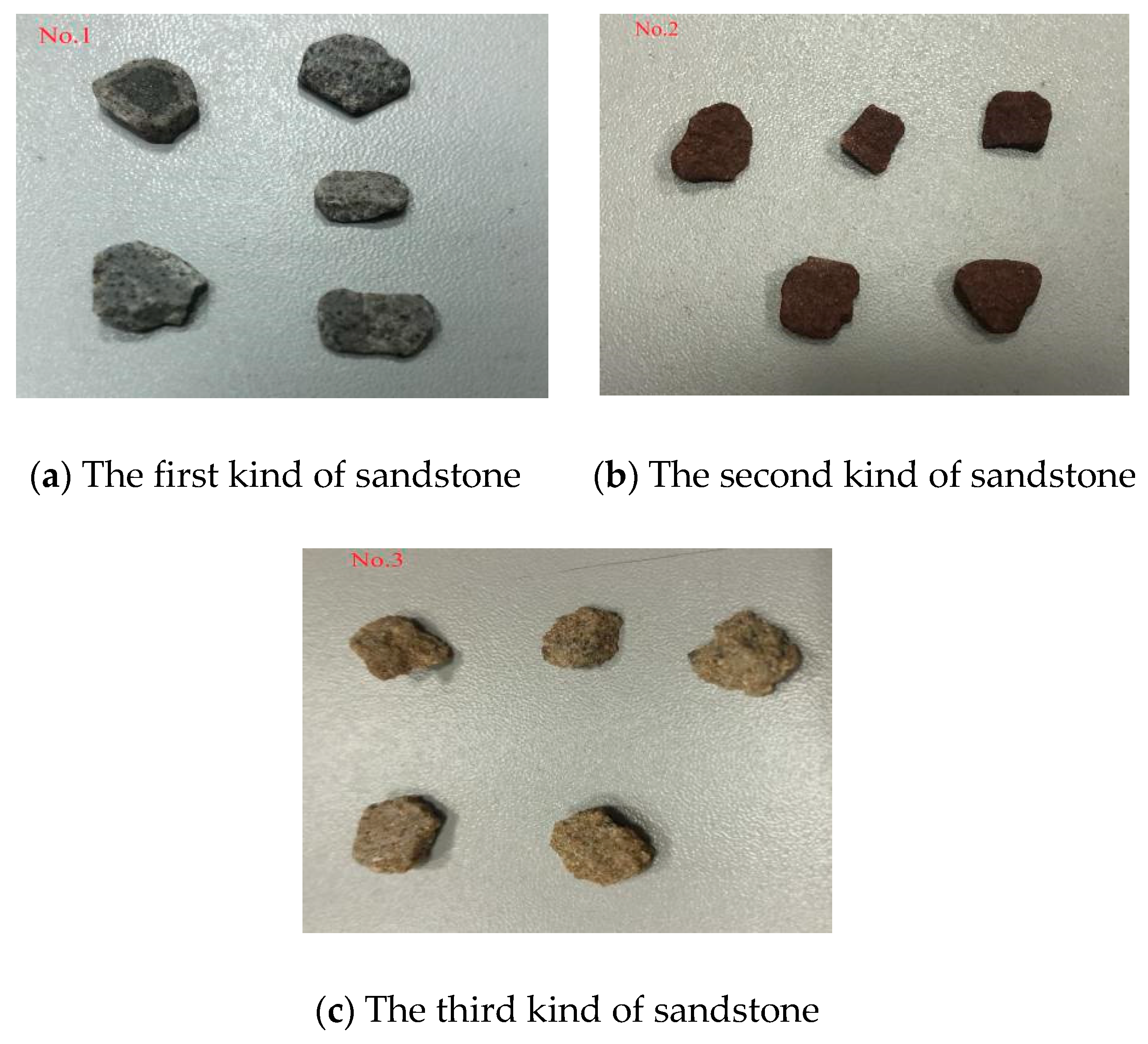
2. Typical Sandstone and Sample Preparation

Three types of typical sandstone (No.1,2,3) in Xinwen coalfield were selected (Figure 1). The mechanical properties of these three types of sandstone were obviously different. The samples are taken from Shanxi formation of the North China type Carboniferous Permian, mainly composed of sandstone, siltstone, mudstone and coal seam. The first kind of sandstone is gray black, containing plant root fossil fragments, massive structure; the second kind is dark red, mainly composed of quartz particles, with stable structure; the third type is brown-yellow with a fine-grained structure. Using Shimadzu electronic universal testing machine and SEM, the macro-mechanical parameters and meso-structure of them were studied.

2.1. Typical Sandstone Geological Conditions

Sandstone is a kind of sedimentary rock, which is mainly cemented by various sand particles. The sand accounts for more than 50% of the component, and the diameter is between 0.05 and 2 mm. It is usually light brown or red. The sandstone structure is stable, resistant to damage and easy to clean. It is a kind of high-quality natural stone.
Xinwen Mining Group, founded in 1956, located in Xintai City, Shandong Province, with 28.3 billion tons of coal geological reserves and 112 million tons of designed production capacity. The mining area is mainly composed of sandstone, siltstone, mudstone and coal seam, etc. The coal types mainly include fat coal, gas coal, gas fat coal and coking coal. The average mining depth of the coal mine is more than 1000 m, the geological structure is complex, and the mining support is very difficult.

2.2. Rock Sample Preparation

Three types of sandstone from the above coal mines were selected; they are of different geological conditions. After sealing, the rock samples are sent to the laboratory immediately. In the laboratory, the vertical diamond machine is used to drill the rock samples with the same diameter, and then the cutting machine is used to cut off the rock samples with the same height. Finally, the two end faces of the rock samples are ground with a double end face grinder. The Standard sample is 50 mm×100 mm, the size error is within 0.3 mm.

3. Meso Structural Characteristics of Rock Mass

From the meso structure, sandstone is a typical porous material, which has been proved by many scholars through experiments [20,25,26]. There are a lot of micropores and microcracks in sandstone, and their types, sizes and structures are different. According to the genesis, the types of pores and fractures can be divided into primary pore cracks, secondary pores and micro-fractures. Moreover, rock mass will change its pore and fracture structure under long-term geological action. It is necessary to study the micro-structure characteristics of rock mass.

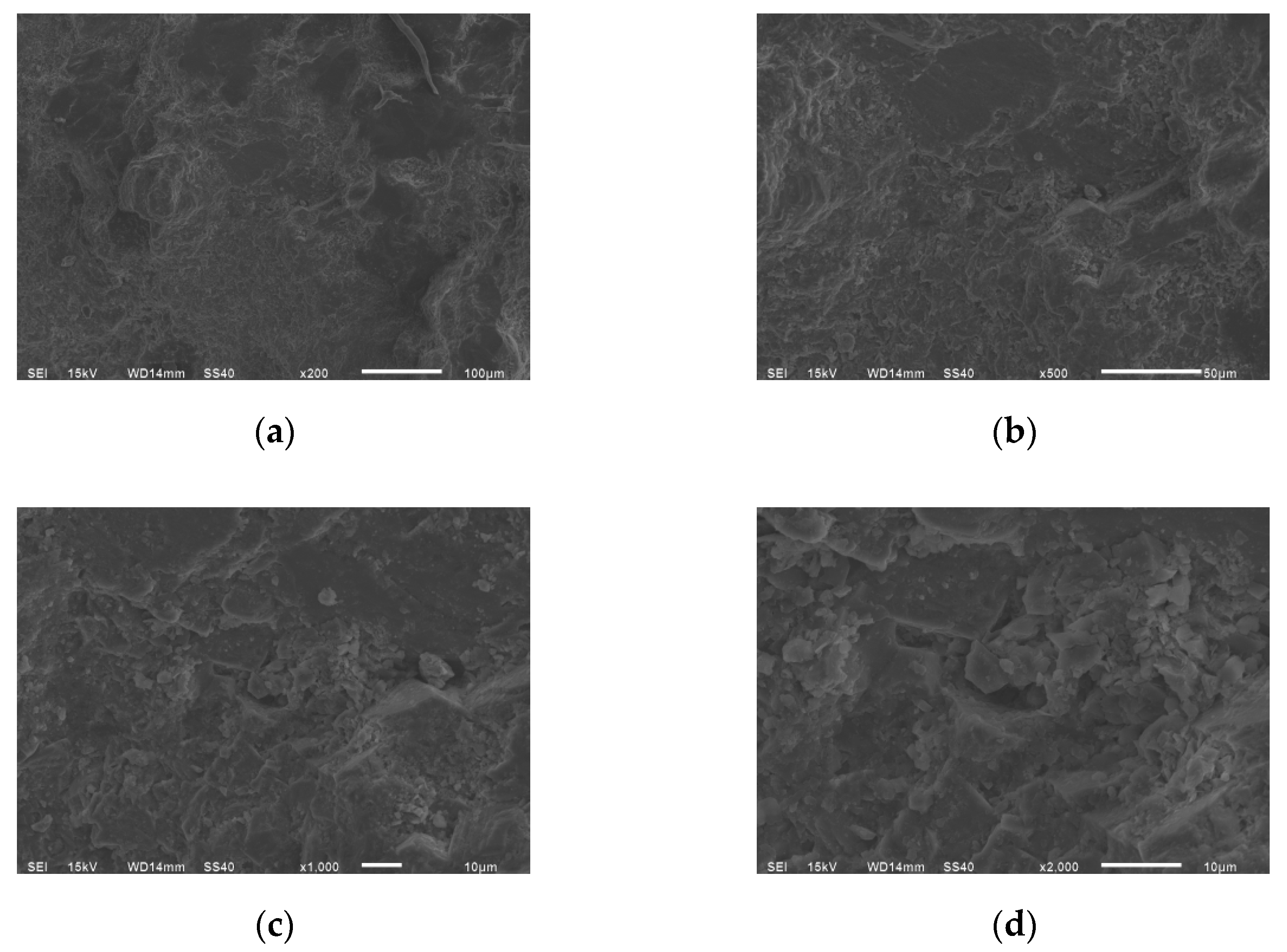
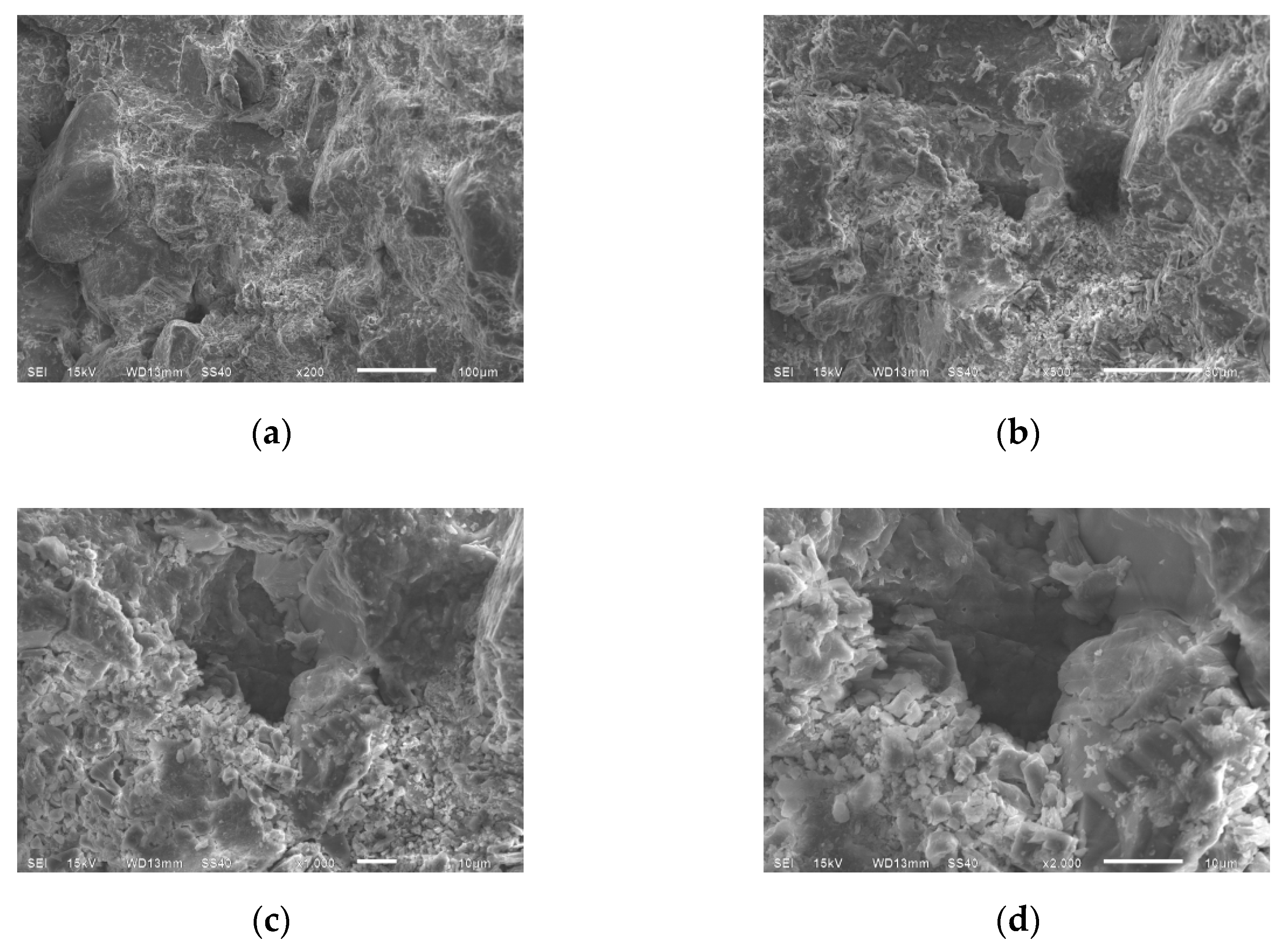
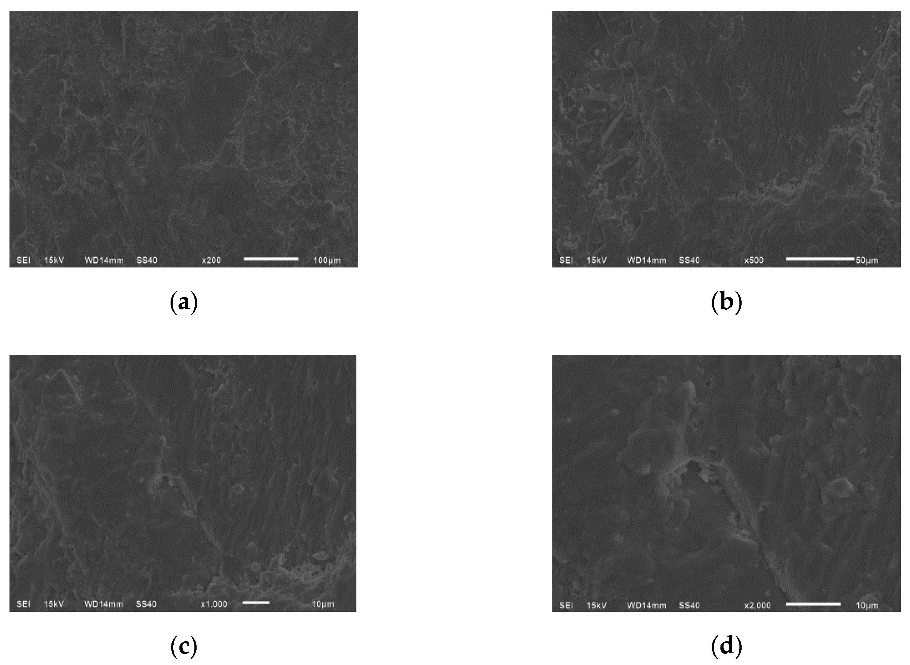
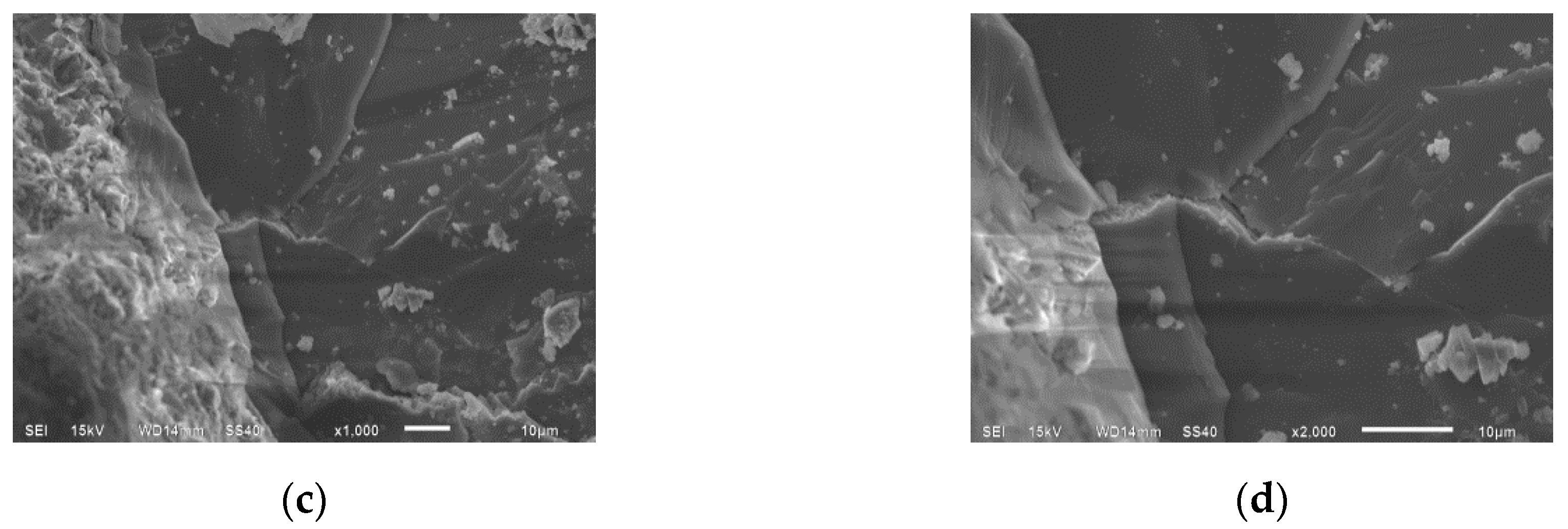
3.1. Micropores and Distribution Characteristics of Sandstone

The meso-structure of sandstone is characterized by granulation, and its internal pores are mainly caused by sand accumulation. The pore structure includes pore and roar channel. Sedimentation and diagenesis will make the micro-pores of sandstone complicated.
The meso-morphology of three types of sandstone was observed by SEM [27,28]. When using SEM to observe, it is necessary to keep the sample dry enough and do gold plating on the observation surface. Images with magnification of 200, 500, 1000 and 2000 were selected (Figure 2, Figure 3 and Figure 4).
From Figure 2, Figure 3 and Figure 4, it can be observed that the meso-surface of the first type of sandstone is flat, the flake particle structure is less and orderly stacked, and the particles at the pore position are relatively loose. The overall structure of the second type of sandstone is more obvious, but the particles are obvious, stacked tightly, and the pore structure is more. The third type of sandstone shows a honeycomb porous loose structure, the overall meso-structure is relatively loose, there are many particles, and, between particles, the cementation degree is not good, the pores are more obvious, and there are small particles filling into the pore structure.

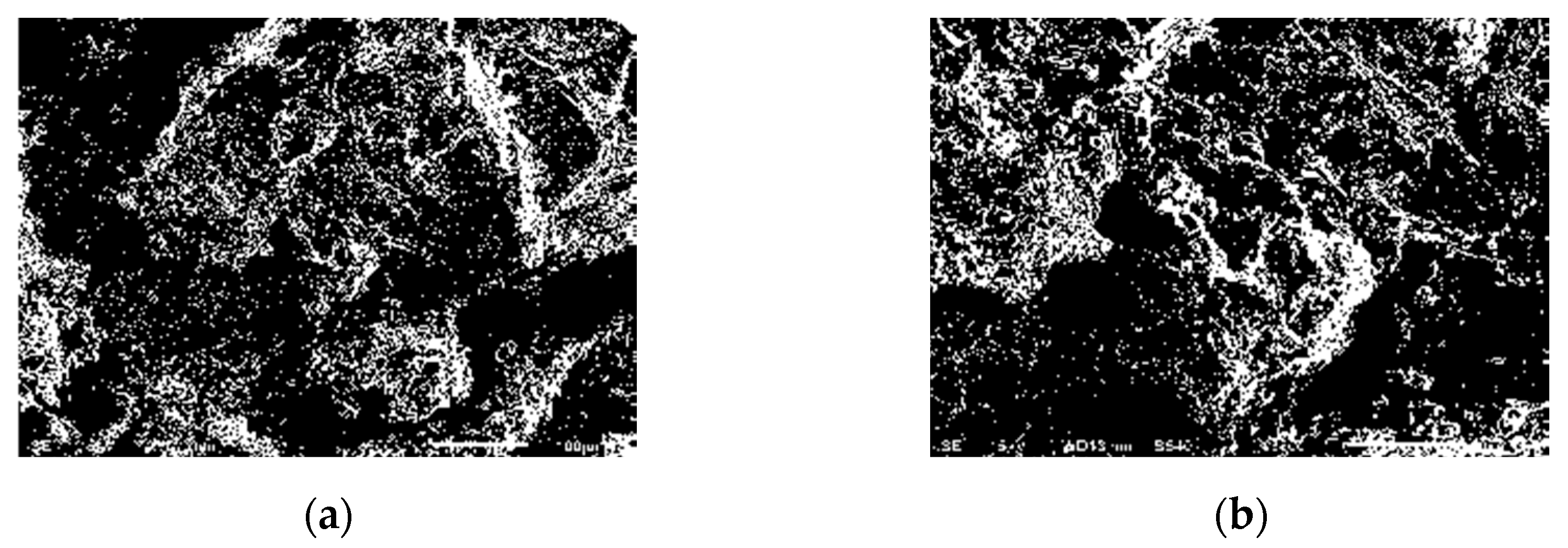
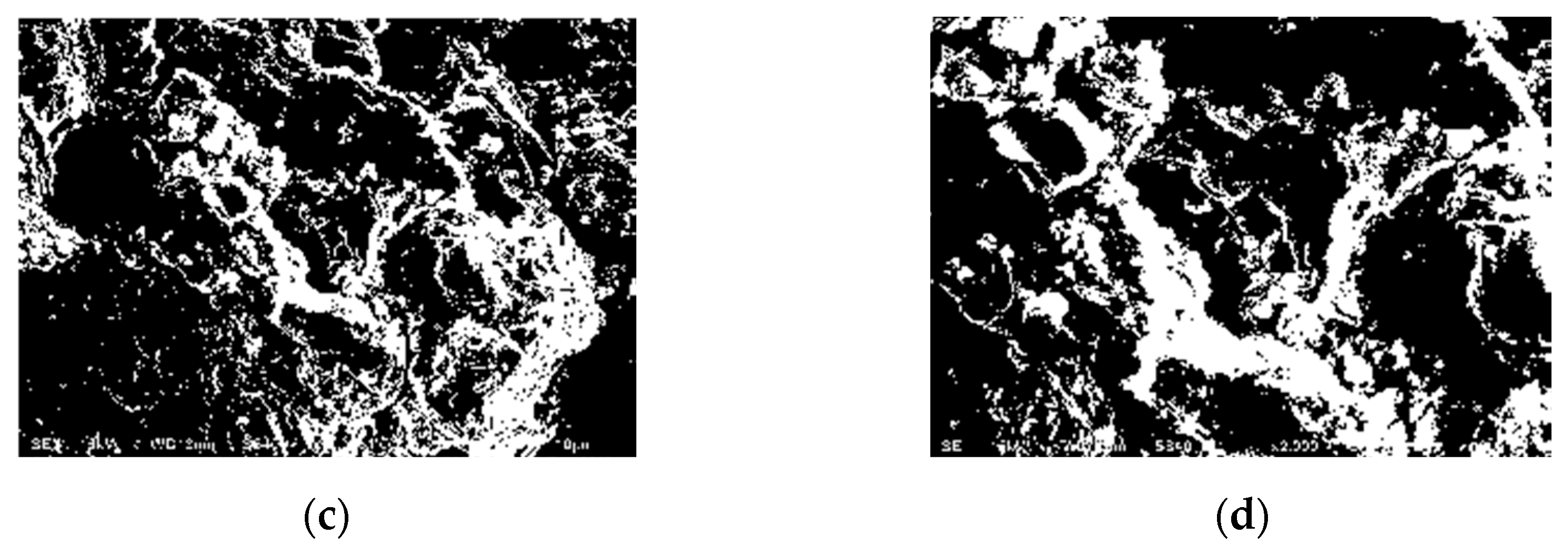
3.2. Microfractures Characteristics of Sandstone

The structure of sandstone has obvious joint layered characteristics. The development of sandstone fractures has directionality. Due to the influence of geological movement and man-made mining activities, sandstone will also produce fractures, accompanied by small micro-fractures. SEM was used to observe the micro-crack structure development of three types of sandstone under four magnification (×200, ×500, ×1000, ×2000), as shown in Figure 5, Figure 6 and Figure 7.
The first type of sandstone has a smooth structure, the crack structure is not obvious, the surface is relatively flat, and the particles are less and evenly distributed. The second type of sandstone has more broken particles scattered, the crack surface is sunken, there are more broken particles, and there are some small pore structures. The third type of sandstone surface structure has obvious faults, fewer broken particles, obvious and numerous cracks, and complex surface structure.

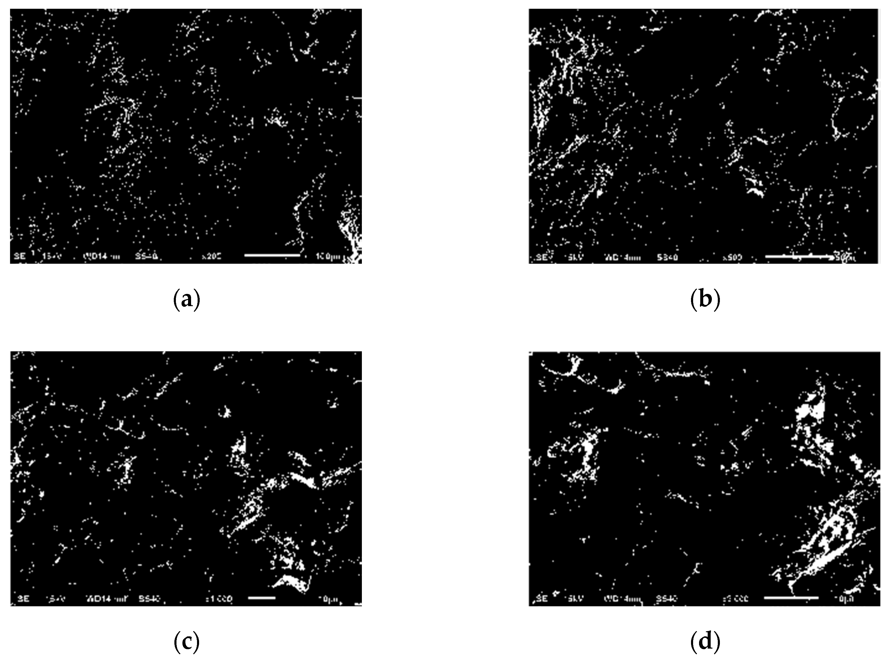
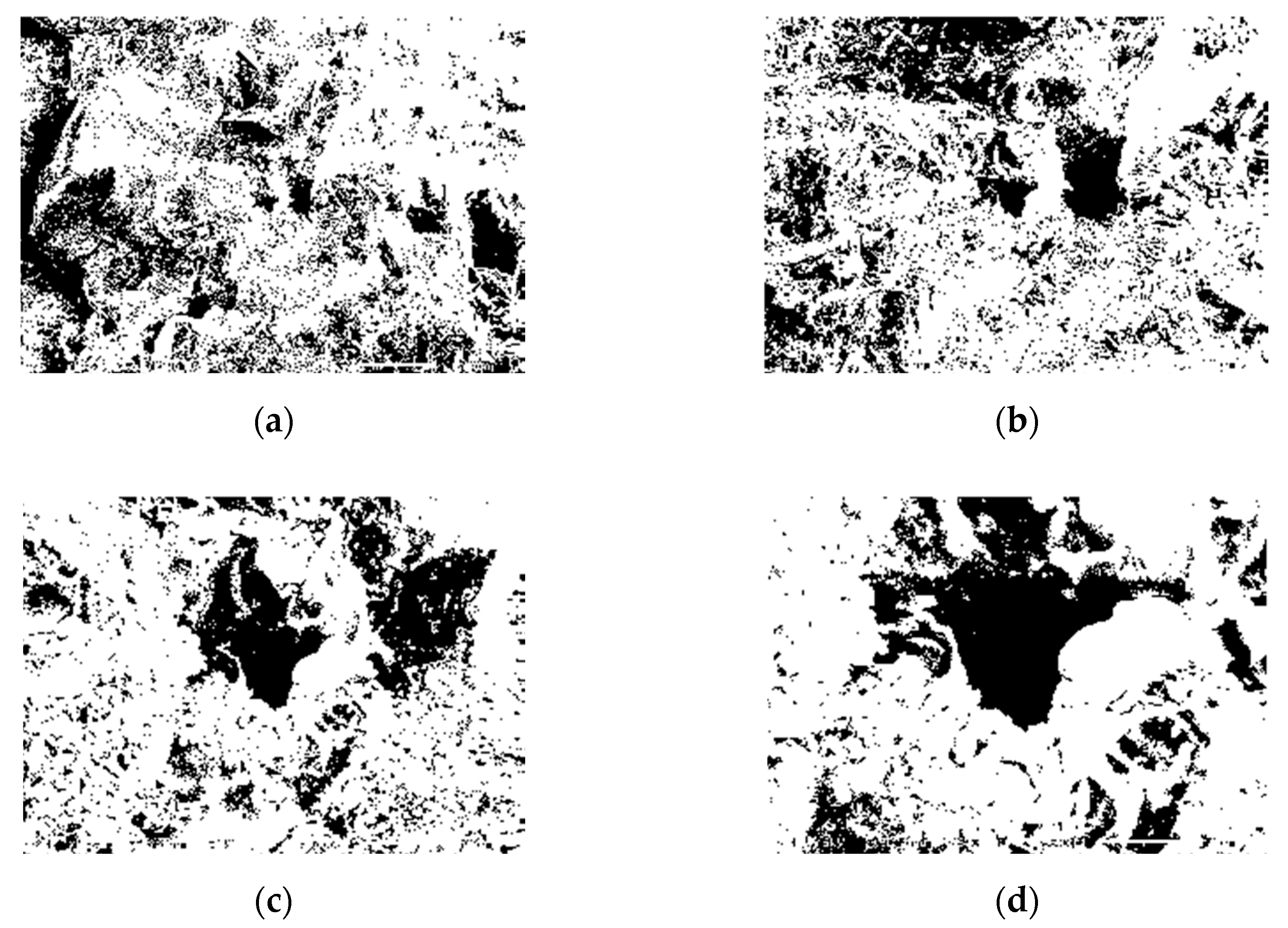
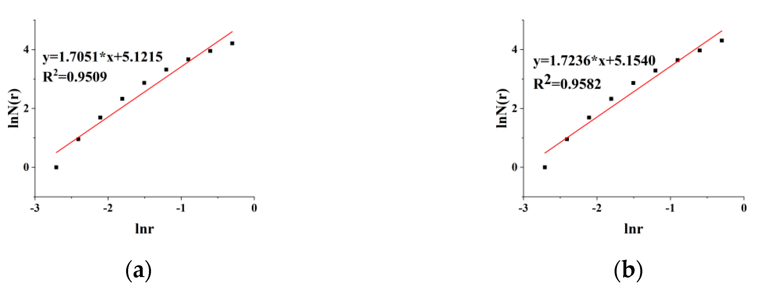
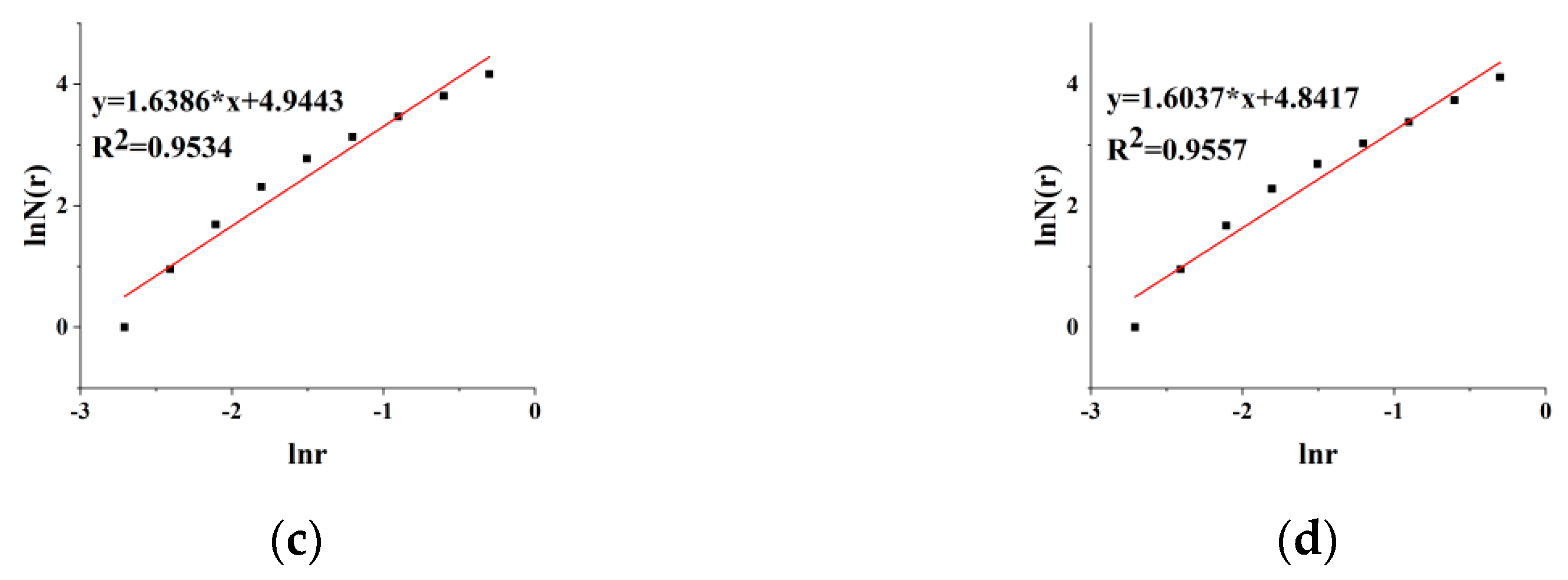
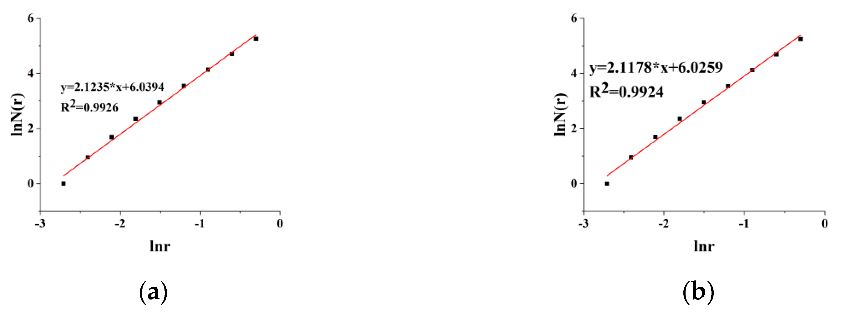
3.3. Fractal Characteristics of Sandstone Micropores

Fractal theory [29,30,31] was first proposed by B.B. Mandelbrot, which has become an important method to study the pore structure of porous materials. There are a lot of micropores and microcracks in the rock, which has fractal structure [32,33,34]. It is proved to be feasible to calculate the fractal dimension and study the meso-mechanism of rock failure [35,36]. The common fractal dimensions include Hausdorff dimension, box dimension, correlation dimension, information dimension, generalized dimension, and self-similar dimension [37]. Among many fractal dimensions, box dimension has its own characteristics: the calculation method is simple and the results are accurate. Therefore, the box dimension method [38,39] is used to calculate the fractal dimension in this study. Suppose that F is an arbitrary nonempty bounded subset on Rn, and N δ ( F ) is the minimum number of sets whose diameter is the largest δ and can cover F, then the box dimension of F is defined as
D F = lim δ 0 ln N δ ( F ) ln δ
The fractal characteristics of sandstone meso-pore structure images are analyzed [40,41], and the obtained micro-images are processed into binary images by Matlab (Figure 8, Figure 9 and Figure 10). The image can be regarded as a numerical matrix of M × N. The data matrix is divided into several blocks with side length r. the box number N ( r ) occupied by black blocks in the matrix is calculated. Generally, r = 1, 2, 4, … 2i (i = 0,1,2,…). The logarithm of the box number ln N ( r ) and the box side length ln r is taken, and the linear fitting is performed. The slope of the straight line is the fractal dimension of the image (Figure 11, Figure 12 and Figure 13). This step can also be realized by Matlab. In order to obtain a more accurate fractal dimension, take the average as the final fractal dimension, as shown in Table 1.
By observing the change in fractal dimension under different magnification (Figure 14), it can be found that with the increase in image magnification, the calculated fractal dimension generally shows a decreasing trend. The analysis is that with the increase in image magnification, the observable rock surface range decreases, so the number of observable pores will also decrease, and the complexity of surface structure will decrease, so the fractal dimension will decrease. At the same time, the fractal dimension values of different types of sandstone are also different.
According to the results of SEM and fractal dimension, sandstone has different structural characteristics at the meso level, which is mainly cemented by various sand particles, with obvious grain structure and primary fine cracks. Sandstone has self-similar characteristics, which can be expressed by fractal dimension. Different types of sandstone have obvious differences in fractal dimension.

4. Relationship between Fractal Dimension and Mechanical Properties of Sandstone

The strength of rock is an important parameter in geotechnical engineering, and the strength often determines the engineering properties of rock. Based on the damage mechanics theory [42,43,44], fractal dimension is fitted with the mechanical parameters, and the influence of meso-parameters on the strength characteristics and damage to sandstone is analyzed according to the results.

4.1. Strength Characteristics of Sandstone

The samples are taken from the Shanxi formation of North China type Carboniferous Permian. The first type of sample is light grayish green sandstone with particle size of 0.25~0.125 mm. Its main components are quartz, feldspar, cement and recrystallized minerals. It has fine sand structure, compact massive structure and undeveloped interlayer. The coefficient of firmness is 6~6.5. The second kind of sample is dark red sandstone with particle size less than 0.05 mm. Its main component is quartz, which is hard in texture, simple in structure, and not obvious in bedding development. The coefficient of firmness is about 4. The third type of sandstone is brown-yellow sandstone with fine grain. Its main mineral composition is quartz, chlorite and mica. The bedding is developed and the general coefficient of firmness is 2.5~3. The uniaxial compression tests of three kinds of sandstone were carried out on Shimadzu servo rock testing machine, under the uniaxial compression stress, with the sandstone deformed and destroyed. Thus, the stress–strain curve of the whole process can be drawn (Figure 15), and the compressive strength Rc, elastic modulus E and Poisson’s ratio μ can be obtained (Table 2), then the relationship between fractal dimension and strength characteristics is analyzed.
According to the uniaxial compression test and fitting diagram, the mechanicals are greatly affected by the meso-structure of three types sandstone. The specimens with different strengths have different fractal dimensions; with the increase in uniaxial compressive strength or elastic modulus, the fractal dimension of sandstone decreases, while Poisson’s ratio has no specific relationship with fractal dimension. Therefore, the macroscopic strength characteristics of sandstone are inversely proportional to the meso-structure characteristics.

4.2. Damage Mechanics Analysis of Sandstone

There are meso-scale defects in sandstone, which will lead to a decline in the mechanical properties; they become a hidden danger in geotechnical engineering. It is necessary to understand the damage mechanism of sandstone to ensure the stability of geotechnical engineering. Damage refers to the deterioration of mechanical properties of materials under stress, mainly manifested in the initiation and propagation of microcracks in sandstone. Damage mechanics is an effective means to evaluate the rock mechanical properties. The key is to define the rock damage variable. Wu et al. [45] found that the calculation formula of rock damage variable D can be expressed as follows
D = 1 exp [ ( ε a ) m ]
where, ε is the strain; a is the Weibull distribution parameter; m is the shape parameter. At the same time, the maximum damage of materials on ordinary testing machines is only related to the mechanical parameters of materials, including elastic modulus E, peak stress σc and peak strain εc, the critical damage value is defined
D r = 1 exp ( ln ( σ c E ε c ) )
Based on the strength parameters obtained in the previous section, the critical damage values can be calculated, as shown in Table 3.
The fractal dimension, compressive strength and critical damage value were fitted to study the relationship between sandstone meso-structure and damage characteristics (Figure 16).
According to Table 3 and Figure 16, the fractal dimension of sandstone is directly proportional to the critical damage. The larger the fractal dimension is, the stronger the self-similarity of sandstone is and the more complex the internal structure is, and the smaller the macro-strength parameter is, the greater the internal damage of sandstone is, which is consistent with the actual situation. On the contrary, the smaller the fractal dimension, the smaller the critical damage.

5. Conclusions

On the basis of uniaxial compression experiment and SEM, taking sandstone as an example, according to the box dimension algorithm, combined with digital image processing and Matlab, a meso-image fractal dimension calculation program is developed. Through the fractal dimension and the sandstone mechanical characteristics, the following conclusions are drawn:
(1)
The study of micro-damage to sandstone can analyze its macro mechanical characteristics in depth. According to the observation of SEM, there are many micropores and microcracks in the three kinds of sandstones, the overall structure is undulating, and there are many particles on the surface, which are typical materials with original damage. The macroscopic mechanical properties of sandstone can be analyzed according to its meso-structure;
(2)
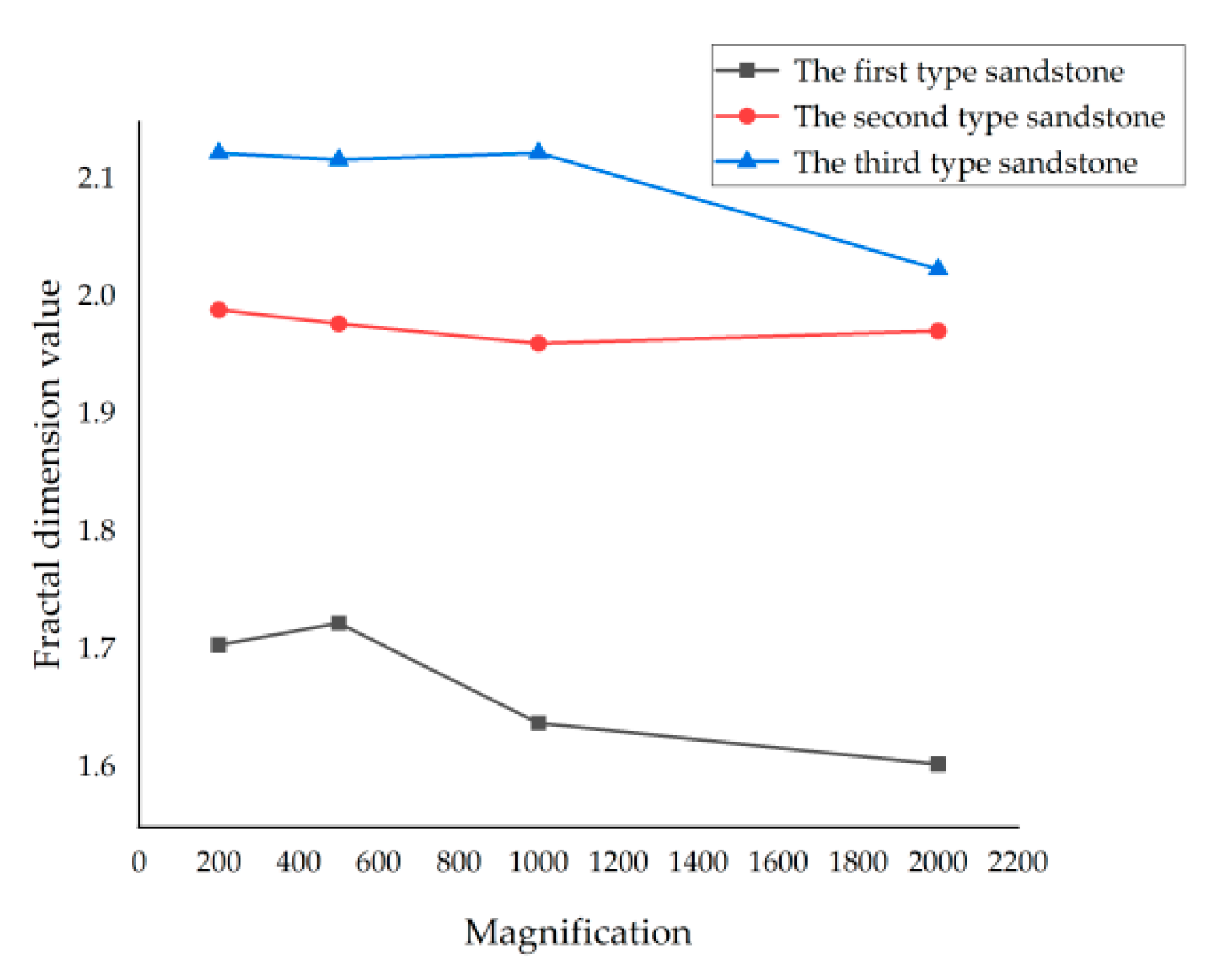
Matlab is used to calculate the fractal dimension. In order to exclude the particularity, the average value of fractal dimension is selected as the final fractal dimension value. The average fractal dimensions of the three kinds of sandstones are 1.6678, 1.9756 and 2.0974, the micro-morphology of different sandstones varies greatly. The more obvious the micro-fractures and micropores are, the larger the fractal dimension is. Therefore, it is appropriate to use the fractal dimension to describe the characteristics of sandstone. The fractal dimension decreases with the increase in magnification;
(3)
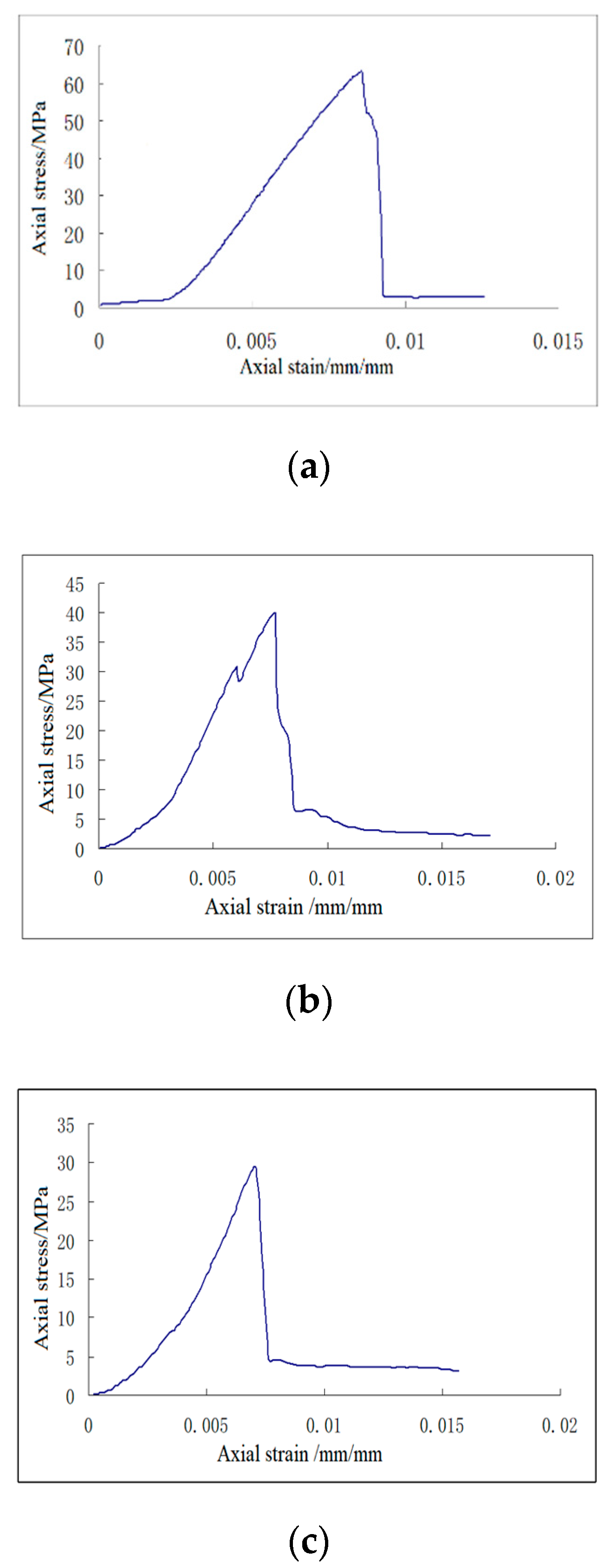
According to the results of uniaxial compression test, the compressive strength of the three sandstones are 63.25, 40.93 and 29.54 Mpa, and the elastic modulus are 9.97, 8.46 and 7.06 GPa. The strength characteristics of sandstone are inversely proportional to the fractal dimension. The larger the uniaxial compressive strength and elastic modulus are, the smaller the fractal dimension is;
(4)
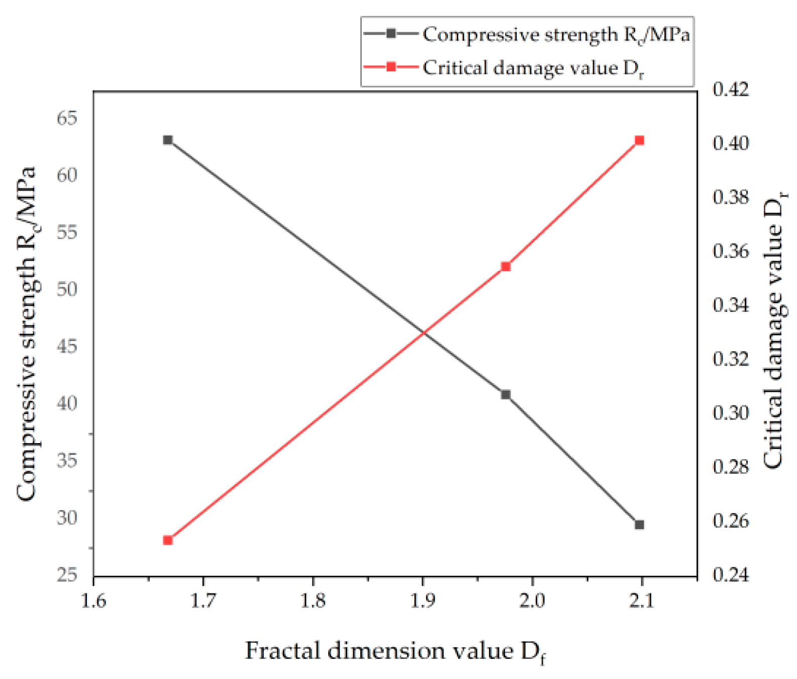
Based on the damage mechanics theory, the relationship between the critical damage value, compressive strength and fractal dimension is studied. It is found that the critical damage values of the three sandstones are in direct proportion to their fractal dimensions. The larger the fractal dimension of sandstone is, the more micropores and microcracks there are, and the more serious the damage is, the lower the compressive strength is. This shows that the strength and deformation characteristics of sandstone are directly related to its meso-damage, which is consistent with the actual situation.

Author Contributions

Conceptualization and methodology, D.H. and X.C.; X.C.; data analysis, C.G.; writing—original draft preparation, D.H.; writing—review and editing; H.X.; validation; D.H. and X.C.; funding acquisition. Correspondence should be addressed to D.H. and X.C. All authors have read and agreed to the published version of the manuscript.

Funding

This research was funded by the National Natural Science Foundation of China (51704179), the Natural Science Foundation of Shandong Province (ZR2016EEB23), the first-class discipline construction projects (01AQ03703,01CK05902), and was supported by the SDUST Research Fund.

Conflicts of Interest

The authors declare no conflict of interest.

References

  1. Fan, P.K. Study on Microstructural Evolution and Strength Model of Cemented Backfill. Ph.D. Thesis, North China University of Science and Technology, Qinhuangdao, China, 2019; pp. 3–8. [Google Scholar]
  2. Chen, L.J.; Zhang, M. Numerical simulation of hydraulic fracturing of brittle rock with three-dimensional deep crack. Hydropower 2019, 45, 34–38. [Google Scholar]
  3. Yin, Y.C.; Zhao, T.B.; Tan, Y.L.; Yu, F.H. Rock meso model reconstruction and numerical experiment based on Otsu image processing. Geotech. Mech. 2015, 36, 2532–2540. [Google Scholar]
  4. Wetzel, M.; Kempka, T.; Kuhn, M. Predicting macroscopic elastic rock properties requires detailed information on microstructure. Energy Procedia 2017, 125, 561–570. [Google Scholar] [CrossRef]
  5. Zhu, F.; Hu, W.; Cao, J.; Sun, F.; Liu, Y.; Sun, Z. Micro/nanoscale pore structure and fractal characteristics of tight gas sandstone: A case study from the Yuanba area, northeast Sichuan Basin, China. Mar. Pet. Geol. 2018, 98, 116–132. [Google Scholar] [CrossRef]
  6. Hammes, D.M.; Peternell, M. FAME: Software for analysing rock microstructures. Comput. Geosci. 2016, 90, 24–33. [Google Scholar] [CrossRef]
  7. ASabatakakis, N.; Tsiambaos, G.; Ktena, S.; Bouboukas, S. The effect of microstructure on mi strength parameter variation of common rock types. Bull. Eng. Geol. Environ. 2018, 77, 1673–1688. [Google Scholar] [CrossRef]
  8. Regnet, J.B.; David, C.; Robion, P.; Menéndez, B. Microstructures and physical properties in carbonate rocks: A comprehensive review. Mar. Pet. Geol. 2019, 103, 366–376. [Google Scholar] [CrossRef]
  9. Jeng, F.S.; Weng, M.C.; Lin, M.L.; Huang, T.H. Influence of petrographic parameters on geotechnical properties of tertiary sandstones from Taiwan. Eng. Geol. 2004, 73, 71–91. [Google Scholar] [CrossRef]
  10. Stück, H.; Koch, R.; Siegesmund, S. Petrographical and petrophysical properties of sandstones: Statistical analysis as an approach to predict material behaviour and construction suitability. Environ. Earth Sci. 2013, 69, 1299–1332. [Google Scholar] [CrossRef] [Green Version]
  11. Strzałkowski, P.; Kaźmierczak, U.; Wolny, M. Assessment of the method for abrasion resistance determination of sandstones on Böhme abrasion test apparatus. Bull. Eng. Geol. Environ. 2020, 79, 4947–4956. [Google Scholar] [CrossRef]
  12. Ulusay, R.; Türeli, K.; Ider, M.H. Prediction of engineering properties of a selected litharenite sandstone from its petrographic characteristics using correlation and multivariate statistical techniques. Eng. Geol. 1994, 38, 135–157. [Google Scholar] [CrossRef]
  13. Niu, S.; Ge, S.; Yang, D.; Dang, Y. Mechanical properties and energy mechanism of saturated sandstones. J. Cent. South Univ. 2019, 25, 1447–1463. [Google Scholar] [CrossRef]
  14. Liu, H.Y.; Li, J.F.; Pei, X.L. Dynamic damage constitutive model of intermittent jointed rock mass under uniaxial compression. Explos. Impact 2018, 38, 316–323. [Google Scholar]
  15. Li, K. Study on Fracture Characteristics and Damage Model of Rock Mass with Discontinuous Joints; China University of Geosciences: Beijing, China, 2018. [Google Scholar]
  16. Gooya, R.; Bruns, S.; Muter, D.; Moaddel, A.; Harti, R.P.; Stipp, S.L.S.; Sørensen, H.O. Effect of tomography resolution on the calculated microscopic properties of porous materials: Comparison of sandstone and carbonate rocks. Appl. Phys. Lett. 2016, 109, 104102. [Google Scholar] [CrossRef] [Green Version]
  17. Xie, C. Study on Macro Meso Damage and Failure Characteristics and Numerical Method of Fractured Rock Mass. Ph.D. Thesis, Shandong University, Jinan, China, 2018; pp. 22–38. [Google Scholar]
  18. Zhang, P.; Chen, C. Damage constitutive model of rock mass considering mechanical properties of intermittent joints. Highw. Automob. Transp. 2017, 04, 180–184. [Google Scholar]
  19. Zhang, S.; Liu, W.; Lv, H. Creep energy damage model of rock graded loading. Results Phys. 2019, 12, 1119–1125. [Google Scholar] [CrossRef]
  20. Mohammadi, H.; Pietruszczak, S. Description of damage process in fractured rocks. Int. J. Rock Mech. Min. Sci. 2019, 113, 295–302. [Google Scholar] [CrossRef]
  21. Zhu, Y. A micromechanics-based damage constitutive model of porous rocks. Int. J. Rock Mech. Min. Sci. 2017, 91, 1–6. [Google Scholar] [CrossRef]
  22. Haddad-Martim, P.M.; Carranza, E.J.M.; De Souza Filho, C.R. The fractal nature of structural controls on ore formation: The Case of the Iron oxide copper-gold deposits in the carajás mineral province, Brazilian amazon. Econ. Geol. 2018, 113, 1499–1524. [Google Scholar] [CrossRef]
  23. Gao, D.S. Application of Fractal Models to Research Mineral Particles Surface Morphology: Magnetite in Luoyang iron Deposit of Fujian Province; China University of Geosciences: Beijing, China, 2020. [Google Scholar]
  24. Du, G.S. Brazilian Splitting Test of Rock Based on the Characteristics of Micro Fabric; Inner Mongolia University of Science&Technology: Baotou, China, 2020. [Google Scholar]
  25. Jin, P.H.; Hu, Y.Q.; Shao, J.X.; Liu, Z.J.; Feng, G.; Song, S. Influence of Temperature on the Structure of Pore—Fracture of Sandstone. Rock Mech. Rock Eng. 2020, 53, 1–12. [Google Scholar] [CrossRef]
  26. Lai, J.; Wang, G.W.; Wang, Z.Y.; Chen, J.; Pang, X.J.; Wang, S.C.; Zhou, Z.L.; He, Z.B.; Qin, Z.Q.; Fan, X.Q. A review on pore structure characterization in tight sandstones. Earth Sci. Rev. 2018, 177, 436–457. [Google Scholar] [CrossRef]
  27. Kuzmin, V.A. Microstructural Effects in Electron-Microscopic Studies of Carbonate Rocks. J. Surf. Investig. X-Ray Synchrotron Neutron Tech. 2018, 12, 953–956. [Google Scholar] [CrossRef]
  28. Pedrosa, E.T.; Boeck, L.; Putnis, C.V.; Putnis, A. The replacement of a carbonate rock by fluorite: Kinetics and microstructure. Am. Mineral. 2017, 102, 126–134. [Google Scholar] [CrossRef]
  29. Chen, T.X.; Chen, Y.; Sha, Y.B.; Bai, W.S.; Wu, Z.H.; Wang, T.T. Microstructure and fractal characteristics of red clay particles in existing foundation based on SEM images. Carsologica Sin. 2019, 38, 635–642. [Google Scholar]
  30. Zhou, L.; Du, W.W.; Han, X.; Li, C.F. Fractal characteristics of micro pore structure of clay minerals. J. Heilongjiang Univ. Sci. Technol. 2009, 19, 94–96, 120. [Google Scholar]
  31. Lu, C.; Mai, Y.W.; Xie, H. A sudden drop of fractal dimension: A likely precursor of catastrophic failure in disordered media. Philos. Mag. Lett. 2005, 85, 33–40. [Google Scholar] [CrossRef]
  32. Li, D.J.; Wang, G.L.; Han, L.Q.; Liu, P.Y.; He, M.C.; Yang, G.X.; Tai, Q.M. Analysis of microscopic pore structures of rocks before and after water absorption. Min. Sci. Technol. 2011, 21, 287–293. [Google Scholar]
  33. Chen, C.X.; Liu, X.M.; Liu, C.C. Application of digital image technology in rock micromechanics. Geotech. Mech. 2010, 31, 53–60. [Google Scholar]
  34. Yin, S.; Dong, L.; Yang, X.; Wang, R.Y. Experimental investigation of the petrophysical properties, minerals, elements and pore structures in tight sandstones. J. Nat. Gas Sci. Eng. 2020, 76, 103189. [Google Scholar] [CrossRef]
  35. Liang, C.Y.; Wu, S.R.; Li, X. Study on the micro and micro fracture characteristics of granite under uniaxial compression in the range of low and medium strain rates. J. Rock Mech. Eng. 2015, 34, 2977–2986. [Google Scholar]
  36. Zhang, K.X.; Lai, J.; Bai, G.P.; Pang, X.J.; Ma, X.Q.; Qin, Z.Q.; Zhang, X.S.; Fan, X.C. Comparison of fractal models using NMR and CT analysis in low permeability sandstones. Mar. Pet. Geol. 2020, 112, 104069. [Google Scholar] [CrossRef]
  37. Huang, D.M.; Chang, X.K.; Tan, Y.L.; Fang, K.; Yin, Y.C. From rock microstructure to macromechanical properties based on fractal dimensions. Adv. Mech. Eng. 2019, 11, 3. [Google Scholar] [CrossRef] [Green Version]
  38. Liu, C.X.; Jiang, J.Q.; Liu, F.S.; Wang, S.H. Fractal study of scale effect in microscopic, mesoscopic and macroscopic states for fracture mechanism of rock materials. Rock Soil Mech. 2008, 29, 2619–2622. [Google Scholar]
  39. Huang, D.M.; Chang, X.K.; Lin, X.F.; Fu, Y.X. Fractal characteristics of rock fracture cracks under uniaxial compression. J. Shandong Univ. Sci. Technol. 2014, 33, 58–62. [Google Scholar]
  40. Alfonso, I.; Beltrán, A.; Abatal, M.; Castro, I.; Fuentes, A.; Vázquez, L.; Garcia, A. Fractal Dimension Determination of Rock Pores by Multi-Scale Analysis of Images Obtained Using OM, SEM and XCT. Fractals 2018, 26, 1850067. [Google Scholar] [CrossRef]
  41. Holgado, M.A.; Fernandez-Hervas, M.J.; Rabasco, A.M.; Fini, A. Characterization study of a diclofenac salt by means of SEM and fractal analysis. Int. J. Pharm. 1995, 120, 157–167. [Google Scholar] [CrossRef]
  42. Zhao, D.L.; Zuo, S.Y.; Huang, C.; Wang, S.; Yang, C. Experimental study on anisotropic damage mechanical characteristics of layered rock mass. Hydropower Energy Sci. 2019, 37, 144–147. [Google Scholar]
  43. Zhao, X.; Ma, S.Q. Theoretical analysis and numerical simulation of coal micro damage. J. North China Univ. Sci. Technol. 2016, 13, 53–57, 61. [Google Scholar]
  44. Yang, Y.J.; Wang, D.C.; Wang, K.; Huang, D.M. Micromechanical damage mechanism of coal and rock strength and deformation characteristics. J. Beijing Univ. Sci. Technol. 2011, 33, 653–657. [Google Scholar]
  45. Wu, Z.; Zhang, C.J. Damage model and mechanical properties of rock under uniaxial loading. Acta Petromech. Eng. 1996, 15, 55–61. [Google Scholar]
Figure 1. Type of sandstone samples.
Symmetry 12 01815 g001
Figure 2. Development of micro-pores in the first type of sandstone. (a) Meso-image with magnification of 200; (b) Meso-image with magnification of 500; (c) Meso-image with magnification of 1000; (d) Meso-image with magnification of 2000.
Symmetry 12 01815 g002
Figure 3. Development of micro-pores in the second type of sandstone. (a) Meso-image with magnification of 200; (b) Meso-image with magnification of 500; (c) Meso-image with magnification of 1000; (d) Meso-image with magnification of 2000.
Symmetry 12 01815 g003
Figure 4. Development of micro-pores in the third type of sandstone. (a) Meso-image with magnification of 200; (b) Meso-image with magnification of 500; (c) Meso-image with magnification of 1000; (d) Meso-image with magnification of 2000.
Symmetry 12 01815 g004
Figure 5. Micro-fracture development of the first type sandstone. (a) Meso-image with magnification of 200; (b) Meso-image with magnification of 500; (c) Meso-image with magnification of 1000; (d) Meso-image with magnification of 2000.
Symmetry 12 01815 g005
Figure 6. Micro-fracture development of the second type sandstone. (a) Meso-image with magnification of 200; (b) Meso-image with magnification of 500; (c) Meso-image with magnification of 1000; (d) Meso-image with magnification of 2000.
Symmetry 12 01815 g006aSymmetry 12 01815 g006b
Figure 7. Micro-fracture development of the third type sandstone. (a) Meso-image with magnification of 200; (b) Meso-image with magnification of 500; (c) Meso-image with magnification of 1000; (d) Meso-image with magnification of 2000.
Symmetry 12 01815 g007
Figure 8. The binary diagram of the first kind of sandstone. (a) Binary graph at 200 magnification; (b) Binary graph at 500 magnification; (c) Binary graph at 1000 magnification; (d) Binary graph at 2000 magnification.
Symmetry 12 01815 g008
Figure 9. The binary diagram of the second kind of sandstone. (a) Binary graph at 200 magnification; (b) Binary graph at 500 magnification; (c) Binary graph at 1000 magnification; (d) Binary graph at 2000 magnification.
Symmetry 12 01815 g009aSymmetry 12 01815 g009b
Figure 10. The binary diagram of the third kinds of sandstone. (a) Binary graph at 200 magnification; (b) Binary graph at 500 magnification; (c) Binary graph at 1000 magnification; (d) Binary graph at 2000 magnification.
Symmetry 12 01815 g010
Figure 11. Fractal dimension fitting diagram of the first type sandstone. (a) Fitting diagram at 200 magnification; (b) Fitting diagram at 500 magnification; (c) Fitting diagram at 1000 magnification; (d) Fitting diagram at 2000 magnification.
Symmetry 12 01815 g011aSymmetry 12 01815 g011b
Figure 12. Fractal dimension fitting diagram of the second type sandstone. (a) Fitting diagram at 200 magnification; (b) Fitting diagram at 500 magnification; (c) Fitting diagram at1000 magnification; (d) Fitting diagram at 2000 magnification.
Symmetry 12 01815 g012
Figure 13. Fractal dimension fitting diagram of the third type sandstone. (a) Fitting diagram at 200 magnification; (b) Fitting diagram at 500 magnification; (c) Fitting diagram at 1000 magnification; (d) Fitting diagram at 2000 magnification.
Symmetry 12 01815 g013aSymmetry 12 01815 g013b
Figure 14. The relationship between fractal dimension and magnification.
Symmetry 12 01815 g014
Figure 15. Stress–strain curves of three types of sandstone. (a) The stress-strain curve of the first kind of sandstone; (b) The stress–strain curve of the second kind of sandstone; (c) The stress–strain curve of the third kind of sandstone.
Symmetry 12 01815 g015
Figure 16. Fitting graph of fractal dimension, strength and critical damage value.
Symmetry 12 01815 g016
Table 1. Average value of fractal dimension.
Sandstone Type Fractal Dimension Values Under Different Magnification Average Fractal Dimension
×200×500×1000×2000
11.70511.72361.63861.60371.6678
21.99021.97841.96151.97221.9756
32.12352.11782.12372.02472.0974
Table 2. Strength parameters of the sandstone.
Sandstone TypeCompressive Strength Rc/MPaElastic Modulus E/GPaPoisson’s Ratio μFractal Dimension Value Df
163.259.970.041.6678
240.938.460.161.9756
329.547.060.182.0974
Table 3. Strength parameters of type 3 sandstone.
Sandstone TypeCritical Damage Value DrCompressive Strength Rc/MPaFractal Dimension Value Df
125.35%63.251.6678
235.50%40.931.9756
340.18%29.542.0974
Publisher’s Note: MDPI stays neutral with regard to jurisdictional claims in published maps and institutional affiliations.

