Article

Classification of Body Constitution Based on TCM Philosophy and Deep Learning

Department of Computer Science and Information Engineering, National Central University, Taoyuan 32001, Taiwan
*
Author to whom correspondence should be addressed.
Symmetry 2020, 12(5), 803; https://doi.org/10.3390/sym12050803
Submission received: 8 April 2020 / Revised: 29 April 2020 / Accepted: 6 May 2020 / Published: 12 May 2020

Abstract

:
There is a growing demand for alternative or complementary medicine in health care disciplines that uses a non-invasive instrument to evaluate the health status of various organs inside the human body. In this regard, we proposed a real-time, non-invasive, and painless technique to assess an individual’s health condition. Our approach is based on the combination of iridology and the philosophy of traditional Chinese medicine (TCM). The iridology chart presents perfect symmetry between the left and right eyes, and such a unique representation reveals the body constitution based on TCM philosophy, which classifies the aforementioned body constitution into a combination of nine categories to describe the varieties of genomic traits. In addition, we applied a deep-learning method along with the combination of iridology and TCM to predict the possible physiological or psychological strength or weakness of the subjects and give advice to them about how to take care of their health according to the body constitution assessment. We used several pre-trained convolutional neural networks (CNNs, or ConvNet), such as a residual neural network (ResNet50), InceptionV3, and dense convolutional network (DenseNet201), to classify the body constitution using iris images. In the experiments, the CASIA-Iris-Thousand database was used to perform this task. The experimental results showed that the proposed iris-based health assessment method achieved an 82.9% accuracy.

1. Introduction

In recent years, there has been an increasing interest in non-invasive, real-time, and painless approaches for health status assessment. Because of its painless and non-invasive nature, it has gained tremendous attention and demand in the various healthcare fields.
The human eye plays a pivotal role in such approaches due to having a unique appearance, shape, and color of the iris. The iris is considered an internal human body organ, but externally, it may be easily observed [1]. Each person has a different spatial shape of the iris; therefore, the iris texture has been used extensively for biometrics for the last several decades [1,2,3]. Besides the biometric identification, iris textures and colors are related to the healthiness of the internal organs of a person. The place of the iris is near the human nervous system, and after examining the iris, it can reveal significant information about the human health imbalance and nature of possible disturbances in internal body organs, which is called iris diagnosis (iridology) [2,4]. Additionally, extensive research has shown that the human iris can be used as a manifestation of health status [2,3]. Therefore, the human eyes are referred to as the window to the soul and everything related to health is encrypted in our eyes. The iris has definite characteristics, e.g., fine lines, crypts, and lesions, which are associated with the body constitution, inherent weaknesses, health levels, and changes that occur in the human body [5]. The iris is formed before birth and it remains the same throughout one’s lifetime [
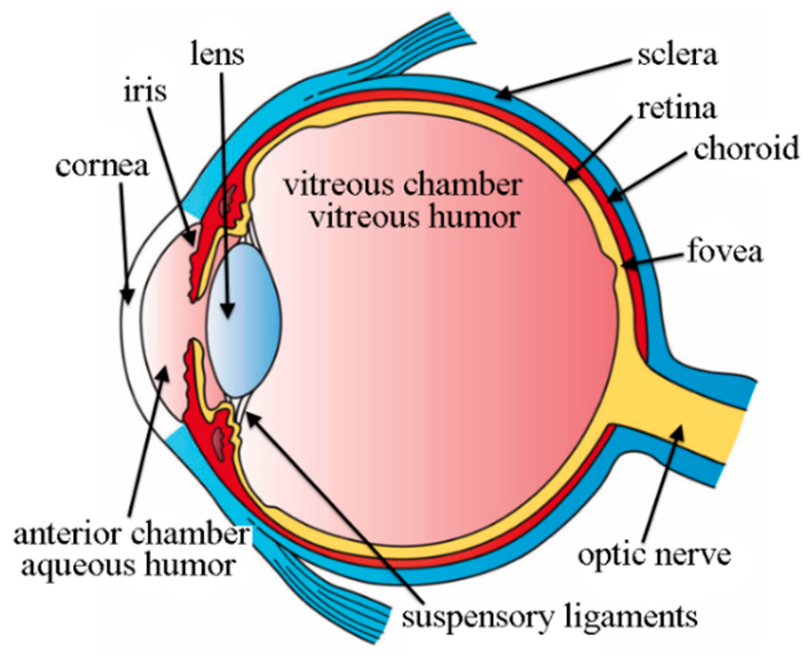
6]. The iris is a thin spherical plate between the pupil and cornea, as shown in Figure 1.
Iridology, also known as iridodiagnosis or iridiagnosis, is an approach that uses the patterns, colors, and many other visible characteristics of the iris to assess an individual’s general health status. Such visible characteristics include pigmentary change, color, and pathological lesions, etc. [8]. In iridology, the iris structure, color, and unique markings provide useful information about the health status [4]. Iridologists believe that every organ in the human body has its corresponding area in the iris. Therefore, the assumption is that the status of body organs can be observed by simply examining the corresponding regions of the iris.
According to iridologists’ investigations, when any change happens in the human body, such as weaknesses and strengths, a spot or mark appears in the iris. These marks on the iris indicate possible diseases or dysfunctions of specific body organs [5]. For instance, changes in kidneys will appear at the bottom edge of the iris. The right kidney affects the right iris, while the left kidney affects the left iris. Changes in the stomach can cause changes in the iris just above the pupil. Thus, iridology reveals any transition in a human body organ because the iris has many regions that are associated with different body parts. The spot or mark will not affect the result of the iris recognition because there is a fault tolerance mechanism built in the iris recognition algorithm. For the iris recognition algorithm, most of the time, researchers set the hamming distance (HD) threshold value to 0.33 for the matching criteria (HD = 0 means perfect match) and this will tolerate a little bit of mismatch in the iris texture. This is why iridologists claim that the iris color may change but it will not affect iris recognition.
In order to identify the corresponding region for different body organs, two schematic diagrams have been developed. First, there are seven concentric zones in the iris, and the pupil is their common axis, as shown in Figure 2 [9]. The ring nearest to the pupil reflects the healthiness condition of the stomach, whereas the most external circle reflects the skin. Second, the iris is divided into radical zones (also known as an iris chart). There are 12 equal sections radiating out from the pupil to the iris border, which are similar to the face of a clock, as shown in Figure 3 [9].
The iris chart in Figure 3 is very helpful in identifying imbalanced body parts. The iris chart divides the iris surface into a number of zones, and each segment is related to an inner organ or a human body function [4,10,11]. From this chart, we can see the corresponding regions of the organs, such as the digestive system, brain, kidney, and heart. Amazingly, the iridology chart for the left and right eyes is symmetric, demonstrating the mysterious balancing nature of the body.
Conventionally, iridologists use a camera and microscope to examine the subject’s iris and then observe any irregularities based on their professional experience. Finally, practitioners compare their observations to the iris chart.
Furthermore, iridology has become particularly the most valuable and widely used diagnostic tool in complementary and alternative medicine (CAM), especially in the United States and Germany, where there are several iridology institutes and societies [12]. Complementary medicine is a research branch consisting of diverse medical and health care systems that claim to improve the standard of health, prevent disease, and address conditions that orthodox medicine has had limited success in remedying, like chronic back pain and certain cancers [13]. Today, in the USA and Europe, the use of complementary medicine in health care has increased; for instance, around two-thirds of the general population now use CAM [14]. Some therapists practicee iridology as a basis for prescribing dietary supplements [15].
Iridology also plays a vital role in traditional Chinese medicine (TCM) treatment. TCM is a type of CAM that practices customized therapies that are based on body constitution theory. However, TCM theory is based on a philosophical framework, e.g., the five-element postulate [16]. Ancient Chinese philosophers hypothesized that nature is composed of five primary elements, similar to how Greek scholars presented the creation of all the elements in the universe. TCM classifies the human organic system into five organs and links them with five elements. The five elements or five states are sorted in order according to five materials, such as wood, fire, earth, metal, and water. Every component has its own literal and logical meaning [17]. TCM practitioners observe human eyes as a diagnostic tool. Physicians who practice TCM have put the five-components assumption into practice, spreading it to clinical practice as a kind of five-element acupuncture (FEA), which was presented by a British acupuncturist, Dr. J.R. Worsley [17,18].
Generally, more than one characteristic of the five elements can be observed in the same person’s iris image. Based on the suggestions from the experts of Taiwan International Institute of Iridology (TIII), for each person, the body constitution can be classified into a primary type and secondary type of the five materials. Therefore, the five elements of categorization are not exclusive. They can overlap and co-exist in one iris. Therefore, we adopted the categorization suggested by TIII to derive nine categories of body constitution classification for each subject [19]. These nine categories are formed by combining five elements in an order, such as primary and secondary types, as shown in Table 1.
In this study, we introduce a non-invasive iris-based health assessment system that adopts the views of experts from TIII to classify the human body into nine classes, which may give health care advice to a subject based on body constitution evaluation. We apply deep learning techniques that can categorize a subject and possible organ dysfunction based on the iris images. Deep learning (DL) is so far the most powerful and robust classification technique among all of the machine learning methods. DL has received significant interest due to its supremacy regarding accuracy when trained with a large volume of data. Starting from 2012 to 2017, the campion of the ImageNet competition was always won by DL approaches, especially the convolutional neural network (CNN) [20,21,22,23,24]. Furthermore, we utilize several existing CNN models to classify body composition using the iris image. In recent years, CNN has made the most significant novelties in the field of computer vision and image processing and gained the researcher’s attention. A CNN is a particular kind of multi-layer neural network, built to identify visual patterns directly from raw images with the least preprocessing. By using DL and CNN, we can achieve end-to-end learning, which means we do not need to care too much about image pre-processing. As long as we collect enough data and send it to the network, it will learn by itself. In this way, it saves a lot of time for the researcher.
The proposed system takes a colored iris image of the subject, processes the image using the proposed DL algorithm, classifys the subject’s body constitution into one of the nine categories, and brings up the warnings about the potential organ disorder with suggestions and advice. In our experiments, the CASIA-Iris-Thousand database was used to perform training, testing, and cross-validation for the proposed CNN models.
The motivation of this work was to develop an online useable practical system that can be used in clinics and homes to perform body assessment without the need to visit a doctor or a hospital and get results immediately. With our system, medical and travel expenses can be greatly reduced and assessment of the health condition can be sped up.
The major contribution of this study is that we combined the image processing technique based on the iris recognition framework (including iris image acquisition, image processing, iris segmentation) and the powerful image classification technique based on CNN (including ResNet 50, Inception V3, and DenseNet 201) to create a practically useful system, which is able to capture iris images and perform nine body constitution classifications in real-time, with a classification accuracy up to 82.9%. Such a system is interdisciplinary research consisting of iris image processing, DL, TCM, and iridology. This paper makes the following contributions:
We built the first largest scale iridology database using CASIA-Iris-Thousand, which contains 20,000 iris images, with help from TIII experts.
The powerful DL model was applied to perform body constitution classification and was able to achieve a very high accuracy.
A practically proven system was built, which can work in a real-time environment. This shows that the designed system is not only theoretically superior but practically useful.
The paper is organized into the following sections. The related work is described in Section 2 and Section 3 discusses the materials and methods; Section 4 presents the experimental results; Section 5 is about the demonstration of the system; and Section 6 draws conclusions.

2. Related Work

Several studies have been performed on human eye iris examinations (iridology) to evaluate the human body organ abnormalities and diagnostic effectiveness of iridology.
Othman et al. [4] presented preliminary research on iris recognition methods to identify abnormalities in the human body organs. They used a novel iris segmentation approach termed the water flow method due to its geometrical resilience and topological rubberiness. Though, the study was preliminary research work with no significant conclusion. Hussain et al. [5] proposed an iris algorithm for obstructive lung disease (OLD) by a non-invasive diagnosing approach. They developed a real-time iris-based lung pre-diagnostic system, which uses a machine learning algorithm and an iridology chart to detect lung disorder. Features were obtained by using a Gabor filter and support vector machine (SVM). In this study, 50 lung patients and 50 healthy subjects were examined, and the system accuracy for lung disorder detection was 88%.
Anna et al. [25] introduced an adaptive resonance theory 1 (ART1), a type of artificial neural network that uses unsupervised learning algorithms to analyze lung disorder through an iris image. In this paper, they designed a lung imbalance detection system that contains several phases, such as iris segmentation, extraction of color dissimilarities, a transformation of the lung and pleura representation region in the iris image as the input of ART1, and pattern recognition by the ART1 neural network framework. In the changing environment of pattern recognition, ART1 is considered one of the most durable and flexible solutions. Adelina et al. [26] proposed a method to identify diabetes by analyzing pancreatic signs through iridology. A backpropagation method was used to identify the condition of the pancreas organ. A Gaussian filter was used to minimize the noise on the image while iris segmentation was done by using the Hough circle transfer method. They used the gray level co-occurrence matrix (GLCM) for region of interest (ROI) feature extraction.
Hussain et al. [13] studied the diagnostic effectiveness of iridology to diagnose abnormalities of the kidneys. They designed an automated approach with an artificial intelligence technique that goes through several steps starting from iris image acquisition, pre-processing, normalization, segmentation, feature extraction, and an adaptive neuro-fuzzy inference system (ANFIS) method for the classification of features from a normal and chronic renal failure subjects group. They examined data from 168 individuals free from kidney disease and 172 patients with chronic renal failure. They obtained an 82% and 93% classification accuracy for normal and kidney patients, respectively. The proposed method was performed on a systemic disease with ocular manifestations that showed promising results. Lim et al. [8] conducted a longitudinal study to investigate the validity of iris parameters, temperament characteristics (TCs), and the association among them over 1 year. They use an intra-class coefficient (ICC) to examine inter-rater consistencies and the robustness of the iris parameters. They performed decision tree modeling that used the iris parameters as a component for high-level and low-level temperament features. This research has some drawbacks, such as it was accomplished only in healthy subjects, and the duration of 1 year could not ensure the efficacy of the iris ever. Hernandez et al. [6] investigated alternative techniques of Alzheimer’s disease detection from the iris image using digital image processing. They used a mathematical model (based on Matlab software) to learn about the presence of Alzheimer’s disease by analyzing the iris pattern. In their proposed model, a Fourier transform was used to normalize the iris image and Hough transform for the detection of the pupil and iris in an image. Several methods based on three multilayer classifiers were used, including naive Bayes, ZeroR, and multilayer perceptron. The performance of the naive Bayes classifier was better than others and achieved a 61.96% diagnostic accuracy. Permatasari et al. [27] proposed a method of heart condition detection from iris classification using the support vector machine (SVM). Feature extraction was done by using a principal component analysis (PCA) method. They obtained a classification accuracy rate of 80%. Herlambang et al. [28] introduced liver disorder in the iris image. They proposed a liver disease detection technique using a back-propagation neural network and gray level co-occurrence matrix (GLCM) for feature extraction. They used Matlab to build a liver disorder diagnosis application. In this study, 60 individuals’ images of the right eye iris were taken, of which 34 were healthy iris images and 26 were abnormal images.
The validity of this approach was tested on 35 liver patients and obtained a 91.42% accuracy rate in detecting liver disease. Miranda et al. [29] developed an automated algorithm technique to detect intestinal system pathologies, such as a distended colon, healthy intestinal tract, prolapse of transverse colon, and spastic colon, by using the human iris parameters. However, this was a pilot study, and thus requires further examination of its efficacy.
In this paper, we evaluated the human organic system based on the concept of TIII experts and iridology together. Furthermore, we applied powerful DL classification algorithms to iridology and built a practically useful system for the general public. All the previous methods mainly focused on specific body organ dysfunction detection and used a smaller amount of iris images in the experiments. On the other hand, our approach performed a holistic health assessment by using the largest scale dataset of iris images. Moreover, the designed system is a real-time, user-friendly, and portable device that can perform the assessment very quickly. The summary of the proposed method and the existing studies of iridology is shown in Table 2. Since the classification goal and the datasets used in our work are different than any of the prior studies, it is inappropriate to directly compare our work with the prior works. Thus, Table 2 is simply a summary that lists all related prior works and also our work.

3. Materials and Methods

This section describes the proposed real-time iris-based health assessment system according to the view of TCM and iridology.

3.1. Iridology with Traditional Chinese Medicine (TCM)

Traditional Chinese medicine (TCM) is a conventional style of Chinese medicine practice, which is based on a unique hypothesis along with diagnostic and treatment approaches [30] and has a history of more than 2500 years [31]. This is the principle of Chinese philosophy, history, and medical knowledge. According to the speculations of ancient Chinese scholars, the creation of the world rotates around the five fundamental elements of daily life, and the formation of the human body is the combination of the five elements. Each element represents a dissimilar attribute, function, or appearance, according to which everything in the universe can be classified. Therefore, TCM practitioners categorize the inner body functioning system into a five-organ system, namely the heart, liver, kidney, stomach (including spleen), and lung, which are further linked to fire, wood, water, earth, and metal, respectively, based on the morphological or functional similarity [17]. Table 3 presents the correspondence between the five elements and the human body organs.
In TCM, the concept of five elements has been practiced to describe the universe and the connections of the main organs of the human body [32]. The imbalance between the five elements is supposed to cause diseases.
In this study, we propose a practical health assessment system that combines DL with the philosophy of TCM and iridology, which evolves into a convenient and portable tool that is able to predict possible organ dysfunction and give advice to subjects about how to take care of their health according to the body constitution assessment. According to the advice of iridology experts from TIII, the key to body constitution using iridology lies in the third zone (ring) of the iris chart. We summarized the classification rules and present them in the form of a table. Table 4 shows the classification of five elements based on the third zone (ring) of the iris chart. Figure 4 shows 15 examples of iris images corresponding to five TCM elements, based on the classification criterion in Table 4. Figure 5 shows the samples of iris images of the derived nine categories (with primary and secondary type), which were used in our experiments.

3.2. Overview of the Proposed System

The proposed real-time iris-based health assessment system contains various stages to perform human body type classification based on the nine classes of body constitution. The overall flowchart of the proposed system is shown in Figure 6.

3.2.1. Iris Image Acquisition

The proposed system takes an iris image as input that is retrieved from an existing database or captured by a handheld high-resolution iris camera.

3.2.2. Image Processing

In the image processing stage, the color image is transformed into a grayscale image and then contrast enhancement is performed. The color-to-grayscale transformation aims to simplify the information contained in the image and reduce the computational complexity. After that, contrast-limited adaptive histogram equalization (CLAHE) is applied to further enhance the contrast of the iris image.

3.2.3. Iris Segmentation

Iris segmentation is a difficult and challenging task because the anatomy of the iris is very complex. Even iris and pupil boundaries are sometimes hard to distinguish. The purpose of iris segmentation is to locate the boundary between the iris area and the sclera or the pupil. The detection of the iris center is an essential section of the segmentation process because it contains useful information of each body organ and helps iridologists to examine the human health status. Different segmentation methods and approaches have been proposed by researchers in the past. In this study, we applied the iris segmentation method proposed in [33], which is a DL-based method. This iris segmentation algorithm is a combination of learning-based and edge-based algorithms. The algorithm consists of three phases that include eye detection, pupillary edge estimation, and limbus border assessment. A well-trained faster R-CNN (Regions with CNN features) model is used to detect and classify the position of the eye in a given image while a Gaussian mixture model (GMM) is used to find the pupillary area. Then, the circular boundary of the pupil area is fitted based on the five key boundary points. The limbus boundary points are located using a boundary point selection algorithm, and the circular boundary of the limbus is created using these boundary points. In such a way, the iris area can be accurately positioned. Interested readers can find more details of the iris segmentation algorithm in [33].

3.2.4. Body Constitution Classification Using CNN Models

In this step, we performed experiments with several existing CNN models to classify the type of body constitution using iris images. The models were trained based on the transfer learning technique. We froze the parameters of most layers in the pre-trained CNN models, and only the parameters of the last few layers were fine-tuned and retrained using annotated iris images. The classification accuracy for the testing datasets was recorded to benchmark their performance. Figure 6 shows the flowchart of the proposed real-time iris-based health assessment system.
For the practical system, it acquires the real-time iris image of the person with a handheld iris camera, classifies the body constitution of the person with a trained CNN model, and displays the classification results of the body constitution along with some useful advice for the subject to take care of their health.

4. Experimental Results

This section presents the results of the experiment on the task of the iris-based health assessment system.

4.1. Database

We used the CASIA-Iris-Thousand (a publicly accessible) database [34] to train the CNN models. The database has been widely used in iris recognition research but has not yet been used in iridology. It contains a total of 20,000 annotated iris images from 1000 subjects. All the iris images were annotated (such as body constitution detail) and precisely labeled by experts from Taiwan International Institute of Iridology (TIII).

4.2. Data Labeling and Partition

For the iris image annotation, we collaborated with experts from TIII. Iris segmentation and image enhancement were performed on each image in the CASIA-Iris-Thousand database. Then, all the segmented iris images were handed over to a few experts from TIII, who inspected each iris image and gave a ground-truth label to each of them. The ground-truth label includes one of the nine categories of body constitution as shown in Table 1. There were five experts who participated in performing this annotation task. The labeling process consisted of two experts for an image. One expert gave the category for one image and then the result of the label image was reviewed by the other expert, such as a double-blind review.
The total number of iris images in the CASIA-Iris-Thousand database is 20,000. However, some of them were removed because those images were not clear enough for the experts to read and give a correct ground-truth label. In the end, the number of usable images was 5538. For data partition, we partitioned all of the labeled data into 10 disjoint sets, which enabled us to perform 10-fold cross-validation in the experiments. The number of images for each class is described in Table 5.

4.3. Model Training

The pre-trained CNN models we tried included ResNet50 [21], InceptionV3 [22], and DenseNet201 [23]. Each model was trained with a transfer learning technique. Our work mainly used Keras and TensorFlow as deep learning frameworks to obtain pre-trained models, which were originally trained on the ImageNet database [24].
To correctly assess the validity of the proposed method, the protocol of k-fold cross-validation was used, which means that the experiments were repeated K times, and for each time, one partition of data was held out as the test set and the other data partition was used as the training set to train the model [35]. The model was tested using the test set and the accuracy was recorded. Such a procedure was repeated k times until all data partitions were used as a test set. The final accuracy was reported by calculating the mean of the k results from the folds. We set k=10 so that each partition contained an approximately equal size of instances.

4.3.1. Inception V3

In 2014, Google scholar Szegdy et al. [22] proposed the inception network, which ranked first in the classification and detection challenges in the 2014 ImageNet contest. Inception V3 is an inherited and upgraded form of inception V1. The motivation of inception V3 is to bypass a demonstrative obstruction, which means exceedingly decreasing the input size of the next layer. This is one of the early adopters of the batch normalization features. It also uses factorization approaches for more effective computations.

4.3.2. Residual Neural Network (ResNet)

The residual neural network (ResNet) was introduced by Kaiming He et al. [21]. ResNet is the winner of the 2015-ILSVRC competition and became the first deeper network with 152 layers. ResNet gained popularity through the use of skip connections and gives a technique for researchers to design even deeper CNNs without affecting the generalization capabilities and accuracy. Furthermore, it was among the foremost CNNs to use batch normalization characteristics.

4.3.3. Dense Convolutional Network (DenseNet)

Gao Huang et al. [23] proposed a novel convolutional network architecture, namely dense convolutional network (DenseNet) to minimize the vanishing gradient problem. In DenseNet, the feature map of every layer is directly connected to the next upcoming layer in a dense block. DenseNet has various compelling benefits, such as it mitigates vanishing gradient problems, enhances feature propagation, encourages feature reuse, and considerably reduces the number of parameters. Compared with ResNet, DenseNet has more intermediate connections.

4.4. Hyperparameters and Hardware

In our experiments, a fixed learning rate of 0.01 was used with the Stochastics Gradient Descent (SGD) optimizer to train all the networks. All networks were trained using 100 epochs and a batch size of 64. The training and testing stages utilized eight Nvidia Titan X (Pascal), two Intel Xeon E5-2620 V4, and 128 GB Memory. We used Python as a programming language. Table 6 shows the hyperparameter settings of the experimented CNN models.

4.5. Performance Analysis on Proposed Networks

This section analyzes the validity and performance of the proposed networks. Table 7 presents the experimental results.
Table 7 shows the test accuracy of the three CNN models. The DenseNet201 network outperforms the others and achieves the highest accuracy, i.e., 82.9% compared with the other models. The accuracy of the ResNet50 and InceptionV3 models is 79.5% and 77.5%, respectively. The receiver operation characteristics (ROC) and area under curve (AUC) are shown in Figure 7. The precision, recall, and F1 score bar graph is presented in Figure 8.
Table 8 shows the performance of the ResNet50 model. In this table, we present the confusion matrix, precision, recall, F1 scores, and average accuracy for each class. The ResNet model obtained a 79.5% total accuracy. The model performs well compared to inceptionV3.
Table 9 demonstrates the performance of the InceptionV3 model. In the table, we display the confusion matrix, precision, recall, F1 scores, and average accuracy for each class. In the inceptionV3 model, we obtained a 77.5% accuracy. The performance of the model is not as good compared to ReNet50 and Densenet201.
Table 10 illustrate the performance of the DenseNet201 model. The table contains the confusion matrix, precision, recall, and F1 scores and average accuracy for each class. The overall performance of the DenseNet201 model is much better than ResNet50 and InceptionV3. This model achieved an 82.9% accuracy.

5. System Real-Time Demo

We developed a real-time body constitution classification system based on the proposed method. We used MATLAB R2019a to design a graphical user interface (GUI) of the system, which is shown in Figure 9.
Because this system was developed in an early stage of this study, it only classifies the body constitution based on the primary attribute, which means it performs five body-type classifications, instead of nine. Firstly, the system acquires an input iris image captured by a handheld iris camera. Then, the system processes an iris image using a deep learning algorithm to find the relationship between five elements and the internal organs. At the final stage, the system performs the body constitution classification, determines the possible most important body organ, and provides healthcare advice.
Let us consider that the system identifies a person who belongs to the earth element. According to the TCM principle, the internal organs related to earth are the stomach and spleen. The system will then perform a TCM diagnosis and display suggestions and advice to keep the body healthy.

Health Assessment Results

In this section, we present the health assessment results of the human body constitution based on the TCM five-elements theory and particularly important body parts with characteristics and suggestions. The results are shown in Figure 10.

6. Conclusions and Future Work

In this paper, we proposed a non-invasive holistic iris-based human health assessment system based on the philosophy of TCM. This study combined the technique of iris image processing, TCM, and iridology to classify the human body constitution using a deep-learning-based algorithm. Furthermore, a real-time operable system based on the proposed method in this study was implemented to show the effectiveness and practicability of this study. The main advantage of the proposed system is that it helps practitioners to make a quick analysis and prompt classification of the subject’s body constitution and also gives instant feedback about how to maintain health. The system is a user-friendly and portable tool that can predict potential internal organ disorders and provide personal health care suggestions. The experimental results demonstrated that our iris-based method is effective and efficient on the CASIA-Iris-Thousand database. The highest achieved accuracy was 82.9% on the DenseNet201 CNN model.
In the future, we will attempt to develop new CNN architectures that will be able to learn the features of the nine classes more efficiently so that we can train our model with a lesser amount of data. In addition, we plan to publish our annotated database of the nine classes so that the research community of iridology can benefit from the labeled data.

Author Contributions

Conceptualization, Y.-H.L.; methodology, Y.-H.L.; software, C.-A.K. and S.-Y.T.; validation, K.-L.Y.; formal analysis, K.-L.Y.; investigation, K.-L.Y.; resources, K.-L.Y.; data curation, K.-L.Y.; writing—original draft preparation, M.S.A.; writing—review and editing, M.S.A.; supervision, Y.-H.L.; project administration, Y.-H.L.; funding acquisition, Y.-H.L. All authors have read and agreed to the published version of the manuscript.

Funding

This research is funded by Ministry of Science and Technology in Taiwan under Contract no. MOST 108-2221-E-008-071.

Acknowledgments

We give great appreciation to the Taiwan International Institute of Iridology (TIII) for helping us labeling the Ground truth for iris images of CASIA-Iris-Thousand.

Conflicts of Interest

The authors declare no conflict of interest.

References

  1. Ma, L.; Zhang, D.; Li, N.; Cai, Y.; Zuo, W.; Wang, K. Iris-based medical analysis by geometric deformation features. IEEE J. Biomed. Health Inform. 2012, 17, 223–231. [Google Scholar] [PubMed]
  2. Demea, A.L. Medical Diagnosis System based on iris analysis. Acta Tech. Napoc. Electron. Telecommun. 2009, 50. [Google Scholar]
  3. Pushpalatha, M.; Muthurani, K. An Labeled Observations Iridology For Diagnosing Kidney Disease. Int. J. Emerg. Technol. Comput. Sci. Electron. 2018, 25. [Google Scholar]
  4. Othman, Z.; Prabuwono, A.S. Preliminary study on iris recognition system: Tissues of body organs in iridology. In Proceedings of the 2010 IEEE EMBS Conference on Biomedical Engineering and Sciences (IECBES), Kuala Lumpur, Malaysia, 30 November–2 December 2010; pp. 115–119. [Google Scholar]
  5. Hussain, T.; Haider, A.; Muhammad, A.M.; Agha, A.; Khan, B.; Rashid, F.; Raza, M.S.; Din, M.; Khan, M.; Ullah, S. An Iris based Lungs Pre-diagnostic System. In Proceedings of the 2019 2nd International Conference on Computing, Mathematics and Engineering Technologies (iCoMET), Sukkur, Pakistan, 30–31 January 2019; pp. 1–5. [Google Scholar]
  6. Hernandez, F.; Vega, R.; Tapia, F.; Morocho, D.; Fuertes, W. Early detection of Alzheimer’s using digital image processing through iridology, an alternative method. In Proceedings of the 2018 13th Iberian Conference on Information Systems and Technologies (CISTI), Cáceres, Spain, 13–16 June 2018; pp. 1–7. [Google Scholar]
  7. Commons, W. File: Three Main Layers of the Eye.png—Wikimedia Commons, the Free Media Repository. Available online: https://commons.wikimedia.org/w/index.php?title=File:Three_Main_Layers_of_the_Eye.png&oldid=371960677 (accessed on 20 February 2020).
  8. Lim, Y.W.; Park, Y.B.; Park, Y.J. A longitudinal study of iris parameters and their relationships with temperament characteristics. Eur. J. Integr. Med. 2016, 8, 991–1000. [Google Scholar] [CrossRef]
  9. Holl, R.M. Iridology: Another look. Altern. Health Pract. 1999, 5, 35–43. [Google Scholar] [CrossRef]
  10. Um, J.Y.; An, N.H.; Yang, G.B.; Lee, G.M.; Cho, J.J.; Cho, J.W.; Hwang, W.J.; Chae, H.J.; Kim, H.R.; Hong, S.H.; et al. Novel approach of molecular genetic understanding of iridology: Relationship between iris constitution and angiotensin converting enzyme gene polymorphism. Am. J. Chin. Med. 2005, 33, 501–505. [Google Scholar] [CrossRef] [PubMed]
  11. Lodin, A.; Demea, S. Design of an Iris-Based Medical Diagnosis System. In Proceedings of the International Symposium on Signals, Circuits and Systems, Lasi, Romania, 9–10 July 2009; pp. 1–4. [Google Scholar]
  12. Münstedt, K.; El-Safadi, S.; Brück, F.; Zygmunt, M.; Hackethal, A.; Tinneberg, H.-R.; Medicine, C. Can iridology detect susceptibility to cancer? A prospective case-controlled study. J. Altern. Complement. Med. 2005, 11, 515–519. [Google Scholar] [CrossRef] [PubMed]
  13. Hussein, S.E.; Hassan, O.A.; Granat, M.H. Control. Assessment of the potential iridology for diagnosing kidney disease using wavelet analysis and neural networks. Biomed. Signal Process. Control 2013, 8, 534–541. [Google Scholar] [CrossRef]
  14. Ernst, E. Complementary/alternative medicine: Engulfed by postmodernism, anti-science and regressive thinking. Br. J. Gen. Pract. 2009, 59, 298–301. [Google Scholar] [CrossRef]
  15. Ernst, E. Iridology: Not useful and potentially harmful. Arch. Ophthalmol. 2000, 118, 120–121. [Google Scholar] [CrossRef] [PubMed] [Green Version]
  16. Zhao, C.; Li, G.-Z.; Wang, C.; Niu, J. Advances in patient classification for traditional Chinese medicine: A machine learning perspective. Evid. Based Complement. Altern. Med. 2015, 2015, 376716. [Google Scholar] [CrossRef] [PubMed]
  17. Chung, S.; Cha, S.; Lee, S.-Y.; Park, J.-H.; Lee, S. The five elements of the cell. Integr. Med. Res. 2017, 6, 452–456. [Google Scholar] [CrossRef] [PubMed]
  18. Worsley, J.R. Classical Five-Element Acupuncture: The Five Elements and the Officials; Redwing Book Company: Damascus, OR, USA, 1998. [Google Scholar]
  19. Irisology Talk. Available online: https://www.youtube.com/watch?v=oogtGRKxU2o&list=PL8OTrtNdZxX31ArC5iMzSYaikP2POSqYF (accessed on 21 April 2020).
  20. Krizhevsky, A.; Sutskever, I.; Hinton, G.E. Imagenet Classification with Deep Convolutional Neural Networks. In Proceedings of the Advances in Neural Information Processing Systems, Las Vegas, NV, USA, 3–8 December 2012; Springer: Berlin/Heidelberg, Germany, 2012; pp. 1097–1105. [Google Scholar]
  21. He, K.; Zhang, X.; Ren, S.; Sun, J. Deep Residual Learning for Image Recognition. In Proceedings of the IEEE Conference on Computer Vision and Pattern Recognition, Las Vegas, NV, USA, 27–30 June 2016; pp. 770–778. [Google Scholar]
  22. Szegedy, C.; Vanhoucke, V.; Ioffe, S.; Shlens, J.; Wojna, Z. Rethinking the Inception Architecture for Computer Vision. In Proceedings of the IEEE Conference on Computer Vision and Pattern Recognition, Las Vegas, NV, USA, 26 June–1 July 2016; pp. 2818–2826. [Google Scholar]
  23. Huang, G.; Liu, Z.; Van Der Maaten, L.; Weinberger, K.Q. Densely Connected Convolutional Networks. In Proceedings of the IEEE Conference on Computer Vision and Pattern Recognition, Honolulu, HI, USA, 21–26 July 2017; pp. 4700–4708. [Google Scholar]
  24. Deng, J.; Dong, W.; Socher, R.; Li, L.-J.; Li, K.; Fei-Fei, L. Imagenet: A Large-Scale Hierarchical Image Database. In Proceedings of the 2009 IEEE Conference on Computer Vision and Pattern Recognition, Miami, FL, USA, 20–25 June 2009; pp. 248–255. [Google Scholar]
  25. Triwijayanti, A.; Suwastio, H.; Damayanti, R. Lung disorders detection based on irises image using computational intelligent art. TEKTRIKA-Jurnal Penelitian dan Pengembangan Telekomunikasi, Kendali, Komputer, Elektrik, dan Elektronika 2003, 8. [Google Scholar] [CrossRef]
  26. Adelina, D.C.; Sigit, R.; Harsono, T.; Rochmad, M. Identification of diabetes in pancreatic organs using iridology. In Proceedings of the 2017 International Electronics Symposium on Knowledge Creation and Intelligent Computing (IES-KCIC), Surabaya, Indonesia, 26–27 September 2017; pp. 114–119. [Google Scholar]
  27. Permatasari, L.I.; Novianty, A.; Purboyo, T.W. Heart disorder detection based on computerized iridology using support vector machine. In Proceedings of the 2016 International Conference on Control, Electronics, Renewable Energy and Communications (ICCEREC), Bandung, Indonesia, 13–15 September 2016; pp. 157–161. [Google Scholar]
  28. Herlambang, R.A.N.P.; Isnanto, R.R.; Ajub, A.Z. Application of liver disease detection using iridology with back-propagation neural network. In Proceedings of the 2015 2nd International Conference on Information Technology, Computer, and Electrical Engineering (ICITACEE), Semarang, Indonesia, 16–18 October 2015; pp. 123–127. [Google Scholar]
  29. Miranda, J.D.; Salinas, S.A. Computational Measuring Approach for the Identification of Probable Intestinal System Pathologies through the Human Iris Parameters. In Proceedings of the 2019 XXII Symposium on Image, Signal Processing and Artificial Vision (STSIVA), Bucaramanga, Colombia, 24–26 April 2019; pp. 1–5. [Google Scholar]
  30. Tang, H.; Huang, W.; Ma, J.; Liu, L. SWOT analysis and revelation in traditional Chinese medicine internationalization. Chin. Med. 2018, 13, 5. [Google Scholar] [CrossRef] [PubMed] [Green Version]
  31. Huan, E.-Y.; Wen, G.-H.; Zhang, S.-J.; Li, D.-Y.; Hu, Y.; Chang, T.-Y.; Wang, Q.; Huang, B.-L. Deep convolutional neural networks for classifying body constitution based on face image. Comput. Math. Methods Med. 2017, 2017. [Google Scholar] [CrossRef] [PubMed]
  32. Wang, K.-C. The Five Elements Theory in Business Research. In Proceedings of the 46th Annual Meeting of the Association of International Society for the Systems Sciences, Shanghai, China, 2–6 August 2002. [Google Scholar]
  33. Li, Y.-H.; Huang, P.-J.; Juan, Y. An Efficient and Robust Iris Segmentation Algorithm Using Deep Learning. Mob. Inf. Syst. 2019, 2019, 4568929. [Google Scholar] [CrossRef]
  34. CASIA Iris Image Database. Available online: http://biometrics.idealtest.org/ (accessed on 17 January 2020).
  35. Alpaydin, E. Introduction to Machine Learning; MIT Press: Cambridge, MA, USA, 2020. [Google Scholar]
Figure 1. Human iris image [7].
Symmetry 12 00803 g001
Figure 2. The seven zones of the iris.
Symmetry 12 00803 g002
Figure 3. Iridology chart is shown for both the right and left iris and their relations to body organs [5].
Symmetry 12 00803 g003
Figure 4. Samples of iris images based on the third ring on the iris. (a) Wood type iris image. (b) Water type iris image. (c) Metal type iris image. (d) Earth type iris image. (e) Fire type iris image.
Symmetry 12 00803 g004
Figure 5. Samples of iris images of the derived nine categories (with primary and secondary type). (a) Fire and wood; (b) Earth and Metal; (c) Earth and Water; (d) Earth and Wood; (e) Fire and Metal; (f) Fire and Water; (g) Metal and Water; (h) Water and Wood; (i) Wood and Metal.
Symmetry 12 00803 g005
Figure 6. Flowchart of the proposed iris-based health assessment system.
Symmetry 12 00803 g006
Figure 7. The Receiver Operation Characteristics (ROC) curve of the experiment results. (a) DenseNet201 model, (b) InceptionV3 model, and (c) ResNet502 model.
Symmetry 12 00803 g007
Figure 8. Precision, recall, and F1 score of all three models in our experiments. (a) F1 score, (b) Precision, (c) Recall.
Symmetry 12 00803 g008
Figure 9. The graphical user interface (GUI) demonstration of the health assessment system. (a) The first window of the system. (b) Taking the iris image. (c) Obtained iris image. (d) Health prediction.
Symmetry 12 00803 g009
Figure 10. Health assessment results of five different types of body constitution according to the TCM view. (a) Earth type body constitution. (b) Wood type body constitution. (c) Water type body constitution. (d) Fire type body constitution. (e) Metal type body constitution.
Symmetry 12 00803 g010
Table 1. Categorization of nine subclasses of body constitution based on Traditional Chinese Medicine (TCM) philosophy.
PrimarySecondary
EarthMetal
Wood
Water
FireMetal
Wood
Water
WaterWood
WoodMetal
MetalWater
Table 2. Summary of the existing studies using iridology and the proposed method.
MethodsNumber of ClassesNumber of SubjectsDisease TypeAccuracy %
Feature extracted by Gabor filter and SVM Hussain et al. [5] 2100Lung88
Adaptive Resonance Theory (ART) Anna et al. [25]232Lung91.40
Feature extraction by Gray Level Co-Occurrence Matrix (GLCM) Adelina et al. [26]220Diabetes81.35
Adaptive Neuro-Fuzzy Inference System (ANFIS) for classification Hussain et al. [13]2340Kidney82/93
Decision tree model for feature extraction Lim et al. [8]270-77.5/88.9
Fourier Transform for normalization, and Hough Transform for detection of a pupil. For classification: Naive Bayes, ZeroR, and multilayer perceptron Hernandez et al. [6]2-Alzheimer’s61.96
Feature extracted by (PCA) method and Support Vector Machine (SVM) for Classification Permatasari et al. [27]240Heart80
Feature extraction by Gray Level Co-Occurrence Matrix (GLCM) Herlambang et al. [28]235Liver91.42
Deep learning edge-based algorithms for Iris segmentation. CNN models for Classification (Proposed Method)91000Holistic Health Assessment82.9
Table 3. Five elements of Traditional Chinese medicine (TCM) and its correspondence to the human internal organs.
Elements of TCMCorresponding Human Organs
WoodLiver
WaterKinder and Bladder
MetalLungs and Large Intestine
EarthStomach and Spleen
FireHeart and Small Intestine
Table 4. Classification criteria of the five body constitutions based on the third zone (ring) of the iris chart (autonomic nervous system).
TCM ElementsRing Shape
WoodIt has no explicit ring pattern.
WaterIt has an explicit ring pattern and very small acute angles acceptable.
MetalIt has acute angles around itself.
EarthIts radius is bigger than the radius of 1/3 iris.
FireIts radius is smaller than the radius of 1/3 iris.
Table 5. Number of images for each category.
Class Class NameNumber of Images
0Earth, Wood616
1Earth, Water585
2Earth, Metal659
3Wood, Metal603
4Water, Wood598
5Fire, Wood611
6Fire, Water639
7Fire, Metal640
8Metal, Water587
Total 5538
Table 6. Hyperparameter settings of the experimented CNN models.
ModelsImage SizeBatch SizeEpochLearning RateOptimizerModel Size
Inception V3224 × 224 × 3641000.01SGD80.2MB
ResNet50229 × 229 × 3641000.01SGD87.6MB
DenseNet201224 × 224 × 3641000.01SGD74.2MB
Table 7. Performance of the CNN models.
ModelsMean Accuracy %
ResNet5079.5
InceptionV377.5
DenseNet20182.9
Table 8. Performance analysis of the ResNet 50 model.
Ground TruthPredicted
012345678
057131024141110
175517253541
22610514861652945
3117134671036182714
46712539161494
525993628368507412
62326928549364
7810292423614140836
8131326159172233439
Precision0.8530.8980.8410.8300.8510.6580.77870.6570.776
Recall0.9260.9410.7790.7740.9010.6020.8590.6370.747
F1-Score0.8880.9190.8090.8010.8750.6290.8160.6470.762
Total Acc0.795
Table 9. Performance analysis of the Inception V3 model.
Ground TruthPredicted
012345678
05585117519335
1953510639346
213135272281112836
35413464114093126
443512528179155
51515104027361487619
66731511325232715
7512423416754037541
81712332212191829425
Precision0.8820.8820.8050.7450.8500.6190.7990.6370.735
Recall0.9050.9140.7990.7690.8820.59080.8180.5850.724
F1-Score0.8940.8980.80270.7570.8660.6040.8080.6100.729
Total Acc0.775
Table 10. Performance analysis of the DenseNet201 model.
Ground TruthPredicted
012345678
058718226424
125627136211
2136564154931926
310617485924152215
4632255681065
5156162324409525610
620166305622210
7813461316764139829
8993411391625471
Precision0.900 0.9273 0.811 0.869 0.892 0.708 0.797 0.722 0.824
Recall0.9520.9600.8550.8040.92970.6690.8790.6210.802
F1-Score0.9250.9430.8330.8350.9100.6880.8360.6680.813
Total Acc0.829

