Article

Assessment of the Orbital and Auricular Asymmetry in Italian and Sudanese Children: A Three-Dimensional Study

1
Department of Biomedical Sciences for Health, Università degli Studi di Milano, 20133 Milano, Italy
2
Khartoum Centre for Research and Medical Training, Khartoum 11111, Sudan
3
Institute of Dentistry, Barts and The London School of Medicine and Dentistry, Queen Mary University of London, London E1 2AD, UK
4
U.O. Laboratorio di Morfologia Umana Applicata, IRCCS Policlinico San Donato, 20097 San Donato Milanese, Italy
*
Author to whom correspondence should be addressed.
These authors contributed equally to this work.
Symmetry 2021, 13(9), 1657; https://doi.org/10.3390/sym13091657
Submission received: 16 May 2021 / Revised: 10 August 2021 / Accepted: 1 September 2021 / Published: 8 September 2021
(This article belongs to the Section Life Sciences)

Abstract

:
The evaluation of the symmetry of orbital and ear soft tissues is important for aesthetic and reconstructive surgery. However, little information is available for these facial regions, especially in children. We analyzed the orbital and auricular symmetry in 418 Italian and 206 Sudanese subadult males divided into three age groups (8–11, 12–15, and 16–19 years old). Orbital and auricular height and width were measured for calculating fluctuating and directional asymmetry indices. Differences in asymmetry indices according to ethnicity and age group were assessed through the two-way ANOVA test (p < 0.01), while differences in the prevalence of right or left asymmetry according to ethnicity were assessed through the chi-square test. On average, directional asymmetry indices ranged from −2.1% to 1.1%, while fluctuating asymmetry indices ranged between 2.9% and 5.4%, corresponding to a small effect size and to 1.06–2.34 mm actual dimensions. Sudanese subjects showed a greater asymmetry for all the indices except for the fluctuating asymmetry of orbital height (p < 0.01). The directional asymmetry of auricular width increased with age. A prevalent right-side asymmetry was found for all the orbital indices (p < 0.001) in both populations, although significantly more prevalent in Sudanese individuals (over 83% for both measures), while auricular measures showed a prevalent left asymmetry exclusively in the Sudanese but with lower percentages. Aside from the limited effect size, the results proved the ethnic variability of asymmetry of orbital and auricle regions in children and suggest the need to collect more population data.

Graphical Abstract

1. Introduction

Information about precise proportions and symmetry of facial structures has important applications in several fields of research and clinical care, especially in the area of reconstructive and aesthetic surgery [1,2,3,4,5,6]. The goal of an intervention is to obtain an aesthetically pleasant result, having to be consistent with gender, age, ethnicity, and facial features of the patient, and taking needs and expectations into account. In the case of injuries of paired and symmetric structures, the contralateral is used as the reference for the reconstruction of the injured element [7,8]. However, this approach should be based on detailed knowledge of the actual level of symmetry of the corresponding anatomical structures: the hypothesis of a perfect bilateral symmetry (between paired structures) has never been supported by experimental data [1,6]. Even attractive faces show a certain degree of asymmetry [9,10,11]. The influence of facial symmetry on attractiveness has been studied extensively with inconclusive results in the past decades. Effectively, depending on the studies, facial symmetry was found to be alternatively essential for attractiveness, irrelevant (if slight), or even a pejorative factor [11,12,13]. To explain these conflicting findings, in a recent study, Zheng et al. hypothesized that facial symmetry increases attractiveness only when it improves the perception of normality of the face, considering ‘normal’ a face whose features and configuration are within the normal range of values appropriate for sex, age, and ethnicity [13].
Therefore, the assessment of facial symmetry degree as a characteristic of a population is fundamental to provide data useful to improve the efficacy and the expectations of reconstructive surgical procedures [1]. For example, data concerning facial asymmetry in relation to sex, age, and ethnicity could be usefully included among the characteristics used for the three-dimensional simulation of a reconstructive/aesthetic surgical intervention, not only for a better communication with patients [14] but also as a valuable part of the strategies used in plastic surgery [15]. Besides the surgical field, the design of prostheses and medical and professional devices such as respiratory protective equipment also needs a detailed description of facial asymmetry and its range in normal people [6].
From a general point of view, asymmetry can be classified as fluctuating and directional [12,13,16,17,18,19]. Fluctuating asymmetry does not favor one side of the body over the other, and it is considered as the random developmental variation in a trait that is perfectly symmetrical, on average [20]. Specifically, it is the variance in difference between left and right sides with values distributed around 0 for a potentially perfectly symmetrical characteristic. Fluctuating asymmetry is believed to evidence the imprecise expression of development instability: according to Van Valen [21], the term ‘fluctuating’ indicates that the direction of symmetry is not under genetic control but may ‘fluctuate’ from one generation to the next. In other terms, fluctuating asymmetry is the result of random perturbations during the developmental process [16]. Less stressed individuals or individuals with a larger buffering capacity should show a reduced level of asymmetry [20,22].
If, on the one hand, fluctuating asymmetry can be seen as a small difference between the left and the right sides due to random errors in the individual development, and so it is the amount of deviation from the genetically determined ‘target phenotype’ [18], on the other hand, directional symmetry indicates a systematic difference in the development in a population sample (the average asymmetry between the two sides in that population). Thus, directional asymmetry expresses the prominence of a side in a population, gaining the value of being a marker of the evolutionary pressure under genetic components and environmental control [18,23].
Indeed, the investigation of both types of symmetry has gathered momentum, particularly in the ecological and evolutionary contexts, with the aim to suggest evolutionary mechanisms behind the left–right dominance in many vertebrate species, including humans [16,17]. For instance, in humans, all of the abovementioned discussion about facial symmetry, attractiveness, and perceived good health [13] has been related to mate choice and the transmission of good genes to the next generation [11,17]. In other vertebrates, investigations about limb and body symmetry, brain dominance, and occurrence of injuries lead to hypotheses that consider left-sided individuals to have a higher mortality rate than right-sided ones. Studies extensively analyzed some reptile species [19,24,25] and proposed left dominance in ‘limbs’ (in comparison to right dominance) as stress-related or functional [24]. The hypothesis may be relevant for Homo sapiens [19].
Switching from reptiles to humans, studies in the literature show extensive research on fluctuating and directional symmetry of the face and body with both evolutionary and biomedical perspectives. Among others, the periorbital and auricle soft tissues are often involved in reconstructive surgical interventions [26,27]. For what concerns these facial structures, several studies have provided information about their metric and morphological variability among individuals of different sex, age, and ethnicity [8,26,27,28,29], but very few authors have specifically investigated their bilateral symmetry, also known as matching symmetry [30]. If, on the one hand, some information is available for single facial structures such as the auricle, on the other hand, this kind of data is still limited to the adult age [6,7], whilst data concerning the overall facial asymmetry is the only reported for subadult ages (children and adolescents) [1,2,9,31]. Facial plastic and reconstructive surgery, as well as maxillofacial surgery, are performed in different percentages in patients of different ages, and the needs greatly vary across the world, requiring appropriate information for each age [32].
Moreover, data about symmetry in human populations of different geographical origin are still scarce in the literature; yet, ethnicity may influence metric and morphological features of the face, with obvious consequences on the choice of the reference standards used by reconstructive surgery. The first studies focused on European Whites, Africans, and Latin Americans, with Asian subjects being present only in more recent investigations [1,2,6,9,22,33]. This implies the collection of reference data pertaining to different populations, as the analysis of symmetry should consider factors such as sex, age, growth, health status, and ethnicity.
A further consideration is about the instruments and methods used to quantify facial symmetry; recent investigations applied geometric morphometrics and similar methods to both two- and three-dimensional data and obtained global estimates of the differences between the two sides of the face [1,2,6,9,10,16,17,22]. In addition, in these studies, the facial surface was often sampled using optical instruments that collect a wealth of data; the specialized expertise necessary for their analysis and interpretation may not be so widespread as it should. Recently, Ekrami et al. [16,17] developed an elegant method to quantify fluctuating asymmetry in the face of adult subjects. While these methods can provide precious information to gain a deeper understanding of the underlying biological processes, they may be of difficult practical application within clinical settings, where neither the instruments nor the know-how of using computer-assisted three-dimensional software enabling three-dimensional morphometric analyses are available, and where the main demands are often limited to the actual quantification of asymmetry through linear distances [10,33,34].
Therefore, this study aimed to partially fill some of the gaps existing in literature; in particular, data on orbital and auricular symmetry were compared in two subadult populations of different ethnicity: Italian (White European) and Sudanese (Arab descent) children and adolescents aged between 8 and 19 years. Both ethnic groups have already been investigated in our laboratory, but asymmetry was not studied [26,27,35]. For this purpose, data collectable also by simply using a caliper (linear distances) were considered and thus analyzed with basic calculations without requiring specific technical competencies. The results will be useful to verify the possible modification of symmetry of linear distances with age and the existence of ethnic variability among the compared populations, thus providing important and new data paving the way to additional studies on facial region symmetry in populations of ethnicity different from the ones examined here. In addition, some initial, nonexhaustive considerations dealing with the current debate about symmetry and its evolutionary role are reported.

2. Materials and Methods

Three-dimensional facial coordinates were acquired on 418 Italian and 206 Sudanese male subjects aged between 8 and 19 years (mean age, Italian: 13.4 ± 3.3 years; Sudanese: 13.7 ± 3.1 years). Possible differences of age between the two populations were assessed through Student’s t-test. The study followed the guidelines provided by the Helsinki Declaration and was approved by the University ethical committees (Italy: 26.03.14, no. 92/14; Sudan: Elrazi dental School, no. Dent/01). Part of the data of the Sudanese subjects were published elsewhere [35], while data of the Italian subjects were obtained by our laboratory database.
Acquisition was performed through a stereophotogrammetric system (VECTRA-3D®: Canfield Scientific, Inc., Fairfield, NJ, USA) and a portable handheld laser scanner (FastSCAN Cobra, Polhemus Inc., Colchester, VT, USA), respectively, in the Italian and Sudanese groups. Both the acquisition techniques are noninvasive and provide no risks [36,37,38,39,40,41]. All the recruited individuals were not affected by any genetic diseases and with no facial congenital or acquired deformities. Their face was also considered symmetrical at visual inspection. Repeated landmark identifications and facial digitizations using both stereophotogrammetry and the laser scanner were found to be free from statistically significant systematic errors, with mean random errors (technical error of measurement) of 0.722 and 0.755 mm, respectively [36,37,38].
Four landmarks were assessed both for the orbital and the auricular regions (as described in Table 1 and shown in Figure 1) according to Farkas [42]. Four measurements were then automatically calculated from the coordinates of the abovementioned landmarks both on the right and the left side: orbital height (os-or), orbital width (en-ex), auricular height (sa-sba), and auricular width (pra-pa).
For the Italian group, the landmarks were manually identified on the stereophotogrammetric reproduction of the face, where a textured photograph was obtained together with the mesh of the digitized points. Details about the procedures can be found in recent reports [34,41,43]. For the Sudanese group, the acquired facial image was analyzed through an interactive three-dimensional modeler software (Rhinoceros NURBS modeling for Windows 4.0, RobertMcNeel & Associates, Seattle, WA). The software is based on nonuniform Rational B-Splines and can model the geometric shape as a three-dimensional parameter that accurately reflects the facial shape. The coordinates (x, y, z) of biologically relevant landmarks can then be extracted for further three-dimensional measurements [35,38].
For each measurement, an asymmetry index was calculated as follows:
( R L )   100 R + L
where R is the right- and L the left-side measurement (directional asymmetry): positive values are obtained when the right-side distance is longer than the left-side one, and negative values are obtained in the opposite situation, with a predominant left-side value. Moreover, the absolute value of the above formula was calculated as well to obtain the fluctuating asymmetry [44]. Mean asymmetry indices and corresponding standard deviations were calculated in both the populations according to three age groups: 8–11 years, 12–15 years, and 16–19 years, and their difference from an expected value of 0 (perfect symmetry) was assessed by paired Student’s t tests (p < 0.01). The prevalent side of directional symmetry, either right- or left-side asymmetry (i.e., positive or negative asymmetry index, respectively), was recorded for both the populations.
Possible significant differences in asymmetry indices for orbital and auricular height and width according to ethnicity and age group were assessed through a two-way ANOVA test (p < 0.01); a post hoc assessment according to age groups was performed through Tukey’s test applying Bonferroni correction for the degrees of freedom (p < 0.003). To estimate the effect size of significant main effects and interactions, the eta squared value was computed. This value describes the practical importance of the statistically significant difference: values lower than 0.02 are considered very small, up to 0.13 small, between 0.13 and 0.26 medium, and from 0.26 to 1.00 large [45].
Possible significant differences in prevalence of right or left directional asymmetry for orbital and auricular height and width were assessed through the chi-square test using a hypothetical sample with right- (positive asymmetry index) and left- (negative asymmetry index) side asymmetry equally represented (p < 0.01). Possible differences in the prevalence of right- and left-side directional asymmetry for all the measurements according to ethnicity were assessed through the chi-square t-test (p < 0.01). For all comparisons, two-tailed tests were used.

3. Results

No significant differences in age were found according to ethnicity (p > 0.05). Mean values of the four linear distances are shown in Table 2; on average, the orbital height and width were about 37.5 mm in the Italian population and 44 mm in the Sudanese sample. In the Italian population, the auricular height and width were, on average, 56 mm and 34 mm, respectively, while the same measurements in the Sudanese sample were, on average, 51 mm and 32 mm. All symmetry indices were significantly different from 0 (p < 0.001), except ear width in the Italian subjects (p = 0.205).
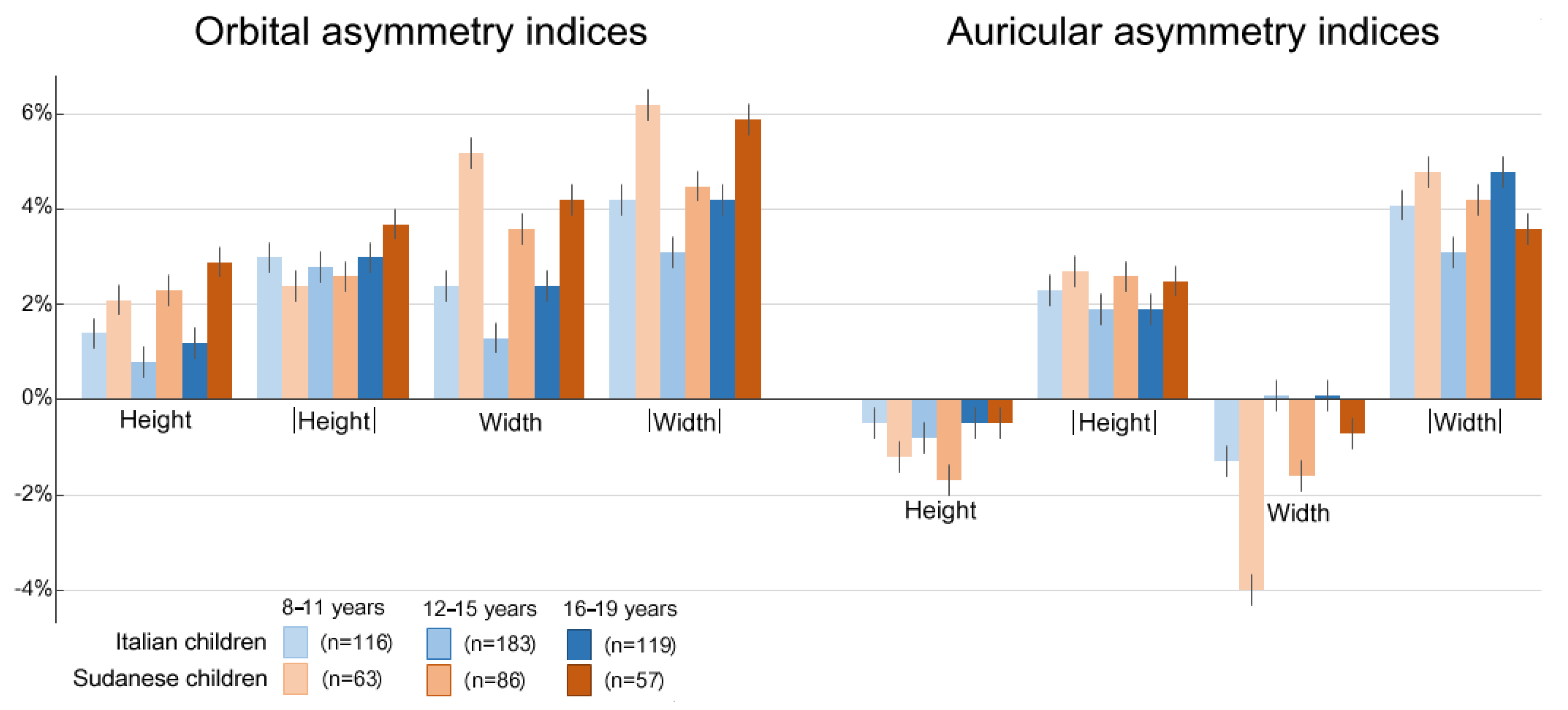
General results concerning indices are shown in Figure 2; the directional asymmetry index of orbital height and width was, on average, 1.1 ± 3.5% (mean ± SD%) and 1.9 ± 4.8% for the Italian and 2.4 ± 2.6% and 4.3 ± 5.2% for the Sudanese groups, respectively; the corresponding values of the fluctuating asymmetry index were 2.9 ± 2.2% and 3.7 ± 3.6% for the Italian and 2.9 ± 2.2% and 5.4 ± 4.0% for the Sudanese groups, respectively.
The directional asymmetry index of auricular height and width was, on average, −0.6 ± 2.6% and −0.3 ± 4.5% for the Italian and −1.2 ± 3.1% and −2.1 ± 5.1% for the Sudanese groups, respectively; the corresponding values of fluctuating asymmetry were 2.0 ± 1.8% and 3.4 ± 3.0% for the Italian and 2.6 ± 2.1% and 4.2 ± 3.6% for the Sudanese groups, respectively. Overall, the actual effect size for all the average asymmetry values ranged between 1.06 mm (Italian group, eye height) and 2.54 mm (Sudanese group, eye width).
Results of the one-way ANOVA test are shown in Table 3. Sudanese subjects showed a higher asymmetry than the Italian ones for all the indices except the fluctuating asymmetry of orbital height (p < 0.01). On the other hand, only the auricular width showed differences according to age groups, being lower in the 8–11 years age group than in the 16–19 years age group (post hoc test, p < 0.003). No significant ethnicity and age interactions were found. In all occasions, the eta squared value was lower than 0.043, indicating small or very small effect sizes.
Data concerning the right- and left-side prevalence of asymmetry are reported in Table 4. Right asymmetry was prevalent both in Italian and Sudanese populations for all the orbital measurements (p < 0.001) with higher percentages for the Sudanese population (86.8% and 83.4% for orbital height and width, respectively) in comparison with those of the Italian sample (67.0% and 64.0% for orbital height and width, respectively) (p < 0.001). Conversely, for auricular measurements, no prevalence of right- or left-side asymmetry was found in the Italian sample (p > 0.05), whereas the left asymmetry was more frequently observed in the Sudanese group (63.7% and 66.2% for auricular height and width, respectively, p < 0.001). Finally, a significant difference was found between the two populations only for auricular width (p < 0.001).

4. Discussion

Reconstructive surgery in the face is often based on the restoration of anatomical structures according to the contralateral one, usually on the base of a hypothesized and undemonstrated symmetry between the two sides. From this point of view, the assessment of the degree of symmetry (left–right mismatch) in a normal population is fundamental as it provides reference data appropriate for sex, age, and ethnicity [9]. Treatment planning should consider these levels of symmetry to regain (or increase) facial normality and, subsequently, its attractiveness [1,10,13]. A recent paper by Zheng et al. [13] explained this link well among actual and perceived characteristics of a face: the closer to ‘normal,’ the more pleasant the individual. Symmetry/asymmetry would augment attractiveness as far as it increases normalcy.
Although the study of asymmetry has gathered much attention in the past decades in many fields and with different objectives, and regardless of the ambiguity noticeable in the literature, the dispute on its role, its measurement, meaning, and interpretation, to our knowledge, few studies have investigated the asymmetry of singular facial regions/structures, and, in particular, none have analyzed the asymmetry of orbital and auricle soft tissues in subadult individuals (children and adolescents).
In addition to age, the ethnic variability is a further factor scantily considered by researchers. Furthermore, some of the partial data already existing in the literature have been derived from recent studies where the data collection procedures and the employed mathematical methods may be not so suitable for all clinical conditions [1,2,6,9]. In particular, the use of geometric morphometrics and advanced mathematical and statistical methods appears to be limited to research settings, with scanty clinical applications [1,2,6,9,17,22]. From this point of view, only optical instruments such as laser scanners and stereophotogrammetry units can provide the necessary dense two- or three-dimensional facial scans. In contrast, clinicians may require simplified approaches focusing on the quantification of asymmetry through linear distances [22].
Therefore, the present study aimed to provide more information about the orbital and auricular symmetry (1) in a subadult population, (2) in two different ethnic groups, and (3) with simple approaches and calculations repeatable in all clinical settings, in order to verify the symmetry of these two facial regions and their variation with age and according to ethnic variability.
The mean values of the orbital and auricular distances observed in the Italian population by the present study are well in line with previous literature reports about the same ethnic group, age, and sex [26,27]. However, data reported for Sudanese subjects were published from our laboratory only and cannot be compared to other reports [35]. Differences in linear distances were found between the two different ethnic groups, confirming the role of ancestry in both orbital and auricular size [8,26,27,31,32,35,42], but no study has reported orbital and auricular asymmetry values in these populations. As any surgical procedure in children and teens should consider the estimated residual growth, we selected children and teens aged 8 years or more because, in both ethnic groups, periorbital and auricular areas have attained 90% or more of their adult dimensions around 8–9 years of age [26,27,35].
As far as facial asymmetry is concerned, the current results provided interesting hints related to evolutionary mechanisms behind the left–right dominance, observed in many vertebrate species, including humans [10,16,17,18]. For instance, studies on reptiles have been conducted on the asymmetry of head traits (eye): some authors have proposed that brain laterality in lizards is also reflected in asymmetrical eyes [46,47]. A possible hypothesis considers morphological directional asymmetry as a proxy for brain laterality, in other words, the existence of an evolutionary mechanism involving the laterality of the brain as an explanation of the stress and risk behavior manifested in individuals with an asymmetry of certain structures [25].
Biological and clinical future research should focus on some of the findings; first, asymmetry indices of the orbital and auricular area were significantly different from 0, except the ear width in Italian boys and adolescents; second, asymmetry was more pronounced in the Sudanese than in the Italian sample; third, most of asymmetry indices were weakly related with age, except for the auricular width, which showed a significant difference between the extreme age groups (8–11 years and 16–19 years). We have no specific explanations about the peculiar results obtained for auricular width, which should be attentively reconsidered.
Cho et al. [1] investigated a group of children and adolescents aged between 0 and 18 years and found that neither age nor ethnicity influenced facial and cranial asymmetry, a datum also reported for reptiles [19]. The lack of age-related differences could be explained by a genetic origin of asymmetry, also modeled by prenatal stress. On the contrary, on adult subjects, both age and ethnicity were reported in the literature to influence facial asymmetry; in a group of males and females aged between 18 and 63 years, Quinto-Sanchez et al. [22] found that asymmetry increased as a function of age; one explanation may be a cumulative effect of postnatal stress. Sajid et al. [6] investigated the impact of sex and ethnicity on the facial asymmetry variations on two-dimensional frontal face images of a large number of adult subjects and reported a significantly higher level of asymmetry for most of the facial dimensions of African compared to European individuals. They tentatively explained the finding as a different masticatory activity in the groups.
Another explanation may be the different level of socioeconomical conditions between Italy and Sudan and the possible effect of a larger developmental instability in the Sudanese subjects, even if recent studies performed in Latin America and in Great Britain rejected this hypothesis, at least for adult people and adolescents [9,22].
Notwithstanding the several significant differences found in the current asymmetry indices, their actual, practical value seems to be limited, as shown by the negligible values of eta squared. According to Seligmann [24], in lizards, a minor directional asymmetry of the limbs may be produced by an evolutionary process relating morphological asymmetry and injury rate: animals may show a higher survival after injury of the morphologically dominant side, thus producing a directional asymmetry in the long run. For what concerns the side of orbital directional asymmetry, the right side is prevalent in both the Italian and the Sudanese sample; moreover, this characteristic is more pronounced in Sudanese subjects (more than 80% of subjects) than in Italian subjects. On the other hand, the auricular measurements did not show a predominant side, but only in the Italian group; in the Sudanese population, the left-side symmetry was significantly prevalent (in more than 60% of cases). It is difficult to comment on this result as no literature information is available; some possible contributing factors may be facial sidedness [8], which might influence orbital soft tissues. Haraguchi et al. [33] reported a significant right-sided dominance of the face in a group of Japanese orthodontic patients aged 2–59 years; neither sex nor skeletal jaw relationships influenced the asymmetry, even if a wider left hemiface was found more frequently in older patients and a wider right hemiface was observed more frequently in younger patients. A larger left-side total face was found by Ercan et al. [10] in young adults. Large left-side eyes were reported by Razzetti et al. [19] for Amiota and were related to brain activity for stimuli given by food (right side eye, less aggressive) or enemies (left side eye, more aggressive) [23,48].
Motion artefacts may also be a factor, especially for the Sudanese group. Indeed, while stereophotogrammetry collects facial data in some milliseconds, laser scanning performed with a handheld instrument may require 60–90 s, thus introducing some possible minor bias [35] that passed the laboratory quality check.
A limitation of the present study is that it did not take into consideration all the wealth of data provided by the optical facial scans and the possibilities of geometric morphometrics [10,16,17]. Thus, the current investigation was limited to some asymmetry indices, and multivariate studies that may unravel some of the complexity of human facial asymmetry have not been performed. Nonetheless, all analyses were based on linear measurements of soft tissues of two facial areas, which can also be easily acquired through direct anthropometry; both photographic and caliper measurements can be used, thus permitting the clinical analysis of asymmetry even when expensive three-dimensional acquisition devices are not available. In our laboratory, we have already tested the compatibility of several data acquisition protocols starting from caliper measurements [49]. However, to perform a global analysis of asymmetry through surface superimposition, good-quality three-dimensional optical digitizers are necessary as surfaces are poorly reproducible without adequate instruments [37,50].
The present study has the limit that measurements were acquired through the use of two different three-dimensional image acquisition systems: the comparison of facial virtual models obtained through different devices may lead to discordant data, but only for what concerns volume measurement and the registration of three-dimensional surfaces [50]. Among studies on the similarity between facial measurements obtained by stereophotogrammetry and laser scanning, Gibelli et al. compared facial virtual models acquired from living subjects. They found a high degree of agreement for most of the calculated linear distances (except for the mandibular ramus length), angles, and surface areas, whilst volume measurements and registrations of three-dimensional surfaces were poorly comparable [50]. As in the present study facial models were used for the extraction of landmark coordinates and the calculation of linear distances concerning the orbital and auricular regions, the repeatability of measurements between laser scanning and stereophotogrammetry can be confirmed [50].
Other limitations are related to the analyzed groups; data were collected only in two ethnic groups of males aged between 8 and 19 years, and the current results cannot be extended to subjects with different characteristics. Female subjects, individuals from other age groups, and those of different geographical origins might in fact display a degree of asymmetry different from that reported for our study sample [6,22]. In this perspective, the present study should be considered as a preliminary investigation made in a convenience sample.

5. Conclusions

The present study verified the existence of a clear asymmetry in orbital and auricular measurements on healthy Italian and Sudanese male children and adolescents. Moreover, asymmetry showed to be significantly different in the two analyzed groups, thus demonstrating that ethnicity is a crucial influencing factor for the fluctuating and directional symmetry of some facial regions. Therefore, results highlight the importance of collecting population data for improving the reference standards useful for reconstructive surgery.
Nonetheless, even if statistically significant, on average, asymmetry was limited (mean indices up to 5.4%), with actual effect size values lower than 2.6 mm. Surgical treatments should consider these findings to plan interventions regaining facial characteristics closer to the normal aspect of the patient’s reference group, thus enhancing attractiveness. Additionally, the industrial design of protective equipment should consider appropriate reference values and their variability to produce more comfortable and efficacious specimens.
From a broader point of view about the meaning of facial asymmetry within evolutionary mechanisms that may help the understanding of left–right brain laterality, morphological and functional dominance, or the effect of stress, more specific data collections should be planned by taking advantage of the newest instruments and mathematical modeling.

Author Contributions

Conceptualization, C.S., C.D. and F.E.; methodology, D.M.G., A.C. and L.B.; formal analysis, C.D., A.C. and D.M.G., investigation, C.D., F.E., A.C., D.M.G., L.B. and C.S.; resources, F.E. and C.S.; data curation, C.D., F.E., AC. and D.M.G.; writing—original draft preparation, A.C., L.B. and C.S.; writing—review and editing, C.D., F.E. and D.M.G.; supervision, C.S.; project administration, F.E. and C.S.; funding acquisition, F.E., C.D., D.M.G. and C.S. All authors have read and agreed to the published version of the manuscript.

Funding

This research received no external funding.

Institutional Review Board Statement

The study was conducted according to the guidelines of the Declaration of Helsinki, and approved by the University ethical committees (Italy: 26.03.14, no. 92/14; Sudan: Elrazi dental School, no. Dent/01).

Informed Consent Statement

Informed consent was obtained from all subjects and their parents/legal guardians if they were under 18 years of age.

Data Availability Statement

Due to privacy reasons, data supporting reported results are not publicly available. Requests can be addressed to the corresponding author.

Conflicts of Interest

The authors declare no conflict of interest.

References

  1. Cho, M.J.; Hallac, R.R.; Ramesh, J.; Seaward, J.R.; Hermann, N.V.; Darvann, T.A.; Lipira, A.; Kane, A.A. Quantifying Normal Craniofacial Form and Baseline Craniofacial Asymmetry in the Pediatric Population. Plast. Reconstr. Surg. 2018, 141, 380e–387e. [Google Scholar] [CrossRef] [PubMed]
  2. Chou, P.Y.; Denadai, R.; Chen, S.H.; Tseng, H.J.; Hsu, C.K.; Wang, S.W.; Hallac, R.; Chen, C.H.; Kane, A.A.; Lo, L.J. Identifying Three-Dimensional Facial Fluctuating Asymmetry in Normal Pediatric Individuals: A Panel Assessment Outcome Study of Clinicians and Observers. J. Clin. Med. 2019, 8, 648. [Google Scholar] [CrossRef] [PubMed] [Green Version]
  3. Coward, T.J.; Scott, B.J.J.; Watson, R.M.; Richards, R. Laser scanning of the ear identifying the shape and position in subjects with normal facial asymmetry. Int. J. Oral Maxillofac. Surg. 2000, 29, 18–23. [Google Scholar] [CrossRef]
  4. Coward, T.J.; Scott, B.J.J.; Watson, R.M.; Richards, R. Identifying the position of an ear from a laser scan: The significance for planning rehabilitation. Int. J. Oral Maxillofac. Surg. 2000, 31, 244–251. [Google Scholar] [CrossRef] [Green Version]
  5. Kalcioglu, M.T.; Miman, M.C.; Toplu, Y.; Yakinci, C.; Ozturan, O. Anthropometric growth study of normal human auricle. Int. J. Pediatr. Otorhinolaryngol. 2003, 67, 1169–1177. [Google Scholar] [CrossRef]
  6. Sajid, M.; Shafique, T.; Riaz, I.; Imran, M.; Jabbar Aziz Baig, M.; Baig, S.; Manzoor, S. Facial asymmetry-based anthropometric differences between gender and ethnicity. Symmetry 2018, 10, 232. [Google Scholar] [CrossRef] [Green Version]
  7. Azaria, R.; Adler, N.; Silfen, R.; Regev, D.; Hauben, D.J. Morphometry of the adult human earlobe: A study of 547 subjects and clinical application. Plast. Reconstr. Surg. 2003, 111, 2398–2402. [Google Scholar] [CrossRef]
  8. Brucker, M.J.; Patel, J.; Sullivan, P.K. A morphometric study of the external ear: Age and sex-related differences. Plast. Reconstr. Surg. 2003, 112, 647–652. [Google Scholar] [CrossRef] [PubMed]
  9. Pound, N.; Lawson, D.W.; Toma, A.M.; Richmond, S.; Zhurov, A.I.; Penton-Voak, I.S. Facial fluctuating asymmetry is not associated with childhood ill-health in a large British cohort study. Proc. R. Soc. B 2014, 281, 20141639. [Google Scholar] [CrossRef] [PubMed] [Green Version]
  10. Ercan, I.; Ozdemir, S.T.; Etoz, A.; Sigirli, D.; Tubbs, R.S.; Loukas, M.; Guney, I. Facial asymmetry in young healthy subjects evaluated by statistical shape analysis. J. Anat. 2008, 213, 663–669. [Google Scholar] [CrossRef] [PubMed]
  11. Zaidel, D.W.; Aarde, S.M.; Baig, K. Appearance of symmetry, beauty, and health in human faces. Brain Cogn. 2005, 57, 261–263. [Google Scholar] [CrossRef] [Green Version]
  12. Van Dongen, S.; Ekrami, O.; Claes, P. Lack of Correlation between Facial Sexual Dimorphism, Fluctuating Asymmetry and Self-Perceived Attractiveness in Men and Women. Symmetry 2020, 12, 236. [Google Scholar] [CrossRef] [Green Version]
  13. Zheng, R.; Ren, D.; Xie, C.; Pan, J.; Zhou, G. Normality mediates the effect of symmetry on facial attractiveness. Acta Psychol. 2021, 217, 103311. [Google Scholar] [CrossRef] [PubMed]
  14. Persing, S.; Timberlake, A.; Madari, S.; Steinbacher, D. Three-Dimensional Imaging in Rhinoplasty: A Comparison of the Simulated versus Actual Result. Aesthetic Plast. Surg. 2018, 42, 1331–1335. [Google Scholar] [CrossRef] [PubMed]
  15. Weissler, J.M.; Stern, C.S.; Schreiber, J.E.; Amirlak, B.; Tepper, O.M. The Evolution of Photography and Three-Dimensional Imaging in Plastic Surgery. Plast. Reconstr. Surg. 2017, 139, 761–769. [Google Scholar] [CrossRef] [PubMed]
  16. Ekrami, O.; Claes, P.; White, J.D.; Weinberg, S.M.; Marazita, M.L.; Walsh, S.; Shriver, M.D.; van Dongen, S. A Multivariate Approach to Determine the Dimensionality of Human Facial Asymmetry. Symmetry 2020, 12, 348. [Google Scholar] [CrossRef] [Green Version]
  17. Ekrami, O.; Claes, P.; van Assche, E.; Shriver, M.D.; Weinberg, S.M.; Marazita, M.L.; Walsh, S.; van Dongen, S. Fluctuating Asymmetry and Sexual Dimorphism in Human Facial Morphology: A Multi-Variate Study. Symmetry 2021, 13, 304. [Google Scholar] [CrossRef] [PubMed]
  18. Klingenberg, C.P. Phenotypic Plasticity, Developmental Instability, and Robustness: The Concepts and How They Are Connected. Front. Ecol. Evol. 2019, 7, 56. [Google Scholar] [CrossRef] [Green Version]
  19. Razzetti, E.; Faiman, R.; Werner, Y.L. Directional asymmetry and correlation of tail injury with left-side dominance occur in Serpentes (Sauropsida). Zoomorphology 2007, 126, 31–43. [Google Scholar] [CrossRef]
  20. Graham, J.H.; Özener, B. Fluctuating Asymmetry of Human Populations: A Review. Symmetry 2016, 8, 154. [Google Scholar] [CrossRef] [Green Version]
  21. Van Valen, L. A study of fluctuating asymmetry. Evolution 1962, 16, 125–142. [Google Scholar] [CrossRef]
  22. Quinto-Sanchez, M.; Cintas, C.; Silva de Cerqueira, C.C.; Ramallo, V.; Acuña-Alonzo, V.; Adhikari, K.; Castillo, L.; Gomez-Valdès, J.; Everardo, P.; de Avila, F.; et al. Socioeconomic status is not related with facial fluctuating asymmetry: Evidence from Latin-American populations. PLoS ONE 2017, 12, e0169287. [Google Scholar]
  23. Werner, Y.L.; Seifan, T. Eye Size in Geckos: Asymmetry, Allometry, Sexual Dimorphism, and Behavioral Correlates. J. Morphol. 2006, 267, 1486–1500. [Google Scholar] [CrossRef]
  24. Seligmann, H. Evidence that minor directional asymmetry is functional in lizard hindlimbs. J. Zool. 1998, 245, 205–208. [Google Scholar] [CrossRef]
  25. Seligmann, H. Behavioral and morphological asymmetries in hindlimbs of Hoplodactylus duvaucelii (Lucertilia: Gekkon-omorpha: Gekkota: Diplodactylinae). Laterality 2002, 7, 277–283. [Google Scholar] [CrossRef] [PubMed]
  26. Sforza, C.; Grandi, G.; Binelli, M.; Tommasi, D.G.; Rosati, R.; Ferrario, V.F. Age- and sex-related changes in the normal human ear. Forensic Sci. Int. 2009, 187, 110.e1–110.e7. [Google Scholar] [CrossRef]
  27. Sforza, C.; Grandi, G.; Catti, F.; Tommasi, D.G.; Ugolini, A.; Ferrario, V.F. Age- and sex-related changes in the soft tissues of the orbital region. Forensic Sci. Int. 2009, 185, 115.e1–115.e8. [Google Scholar] [CrossRef]
  28. Farkas, L.G.; Hreczko, T.A.; Katic, M.J. Craniofacial norms in North American Caucasians from birth (one year) to young adulthood. In Anthropometry of the Head and Face; Farkas, L.G., Ed.; Raven Press: New York, NY, USA, 1994; pp. 235–241. [Google Scholar]
  29. Ito, I.; Imada, M.; Ikeda, M.; Sueno, K.; Arikuni, T.; Kida, A. A morphological study of age changes in adult human auricular cartilage with special emphasis on elastic fibers. Laryngoscope 2001, 111, 881–886. [Google Scholar] [CrossRef]
  30. Claes, P.; Reijniers, J.; Shriver, M.D.; Snyders, J.; Suetens, P.; Nielandt, J.; de Tré, G.; Vandermeulen, D. An investigation of matching symmetry in the human pinnae with possible implications for 3D ear recognition and sound localization. J. Anat. 2015, 226, 60–72. [Google Scholar] [CrossRef] [PubMed] [Green Version]
  31. Launonen, A.M.; Vuollo, V.; Aarnivale, H.; Heikkinen, T.; Pirttiniemi, P.; Valkama, A.M.; Harila, V. Craniofacial asymmetry from one to three years of age: A prospective cohort study with 3D imaging. J. Clin. Med. 2020, 9, 70. [Google Scholar] [CrossRef] [PubMed] [Green Version]
  32. Borrelli, M.R. What Is the Role of Plastic Surgery in Global Health? A Review. World J. Plast. Surg. 2018, 7, 275–282. [Google Scholar] [CrossRef] [PubMed] [Green Version]
  33. Haraguchi, S.; Iguchi, Y.; Takada, K. Asymmetry of the face in orthodontic patients. Angle Orthod. 2008, 78, 421–426. [Google Scholar] [CrossRef] [Green Version]
  34. Gibelli, D.; Dolci, C.; Cappella, A.; Sforza, C. Reliability of optical devices for three-dimensional facial anatomy description: A systematic review and meta-analysis. Int. J. Oral Maxillofac. Surg. 2020, 49, 1092–1106. [Google Scholar] [CrossRef] [PubMed]
  35. Sforza, C.; Elamin, F.; Tommasi, D.G.; Dolci, C.; Ferrario, V.F. Morphometry of the soft tissues of the orbital region in Northern Sudanese persons. Forensic Sci. Int. 2013, 228, 180.e1-11. [Google Scholar] [CrossRef]
  36. De Menezes, M.; Rosati, R.; Ferrario, V.F.; Sforza, C. Accuracy and reproducibility of a 3-dimensional stereophotogrammetric imaging system. J. Oral Maxillofac Surg. 2010, 68, 2129–2135. [Google Scholar] [CrossRef]
  37. Gibelli, D.; Pucciarelli, V.; Cappella, A.; Dolci, C.; Sforza, C. Are Portable Stereophotogrammetric Devices Reliable in Facial Imaging? A Validation Study of VECTRA H1 Device. J. Oral Maxillofac Surg. 2018, 76, 1772–1784. [Google Scholar] [CrossRef] [Green Version]
  38. Sforza, C.; Elamin, F.; Rosati, R.; Lucchini, M.A.; Tommasi, D.G.; Ferrario, V.F. Three-dimensional assessment of nose and lip morphology in north Sudanese subjects with Down’s syndrome. Angle Orthod. 2011, 81, 109–116. [Google Scholar] [CrossRef] [Green Version]
  39. Aung, S.C.; Ngim, R.C.; Lee, S.T. Evaluation of the laser scanner as a surface measuring tool and its accuracy compared with direct facial anthropometric measurements. Br. J. Plast. Surg. 1995, 48, 551Y558. [Google Scholar] [CrossRef]
  40. Hennessy, R.J.; Kinsella, A.; Waddington, J.L. 3D laser surface scanning and geometric morphometric analysis of craniofacial shape as an index of cerebro-craniofacial morphogenesis: Initial application to sexual dimorphism. Biol. Psychiatry 2002, 51, 507Y514. [Google Scholar] [CrossRef]
  41. Othman, S.A.; Saffai, L.; Wan Hassan, W.N. Validity and reproducibility of the 3D VECTRA photogrammetric surface imaging system for the maxillofacial anthropometric measurement on cleft patients. Clin. Oral Investig. 2020, 24, 2853–2866. [Google Scholar] [CrossRef]
  42. Farkas, L.G. (Ed.) Anthropometry of the Head and Face; Raven Press: New York, NY, USA, 1994. [Google Scholar]
  43. Guo, Y.; Schaub, F.; Mor, J.M.; Jia, R.; Koch, K.R.; Heindl, L.M. A Simple Standardized Three-Dimensional Anthropometry for the Periocular Region in a European Population. Plast. Reconstr. Surg. 2020, 145, 514e–523e. [Google Scholar] [CrossRef]
  44. Tomkinson, G.R.; Olds, T.S. Physiological correlates of bilateral symmetry in humans. Int. J. Sports Med. 2000, 21, 545–550. [Google Scholar] [CrossRef] [PubMed]
  45. Ben-Shachar, M.S.; Lüdecke, D.; Makowski, D. Effect size: Estimation of effect size indices and standardized parameters. J. Open Source Softw 2020, 5, 2815. [Google Scholar] [CrossRef]
  46. Hews, D.K.; Castellano, M.; Hara, E. Aggression in females is also lateralized: Left-eye bias during aggressive courtship rejection in lizards. Anim. Behav. 2004, 68, 1201–1207. [Google Scholar] [CrossRef]
  47. Lusting, A.; Ketter-Katz, H.; Katzir, G. Lateralization of visually guided detour behavior in the common chameleon, (Chamaeleo chameleon): A reptile with highly independent eye movements. Behav. Process. 2013, 100, 110–115. [Google Scholar] [CrossRef]
  48. Sion, G.; Tal, R.; Meiri, S. Asymmetric behavior in Ptyodactylus guttatus: Can a digit ratio reflect brain laterality? Symmetry 2020, 12, 1490. [Google Scholar] [CrossRef]
  49. Sforza, C.; Dellavia, C.; Colombo, A.; Serrao, G.; Ferrario, V.F. Nasal dimensions in normal subjects: Conventional anthropometry versus computerized anthropometry. Am. J. Med. Genet. A 2004, 130A, 228–233. [Google Scholar] [CrossRef]
  50. Gibelli, D.; Pucciarelli, V.; Poppa, P.; Cummaudo, M.; Dolci, C.; Cattaneo, C.; Sforza, C. Three-dimensional facial anatomy evaluation: Reliability of laser scanner consecutive scans procedure in comparison with stereophotogrammetry. J. Craniomaxillofac. Surg. 2018, 46, 1807–1813. [Google Scholar] [CrossRef]
Figure 1. Landmarks used for the chosen measurements: os: sovraorbitale; or: orbitale; en: endocanthion; ex: exocanthion; sa: superaurale; sba: subaurale; pra: preaurale; pa: postaurale.
Symmetry 13 01657 g001
Figure 2. Mean values of asymmetry indices of orbital and auricular region and corresponding standard errors according to nationality and age groups.
Symmetry 13 01657 g002
Table 1. Definition and abbreviation of analyzed facial landmarks.
LandmarkAbbreviationDefinition
Orbital regionSopraorbitaleOsPoint of the superior orbital edge corresponding to the supraorbital notch
OrbitaleOrMedial point of the inferior orbital edge
EndocanthionEnPoint at which the inner end of the upper and lower eyelid meet
ExocanthionExPoint at which the outer end of the upper and lower eyelid meet
Auricular regionSuperauraleSaMost superior point of the auricle
SubauraleSbaMost inferior point of the auricle
PreauralePraMost anterior point of the auricle
PostauralePaMost posterior point of the auricle
Table 2. Orbital and auricular measurements and corresponding standard deviations in the Italian and Sudanese samples.
Orbital MeasurementsAuricular Measurements
Height (mm)Width (mm)Height (mm)Width (mm)
Italian8–11 years
(N = 116)
Right side36.3 (7.9)36.9 (7.8)53.3 (7.3)32.5 (4.9)
Left side35.2 (7.2)35.0 (6.4)53.8 (6.9)33.2 (4.4)
12–15 years
(N = 183)
Right side36.2 (7.8)37.5 (6.8)56.2 (5.9)34.4 (4.0)
Left side35.5 (6.8)36.4 (5.7)57.1 (5.7)34.3 (3.7)
16–19 years
(N = 119)
Right side40.6 (7.5)40.8 (8.4)55.6 (5.3)33.2 (3.8)
Left side39.5 (6.5)38.6 (6.9)56.2 (5.5)33.1 (3.2)
Sudanese8–11 years
(N = 63)
Right side43.0 (3.0)43.2 (4.5)49.2 (3.9)30.2 (3.1)
Left side41.2 (2.4)39.2 (4.6)50.4 (3.9)32.8 (3.6)
12–15 years
(N = 86)
Right side44.8 (3.9)45.1 (5.0)50.3 (3.4)31.2 (2.9)
Left side42.8 (4.1)41.6 (5.2)52.0 (3.7)32.2 (3.3)
16–19 years
(N = 57)
Right side46.4 (4.2)47.0 (5.6)53.3 (4.2)32.1 (3.4)
Left side43.8 (4.3)43.3 (5.6)53.9 (4.9)32.5 (2.7)
Table 3. Results of two-way ANOVA test applied to asymmetry indices according to nationality and age group: in bold: p < 0.01; # differences between 8–11 years and 16–19 years (<0.001).
EthnicityAge GroupEthnicity × Age Group
FPEta SquaredFPEta SquaredFpEta Squared
Orbital asymmetry indicesHeight19.076<0.0010.0301.6110.2010.0051.9010.3360.004
|Height|0.6360.4260.0013.9590.0200.0132.6150.0740.008
Width28.093<0.0010.0431.8820.1530.0060.4760.6210.002
|Width|24.218<0.0010.0383.5000.0310.0112.8500.2040.001
Auricular asymmetry indicesHeight7.1130.0080.0101.5820.2060.0050.6320.5320.002
|Height|13.045<0.0010.0201.3670.2560.0040.0520.9500.000
Width24.477<0.0010.03610.971<0.001 #0.0334.3790.0130.013
|Width|10.2600.0010.0154.1580.0160.0130.2100.8100.001
Table 4. Frequency of right and left asymmetry for all measurements: in bold: chi-square test, p < 0.01.
Eye Asymmetry IndicesEar Asymmetry Indices
Height Width Height Width
Italian (%)Right67.0
(p < 0.001)
64.0
(p < 0.001)
43.9
(p: 0.0714)
52.0
(p: 0.5484)
Left33.0
(p < 0.001)
36.0
(p < 0.001)
56.1
(p: 0.0714)
48.0
(p: 0.5484)
Sudanese (%)Right86.8
(p < 0.001)
83.4
(p < 0.001)
36.3
(p: 0.0053)
33.8
(p < 0.001)
Left13.2
(p < 0.001)
16.6
(p < 0.001)
63.7
(p: 0.0053)
66.2
(p < 0.001)
Differences according to ethnicity<0.001<0.0010.0718<0.001
Publisher’s Note: MDPI stays neutral with regard to jurisdictional claims in published maps and institutional affiliations.