Review

Acquired Facial, Maxillofacial, and Oral Asymmetries—A Review Highlighting Diagnosis and Management

1
McGill Craniofacial Tissue Engineering and Stem Cells Laboratory, Faculty of Dentistry, McGill University, 3640 University Street, Montreal, QC H3A 0C7, Canada
2
Department of Orthodontics, Faculty of Dentistry, Ho Chi Minh University of Medicine and Pharmacy, Ho Chi Minh City 700000, Vietnam
*
Author to whom correspondence should be addressed.
Symmetry 2021, 13(9), 1661; https://doi.org/10.3390/sym13091661
Submission received: 25 June 2021 / Revised: 15 August 2021 / Accepted: 1 September 2021 / Published: 9 September 2021
(This article belongs to the Special Issue Symmetry in Dentistry: From the Clinic to the Lab)

Abstract

:
Facial asymmetry refers to the absence of, or the deviation from the regular mirror image of facial structures, relative to a referenced midline axis. It can be attributed to a wide spectrum of deformities, including congenital, developmental, or acquired conditions, which can originate either prenatally or postnatally. Though highly prevalent, asymmetry commonly goes undiagnosed due to its subtle or relative nature. Among the spectrum of conditions, acquired cases are triggered postnatally, in previously normal individuals, thus subjecting them to sudden, eventful psychological and psychosocial disharmony. When detected early, timely management may help intervene progressive growth of these conditions. This, therefore, emphasizes the need for a thorough diagnostic workup including medical/dental history, clinical examinations, study models, photographic and radiographic records for a case-by-case basis to prevent severe functional and aesthetic complications. Recently, advanced diagnostic procedures, such as stereophotogrammetry, 3D stereolithographic models, skeletal scintigraphy (radionucleotide scans), 3D computed tomographic scans, cone-beam computed tomography, and magnetic resonance imaging, have provided innovative diagnostic instruments for numerous craniofacial defects. This descriptive review aims at focusing on the factors leading to frequently encountered conditions of acquired facial asymmetry and highlights their clinical evaluation, conservative and surgical interventions by a multi-disciplinary team of clinicians.

Graphical Abstract

1. Introduction

Many multicellular organisms develop bilaterally and exhibit a certain degree of the phenomenon of ‘Symmetry’, described as the presence of equality or lack of clinically significant variation between two halves of the body, demarcated by an imaginary central axis [1,2]. Facial asymmetry (FA) refers to the absence of, or the deviation from the regular mirror image of facial structures, relative to a referenced midline axis, and it is clinically perceived as an imbalance or difference in the proportion of two sides of the face [1,3].
Symmetrical appearances frequently show variations, owing to embryonic tissue development and evolutionary and environmental factors [3]. Embryonic development of the face is initiated at the third week of intrauterine life, largely by the organization of the frontonasal, maxillary, and mandibular prominences. Relative or subclinical asymmetry is often encountered due to the failure in coordinated development and maturation of the craniofacial structures originating from these prominences [3]. The literature reveals more asymmetry away from the cranium; therefore, it is frequently encountered in mandibular regions with a dominance to the right side [1,4,5,6]. Ancient Greek artists have been acknowledged to have initially observed FA and its associated impacts [7]. A vital aspect in studying FA is to concurrently evaluate the skeletal, soft tissue, dental, and functional structures causing facial, maxillofacial, and oral asymmetry. Although oral asymmetry, such as malposed, congenitally missing, or supernumerary teeth, may not cause significant FA, they may contribute to the facial musculature asymmetrically [3]. Skeletal asymmetry, however, may present as single or multiple bone involvement, producing significant FA, owing to compensatory resorption and appositional growth of the adjacent bony structures [3]. Severt and Proffit suggested a 5%, 36%, and 74% deviation in the upper, middle, and lower thirds of the face, respectively [1,8]. This discrete deviation could be attributed to the comparatively longer and delayed growth curve of the mandible [9]. These studies suggest the cranial base be applied as a reference plane in determining the level of middle and lower FAs, as mandibular asymmetries often lead to secondary maxillary asymmetries, owing to compensatory skeletal growth and development [3,10].
FA can be attributed to a wide spectrum of deformities, including congenital, developmental, or acquired conditions, which can originate either prenatally or postnatally, from hard or soft tissue facial, maxillofacial, and oral structures. They may be perceived at rest (a.k.a. Static) or during facial expressions (a.k.a. Dynamic), and may pose as aesthetic, functional, and psychosocial concerns [3,9]. Classifications of FA are ever evolving (as elaborated in Table 1), given the deeper understanding of the development of the maxillofacial structures and associated environmental factors. Though highly prevalent, asymmetry commonly goes undiagnosed due to its subtle or relative nature. Conditions causing acquired FAs are triggered postnatally, through pathologies or external trauma, in previously normal individuals. These patients are subjected to sudden, eventful physical, psychological, and psychosocial distress, in the formative years of life. This, therefore, emphasizes the need for a thorough assessment and management for case-specific evaluation in preventing severe functional and aesthetic complications. In this review, we delve into the factors leading to acquired FA with related diagnostic protocols and therapeutic interventions.

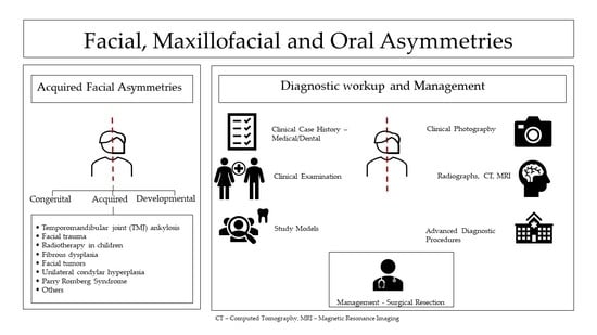
2. Acquired Facial, Maxillofacial, and Oral Asymmetries

Acquired FAs not only manifest during the developmental and growing stages of life, but also in adulthood. They are predisposed by certain traumatic events, infections, or pathologies. These conditions lead to unexpected asymmetry, thereby altering function and aesthetics and causing psychological trauma to the patient. Early evaluation and detection of acquired FAs could assist in preventing gross deformities, through timely intervention. Cheong and Lo categorized acquired FA, which occurs frequently due to injury or disease, as [9]:
  • Temporomandibular joint (TMJ) ankylosis;
  • Facial trauma;
  • Radiotherapy in children;
  • Fibrous dysplasia;
  • Facial tumors;
  • Unilateral condylar hyperplasia;
  • Parry Romberg Syndrome;
  • Others.

2.1. Diagnostic Evaluation for Acquired Facial, Maxillofacial, and Oral Asymmetries

FAs happen to the majority of the population; however, most of them are functionally and aesthetically indiscernible. Acquired FAs are predisposed by various external factors; hence, an accurate diagnosis of these cases consists of a thorough medical/dental history check-up, clinical examinations, study models, photographic and radiographic records.

2.1.1. Medical and Dental History

Following the patient’s chief complaint and evaluation of their medical and dental history helps clinicians identify the precise cause of the asymmetry. Childhood traumas or infections related to the craniofacial area and records of dental history, such as abnormal eruption or premature loss of the dentition, should also be taken into account [1].

2.1.2. Clinical Examinations

  • Extra oral evaluation
This remains one of the foremost steps in diagnosing acquired FA. Direct frontal assessment is performed with both visual and palpable inspection of the facial framework, to detect any differences between the two halves of the face. In several cases, patients tend to mask their asymmetric appearance by titled head postures or customized hairstyles [11]. Thus, during the extra-oral evaluation, clinicians should keep the patient’s face unveiled and the head in the standard position, as per the clinical Frankfort plane or the interpupillary line, and parallel to the floor. As previously discussed, clinical FA may be due to disharmony in the skeletal, soft tissue, dental, and functional structures of the face. The contribution of dental structures may cause minimal FA. However, asymmetries of skeletal origin attribute to significant changes in an individual’s facial harmony, owing to extensive involvement of the adjacent hard tissue structures [3]. Commonly encountered mandibular asymmetries, for instance, may produce changes in size, volume, and position of the condyle or ramus, leading to secondary skeletal changes in the maxillary counterparts [3]. The gold standard for visible FA is considered as a skeletal deviation equal to or greater than 4 mm [3]. However, the degree of skeletal asymmetry may also be compensated by individual-specific soft tissue thickness; thus, some authors suggest a craniofacial deviation of 2 mm deviation as clinically discernible asymmetry [3].
Moreover, the first step in examining facial symmetry is determining the reference midlines of the face. The reference upper midline is often established by the line connecting through soft tissue Glabella (G’—the center point of the eyebrows) and Subnasale (Sn—the central point of the nasal septum and the upper lip) points [12]. Following this reference line, clinicians can assess the upper facial symmetry, and any remarkable deviations from this line should be noted and examined carefully. Regarding the lower midline, some authors consider a reference line through Sn and Pogonion (Pg’—the most projecting median point on the anterior surface of the chin.) [12]. If the reference lower midline is aligned with the upper midline, the chin is centered, otherwise, the mandible is considered shifted and further investigation should be made to determine whether the mandibular deviation is functional or not. In addition, the imaginary mid-pupillary line and the facial midline can divide the face into four parts for gross evaluation, as displayed in Figure 1 [11].
These anatomical landmarks also help to divide the face into three horizontal proportions. The upper third of the face is measured from the mid-point of the hairline (the trichion) to the G′. The distance from the G′point to the Sn point is regarded as the middle third part. Then, the lower third facial proportion is from the Sn to the soft tissue Menton (Me’—the lowest point of the chin) [13,14]. Although the anatomical landmarks are consistent, these parameters and the perceptions of facial beauty may differ regarding gender and ethnicity [15,16].
Another measurement for facial symmetry is “the rule of the fifths”. The transverse proportions of the face are divided into five equal parts. Regarding the ideal facial aesthetics, each vertical fifth should have the approximate width of an eye [14]. The central fifth is measured by the interior intercanthal distance between two eyes, this width is considered ideal if it is corresponding to the width of the interalar base of the nose. The median two-fifths are characterized by the inner and lateral canthus of both eyes. Lastly, the lateral two-fifths of the face are evaluated from the furthest edge of the ears to the lateral canthus of the eyes. While the vertical fifth measurement can be utilized on both genders [14], the ratio may vary depending on ethnicity and race [14]. Wang et al. have demonstrated that Caucasian patients possess a greater mouth-to-nose width ratio than average Asian patients [17]. Some authors also discovered that the nose width in the black population is relatively greater than that of other populations [18].
The occlusal plane canting is performed in both extraoral and intraoral examination. Patients are requested to hold a scale or a tongue blade between their canines to compare the vertical and transverse relation of the occlusal plane to the interpupillary plane. In asymmetric cases, the occlusal plane has a variation of more than 4° from the reference plane [2,10]. The exact cause of occlusal canting can be either due to an uneven dental eruption, a functional shift of the mandible, or the rotation of the entire maxilla and mandible arches, and should be accurately identified [9]. Submental and superior observations are valuable methods of measuring the asymmetry in the head and neck region as these views will allow doctors to note any deviation of midline structures, such as the tip of the nose, the philtrum, the chin point, or any nasal and cheekbone deformities [2].
  • Intraoral evaluation
Various occlusal traits, such as impacted or ankylosed teeth, congenital deformities, dental discrepancy, deep bite, open bite, unilateral/bilateral crossbite, the curve of Spee, the curve of Wilson, and periodontal tissue conditions, are recorded in this step. The upper and lower dental midline are evaluated at rest, in centric relation, centric occlusion, at smiling, and other functional positions of the jaw [1]. Examination during functional movement allows clinicians to identify unhealthy occlusal contacts. In some cases, true bony asymmetries can be induced by a functional mandibular shift, owing to prolonged occlusal interferences during childhood, as illustrated in Figure 2 [2]. Bad oral habits, such as finger or thumb sucking, tongue thrusting, lip biting, and mouth breathing, should also be evaluated, especially in young patients, as these prolonged habits may cause an orofacial myofunctional disorder and result in imbalanced facial development [10].

2.1.3. Diagnostic Records

Comprehensive records of study models, photographic and radiographic assessments are collected as an aid for measuring the degree of FA. The first step is to obtain standardized photographs of the patients, which include extra-oral shots of the frontal, lateral, submental, and superior views, intra-oral ones of each dental arch, and frontal and lateral view of the occlusion in the centric position [2,19]. Conventional cast models or virtual occlusograms of the patients are then taken for a thorough evaluation. Appropriate study models transferred onto an adaptable articulator will provide clinicians with a better perspective of two dental arches and their relationships [2,19]. In addition, radiographic records are also essential to detect underlying hard tissue irregularities that are usually undiscovered by clinical examination. The two principal radiographs that are often prescribed for the diagnosis of asymmetry are the posterior-anterior cephalogram and orthopantomogram. While lateral cephalograms are often utilized in orthodontic treatment planning, this method is less common in measuring the extent of asymmetry [9]. These 2D radiographs also allow clinicians to screen for underlying pathologic conditions in the craniofacial area, such as condylar hyperplasia or maxillofacial tumors [10]. Substantial information can also be gathered by comparative observation of the patients’ photographs, radiographs, and study models, prior to the acquired condition. Other advanced diagnostic procedures, such as stereophotogrammetry, 3D stereolithographic models, and skeletal scintigraphy (radionucleotide scans), have also been promoted as effective tools for FA diagnosis. The application of these three-dimensional imaging techniques for surface asymmetric evaluation has been increasing enormously in recent years, especially digital stereophotogrammetry, owing to its lack of exposure to radiation and lack of requirements for patient compliance. This quantitative measurement, which uses several cameras from different angels to capture and produce a 3D image of the facial area, has quickly been a favorable surface imaging technology [20]. This technology can generate the sophisticated geometric patterns on the face, creating a photorealistic 3D image for digital evaluation. The stereophotogrammetric assessment has also been describes thoroughly and confirmed by numerous studies to be a reliable technique for soft tissue measurements before and after craniofacial surgical procedures [21,22,23]. Recently, more sophisticated techniques, such as 3D computed tomographic scans, cone beam-computed tomography (CBCT), and magnetic resonance imaging (MRI), have provided innovative diagnostic instruments for numerous craniofacial defects [1,24]. The CBCT technique is widely used for the detection of maxillofacial bony changes, such as tumors, fractures, osteitis, idiopathic osteosclerosis, and condylar hyperplasia, as it can produce excellent details of the surrounding tissues, with a relatively low dose of radiation. In addition, MRI also demonstrates detailed visualization of not only hard tissues but also soft ones, which is why it is favored for the diagnosis of soft tissue diseases such as temporomandibular joint (TMJ) disc displacement or de-arrangement and inflammatory conditions of the salivary glands, maxillofacial muscles, and sinuses [2]. The incorporation of these techniques in diagnosis and treatment planning has improved the communication between doctors and patients. It has also allowed clinicians to visualize and perform various surgical procedures more precisely and clearly, which leads to better treatment outcomes [1,2].

2.2. Conditions Presenting with Acquired Facial, Maxillofacial, and Oral Asymmetries

Understanding the underlying etiology is essential to critically evaluate and determine the precise case-specific treatment protocols that result in a better prognosis. As previously elaborated, Cheong and Lo classified common conditions of acquired FA as follows [9].

2.2.1. Temporomandibular Joint Ankylosis

Ankylosis of the TMJ is described as limited mouth opening due to an abnormal adhesion between the glenoid fossa of the temporal bone and mandibular condyle or any component between the mandible and maxilla, zygoma, or base of the skull [25,26]. Prevalent in both children and adults, TMJ ankylosis presents commonly in the first two decades of life [27]. Ankylosis can be classified based on the site of involvement (intra-articular or extra-articular); tissue of involvement (fibrous or osseous or fibro-osseous); degree of involvement (complete or incomplete); and relation to the TMJ (true or false) [28]. This pathologic condition often results from local infections, such as otitis media or mastoiditis (10–49%); traumatic damages, such as condylar trauma at vaginal birth (13–100%); or systemic diseases, such as tuberculosis, syphilis, ankylosing spondylitis, and rheumatoid arthritis (10%), although post-operative cases have also been reported [25,29,30,31]. Traumatic inflammation and infections have been hypothesized to form an intra-articular hematoma and soft tissue scarring, which ultimately develop into excessive abnormal adhesion and loss of function of the TMJ [28].
When a unilateral or bilateral TMJ ankylosis occurs in the early childhood period, it often prohibits the normal growth and functional movement of the mandible, and causes facial disfigurement, which exacerbates as age advances [7,27,32]. Restricted mandibulo-facial growth is associated with impaired speech, mastication, digestion, inability to maintain oral hygiene, rampant dental caries, periodontal diseases, malocclusion, snoring, sleep apnea, and compromised airway [28,31,33]. TMJ ankylosis is commonly accompanied by impaired cosmesis and psychosocial problems, owing to deviated TMJ and micrognathia [28].
Imaging modalities play a very important role in the diagnosis of TMJ ankylosis. Three-dimensional imaging techniques, such as multi-slice computed tomography and CBCT, in combination with conventional panoramic and TMJ radiographs, reveal a loss of joint space due to the fusion of the bone with the TMJ [31,34]. Contemporary treatment procedures of ankylosis usually include surgical intervention with the aid of fixed orthodontics or functional appliances. The treatment should aim at re-establishing functional movement of the joint and the lower jaw, correcting FA, and preventing re-ankylosis [7]. The three major techniques to treat ankylosis are gap arthroplasty, inter-positional arthroplasty, and reconstruction arthroplasty. The first technique involves the resection of the ankylotic mass, without inserting any inter-positional matters between the ramus and the mandibular fossa [35]. The second one is also known as the ‘Gap Technique’, but filled with autogenous materials, such as costochondral graft, iliac crest, temporalis fascial flap, and fat graft or alloplastic materials [33]. Lastly, the third method replaces the whole joint with an artificial joint prosthesis [36,37]. No matter the therapeutic interventions used to treat TMJ ankylosis, the foremost strategy is physiotherapy. Without patient cooperation and aggressive post-operative physical treatment, the surgical outcomes are rarely successful and the incidences of re-ankylosis are considerably high [38]. A long period of follow-up and fixed orthodontic treatment is also recommended for optimal outcomes [39].

2.2.2. Facial Trauma

According to the National Trauma Bank Data, facial trauma takes up a quarter of all forms of injuries yearly [40]. Particularly, these traumatic events that occur at a young age usually interrupt the process of growth in the facial regions. This leads to detrimental influences on the development of the craniofacial skeleton, resulting in asymmetric changes of the face and causing long-term aesthetic, physical, and psychological distress [41]. The World Health Organization (WHO) confirms a death rate of more than nine per minute among those inflicted by facial trauma [42]. Although maxillofacial injuries are predominant in male adults and occur more between the second and fourth decades, they are also the leading cause of mortality in children [43,44]. Published literature revealed road traffic accidents and interpersonal violence, both triggered by excessive alcohol consumption, as the leading cause of facial trauma [41]. The other attributed etiologic factors include accidental falls and sports injuries, which are both common among the elderly and children/young adults, respectively [41]. Facial trauma can also be manifested in 2.3% of child abuse cases and 27% of animal/human bite cases [45,46].
The impact of maxillofacial trauma may range from local soft or hard tissue involvement to subsequent systemic infection, bleeding, airway obstruction, and life-threatening neurological or orthopedic involvement [47]. Soft tissue injury may present as avulsions, bruising, burns, oedema, lacerations, abrasions, puncture wounds with associated neurovascular damage to the head and neck region, special senses, and ophthalmologic traumatic emergencies [42,43]. The underlying craniofacial skeleton is commonly divided into the upper, middle, and lower thirds; consisting of the frontal bone and sinuses, the orbital and zygomaticomaxillary complex, and the mandible. Osseous involvement may include individual or multiple fractures of these bones, with or without brain and vital structures; each presenting differently [42,43]. Among children, however, the most frequent presentations of facial trauma are fractures which occur as mandibular (55%), orbital (30%), dentoalveolar (23%), midface (17%), nasal (15%), complex (14%), and cranial fractures (6%) [43]. Facial fractures commonly encompass symptoms of ocular, auditory, and nasal involvement associated with damage to the base of the skull [43]. Symptoms of mandibular fractures include pain, oedema, hematoma, paresthesia, trismus, open bite, dentoalveolar fractures, malocclusion, and TMJ ankylosis [43]. Laryngeal injuries may present as life-threatening airway obstruction, aspiration, impaired vocal function, and dysphagia [48].
Imaging is also useful for the diagnosis of facial traumas, to determine whether there is an involvement of bone [43]. This should be accompanied by region-specific clinical assessments to check for damage to the underlying structures, such as the facial nerve and salivary and lacrimal glands to ensure their integrity, especially in soft tissue traumas [43]. Visual eye assessments are also essential to rule out any injuries threatening eyesight [43]. Restoring anatomical structures, rebuilding facial aesthetics, and maintaining the normal growth rates of the skull and face are some of the main goals of the treatment which should be least invasive to the patient, wherever possible [44]. The choice of management depends on the patient’s age, the positions of the fractures, and the severity of the bone displacement [43,45,46]. In mild and moderate cases of the frontal sinus, orbital, zygomatic, nasal, and mandibular fractures, the principal management usually involves conservative techniques, fracture reduction, and rigid fixation. However, in severe cases of facial trauma, seriously broken bones are often reconstructed by the technique of Open Reduction Internal Fixation under general anesthesia. After that, mini-plates or mini-screws are placed in the bone to stabilize the fractures [43,49,50]. Surgical intervention is only designated for severely bone displacement. Postoperative follow-up is required to prevent long-term complications and to guarantee the optimal tissue healing of the craniofacial structures [50].

2.2.3. Radiotherapy in Children

Cancer is known to affect 12 in every 100,000 children; commonly leukemia (30.1%), central nervous system tumors (27.8%), and lymphomas (11%) [51]. Despite the debilitating nature of cancer, 1 in every 900 survive; however, 50% of them also experience the long-term effects of treatment [51]. Radiotherapy has been applied in the treatment of craniofacial cancers for decades and it does manage to control and eliminate tumor cells effectively. However, this therapy is also responsible for myriads of growth impairments in craniofacial hard and soft tissues, and these are present among 66% to 100% of premature survivors [52,53]. In growing patients, radiation induces severe disturbances in head and neck areas, such as skeletal bone growth [54], osteoradionecrosis [52,55], neuroendocrine sequelae [56,57], muscle atrophy, and tissue fibrosis [58,59,60,61]. These disruptions lead to FAs and psychosocial disabilities due to radiation [62].
Irradiation of cancerous and healthy tissues causes endarteritis and its sequelae of tissue hypoxia, hypervascularity, hypocellularity, and breakdown [63,64]. This tissue damage occurs due to the intrinsic cytotoxic effects of radiotherapy, resulting in the non-proliferation of cells and cell death [63,64]. The deranged immune system, downstream cytokines, and growth factors lead to late-radiation effects of chronic inflammation [63,64]. Growth centers of the maxilla and mandible may be inhibited, causing facial dysmorphism due to complexed non-synchronous rate of growth [65,66]. The immediate effects of irradiation commonly present are xerostomia, mucositis, oral ulcers with secondary infection, poor dentition, rampant caries, poor oral hygiene, malocclusion, malnutrition, and visual impairment [52]. Other delayed effects of craniofacial radiotherapy are neurocognitive deficits, panhypopituitarism, seizures, strokes, second cancers, and cataracts [64,67].
The management of radiation complications remains an arduous clinical problem. Recently, several studies have proposed the technique of distraction osteogenesis (DO) as a promising treatment for radiated young patients [62,68]. Bianchi et al. have reported that the DO method with microvascular free flap reconstruction could be a suitable therapeutic intervention to treat growing patients with irradiated FA [62]. They concluded that the factors that contribute to the success of this method are the patient’s age, surgical intervention, distraction rate, latency, and consolidation period [62]. Raghoebar et al. have indicated that DO in the irradiated mandibular region in non-growing patients has an unpredictable bone formation outcome; however, the reconstruction of soft tissue in this method can be observed as a mileage [69]. Moreover, a study by Faghahati et al. revealed that fat transfer could also be a favorable alternative to correct the sequelae of craniofacial irradiation. The technique has proved to significantly restore the aesthetic facial appearance, without leaving any long-term complications [70]. Together with surgical interventions, fixed orthodontic appliances and asymmetric functional activators have played a key role in stimulating the growth of facial tissues on the irradiated sides [71]. In short, the management of irradiated-induced FA may involve a series of surgical and non-surgical interventions to achieve satisfactory results for both patients and a multidisciplinary team of clinicians.

2.2.4. Fibrous Dysplasia

Fibrous dysplasia (FD) is a sporadic benign fibro-osseous disorder of bone, constituting 2.5% to 7% of the benign osseous lesion [72,73,74]. It was first identified by Von Recklinghausen in 1891, and described by Lichtenstein and Jaffe in 1942 [72,75]. The lesion presents as either the monostotic (one bone) or polyostotic (multiple bones) type, which may be connected with other systemic manifestations [76,77]. The craniofacial involvement accounts for 50–100% of all FD cases and involves multiple bones of the craniofacial region [73,75]. In total, 90% of craniofacial FD cases clinically manifest under 5 years of age [73]. The origin of FD was associated with the acquired somatic missense mutation of the gene GNAS1, causing replacement of normal bone by fibrous tissue, due to altered osteogenic functioning [73,75,78,79]. This non-inherited mutation occurs postzygomatically, inhibiting intrinsic GTPase activity and, thus, increasing intracellular cAMP [79,80]. The dynamics between mutated and normal osteogenic function play an integral role in postnatal clinical presentation of FD. This cellular ratio of mutated to normal genes may be widely influenced by extrinsic factors, such as growth factors and hormones [74,79]. The molecular pathogenesis of FD could present a plausible explanation for adding this genetic disorder to a list of acquired FAs.
Most FD cases are monostotic, asymptomatic, and discovered by chance; however, other cases present with swelling, bone fractures and curvatures, and soreness [76]. Craniofacial lesions are commonly observed, which induce FA, nasal airway obstruction, and headache [77]. This variant is known to involve the maxilla, mandible, frontal, sphenoidal, ethmoidal, parietal, temporal, and occipital bones, in descending order [75]. Though the majority of cases may be accidental radiographic findings, some symptomatic cases may present localized pain, swelling, weakness, fractures, and visual and hearing impairments [73].
Diagnosis of osseous lesions, such as FD are confirmed after a thorough correlation of clinical, radiographic, and histopathological findings. Radiographically, FD typically demonstrates a ‘ground-glass’ appearance that can be histopathologically correlated by the haphazardly distributed woven bone in the ‘Chinese letter’ pattern [73,75]. The management of craniofacial lesions depends on the clinical manifestations, age, and stages of skeletal development. Asymptomatic lesions could be treated with observation, patient education, and annual clinical evaluations. Photographs, radiographs (X-rays and computed tomography scans), and other pre-clinical assessments should be obtained in follow-up appointments [77,78]. Bisphosphonate intervention is another treatment option for FD, especially polyostotic ones. A combination of intravenous and oral administration of bisphosphonates, which cannot cure the disorder completely, can diminish discomfort, postpone osteoclastic activity, lower the risk of bone fractures, and improve the thickness of the cortical bone [73,76,77,81]. Surgical intervention is indicated to remove symptomatic deformity and prevent functional and aesthetic damage, progressive airway obstruction, and hearing or visual loss [78,82]. The techniques to alleviate the lesions vary from conservative methods, such as curettage, contouring of abnormal bones, and bone grafting, to aggressive ones, which involve a total resection of the lesion and instantaneous bone reconstruction [78]. A variety of biomaterials have been applied as bone scaffolds for tissue reconstruction of facial defects, which include titanium and PLGA (poly lactic-co-glycolic) implants [81]. Owing to the development of the latest technology, FD can be identified and controlled efficiently with conservative or aggressive approaches to restore function and cosmetic appearance for the patients [81].

2.2.5. Facial Tumors

The term ‘craniofacial tumor’ involves a wide variety of disorders, ranging from benign masses, such as cystic hygromas, dermoid cysts, osteoma, neurofibromatosis, and vascular malformation [83], to aggressive cancers, such as squamous cell carcinoma, malignant melanoma, angiofibroma, sebaceous hyperplasia, and Merkel cell carcinoma [84]. The structures most involved are the craniofacial bones, soft tissues, and salivary glands [85]. The latest WHO classification broadly categorizes these tumors into odontogenic tumors, maxillofacial bone and cartilaginous tumors, soft tissue tumors, fibro-osseous tumors, and hemato-lymphoid tumors, which are further divided into benign and malignant counterparts [86,87]. Whether non-cancerous or cancerous, the development of tumors can detrimentally damage functions and aesthetics in the maxillofacial area, which may lower the self-esteem or negatively impact the psychosocial well-being of the patients.
Epidemiological data of these tumors seem to vary based on geographical location. In developing countries, the most common benign craniofacial tumor is ameloblastoma, while the most common malignancy is squamous cell carcinoma, both of which account for 65% of all facial tumors [88]. African studies showed that ameloblastic carcinomas were more common than other malignancies [89,90]. The developed countries, however, illustrate a lower incidence of ameloblastomas, but a higher number of cases of odontomas and malignancies such as osteosarcoma [91,92]. The WHO confirms that most tumors occur in the second to fourth decade of life, with a slightly higher predilection for males overall [92]. Studies also reveal that tumors affecting the lower face are more common than those affecting the mid-face [88].
Each tumor group has a specific etiopathogenesis. Craniofacial malignant tumors, especially involving the oro-pharyngeal regions, are usually associated with the consumption of tobacco and alcohol [93]. Viruses such as the Human Papilloma and Epstein-Barr have also been implicated in oro-pharyngeal tumors [94]. Other known etiological factors include irradiation, genetic, environmental factors, and occupational hazards, causing tumors in the oral, nasal, and salivary regions [93,95,96,97]. Ameloblastoma and Keratocystic Odontogenic Tumor, the most common benign odontogenic tumors, are usually due to genetic alterations involving BRAF and PTCH genes, both components of the MAPK pathway [86]. Further mutations involving TP53 lead to a malignant transformation of these benign tumors [98]. Soft tissue tumors are very poorly understood, but studies exhibit reciprocal translocations and specific driver mutations that lead to chromosomal instability [97].
Facial tumors usually present as unilateral asymmetries, with occasional bilateral occurrences [85]. Riaz and Warriach found that most tumors displayed mandibular predilection, followed by the asymmetry in the buccal mucosa; however, tumors involving the pre-auricular and peri-orbital regions were relatively fewer [88]. Benign tumors usually present with painless, slow growing tumors coupled with the bony expansion, and can be distinguished from malignant tumors, as the latter are usually more aggressive and rapidly growing [88]. They usually present with ulcerations containing discharge and perforate adjacent structures [88,93,99].
Facial tumors and cysts can be differentiated using imaging modalities, clinical assessment, and histopathological analysis. Clinically, benign and malignant tumors can be differentiated based on the patient’s history and clinical features. Benign tumors are usually painless and slow-growing when compared to malignant tumors, which are more aggressive and are often accompanied by ulcerations [88]. Imaging modalities also display the involvement of surrounding structures in malignant tumors. Histopathology also shows the presence of dysplastic cells in malignancies and is the gold standard for diagnosing tumors and cysts [94]. Cysts can be differentiated radiographically, in that they usually present as unilocular or multilocular well-demarcated radiolucencies and by histopathological examination [100].
Interdisciplinary assessment and adequate treatment planning are necessary for the management of these tumors. For benign tumors, the treatment may include several major procedures of complete surgical resection or enucleation, sclerotherapy, cryotherapy, and laser intervention [83,84]. With regards to malignant facial tumors, the principal management consists of chemotherapy, radiotherapy, photodynamic therapy, immunotherapy, cryosurgery, electrodesiccation, and surgical interventions [84,101,102]. Frunza et al. stated that a combination of multiple treatment methods is recommended, especially for large tumors [84]. They believed that the precise excision of a full tumor resulted in better healing and also prevented recurrence. The indication for surgery may also depend on the patient’s age and overall health. Although surgical intervention is the optimal treatment for large tumors, this invasive technique may induce more harm to the patients than the actual tumor, which necessitates less invasive approaches [84]. After a total elimination of the tumor, concomitant plastic surgery is sometimes necessary to reconstruct the appearance and function of the craniofacial area [103,104].

2.2.6. Unilateral Condylar Hyperplasia

Condylar hyperplasia (CH) is an uncommon benign disorder of the mandible resulting in abnormal growth of the condylar head, neck, or mandibular ramus, which leads to asymmetric facial appearance, malocclusion, and TMJ dysfunction [105,106,107]. Obwegeser and Makek classified the disorder into three groups: hemi-mandibular hyperplasia (vertical asymmetry), hemi-mandibular elongation (horizontal asymmetry), and a combination of these two [108]. Hemi-mandibular hyperplasia is characterized by vertical asymmetry, involving enlargement of the condyle, neck, ramus, and body with a tilted occlusal plane [105,108]. Hemi-mandibular elongation, however, is horizontal asymmetry, with enlargement of the condylar neck and displacement of the ramus and body without tilting [105]. Although cited data are relatively limited, CH occurs in children and young adults (11–30 years) [109]. Gender predilection, however, remains debatable, as most studies display higher occurrence in females, but the associated biological basis is yet to be established [110]. Few studies display equal occurrence in males and females with no side predilection [111].
CH occurs due to trauma, hormonal imbalance, infection of the TMJ, arthrosis, hypervascularity, and some evidence of genetic disposition [109]. Obwegeser and Makek postulated that CH presents due to the influence of different growth factors, responsible for hypertrophy and elongation [108]. This was later validated in a study, which elaborated the possible role of Insulin Growth Factor-1 in the pathogenesis [110,112]. Excessive loading of the TMJ was also identified as another possible etiological factor [111]. The typical clinical features of CH include enlargement of the mandibular condyle, condylar neck, and excessive growth of the body of the mandible [109]. The condylar enlargement causes mandibular deviation towards the affected side with malocclusion and articular dysfunction [105]. Additional features include outward bowing, followed by downward growth of the mandibular body and ramus of the affected side [105]. While there is evidence of fullness present on the ipsilateral lower face, the contralateral part shows flattening [110].
CH appears as a symmetrical enlargement of the condylar neck, head, and body of the mandible in the symphyseal region when viewed with conventional radiographs [109]. The treatment of the disorder can only begin once the pathologic growth of the condyle has ceased, else, recurrences of CH will constantly happen [113]. The management of patients with CH is often multidisciplinary, with the involvement of different specialists, such as orthodontists, maxillofacial surgeons, and physiotherapists [113]. Multiple techniques ranging from conservative to multiple-staged surgical interventions have been applied to treat CH, depending on the condylar growth activity, the patient’s age, and the severity of FA. The need for surgery increases as the chin deviates more than 10 mm from the facial midline [114]. Studies have reported the application of several major surgical procedures, such as high condylectomy, maxillar, and mandibular osteotomy or orthognathic surgery [105,106,114,115,116]. In most cases, patients will be treated with fixed orthodontic appliances and active physical therapy post-operatively. A long-term follow-up plan is advisable for patients, as periodic radiographs and clinical assessments will be made to observe the healing of TMJ and to prevent long-term recurrences of CH [106,116,117].

2.2.7. Parry-Romberg Syndrome

Romberg’s disease or Parry-Romberg syndrome (PRS) is a rare, slow, progressive, degenerative disorder, causing atrophy on one side of the face [118]. The clinical manifestation of the disease involves the progressive unilateral decrease in the size of facial tissue such as skin, connective tissues, muscles, and even osteocartilaginous structures [119]. Besides causing FA and other aesthetic problems, PRS also negatively affected the normal functions of the face and the psychosocial well-being of the patients [118]. It was first reported in the literature by Dr. Caleb Hillier Parry in 1825 and then later described in 1846 by Dr. Moritiz Heinrich Romberg [119]. PRS presents as mild, moderate, and severe forms, based on the level of involvement of the Trigeminal Nerve [120]. The mild form involves only the skin and the subcutaneous tissue along with a single sensory branch of the nerve, whereas the moderate and severe forms involve two and three branches, respectively [120].
This progressive, self-limiting syndrome typically presents in children and young adults, more frequently in females than in males, and more on the left side of the face, with a 5–10% bilateral presentation [119,121,122]. PRS is an idiopathic disorder, with multifactorial triggers, involving infection, trauma, sympathetic nervous system dysfunction, vascular abnormalities, inflammatory conditions, and autoimmune disorders [119]. Dental infection, Herpes and Varicella Zoster viral attacks, Lyme disease, and traumatic surgical procedures have been known to initiate PRS [120,123]. Other etiological factors that have been recorded include disturbances in fat metabolism, endocrine disorders, and radiotherapy [123]. PRS presents as characteristic unilateral facial atrophy, involving the forehead, peri-orbital region, teeth, perioral region, mandible, and neck [119,124]. Data also reveal higher involvement of the cheeks (75%) and forehead (63%) compared to the other sites [124]. The initial manifestations of skin discolorations and alopecia are followed by neurological and ophthalmological damages, such as migraine, epilepsy, neuralgia, and enophthalmos [120,123]. The characteristic oral findings, especially in the younger age group, are resorption of teeth, delayed eruption, painful mastication, and inadequate mandibular movements [123]. Dermakarian et al. suggested that atrophy of the optic globe occurs, relative to the involvement of other facial tissues [125].
FA involving multiple regions and systems such as PRS requires a careful diagnostic evaluation with clinical and radiological investigations [121]. MRI can usually reveal atrophy of the facial muscles and adipose tissue [121]. Other radiological investigations show bone resorption in the maxilla and mandible [125]. PRS involves a very detailed ophthalmic examination, such as gonioscopy, which displays the drainage angle of the eye [125]. The findings reveal abnormalities, such as enophthalmos, hyperopia, and globe atrophy [125]. The management of PRS includes myriads of different therapies, such as conservative anti-inflammatory drug application, lipofilling, and reconstructive or orthognathic surgery. Autogenous fat grafts, silicon injections, cartilage grafts, acrylic prostheses, and bovine collagen are several practicable treatment alternatives [119,126,127]. Recently, Kasielska-Trojan et al. have reported autogenous fat grafting (lipofilling) to be one of the most reliable and effective techniques to treat PRS patients [128]. Nowadays, liposuction and lipofilling are widely utilized in mild and moderate cases to restore the aesthetics of the asymmetric face, while in severe cases, a combination of multiple surgical and non-surgical therapies is inevitable to achieve satisfactory outcomes for patients with PRS [129].

2.2.8. Others

Although Cheong and Lo have mentioned a category of ‘Others’ under acquired FAs, they fail to specify these conditions. However, Cohen et al. earlier identified miscellaneous conditions causing acquired FAs due to infections, osseous lesions, cysts, burns, and salivary gland diseases [9,85]. Owing to this consistent gap in literature, elaborating conditions under this category should be addressed. According to Cohen et al., FAs due to bacterial or viral infection include salivary conditions, such as sialadenitis or acute parotitis and dentoalveolar abscesses [130,131]. Cysts of the jaws, of both odontogenic and non-odontogenic origin, commonly arise from epithelial remnants associated with tooth structures or the maxilla and mandible [100]. The most common odontogenic cysts include the dentigerous and radicular cysts and the non-odontogenic cysts include the nasopalatine and median palatine cysts [100]. These cysts usually present in the young to middle age group and cause displacement of adjacent structures, leading to asymmetry [100]. Paget’s disease is characterized by generalized osseous enlargement, bone pain, and asymmetry, with motor and sensory deficits in older adults [100,132].
Other miscellaneous conditions causing asymmetry include mucoceles, burns, and Cancrum Oris [85]. Mucoceles commonly present as painless swellings of the lower lip; however, ranulas may arise secondary to obstruction of the sublingual gland, in the floor of the mouth extending to the neck [131]. Accidental electrical burns, mainly present in children, cause FAs due to necrosis and disfigurement of the lips or tongue [85]. Cancrum Oris, a gangrenous oral infection in children, is predisposed by malnutrition and measles and often presents as an ulcer on the gingiva, which may extend to soft tissue and bone [85]. The medical and dental history is crucial for identifying FA. For salivary gland disorders, a physical examination involves an assessment of the location, onset, and duration of the lesion [96]. Rapid growth is associated with infections, such as viral or bacterial sialadenitis [96]. Bimanual palpation of the salivary glands allows the clinician to assess the extent of inflammation and reduction of salivary flow [131]. Imaging studies, such as ultrasonography, helps to detect the position of associated blood vessels, lymph nodes, and nerve involvement in salivary gland inflammation [131]. Laboratory tests can help distinguish between viral and bacterial sialadenitis [131].
Usually, infection-associated FAs can be managed either by medication, or by conservative surgical treatment [130,131]. Inflammatory diseases of the salivary glands, including mucoceles and ranulas, can usually be managed by surgical incision and drainage, or by complete removal of the gland, if extensive [131]. Dentoalveolar abscesses are usually treated with antibiotics and indicated dental management [130]. Orofacial cysts are managed surgically by enucleation, marsupialization or excision [100]. Paget’s disease, being a very quiescent disorder, is managed by cosmetic contouring of the enlargement [132]. Electrical burns usually occur on the lips of the children, and management is focused on providing functional tissue-borne and tooth-borne appliances to promote healing [85]. Cancrum Oris causes severe soft tissue and bony asymmetry, demanding surgical correction and restoration of speech, aesthetics, and oral competence, with monitored nutritional assessments [85].

3. Conclusions

Over the years, technology and advanced imaging techniques have aided in the accurate detection of facial disproportion. These diagnostic tools allow the clinicians to better understand the etiological and triggering factors leading to facial asymmetries. Apart from cosmetic and functional disharmonies, these conditions are frequently associated with psychological trauma, shifting the focus of management to bi-directional assessment. This enables the multi-disciplinary team of clinicians to realistically meet the patients’ expectations through personalized interventions that are less harmful and more beneficial.

Author Contributions

S.D.T., J.I. and A.H. designed and conceptualized the review. J.I., A.H. and U.M.N.C. collected the information from literature and wrote the manuscript. J.I. and A.H. reviewed and edited the manuscript. S.D.T. supervised the paper. All authors have read and agreed to the published version of the manuscript.

Funding

This research received no external funding.

Institutional Review Board Statement

Not Applicable.

Informed Consent Statement

Not Applicable.

Data Availability Statement

Not Applicable.

Acknowledgments

We thank Tuyet Oanh Nguyen from the Tuyet Oanh Dental Clinic, Ho Chi Minh City, Vietnam, for contributing clinical photographs.

Conflicts of Interest

The authors declare no conflict of interest.

References

  1. Sandhya Maheshwari, S.K.V.; Aditi, G.; Sushma, D. Diagnosis and management of facial asymmetries. J. Orthod. Res. 2015, 3, 81–87. [Google Scholar] [CrossRef]
  2. Srivastava, D.; Singh, H.; Mishra, S.; Sharma, P.; Kapoor, P.; Chandra, L. Facial asymmetry revisited: Part I—Diagnosis and treatment planning. J. Oral Biol. Craniofac. Res. 2018, 8, 7–14. [Google Scholar] [CrossRef] [PubMed] [Green Version]
  3. Thiesen, G.; Gribel, B.F.; Freitas, M.P. Facial asymmetry: A current review. Dental Press J. Orthod. 2015, 20, 110–125. [Google Scholar] [CrossRef]
  4. Peck, S.; Peck, L.; Kataja, M. Skeletal asymmetry in esthetically pleasing faces. Angle Orthod. 1991, 61, 43–48. [Google Scholar] [CrossRef]
  5. Maeda, M.; Katsumata, A.; Ariji, Y.; Muramatsu, A.; Yoshida, K.; Goto, S.; Kurita, K.; Ariji, E. 3D-CT evaluation of facial asymmetry in patients with maxillofacial deformities. Oral Surg. Oral Med. Oral Pathol. Oral Radiol. Endod. 2006, 102, 382–390. [Google Scholar] [CrossRef]
  6. Wang, T.T.; Wessels, L.; Hussain, G.; Merten, S. Discriminative Thresholds in Facial Asymmetry: A Review of the Literature. Aesthet. Surg. J. 2017, 37, 375–385. [Google Scholar] [CrossRef]
  7. Gulsen, A.; Sibar, S.; Ozmen, S. Orthognathic treatment of facial asymmetry due to temporomandibular joint ankylosis. Arch. Plast. Surg. 2018, 45, 74–79. [Google Scholar] [CrossRef] [Green Version]
  8. Severt, T.R.; Proffit, W.R. The prevalence of facial asymmetry in the dentofacial deformities population at the University of North Carolina. Int. J. Adult Orthodon. Orthognath. Surg. 1997, 12, 171–176. [Google Scholar]
  9. Cheong, Y.W.; Lo, L.J. Facial asymmetry: Etiology, evaluation, and management. Chang. Gung Med. J. 2011, 34, 341–351. [Google Scholar] [PubMed]
  10. Andrade, N.N.; Mathai, P.; Aggarwal, N. Facial Asymmetry. In Oral and Maxillofacial Surgery for the Clinician; Bonanthaya, K., Panneerselvam, E., Manuel, S., Kumar, V.V., Rai, A., Eds.; Springer: Singapore, 2021; pp. 1549–1576. [Google Scholar] [CrossRef]
  11. de Lima Lucas, B.; Júnior, R.B.; Gonçalves, L.C.; Gavião, M.B.; Gomes, V.L. Research and Clinical Applications of Facial Analysis in Dentistry, Oral Health Care—Prosthodontics, Periodontology, Biology, Research and Systemic Conditions; IntechOpen: London, UK, 2012. [Google Scholar] [CrossRef] [Green Version]
  12. Haraguchi, S.; Iguchi, Y.; Takada, K. Asymmetry of the face in orthodontic patients. Angle Orthod. 2008, 78, 421–426. [Google Scholar] [CrossRef] [PubMed] [Green Version]
  13. Milutinovic, J.; Zelic, K.; Nedeljkovic, N. Evaluation of facial beauty using anthropometric proportions. Sci. World J. 2014, 428250. [Google Scholar] [CrossRef] [Green Version]
  14. Hashim, P.W.; Nia, J.K.; Taliercio, M.; Goldenberg, G. Ideals of facial beauty. Cutis 2017, 100, 222–224. [Google Scholar] [PubMed]
  15. Porter, J.P. The average African American male face: An anthropometric analysis. Arch. Facial Plast. Surg. 2004, 6, 78–81. [Google Scholar] [CrossRef] [Green Version]
  16. Porter, J.P.; Olson, K.L. Anthropometric facial analysis of the African American woman. Arch. Facial Plast. Surg. 2001, 3, 191–197. [Google Scholar] [CrossRef]
  17. Dawei, W.; Guozheng, Q.; Mingli, Z.; Farkas, L.G. Differences in horizontal, neoclassical facial canons in Chinese (Han) and North American Caucasian populations. Aesthetic Plast. Surg. 1997, 21, 265–269. [Google Scholar] [CrossRef]
  18. Farkas, L.G.; Forrest, C.R.; Litsas, L. Revision of neoclassical facial canons in young adult Afro-Americans. Aesthetic Plast. Surg. 2000, 24, 179–184. [Google Scholar] [CrossRef] [PubMed]
  19. Lee, M.S.; Chung, D.H.; Lee, J.W.; Cha, K.S. Assessing soft-tissue characteristics of facial asymmetry with photographs. Am. J. Orthod. Dentofacial. Orthop. 2010, 138, 23–31. [Google Scholar] [CrossRef] [PubMed]
  20. Pucciarelli, V.; Baserga, C.; Codari, M.; Beltramini, G.A.; Sforza, C.; Giannì, A.B. Three-Dimensional Stereophotogrammetric Evaluation of the Efficacy of Autologous Fat Grafting in the Treatment of Parry-Romberg Syndrome. J. Craniofac. Surg. 2018, 29, 2124–2127. [Google Scholar] [CrossRef]
  21. Baserga, C.; Cappella, A.; Gibelli, D.M.; Sacco, R.; Dolci, C.; Cullati, F.; Giannì, A.B.; Sforza, C. Efficacy of Autologous Fat Grafting in Restoring Facial Symmetry in Linear Morphea-Associated Lesions. Symmetry 2020, 12, 2098. [Google Scholar] [CrossRef]
  22. Primozic, J.; Perinetti, G.; Zhurov, A.; Richmond, S.; Ovsenik, M. Assessment of facial asymmetry in growing subjects with a three-dimensional laser scanning system. Orthod. Craniofac. Res. 2012, 15, 237–244. [Google Scholar] [CrossRef]
  23. Heike, C.L.; Upson, K.; Stuhaug, E.; Weinberg, S.M. 3D digital stereophotogrammetry: A practical guide to facial image acquisition. Head Face Med. 2010, 6, 18. [Google Scholar] [CrossRef] [Green Version]
  24. Katsumata, A.; Fujishita, M.; Maeda, M.; Ariji, Y.; Ariji, E.; Langlais, R.P. 3D-CT evaluation of facial asymmetry. Oral Surg. Oral Med. Oral Pathol. Oral Radiol. Endod. 2005, 99, 212–220. [Google Scholar] [CrossRef] [PubMed]
  25. Sales, S.; Silva, M.; Lehman, L.; Castro, W.; Campos, F. Management of temporomandibular joint ankylosis. Int. J. Oral Maxillofac. Surg. 2019, 48, 284. [Google Scholar] [CrossRef]
  26. Hegab, A.F. Outcome of Surgical Protocol for Treatment of Temporomandibular Joint Ankylosis Based on the Pathogenesis of Ankylosis and Re-Ankylosis. A Prospective Clinical Study of 14 Patients. Int. J. Oral Maxillofac. Surg. 2015, 73, 2300–2311. [Google Scholar] [CrossRef] [PubMed]
  27. De Roo, N.; Van Doorne, L.; Troch, A.; Vermeersch, H.; Brusselaers, N. Quantifying the outcome of surgical treatment of temporomandibular joint ankylosis: A systematic review and meta-analysis. J. Craniomaxillofac. Surg. 2016, 44, 6–15. [Google Scholar] [CrossRef] [PubMed]
  28. Babu, L.; Jain, M.K.; Ramesh, C.; Vinayaka, N. Is aggressive gap arthroplasty essential in the management of temporomandibular joint ankylosis?-a prospective clinical study of 15 cases. Br. Int. J. Oral Maxillofac. Surg. 2013, 51, 473–478. [Google Scholar] [CrossRef]
  29. Zhu, S.; Wang, D.; Yin, Q.; Hu, J. Treatment guidelines for temporomandibular joint ankylosis with secondary dentofacial deformities in adults. J. Craniomaxillofac. Surg. 2013, 41, e117–e127. [Google Scholar] [CrossRef]
  30. Vasconcelos, B.C.; Porto, G.G.; Bessa-Nogueira, R.V.; Nascimento, M.M. Surgical treatment of temporomandibular joint ankylosis: Follow-up of 15 cases and literature review. Med. Oral Patol. Oral Cir. Bucal. 2009, 14, E34–E38. [Google Scholar] [CrossRef] [PubMed] [Green Version]
  31. Chouinard, A.F.; Kaban, L.B.; Peacock, Z.S. Acquired Abnormalities of the Temporomandibular Joint. Oral Maxillofac. Surg. Clin. N. Am. 2018, 30, 83–96. [Google Scholar] [CrossRef]
  32. McFadden, L.R.; Rishiraj, B. Treatment of temporomandibular joint ankylosis: A case report. J. Can. Dent. Assoc. 2001, 67, 659–663. [Google Scholar]
  33. Karamese, M.; Duymaz, A.; Seyhan, N.; Keskin, M.; Tosun, Z. Management of temporomandibular joint ankylosis with temporalis fascia flap and fat graft. J. Craniomaxillofac. Surg. 2013, 41, 789–793. [Google Scholar] [CrossRef]
  34. Swatantra Srivastava, G.S.N. Ramanpal Singh Makkad, Santosh R Patil, Mohammad Khursheed Alam. Evaluation of a Case of Temporomandibular Joint Ankylosis with Conventional and Advanced Imaging Modalities. Int. Med. J. 2020, 27, 639–640. [Google Scholar]
  35. Gundlach, K.K. Ankylosis of the temporomandibular joint. J. Craniomaxillofac. Surg. 2010, 38, 122–130. [Google Scholar] [CrossRef]
  36. Güven, O. Treatment of temporomandibular joint ankylosis by a modified fossa prosthesis. J. Craniomaxillofac. Surg. 2004, 32, 236–242. [Google Scholar] [CrossRef]
  37. Güven, O. A clinical study on temporomandibular joint ankylosis in children. J. Craniofac. Surg. 2008, 19, 1263–1269. [Google Scholar] [CrossRef] [PubMed] [Green Version]
  38. Kaban, L.B.; Bouchard, C.; Troulis, M.J. A protocol for management of temporomandibular joint ankylosis in children. Int. J. Oral Maxillofac. Surg. 2009, 67, 1966–1978. [Google Scholar] [CrossRef]
  39. Hu, Y.; Zhang, L.; He, D.; Yang, C.; Chen, M.; Zhang, S.; Li, H.; Ellis, E., III. Simultaneous treatment of temporomandibular joint ankylosis with severe mandibular deficiency by standard TMJ prosthesis. Sci. Rep. 2017, 7, 45271. [Google Scholar] [CrossRef] [PubMed] [Green Version]
  40. Fildes, M.F. The National Trauma Data Bank Annual Report. American College of Surgeons; NTDB: Washington, DC, USA, 2012. [Google Scholar]
  41. Morris, C.; Kushner, G.M.; Tiwana, P.S. Facial skeletal trauma in the growing patient. Oral Maxillofac. Surg. Clin. N. Am. 2012, 24, 351–364. [Google Scholar] [CrossRef] [PubMed]
  42. Krishnan, D.G. Systematic assessment of the patient with facial trauma. Oral Maxillofac. Surg. Clin. N. Am. 2013, 25, 537–544. [Google Scholar] [CrossRef]
  43. Mccormick, R.; Putnam, G. The management of facial trauma. Surgery 2018, 36, 587–594. [Google Scholar] [CrossRef]
  44. Koltai, P.J.; Rabkin, D. Management of facial trauma in children. Pediatr. Clin. N. Am. 1996, 43, 1253–1275. [Google Scholar] [CrossRef]
  45. Braun, T.L.; Xue, A.S.; Maricevich, R.S. Differences in the Management of Pediatric Facial Trauma. Semin. Plast. Surg. 2017, 31, 118–122. [Google Scholar] [CrossRef] [Green Version]
  46. Choi, J.; Lorenz, H.P.; Spain, D.A. Review of facial trauma management. J. Trauma Acute Care Surg. 2020, 88, e124–e130. [Google Scholar] [CrossRef] [PubMed]
  47. Laine, P.; Kontio, R.; Salo, A.; Mesimäki, K.; Lindqvist, C.; Suuronen, R. Secondary correction of malocclusion after treatment of maxillofacial trauma. Int. J. Oral Maxillofac. Surg. 2004, 62, 1312–1320. [Google Scholar] [CrossRef] [PubMed]
  48. Verschueren, D.S.; Bell, R.B.; Bagheri, S.C.; Dierks, E.J.; Potter, B.E. Management of laryngo-tracheal injuries associated with craniomaxillofacial trauma. Int. J. Oral Maxillofac. Surg. 2006, 64, 203–214. [Google Scholar] [CrossRef] [PubMed]
  49. Cleveland, C.N.; Kelly, A.; DeGiovanni, J.; Ong, A.A.; Carr, M.M. Maxillofacial trauma in children: Association between age and mandibular fracture site. Am. J. Otolaryngol. 2021, 42, 102874. [Google Scholar] [CrossRef]
  50. Mukherjee, C.G.; Mukherjee, U. Maxillofacial trauma in children. Int. J. Clin. Pediatr. Dent. 2012, 5, 231–236. [Google Scholar] [CrossRef]
  51. Huggare, G.D.a.J. Orthodontic Considerations in the Pediatric Cancer Patient: A Review. Semin. Orthod. 2004, 10, 266–276. [Google Scholar]
  52. Gevorgyan, A.; La Scala, G.C.; Neligan, P.C.; Pang, C.Y.; Forrest, C.R. Radiation-induced craniofacial bone growth disturbances. J. Craniofac. Surg. 2007, 18, 1001–1007. [Google Scholar] [CrossRef]
  53. O’Donovan, D.A.; Yeung, I.; Zeman, V.; Neligan, P.C.; Pang, C.Y.; Forrest, C.R. Radiation-induced craniofacial bone growth inhibition: Development of an animal model. J. Craniofac. Surg. 2001, 12, 533–543. [Google Scholar] [CrossRef]
  54. Estilo, C.L.; Huryn, J.M.; Kraus, D.H.; Sklar, C.A.; Wexler, L.H.; Wolden, S.L.; Zlotolow, I.M. Effects of therapy on dentofacial development in long-term survivors of head and neck rhabdomyosarcoma: The memorial sloan-kettering cancer center experience. J. Pediatr. Hematol. Oncol. 2003, 25, 215–222. [Google Scholar] [CrossRef] [PubMed]
  55. Kubota, H.; Miyawaki, D.; Mukumoto, N.; Ishihara, T.; Matsumura, M.; Hasegawa, T.; Akashi, M.; Kiyota, N.; Shinomiya, H.; Teshima, M.; et al. Risk factors for osteoradionecrosis of the jaw in patients with head and neck squamous cell carcinoma. Radiat. Oncol. 2021, 16, 1. [Google Scholar] [CrossRef] [PubMed]
  56. Sun, H.B.; Gao, X.J.; Deng, J.; Li, N.Y.; Lu, H.J. Progress of oral sequelae during head-neck radiotherapy. Chin. J. Dent. Res. 2010, 13, 51–55. [Google Scholar] [PubMed]
  57. Ahadian, H.; Yassaei, S.; Bouzarjomehri, F.; Ghaffari Targhi, M.; Kheirollahi, K. Oral Complications of The Oromaxillofacial Area Radiotherapy. Asian Pac. J. Cancer Prev. 2017, 18, 721–725. [Google Scholar] [CrossRef] [PubMed]
  58. van Leeuwen-Segarceanu, E.M.; Dorresteijn, L.D.; Pillen, S.; Biesma, D.H.; Vogels, O.J.; van Alfen, N. Progressive muscle atrophy and weakness after treatment by mantle field radiotherapy in Hodgkin lymphoma survivors. Int. J. Radiat. Oncol. Biol. Phys. 2012, 82, 612–618. [Google Scholar] [CrossRef] [PubMed]
  59. Kim, J.; Shin, E.S.; Kim, J.E.; Yoon, S.P.; Kim, Y.S. Neck muscle atrophy and soft-tissue fibrosis after neck dissection and postoperative radiotherapy for oral cancer. Radiat. Oncol. J. 2015, 33, 344–349. [Google Scholar] [CrossRef] [Green Version]
  60. Furby, A.; Béhin, A.; Lefaucheur, J.P.; Beauvais, K.; Marcorelles, P.; Mussini, J.M.; Bassez, G.; Créange, A.; Eymard, B.; Pénisson-Besnier, I. Late-onset cervicoscapular muscle atrophy and weakness after radiotherapy for Hodgkin disease: A case series. J. Neurol. Neurosurg. Psychiatry 2010, 81, 101–104. [Google Scholar] [CrossRef]
  61. Zhang, L.L.; Mao, Y.P.; Zhou, G.Q.; Tang, L.L.; Qi, Z.Y.; Lin, L.; Yao, J.J.; Ma, J.; Lin, A.H.; Sun, Y. The Evolution of and Risk Factors for Neck Muscle Atrophy and Weakness in Nasopharyngeal Carcinoma Treated With Intensity-Modulated Radiotherapy: A Retrospective Study in an Endemic Area. Medicine 2015, 94, e1294. [Google Scholar] [CrossRef]
  62. Bianchi, A.; Crimi, S.; Cipriani, R.; De Ponte, F.S.; Cicciù, M.; Marchetti, C. Comprehensive Treatment of Facial Deformity Due to Radiotherapy in Rhabdomyosarcoma Patients: Distraction Osteogenesis and Free Flaps Surgical Technique. J. Craniofac. Surg. 2019, 30, 1275–1279. [Google Scholar] [CrossRef]
  63. Ihde, S.; Kopp, S.; Gundlach, K.; Konstantinović, V.S. Effects of radiation therapy on craniofacial and dental implants: A review of the literature. Oral Surg. Oral Med. Oral Pathol. Oral Radiol. Endod. 2009, 107, 56–65. [Google Scholar] [CrossRef]
  64. Palmer, J.D.; Tsang, D.S.; Tinkle, C.L.; Olch, A.J.; Kremer, L.C.M.; Ronckers, C.M.; Gibbs, I.C.; Constine, L.S. Late effects of radiation therapy in pediatric patients and survivorship. Pediatr. Blood Cancer 2021, 68 (Suppl. S2), e28349. [Google Scholar] [CrossRef]
  65. Martin, P.; Muller, E.; Paulus, C. Alteration of facial growth after radiotherapy: Orthodontic, surgical and prosthetic rehabilitation. J. Stomatol. Oral Maxillofac. Surg. 2019, 120, 369–372. [Google Scholar] [CrossRef] [PubMed]
  66. Khan, S.; Maheshwari, S.; Khan, M.T.; Verma, S.K. Long term dento-facial effects of radiotherapy in a treated patient of retinoblastoma. J. Oral Biol. Craniofac. Res. 2014, 4, 214–217. [Google Scholar] [CrossRef] [PubMed] [Green Version]
  67. Oeffinger, K.C.; Hudson, M.M. Long-term complications following childhood and adolescent cancer: Foundations for providing risk-based health care for survivors. CA Cancer J. Clin. 2004, 54, 208–236. [Google Scholar] [CrossRef] [PubMed] [Green Version]
  68. González-García, R.; Rodríguez-Campo, F.J.; Naval-Gías, L.; Sastre-Pérez, J.; Díaz-González, F.J. The effect of radiation in distraction osteogenesis for reconstruction of mandibular segmental defects. Br. Int. J. Oral Maxillofac. Surg. 2007, 45, 314–316. [Google Scholar] [CrossRef] [PubMed]
  69. Raghoebar, G.M.; Jansma, J.; Vissink, A.; Roodenburg, J.L. Distraction osteogenesis in the irradiated mandible. A case report. J. Craniomaxillofac. Surg. 2005, 33, 246–250. [Google Scholar] [CrossRef] [PubMed]
  70. Faghahati, S.; Delaporte, T.; Toussoun, G.; Gleizal, A.; Morel, F.; Delay, E. [Treatment by fat tissue transfer for radiation injury in childhood facial cancer]. Ann. Chir. Plast. Esthet. 2010, 55, 169–178. [Google Scholar] [CrossRef]
  71. Srivastava, D.; Singh, H.; Mishra, S.; Sharma, P.; Kapoor, P.; Chandra, L. Facial asymmetry revisited: Part II—Conceptualizing the management. J. Oral Biol. Craniofac. Res. 2018, 8, 15–19. [Google Scholar] [CrossRef] [PubMed] [Green Version]
  72. Jaffe, H.L.; Lichtenstein, L. Non-osteogenic fibroma of bone. Am. J. Pathol. 1942, 18, 205–221. [Google Scholar]
  73. Adetayo, O.A.; Salcedo, S.E.; Borad, V.; Richards, S.S.; Workman, A.D.; Ray, A.O. Fibrous dysplasia: An overview of disease process, indications for surgical management, and a case report. Eplasty 2015, 15, e6. [Google Scholar]
  74. Feller, L.; Wood, N.H.; Khammissa, R.A.; Lemmer, J.; Raubenheimer, E.J. The nature of fibrous dysplasia. Head Face Med. 2009, 5, 22. [Google Scholar] [CrossRef] [Green Version]
  75. Valentini, V.; Cassoni, A.; Marianetti, T.M.; Terenzi, V.; Fadda, M.T.; Iannetti, G. Craniomaxillofacial fibrous dysplasia: Conservative treatment or radical surgery? A retrospective study on 68 patients. Plast. Reconstr. Surg. 2009, 123, 653–660. [Google Scholar] [CrossRef]
  76. Parekh, S.G.; Donthineni-Rao, R.; Ricchetti, E.; Lackman, R.D. Fibrous dysplasia. J. Am. Acad. Orthop. Surg. 2004, 12, 305–313. [Google Scholar] [CrossRef]
  77. DiCaprio, M.R.; Enneking, W.F. Fibrous dysplasia. Pathophysiology, evaluation, and treatment. J. Bone Jt. Surg. Am. 2005, 87, 1848–1864. [Google Scholar] [CrossRef]
  78. Lee, J.S.; FitzGibbon, E.J.; Chen, Y.R.; Kim, H.J.; Lustig, L.R.; Akintoye, S.O.; Collins, M.T.; Kaban, L.B. Clinical guidelines for the management of craniofacial fibrous dysplasia. Orphanet. J. Rare Dis. 2012, 7 (Suppl. S1), S2. [Google Scholar] [CrossRef] [Green Version]
  79. Lung, H.; Hsiao, E.C.; Wentworth, K.L. Advances in Models of Fibrous Dysplasia/McCune-Albright Syndrome. Front. Endocrinol. 2020, 10, 925. [Google Scholar] [CrossRef] [PubMed]
  80. Boyce, A.M.; Collins, M.T. Fibrous Dysplasia/McCune-Albright Syndrome: A Rare, Mosaic Disease of Gα s Activation. Endocr. Rev. 2019, 41, 345–370. [Google Scholar] [CrossRef]
  81. Murray, D.J.; Edwards, G.; Mainprize, J.G.; Antonyshyn, O. Advanced technology in the management of fibrous dysplasia. J. Plast. Reconstr. Aesthet. Surg. 2008, 61, 906–916. [Google Scholar] [CrossRef] [PubMed]
  82. Tan, Y.C.; Yu, C.C.; Chang, C.N.; Ma, L.; Chen, Y.R. Optic nerve compression in craniofacial fibrous dysplasia: The role and indications for decompression. Plast. Reconstr. Surg. 2007, 120, 1957–1962. [Google Scholar] [CrossRef]
  83. Stefanelli, S.; Mundada, P.; Rougemont, A.L.; Lenoir, V.; Scolozzi, P.; Merlini, L.; Becker, M. Masses of developmental and genetic origin affecting the paediatric craniofacial skeleton. Insights Imaging 2018, 9, 571–589. [Google Scholar] [CrossRef] [PubMed]
  84. Frunza, A.; Slavescu, D.; Lascar, I. Aggressive tumor of the midface. Eplasty 2014, 14, ic26. [Google Scholar] [PubMed]
  85. Cohen, M.M., Jr. Perspectives on craniofacial asymmetry. III. Common and/or well-known causes of asymmetry. Int. Int. J. Oral Maxillofac. Surg. 1995, 24, 127–133. [Google Scholar] [CrossRef]
  86. Wright, J.M.; Vered, M. Update from the 4th Edition of the World Health Organization Classification of Head and Neck Tumours: Odontogenic and Maxillofacial Bone Tumors. Head Neck Pathol. 2017, 11, 68–77. [Google Scholar] [CrossRef] [PubMed] [Green Version]
  87. Aldelaimi, A.A.; Enezei, H.H.; Aldelaimi, T.N.; Mohammed, K.A. Tumors of Craniofacial Region in Iraq (Clinicopathological Study). J. Res. Med. Dent. Sci. 2021, 9, 66–71. [Google Scholar]
  88. Riaz, N.; Warraich, R.A. Tumors and Tumor – Like Lesions of the Oro – Facial Region at Mayo Hospital, Lahore – A Five Year Study. Ann. King Edw. Med. Univ. 1970, 17, 123. [Google Scholar] [CrossRef]
  89. Natana, G.G.; Kalyanyama, B.M.; Simon, E.N. Orofacial tumours and tumour-like lesions in children treated at Muhimbili National Hospital, Tanzania. South. Sudan Med. J. 2019, 12, 5–8. [Google Scholar]
  90. Aregbesola, B.; Soyele, O.; Effiom, O.; Gbotolorun, O.; Taiwo, O.; Amole, I. Odontogenic tumours in Nigeria: A multicentre study of 582 cases and review of the literature. Med. Oral Patol. Oral Cir. Bucal. 2018, 23, e761–e766. [Google Scholar] [CrossRef]
  91. Pires, A.L.; Nascimento, I.S.; de Souza Assis, A.L.; Hassam, S.F.; de Farias, J.G. Prevalence of tumours of the maxillomandibular complex diagnosed in a reference center in Brazil. Braz. J. Oral Sci. 2021, 20, e211817. [Google Scholar] [CrossRef]
  92. da Silva, L.P.; de Paiva Macedo, R.A.; Serpa, M.S.; Sobral, A.P.; de Souza, L.B. Global frequency of benign and malignant odontogenic tumors according to the 2005 WHO classification. JORDI J. Oral Diagn. 2017, 2, 1–8. [Google Scholar] [CrossRef]
  93. Barnes, L.; Eveson, J.W.; Reichart, P.; Sidransky, D. Pathology and Genetics of Head and Neck Tumours. Chapter 4: Tumors of the Oral Cavity and Oropharynx; IARC Press: Lyon, France, 2005. [Google Scholar]
  94. Westra, W.H.; Lewis, J.S., Jr. Update from the 4th Edition of the World Health Organization Classification of Head and Neck Tumours: Oropharynx. Head Neck Pathol. 2017, 11, 41–47. [Google Scholar] [CrossRef] [Green Version]
  95. Barnes, L.; Eveson, J.W.; Reichart, P.; Sidransky, D. Pathology and Genetics of Head and Neck Tumours. Chapter 1: Nasal Cavity and Paranasal Sinuses; IARC Press: Lyon, France, 2005. [Google Scholar]
  96. Barnes, L.; Eveson, J.W.; Reichart, P.; Sidransky, D. Pathology and Genetics of Head and Neck Tumours. Chapter 5: Salivary Glands; IARC Press: Lyon, France, 2005. [Google Scholar]
  97. Oniscu, A.; Salter, D. Pathology of soft tissue tumours. Surgery 2020, 38, 61–64. [Google Scholar]
  98. Duarte-Andrade, F.F.; Vitório, J.G.; Pereira, T.; Gomes, C.C.; Gomez, R.S. A review of the molecular profile of benign and malignant odontogenic lesions. Oral Surg. Oral Med. Oral Pathol. Oral Radiol. 2020, 129, 357–368. [Google Scholar] [CrossRef]
  99. Farshbaf, A.; Zare, R.; Mohajertehran, F.; Mohtasham, N. New diagnostic molecular markers and biomarkers in odontogenic tumors. Mol. Biol. Rep. 2021, 48, 3617–3628. [Google Scholar] [CrossRef]
  100. Woolgar, J.a.G.H. Cysts and Tumours of the Bony Facial Skeleton. Scott-Brown’s Otorhinolaryngology Head and Neck 769 Surgery, 8th ed.; CRC Press: Boca Raton, FL, USA, 2019; Volume 3. [Google Scholar]
  101. Flanagan, A.M.; Speight, P.M. Giant cell lesions of the craniofacial bones. Head Neck Pathol. 2014, 8, 445–453. [Google Scholar] [CrossRef] [Green Version]
  102. Dahllöf, G. Craniofacial growth in children treated for malignant diseases. Acta Odontol. Scand. 1998, 56, 378–382. [Google Scholar] [CrossRef]
  103. Wollina, U. Reconstruction of Large Facial Defects after Delayed Mohs Surgery for Skin Cancer. Acta Dermatovenerol. Croat. 2015, 23, 265–269. [Google Scholar]
  104. Meaike, J.D.; Dickey, R.M.; Killion, E.; Bartlett, E.L.; Brown, R.H. Facial Skin Cancer Reconstruction. Semin. Plast. Surg. 2016, 30, 108–121. [Google Scholar] [CrossRef] [PubMed] [Green Version]
  105. Bharathi, S.C.; Senthilnathan, S.; Kumar, L.D.; Mohan, A.C.; Taranath, M. Unilateral condylar hyperplasia: A case report and review of literature. J. Int. Soc. Prev. Community Dent. 2014, 4, 67–70. [Google Scholar] [CrossRef] [PubMed] [Green Version]
  106. Kim, J.Y.; Ha, T.W.; Park, J.H.; Jung, H.D.; Jung, Y.S. Condylectomy as the treatment for active unilateral condylar hyperplasia of the mandible and severe facial asymmetry: Retrospective review over 18 years. Int. J. Oral Maxillofac. Surg. 2019, 48, 1542–1551. [Google Scholar] [CrossRef] [PubMed]
  107. Almeida, L.E.; Zacharias, J.; Pierce, S. Condylar hyperplasia: An updated review of the literature. Korean J. Orthod. 2015, 45, 333–340. [Google Scholar] [CrossRef] [PubMed] [Green Version]
  108. Obwegeser, H.L.; Makek, M.S. Hemimandibular hyperplasia—Hemimandibular elongation. J. Maxillofac. Surg. 1986, 14, 183–208. [Google Scholar] [CrossRef]
  109. Arora, K.S.; Bansal, R.; Mohapatra, S.; Pareek, S. Review and Classification Update: Unilateral condylar hyperplasia. BMJ Case Rep. 2019, 12. [Google Scholar] [CrossRef] [PubMed]
  110. Higginson, J.A.; Bartram, A.C.; Banks, R.J.; Keith, D.J.W. Condylar hyperplasia: Current thinking. Br. Int. J. Oral Maxillofac. Surg. 2018, 56, 655–662. [Google Scholar] [CrossRef] [PubMed]
  111. Loli, D.; Coppotelli, E.; Toni, B.; Costantini, A.M.; Germanò, F. Condylar Hyperplasia: Classification over the years. WebMed Cent. Orthod. 2017, 8, 1–6. [Google Scholar]
  112. Chen, Y.; Ke, J.; Long, X.; Meng, Q.; Deng, M.; Fang, W.; Li, J.; Cai, H.; Chen, S. Insulin-like growth factor-1 boosts the developing process of condylar hyperplasia by stimulating chondrocytes proliferation. Osteoarthr. Cartil. 2012, 20, 279–287. [Google Scholar] [CrossRef] [PubMed] [Green Version]
  113. Olate, S.; Almeida, A.; Alister, J.P.; Navarro, P.; Netto, H.D.; de Moraes, M. Facial asymmetry and condylar hyperplasia: Considerations for diagnosis in 27 consecutives patients. Int. J. Clin. Exp. Med. 2013, 6, 937–941. [Google Scholar]
  114. Olate, S.; Netto, H.D.; Rodriguez-Chessa, J.; Alister, J.P.; de Albergaria-Barbosa, J.; de Moraes, M. Mandible condylar hyperplasia: A review of diagnosis and treatment protocol. Int. J. Clin. Exp. Med. 2013, 6, 727–737. [Google Scholar] [PubMed]
  115. Ferreira, S.; da Silva Fabris, A.L.; Ferreira, G.R.; Faverani, L.P.; Francisconi, G.B.; Souza, F.A.; Garcia, I.R., Jr. Unilateral condylar hyperplasia: A treatment strategy. J. Craniofac. Surg. 2014, 25, e256–e258. [Google Scholar] [CrossRef]
  116. Xavier, S.P.; Santos Tde, S.; Silva, E.R.; Faria, A.C.; de Mello Filho, F.V. Two-stage treatment of facial asymmetry caused by unilateral condylar hyperplasia. Braz. Dent. J. 2014, 25, 257–260. [Google Scholar] [CrossRef] [Green Version]
  117. Maniskas, S.A.; Ly, C.L.; Pourtaheri, N.; Parsaei, Y.; Steinbacher, D.M. Concurrent High Condylectomy and Orthognathic Surgery for Treatment of Patients With Unilateral Condylar Hyperplasia. J. Craniofac. Surg. 2020, 31, 2217–2221. [Google Scholar] [CrossRef]
  118. Pinheiro, T.P.; Silva, C.C.; Silveira, C.S.; Botelho, P.C.; Pinheiro, M.; Pinheiro, J.D. Progressive Hemifacial Atrophy—Case report. Med. Oral Patol. Oral Cir. Bucal. 2006, 11, E112–E114. [Google Scholar] [PubMed]
  119. Wong, M.; Phillips, C.D.; Hagiwara, M.; Shatzkes, D.R. Parry Romberg Syndrome: 7 Cases and Literature Review. AJNR Am. J. Neuroradiol. 2015, 36, 1355–1361. [Google Scholar] [CrossRef] [PubMed] [Green Version]
  120. Schultz, K.P.; Dong, E.; Truong, T.A.; Maricevich, R.S. Parry Romberg Syndrome. Clin. Plast. Surg. 2019, 46, 231–237. [Google Scholar] [CrossRef] [PubMed]
  121. Liaquat, A.; Riaz, N.; Shah, A.K. Parry romberg syndrome: A case report. J. Chitwan Med. Coll. 2020, 10, 75–77. [Google Scholar] [CrossRef]
  122. Rangare, A.L.; Babu, S.G.; Thomas, P.S.; Shetty, S.R. Parry-romberg syndrome: A rare case report. J. Oral Maxillofac. Res. 2011, 2, e5. [Google Scholar] [CrossRef]
  123. Arif, T.; Fatima, R.; Sami, M. Parry-Romberg syndrome: A mini review. Acta Dermatovenerol. Alp. Pannonica Adriat. 2020, 29, 193–199. [Google Scholar]
  124. Kumar, N.G.; Maurya, B.S.; Sudeep, C.S. Parry Romberg Syndrome: Literature Review and Report of Three Cases. J. Maxillofac. Oral Surg. 2019, 18, 210–216. [Google Scholar] [CrossRef]
  125. Dermarkarian, C.R.; Sweeney, A.R.; Chambers, C.B.; Chang, S.H. Ophthalmic changes in patients with hemifacial atrophy (Parry-Romberg syndrome). Int. Ophthalmol. 2021, 41, 599–604. [Google Scholar] [CrossRef]
  126. Liapakis, I.E.; Tzouganakis, A.C.; Paschalis, E.I.; Englander, M.; Christopoulos, A.; Gloustianou, G.; Kontoes, P. Parry-Romberg syndrome treatment with fat transfer and a new bleaching formula. J. Cosmet. Dermatol. 2019, 18, 1424–1429. [Google Scholar] [CrossRef]
  127. Alencar, J.C.; Andrade, S.H.; Pessoa, S.G.; Dias, I.S. Autologous fat transplantation for the treatment of progressive hemifacial atrophy (Parry-Romberg syndrome: Case report and review of medical literatute). An. Bras. Dermatol. 2011, 86, S85–S88. [Google Scholar] [CrossRef] [Green Version]
  128. Kasielska-Trojan, A.; Zieliński, T.; Antoszewski, B. Autologous fat transfer for facial recontouring in Parry-Romberg syndrome. J. Cosmet. Dermatol. 2020, 19, 585–589. [Google Scholar] [CrossRef] [PubMed]
  129. Van der Cruyssen, F.; Meeus, J.; Schoenaers, J.; Politis, C. Parry Romberg syndrome: A long-term retrospective cohort study of 10 patients. Oral Maxillofac. Surg. Cases 2018, 4, 73–83. [Google Scholar] [CrossRef]
  130. Dhanvanth, M.; Ganapathy, D.; Jain, A. Choice of Antibiotics in the Management of Dentoalveolar Abscess among Dental 766 Practitioners. Drug Invent. Today 2018, 10, 2390–2394. [Google Scholar]
  131. Sidell, D.; Shapiro, N.L. Chapter 56—Salivary Glands. In Pediatric Surgery, 7th ed.; Coran, A.G., Ed.; Mosby: Philadelphia, PA, USA, 2012; pp. 729–735. [Google Scholar] [CrossRef]
  132. Kushchayeva, Y.S.; Kushchayev, S.V.; Glushko, T.Y.; Tella, S.H.; Teytelboym, O.M.; Collins, M.T.; Boyce, A.M. Fibrous dysplasia for radiologists: Beyond ground glass bone matrix. Insights Imaging 2018, 9, 1035–1056. [Google Scholar] [CrossRef] [PubMed] [Green Version]
Figure 1. Frontal view photographs of two patients at rest and at smiling. An imaginary mid-pupillary line and a facial median line are dividing the face into four parts for asymmetric evaluation. Both patients are diagnosed with FA with regard to these two reference lines. While the first patient’s face (above) only displays indiscernible asymmetric patterns, that can be aesthetically acceptable, the second patient (lower) has a greater degree of FA due to the shift of the mandible to the right side. Therefore, the latter needs both orthodontic treatment and surgical correction for the optimal result of facial symmetry.
Symmetry 13 01661 g001
Figure 2. Intra-oral frontal view of two cases, illustrating the functional mandibular shift owing to occlusal interferences.
Symmetry 13 01661 g002
Table 1. Classifications of acquired facial, maxillofacial, and oral asymmetries [10].
AuthorBased onDetails
Plint (1974)Etiology
  • Laterocclusion
    Apparent asymmetry due to occlusal disharmony
  • Laterognathism
    True facial asymmetry
Obwegeser and Makek (1986)(Mandible only)Morphology
  • Hemi-mandibular elongation
  • Hemi-mandibular hyperplasia
  • Combined/hybrid forms
Bishara (1994)Involved structures
  • Dental
    Congenitally missing tooth or teeth
    Premature loss of deciduous teeth
    Deleterious oral habits such as digit sucking resulting in asymmetric open bite
    Midline discrepancies
    Occlusal discrepancies in first-, second-, or third-order plane
  • Skeletal
    Involving maxilla
    Involving mandible
    Involving number of skeletal structures on one side of face, as in Hemifacial microsomia and Treacher Collins syndrome
  • Muscular
    Hemifacial microsomia
    Mobius syndrome
    Cerebral palsy
    Unilateral masseter or temporal muscle hypertrophy
    Long-term untreated cases of torticollis causing fibrosis of the sternocleidomastoid muscle
  • Functional
    Centric prematurities causing a lateral mandibular displacement of full closure from initial tooth contact position to habitual occlusal position
    Presence of malpositioned tooth, dental crossbite, constricted maxillary arch, or anteriorly displaced articular disc usually results in functional deviations.
  • Combination
Cohen (1995)Morphology
  • Hemi-hyperplasia (hemifacial hypertrophy)
  • Hemi-hypoplasia (hemifacial microsomia)
  • Hemi-atrophy (Parry Romberg syndrome)
  • Miscellaneous entities (hemi-maxillofacial dysplasia)
Chia (2008)Etiology
  • Pathological
  • Functional
  • Traumatic
  • Developmental
Haraguchi (2008)Etiology
  • Hereditary factors of pre-natal origin
  • Acquired factors of post-natal origin
Wolford (2009)Etiology
  • Pseudo-asymmetry
    Occlusal interferences
    Neuromuscular dysfunction
    Habitual posturing
    Condylar dislocation
    Temporary unilateral facial swelling due to trauma/infection
  • Normal facial asymmetry (non-pathologic)
    Genetics
    Intrauterine moulding
    Natural growth variance
  • Unilateral overdevelopment
    Condylar hyperplasia/mandibular
  • hyperplasia/deviant prognathism
    Osteochondroma/osteoma
    Unilateral muscle hyperplasia (masseteric muscle hypertrophy)
    Other benign/malignant tumors
    Neuromuscular disorders (facial nerve trauma, Bell’s palsy, Ramsey-Hunt syndrome, Mobius syndrome, mastoid infections, and cerebral vascular accidents affecting the facial nerve)
  • Unilateral underdevelopment
    Acquired: trauma, infection, TMJ ankylosis, and iatrogenicities (due to tumor resection, radiation, unstable orthognathic procedures and adverse surgical events), failed TMJ alloplastic implants, and failed autogenous tissue grafts
    Congenital deformities (unilateral cleft lip and palate, hemifacial microsomia, and Treacher Collins syndrome)
    Unilateral adolescent idiopathic condylar resorption
    Unilateral TMJ reactive (inflammatory) arthritis
    Connective tissue and autoimmune diseases (juvenile rheumatoid arthritis, ankylosing spondylitis, mixed connective tissue disease, etc)
Reyeneke (2010)Etiology
  • Congenital
  • Developmental
  • Post-traumatic
  • Pathology-related
Cheong (2011)Etiology
  • Congenital factors (pre-natal origin)
  • Acquired factors (injury or disease)
  • Developmental factors
  • Unknown origin
Waite (2012)Etiology
  • Congenital
    Malformation
    Deformities
    Disruptions
  • Developmental
    Primary growth deformities
    Secondary growth deformities
  • Acquired
    Trauma
    Pathology
  • Idiopathic
Adapted from Andrade, N.N.; Mathai, P.; Aggarwal, N. Facial Asymmetry. In Oral and Maxillofacial Surgery for the Clinician, Bonanthaya, K., Ed.; Springer: Singapore, 2021; pp. 1549–1576.
Publisher’s Note: MDPI stays neutral with regard to jurisdictional claims in published maps and institutional affiliations.

