The Association between Symmetrical or Asymmetrical High-Arched Feet and Muscle Fatigue in Young Women
Abstract
:1. Introduction
2. Materials and Methods
2.1. Participants
- -
- age between 20–40 years
- -
- average physical activity
- -
- consent to participate in research
- -
- orthopedic disorders, which, by pain and movement restrictions, may significantly influence the study results, e.g., scoliosis, low back pain, lower limbs joints osteoarthrosis, knee ligaments rupture.
- -
- neurological disorders, which may influence tests performance
- -
- regular professional athletic training
- -
- the acute injury during last 6-months before the study
- -
- no consent to participate in research
2.2. Experimental Procedures
2.2.1. Arch Index Measurement
2.2.2. sEMG Measurement
- Lateral (biceps femoris—BF) and medial (semitendinosus and semimembranosus—SEM) hamstring muscles
- Erector spine muscle at lumbar (LUM) and at thoracic (TH) region
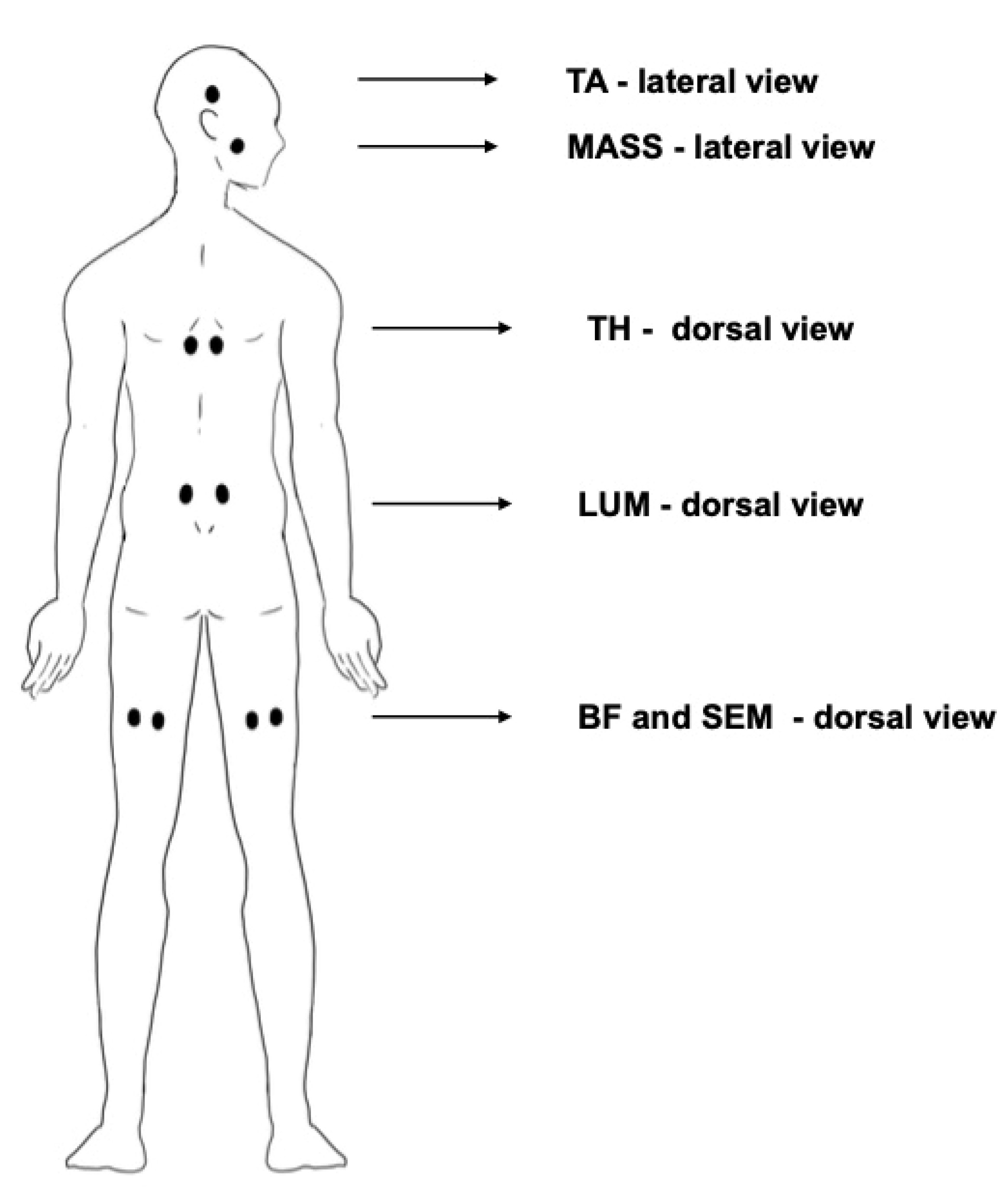
- Masseter (MASS) and temporalis anterior (TA) muscles were measured according to the SENIAM guidelines [35,36,52]. Surface electrodes (Ag/AgCl) (Sorimex, Poland) with a 2 cm center-to-center distance after cleaning the skin with alcohol were attached on the muscles (Figure 3). The sEMG signals were recorded with 16-bit accuracy at a sampling rate of 1500 Hz with the Noraxon G2 TeleMyo 2400 unit (Noraxon USA).
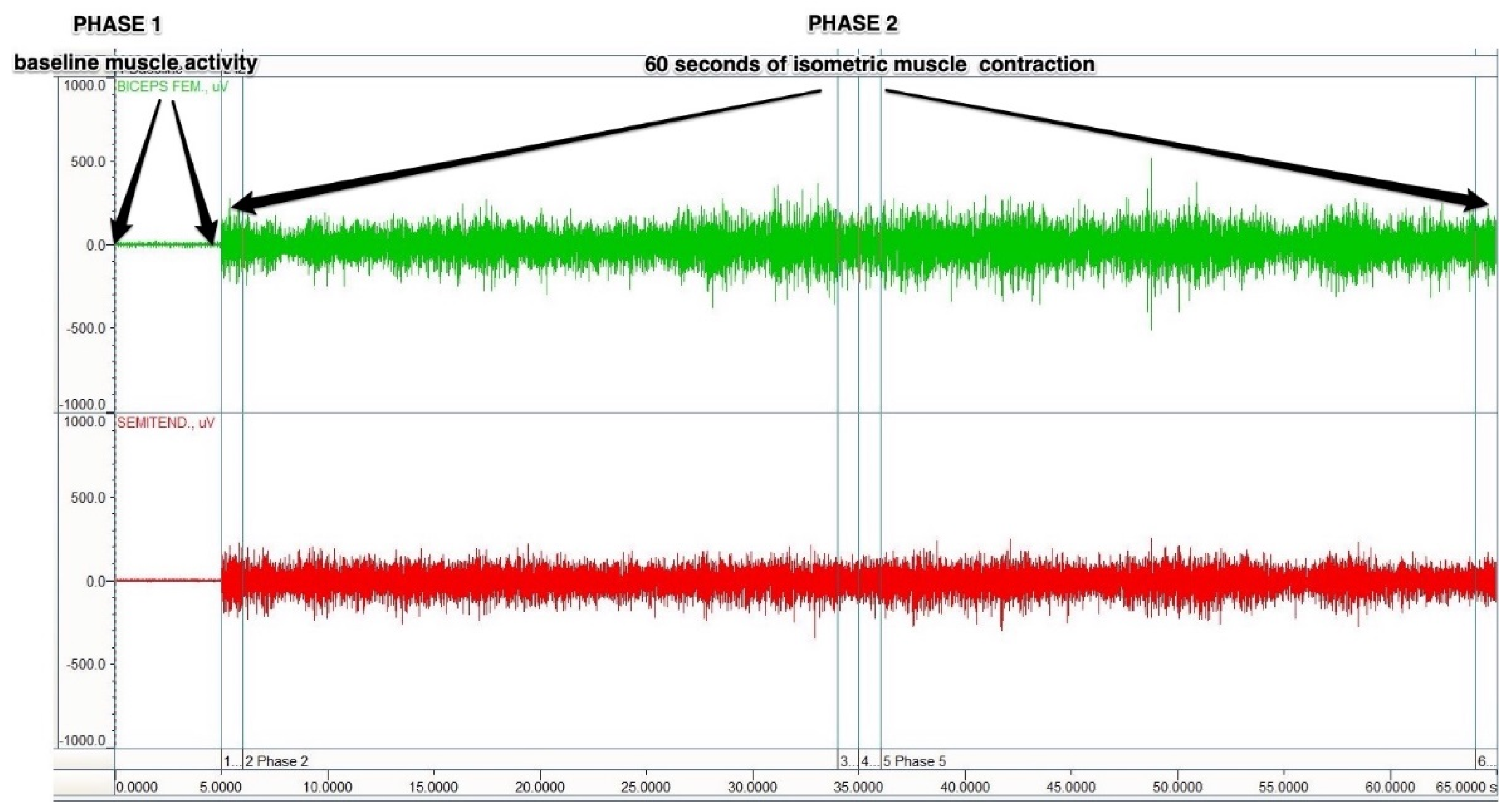
- Biceps femoris (BF) and semitendinosus-semimembranosus (SEM) muscles-Prone position, with the knee flexed to 60 degrees against manually applied resistance; (approximately 50% of maximal effort). Before starting the sEMG signal recording, the subject was asked to flex the evaluated leg against resistance applied by the researcher. The signal from the muscle was, at all times, seen on the computer screen. During the 5–10 s, the researcher adjusted the amount of resistance to the capabilities of the examined person, so the resistance was not too high and allows them to maintain a stable isometric contraction.
- Lumbar (LUM) and thoracic (TH) erector spinae muscles-prone position, with the trunk raised without any additional resistance;
- Masseter (MASS) and temporalis anterior (TA) muscles-sitting position, clenching of the teeth, with a cotton swab placed between the molar teeth; subjects were given a command: “clench the teeth as tight as you can”.
2.3. Statistical Analysis
3. Results
3.1. Fatigue of the Biceps Femoris (BF) and Semimembranosus-Semitendinosus (SEM) Muscles
3.2. Fatigue of the Lumbar (LUM) and Thoracic (TH) Erector Spinae Muscles
3.3. Fatigue of the Masseter (MASS) and Temporalis Anterior (TA) Muscles
4. Discussion
5. Conclusions
Author Contributions
Funding
Institutional Review Board Statement
Informed Consent Statement
Data Availability Statement
Conflicts of Interest
Abbreviations
| MC—Myofascial continuity |
| sEMG—surface electromyography |
| AI—arch index |
| BF—biceps femoris muscle |
| SEM—semitendinosus and semimembranosus hamstring muscles |
| LUM—erector spinae muscle at lumbar region |
| TH—erector spinae muscle at thoracic region |
| MASS—masseter muscle |
| TA—temporalis anterior muscle |
| FFT—Fast Fourier Transform |
| ES—effect size |
| SD—standard deviation |
| SEM—standard error of measurement |
| SBL—superficial back line |
| R—right side |
| L—left side |
References
- Bernabei, M.; Maas, H.; van Dieën, J.H. A lumped stiffness model of intermuscular and extramuscular myofascial pathways of force transmission. Biomech. Modeling Mechanobiol. 2016, 15, 1747–1763. [Google Scholar] [CrossRef] [Green Version]
- Stecco, C.; Porzionato, A.; Macchi, V.; Stecco, A.; Vigato, E.; Parenti, A.; Delmas, V.; Aldegheri, R.; De Caro, R. The expansions of the pectoral girdle muscles onto the brachial fascia: Morphological aspects and spatial disposition. Cells Tissues Organs 2008, 188, 320–329. [Google Scholar] [CrossRef]
- Huijing, P.A. Epimuscular myofascial force transmission: A historical review and implications for new research. International Society of Biomechanics Muybridge Award Lecture, Taipei, 2007. J. Biomech. 2009, 42, 9–21. [Google Scholar] [CrossRef] [PubMed]
- Maas, H.; Sandercock, T.G. Force transmission between synergistic skeletal muscles through connective tissue linkages. J. Biomed. Biotechnol. 2010, 2010, 575–672. [Google Scholar] [CrossRef] [Green Version]
- Schleip, R.; Klingler, W.; Lehmann-Horn, F. Active fascial contractility: Fascia may be able to contract in a smooth muscle-like manner and thereby influence musculoskeletal dynamics. Med. Hypotheses 2005, 65, 273–277. [Google Scholar] [CrossRef] [PubMed]
- Myers, T. Anatomy Trains: Myofascial Meridians for Manual and Movement Therapists, 3rd ed.; Elsevier: Amsterdam, The Netherlands; Churchill Livingstone: London, UK, 2014. [Google Scholar]
- Stecco, A.; Macchi, V.; Stecco, C.; Porzionato, A.; Day, J.A.; Delmas, V.; De Caro, R. Affiliations expand Anatomical study of myofascial continuity in the anterior region of the upper limb. J. Bodyw. Mov. 2009, 13, 53–62. [Google Scholar] [CrossRef] [PubMed]
- Turvey, M.T.; Fonseca, S.T. The medium of haptic perception: A tensegrity hypothesis. J. Mot. Behav. 2014, 46, 143–187. [Google Scholar] [CrossRef] [PubMed]
- Krause, F.; Wilke, J.; Vogt, L.; Banzer, W. Intermuscular force transmission along myofascial chains: A systematic review. J. Anat. 2016, 228, 910–918. [Google Scholar] [CrossRef]
- Masi, A.T.; Hannon, J.C. Human resting muscle tone (HRMT): Narrative introduction and modern concepts. J. Bodyw. Mov. Ther. 2008, 12, 320–332. [Google Scholar] [CrossRef]
- Dischiavi, S.L.; Wright, A.A.; Hegedus, E.J.; Bleakley, C.M. Biotensegrity and myofascial chains: A global approach to an integrated kinetic chain. Med. Hypotheses 2018, 110, 90–96. [Google Scholar] [CrossRef]
- Langevin, H. Connective tissue: A body-wide signaling network? Med. Hypotheses 2006, 66, 1074–1077. [Google Scholar] [CrossRef]
- Kassolik, K.; Jaskólska, A.; Kisiel-Sajewicz, K.; Marusiak, J.; Kawczyński, A.; Jaskólski, A. Tensegrity principle in massage demonstrated by electro- and mechanomyography. J. Bodyw. Mov. Ther. 2009, 13, 164–170. [Google Scholar] [CrossRef] [PubMed]
- Stecco, C.; Porzionato, A.; Lancerotto, L.; Stecco, A.; Macchi, V.; Day, J.A.; De Caro, R. Histological study of the deep fasciae of the limbs. J. Bodyw. Mov.Ther. 2008, 12, 225–230. [Google Scholar] [CrossRef] [PubMed]
- Hyong, I.H.; Kang, J.H. The immediate effects of passive hamstring stretching exercises on the cervical spine range of motion and balance. J. Phys. Ther. Sci. 2013, 25, 113–116. [Google Scholar] [CrossRef] [Green Version]
- Hyong, I.H.; Kim, J.H. The effect of forward head on ankle joint range of motion and static balance. J. Phys. Ther. Sci. 2012, 24, 925–927. [Google Scholar] [CrossRef] [Green Version]
- Nicolakis, P.; Nicolakis, M.; Piehslinger, E.; Ebenbichler, G.; Vachuda, M.; Kirtley, C.; Fialka-Moser, V. Relationship between craniomandibular disorders and poor posture. Cranio 2000, 18, 106–112. [Google Scholar] [CrossRef] [PubMed]
- Cuccia, A.; Caradonna, C. The relationship between the stomatognathic system and body posture. Clinics 2009, 64, 61–66. [Google Scholar] [CrossRef] [PubMed] [Green Version]
- Evcik, D.; Aksoy, O. Correlation of temporomandibular joint pathologies, neck pain and postural differences. J. Phys. Ther. Sci. 2000, 12, 97–100. [Google Scholar] [CrossRef]
- Grieve, R.; Goodwin, F.; Alfaki, M.; Bourton, A.J.; Jeffries, C.; Scott, H. The immediate effect of bilateral self-myofascial release on the plantar surface of the feet on hamstring and lumbar spine flexibility: A pilot randomized controlled trial. J. Bodyw. Mov. Ther. 2015, 19, 544–552. [Google Scholar] [CrossRef]
- Aminian, A.; Sangeorzan, B.J. The anatomy of cavus foot deformity. Foot Ankle Clin. 2008, 13, 191–198. [Google Scholar] [CrossRef]
- Rao, U.B.; Joseph, B. The influence of footwear on the prevalence of flat foot. J. Bone Ankle Surg. 1992, 74, 525–527. [Google Scholar] [CrossRef]
- Stavlas, P.; Grivas, T.B.; Michas, C.; Vasiliadis, E.; Polyzois, V. The evolution of foot morphology in children between 6 and 17 years of age: A cross-sectional study based on footprints in a Mediterranean population. J. Foot Ankle Surg. 2005, 44, 424–428. [Google Scholar] [CrossRef]
- Umbrasko, S.; Vetra, J.; Dulevska, J.; Boka, S.; Gavricenkova, L.; Zagare, R. Specifiers of foot growth among schoolchildren of Riga and Latvian regions. Pap. Anthropol. 2007, 16, 283–292. [Google Scholar]
- Woźniacka, R.; Bac, A.; Matusik, S.; Szczygieł, E.; Ciszek, E. Body weight and the medial longitudinal food arch: High arched foot, a hidden problem? Eur. J. Pediatr. 2013, 172, 683–691. [Google Scholar] [CrossRef] [PubMed] [Green Version]
- Woźniacka, R.; Bac, A.; Matusik, S. Effect of obesity level on the longitudinal arch in 7- to 12-year-old rural and urban children. J. Am. Podiatr. Med. Assoc. 2015, 105, 484–492. [Google Scholar] [CrossRef] [PubMed]
- Buldt, A.K.; Forghany, S.; Landorf, K.B.; Levinger, P.; Murley, G.S.; Menz, H.B. Foot posture is associated with plantar pressure during gait: A comparison of normal, planus and cavus feet. Gait Posture 2018, 62, 235–240. [Google Scholar] [CrossRef]
- Burns, J.; Crosbie, J.; Hunt, A.; Ouvrier, R. The effect of pes cavus on foot pain and plantar pressure. Clin. Biomech. (Bristol Avon) 2005, 20, 877–882. [Google Scholar] [CrossRef] [PubMed]
- Murley, G.S.; Landorf, K.B.; Menz, H.B.; Ouvrier, R. Effect of foot posture, foot orthoses and footwear on lower limb muscle activity during walking and running: A systematic review. Gait Posture 2009, 29, 172–187. [Google Scholar] [CrossRef]
- Levin, D.; Whittle, M.W. The effects of pelvic movement on lumbar lordosis in the standing position. J. Orthop. Sports Phys. Ther. 1996, 24, 130–135. [Google Scholar] [CrossRef]
- Duval, K.; Lam, T.; Sanderson, D. The mechanical relationship between the rearfoot, pelvis and low-back. Gait Posture 2010, 32, 637–640. [Google Scholar] [CrossRef]
- Morris, C.E.; Bonnefin, D.; Darvillem, C. The Torsional Upper Crossed Syndrome: A multi-planar update to Janda’s model, with a case series introduction of the mid-pectoral fascial lesion as an associated etiological factor. J. Bodyw. Mov. Ther. 2015, 19, 681–689. [Google Scholar] [CrossRef]
- Forestier, N.; Teasdale, N.; Nougier, V. Alteration of the position sense at the ankle induced by muscular fatigue in humans. Med. Sci. Sports Exerc. 2002, 34, 117–122. [Google Scholar] [CrossRef]
- Gimmon, Y.; Riemer, R.; Oddsson, L.; Melzer, I. The effect of plantar flexor muscle fatigue on postural control. J. Electromyogr. Kinesiol. 2011, 21, 922–928. [Google Scholar] [CrossRef]
- Merletti, R.; Parker, P. Electromyography: Physiology, Engineering, and Non-Invasive Applications; Wiley-IEEE Press: Hoboken, NJ, USA, 2004. [Google Scholar]
- Hermens, H.J.; Freriks, B.; Merletti, R. SENIAM 8: European Recommendations for Surface Electromyography; Roessingh Research and Development: Enschede, The Netherlands, 1999. [Google Scholar]
- Kasman, G.S.; Cram, J.R.; Wolf, S.L. Clinical Applications in Surface Electromyography, Chronic Musculoskeletal Pain; Aspen Publishers: Gaithersburg, MD, USA, 1998. [Google Scholar]
- Gonzalez-Izal, M.; Malanda, A.; Gorostiaga, E.; Izquierdo, M. Electromyographic models to assess muscle fatigue. J. Electromyogr. Kinesiol. 2012, 22, 501–512. [Google Scholar] [CrossRef] [PubMed]
- Gazzoni, M.; Botter, A.; Vieira, T. Surface EMG and muscle fatigue: Multi-channel approaches to the study of myoelectric manifestations of muscle fatigue. Physiol. Meas. 2017, 38, 27–60. [Google Scholar]
- Pope, R.E. The common compensatory pattern: Its origin and relationship to the postural model. Am. Acad. Osteopathy J. 2003, 14, 19–40. [Google Scholar]
- Tozzi, P. Selected fascial aspects of osteopathic practice. J. Bodyw. Mov. Ther. 2012, 16, 503–519. [Google Scholar] [CrossRef] [PubMed]
- Kouwenhoven, J.W.M.; Vincken, K.L.; Bartels, L.W.; Casteleine, R.M. Analysis of preexistent vertebral rotation in the normal spine. Spine 2006, 31, 1467–1472. [Google Scholar] [CrossRef] [PubMed]
- Hodges, P.W.; Cholewicki, J. Functional Control of the Spine. In Movement, Stability & Lumbopelvic Pain: Integration of Research and Therapy; Vleeming, A., Mooney, V., Stoeckart, R., Eds.; Churchill Livingstone: London, UK; Elsevier: Amsterdam, The Netherlands, 2007. [Google Scholar]
- Rolf, I.P.; Feitis, R. Rolfing and Physical Reality; Inner Traditions/Bear & Co: Rochester, VT, USA, 1990. [Google Scholar]
- Bendová, P.; Růzicka, P.; Peterová, V.; Fricová, M.; Springrová, I. MRI-based registration of pelvic alignment affected by altered pelvic floor muscle characteristics. Clin. Biomech. 2007, 22, 980–987. [Google Scholar] [CrossRef]
- Snijders, C.J.; Vleeming, A.; Stoeckart, R. Transfer of lumbosacral load to iliac bones and legs. Part 1, 2. Clin. Biomech. 1993, 8, 295–301. [Google Scholar] [CrossRef] [Green Version]
- Resende, R.A.; Deluzio, K.J.; Kirkwood, R.N.; Hassan, E.A. Increased unilateral foot pronation affects lower limbs and pelvic biomechanics during walking. Gait Posture 2015, 41, 395–401. [Google Scholar] [CrossRef]
- Hao, Z.; Xie, L.; Wang, J.; Hou, Z. Spatial Distribution and Asymmetry of Surface Electromyography on Lumbar Muscles of Soldiers with Chronic Low Back Pain. Pain Res. Manag. 2020, 26. [Google Scholar] [CrossRef] [PubMed]
- Cavanagh, P.R.; Rodgers, M.M. The arch index: A useful measure from footprints. J. Biomech. 1987, 20, 547–551. [Google Scholar] [CrossRef]
- Murley, G.S.; Menz, H.B.; Landorf, K.B. A protocol for classifying normal- and flat-arched foot posture for research studies using clinical and radiographic measurements. J. Foot Ankle Res. 2009, 4, 2–22. [Google Scholar] [CrossRef] [PubMed] [Green Version]
- Wong, C.K.; Weil, R.; de Boer, E. Standardizing foot-type classification using arch index values. Physiother. Can. 2012, 64, 280–283. [Google Scholar] [CrossRef] [PubMed] [Green Version]
- Hermens, H.J.; Freriks, B.; Disselhorst-Klug, C.; Rau, G. Development of recommendations for SEMG sensors and sensor placement procedures. J. Electromyogr. Kinesiol. 2000, 10, 361–374. [Google Scholar] [CrossRef]
- Cifrek, M.; Medved, V.; Tonković, S.; Ostojić, S. Surface EMG based muscle fatigue evaluation in biomechanics. Clin. Biomech. 2009, 24, 327–340. [Google Scholar] [CrossRef] [PubMed]
- Coorevits, P.; Danneels, L.; Cambier, D.; Ramon, H.; Druyts, H.; Karlsson, J.S.; De Moor, G.; Vanderstraeten, G. Correlations between short-time Fourier- and continuous wavelet transforms in the analysis of localized back and hip muscle fatigue during isometric contractions. J. Electromyogr. Kinesiol. 2008, 18, 637–644. [Google Scholar] [CrossRef]
- Coorevits, P.; Danneels, L.; Cambier, D.; Ramon, H.; Druyts, H.; Karlsson, J.S.; De Moor, G.; Vanderstraeten, G. Test-retest reliability of wavelet and Fourier based EMG (instantaneous) median frequencies in the evaluation of back and hip muscle fatigue during isometric back extensions. J. Electromyogr. Kinesiol. 2008, 18, 798–806. [Google Scholar] [CrossRef]
- Gribble, P.A.; Hertel, J. Effect of hip and ankle muscle fatigue on unipedal postural control. J. Electromyogr. Kinesiol. 2004, 14, 641–646. [Google Scholar] [CrossRef]
- Mueller, M.J.; Maluf, K.S. Tissue adaptation to physical stress: A proposed “Physical Stress Theory” to guide physical therapist practice, education, and research. Phys. Ther. 2002, 82, 383–403. [Google Scholar] [CrossRef] [PubMed]
- Bolívar, Y.A.; Munuera, P.V.; Padillo, J.P. Relationship between tightness of the posterior muscles of the lower limb and plantar fasciitis. Foot Ankle Int. 2013, 34, 42–48. [Google Scholar] [CrossRef] [PubMed]
- Woźniacka, R.; Oleksy, Ł.; Jankowicz-Szymańska, A.; Mika, A.; Kielnar, R.; Stolarczyk, A. The association between high-arched feet, plantar pressure distribution and body posture in young women. Sci. Rep. 2019, 9, 17187. [Google Scholar] [CrossRef] [PubMed]
- Marr, M.; Baker, J.; Lambon, N.; Perry, J. The effects of the Bowen technique on hamstring flexibility over time: A randomized controlled trial. J. Body Mov. Ther. 2011, 15, 281–290. [Google Scholar] [CrossRef] [PubMed]
- Weisman, M.H.; Haddad, M.; Lavi, N.; Vulfsons, S. Surface electromyography recordings after passive and active motion along the posterior myofascial kinematic chain in healthy male subjects. J. Bodyw. Mov. Ther. 2014, 18, 452–461. [Google Scholar] [CrossRef]
- Betsch, M.; Schneppendahl, J.; Dor, L.; Jungbluth, P.; Grassmann, J.P.; Windolf, J. Influence of foot position on the spine and pelvis. Arthritis Care Res. 2011, 63, 1758–1765. [Google Scholar] [CrossRef]
- Søgaard, K.; Blangsted, A.K.; Jørgensen, L.V.; Madeleine, P.; Sjøgaard, G. Evidence of long term muscle fatigue following prolonged intermittent contractions based on mechano- and electromyograms. J. Electromyogr. Kinesiol. 2003, 13, 441–450. [Google Scholar] [CrossRef]
- Rudroff, T.; Staudenmann, D.; Enoka, R.M. Electromyographic measures of muscle activation and changes in muscle architecture of human elbow flexors during fatiguing contractions. J. Appl. Physiol. (1985) 2008, 104, 1720–1726. [Google Scholar] [CrossRef] [Green Version]
- Valentino, B.; Fabozzo, A.; Melito, F. The functional relationship between the occlusal plane and the plantar arches. An EMG study. Surg. Radiol. Anat. 1991, 13, 171–174. [Google Scholar] [CrossRef]
- Cuccia, A.M. Interrelationships between dental occlusion and plantar arch. J. Body Mov. Ther. 2011, 15, 242–250. [Google Scholar] [CrossRef]


| Outcome Measure | Side | Group 1 | p # | Group 2 | p # | Group 3 | p # | p | p * | ES | p ** | ES | p *** | ES |
|---|---|---|---|---|---|---|---|---|---|---|---|---|---|---|
| BF slope (Hz) | R | −0.13 ± 0.15 (0.02) | 0.76 | −0.21 ± 0.18 (0.04) | 0.16 | −0.13 ± 0.18 (0.04) | 0.29 | 0.22 | 0.31 | 0.48 | 0.99 | 0.01 | 0.37 | 0.44 |
| L | −0.14 ± 0.10 (0.02) | −0.13 ± 0.11 (0.03) | −0.17 ± 0.09 (0.03) | 0.57 | 0.98 | 0.09 | 0.63 | 0.31 | 0.72 | 0.39 | ||||
| BF Intercept (Hz) | R | 59.9 ± 11.2 (1.8) | 0.07 | 65.8 ± 13.1 (2.80) | 0.69 | 61.1 ± 9.6 (2.26) | 0.14 | 0.16 | 0.20 | 0.48 | 0.94 | 0.11 | 0.44 | 0.40 |
| L | 65.7 ± 15.2 (2.58) | 64.4 ± 13.3 (2.85) | 64.9 ± 13.1 (3.55) | 0.94 | 0.95 | 0.45 | 0.98 | 0.05 | 0.99 | 0.03 | ||||
| BF difference (%) | R | −20.2 ± 11.3 (1.91) | 0.34 | −22.4 ± 14.4 (3.08) | 0.76 | −19.2 ± 12.5 (2.96) | 0.43 | 0.71 | 0.83 | 0.21 | 0.97 | 0.08 | 0.73 | 0.23 |
| L | −17.6 ± 13.2 (2.23) | −21.1 ± 13.4 (2.86) | −15.8 ± 11.4 (2.71) | 0.52 | 0.72 | 0.26 | 0.93 | 0.14 | 0.54 | 0.42 | ||||
| SEM slope (Hz) | R | −0.20 ± 0.09 (0.01) | 0.47 | −0.25 ± 0.10 (0.02) | 0.23 | −0.20 ± 0.11 (0.02) | 0.41 | 0.14 | 0.23 | 0.52 | 0.99 | 0.01 | 0.26 | 0.47 |
| L | −0.22 ± 0.12 (0.02) | −0.22 ± 0.13 (0.02) | −0.22 ± 0.09 (0.02) | 0.96 | 0.98 | 0.01 | 0.97 | 0.01 | 0.99 | 0.01 | ||||
| SEM Intercept (Hz) | R | 56.6 ± 6.8 (1.15) | 0.69 | 56.8 ± 7.6 (1.63) | 0.14 | 55.5 ± 9.1 (2.15) | 0.44 | 0.84 | 0.99 | 0.02 | 0.89 | 0.13 | 0.86 | 0.15 |
| L | 57.1 ± 5.9 (1.01) | 60.2 ± 9.9 (2.53) | 54.4 ± 7.1 (1.68) | 0.09 | 0.62 | 0.38 | 0.41 | 0.41 | 0.10 | 0.67 | ||||
| SEM difference (%) | R | −25.2 ± 8.9 (1.51) | 0.61 | −29.5 ± 9.1 (1.92) | p = 0.04 ES = 0.52 | −24.2 ± 10.1 (2.38) | 0.25 | 0.13 | 0.27 | 0.47 | 0.94 | 0.10 | 0.20 | 0.55 |
| L | −24.3 ± 10.1 (1.69) | −24.9 ± 8.4 (2.50) | −27.0 ± 12.2 (2.88) | 0.70 | 0.98 | 0.06 | 0.74 | 0.24 | 0.83 | 0.20 |
| Outcome Measure | Side | Group 1 | p # | Group 2 | p # | Group 3 | p # | p | p * | ES | p ** | ES | p *** | ES |
|---|---|---|---|---|---|---|---|---|---|---|---|---|---|---|
| LUM slope (Hz) | R | −0.16 ± 0.08 (0.01) | 0.13 | −0.14 ± 0.05 (0.03) | p = 0.01 ES = 1.32 | −0.13 ± 0.08 (0.01) | 0.43 | 0.75 | 0.89 | 0.29 | 0.80 | 0.35 | 0.97 | 0.14 |
| L | −0.14 ± 0.07 (0.01) | −0.20 ± 0.04 (0.03) | −0.12 ± 0.07 (0.01) | 0.09 | 0.27 | 1.05 | 0.83 | 0.28 | 0.12 | 1.40 | ||||
| LUM Intercept (Hz) | R | 78.3 ± 9.6 (1.63) | 0.15 | 82.7 ± 10.9 (2.28) | 0.55 | 84.5 ± 13.5 (3.10) | 0.33 | 0.11 | 0.37 | 0.42 | 0.19 | 0.52 | 0.86 | 0.14 |
| L | 75.6 ± 10.7 (1.81) | 81.0 ± 11.9 (2.48) | 81.9 ± 13.1 (3.04) | 0.10 | 0.26 | 0.47 | 0.22 | 0.52 | 0.96 | 0.10 | ||||
| LUM difference (%) | R | −12.0 ± 7.1 (1.21) | 0.63 | −4.8 ± 3.4 (1.67) | p = 0.04 ES = 0.92 | −7.4 ± 5.4 (1.47) | 0.50 | 0.09 | 0.05 | 1.29 | 0.35 | 0.72 | 0.71 | 0.57 |
| L | −11.4 ± 6.1 (1.04) | −7.8 ± 3.1 (1.87) | −6.1 ± 4.6 (1.29) | 0.12 | 0.42 | 0.74 | 0.21 | 0.98 | 0.85 | 0.43 | ||||
| TH slope (Hz) | R | −0.15 ± 0.09 (0.01) | 0.73 | −0.20 ± 0.08 (0.02) | 0.73 | −0.10 ± 0.08 (0.01) | 0.17 | 0.005 | 0.02 | 0.58 | 0.26 | 0.58 | 0.01 | 1.25 |
| L | −0.14 ± 0.07 (0.01) | −0.19 ± 0.04 (0.02) | −0.13 ± 0.06 (0.01) | 0.02 | 0.005 | 0.87 | 0.75 | 0.15 | 0.005 | 1.17 | ||||
| TH Intercept (Hz) | R | 66.4 ± 12.7 (2.15) | 0.23 | 68.3 ± 12.1 (2.53) | 0.67 | 65.3 ± 12.9 (2.96) | 0.75 | 0.72 | 0.85 | 0.15 | 0.96 | 0.08 | 0.74 | 0.23 |
| L | 68.5 ± 13.9 (2.35) | 67.2 ± 14.6 (3.05) | 66.1 ± 11.8 (2.72) | 0.82 | 0.94 | 0.09 | 0.85 | 0.18 | 0.96 | 0.08 | ||||
| TH difference (%) | R | −13.9 ± 6.6 (1.13) | 0.34 | −15.4 ± 6.8 (1.85) | 0.68 | −10.4 ± 4.3 (1.44) | 0.66 | 0.03 | 0.77 | 0.22 | 0.30 | 0.62 | 0.01 | 0.87 |
| L | −12.7 ± 6.4 (1.09) | −16.1 ± 6.5 (2.31) | −11.1 ± 3.4 (0.78) | 0.03 | 0.28 | 0.51 | 0.78 | 0.30 | 0.01 | 0.96 |
| Outcome Measure | Side | Group 1 | p # | Group 2 | p # | Group 3 | p # | p | p * | ES | p ** | ES | p *** | ES |
|---|---|---|---|---|---|---|---|---|---|---|---|---|---|---|
| MASS slope (Hz) | R | −0.39 ± 0.15 (0.07) | 0.33 | −0.26 ± 0.15 (0.11) | 0.32 ES = 0.55 | −0.22 ± 0.18 (0.08) | 0.75 | 0.34 | 0.61 | 0.86 | 0.47 | 0.78 | 0.94 | 0.18 |
| L | −0.34 ± 0.16 (0.08) | −0.34 ± 0.14 (0.10) | −0.20 ± 0.19 (0.09) | 0.50 | 0.99 | 0.10 | 0.62 | 0.79 | 0.59 | 0.83 | ||||
| MASS Intercept (Hz) | R | 139.1 ± 20.6 (3.49) | 0.89 | 149.2 ± 30.4 (6.48) | 0.52 | 146.5 ± 22.5 (5.31) | 0.17 | 0.28 | 0.36 | 0.38 | 0.63 | 0.34 | 0.93 | 0.10 |
| L | 138.7 ± 17.4 (2.95) | 146.9 ± 22.5 (4.81) | 141.5 ± 18.9 (4.45) | 0.30 | 0.34 | 0.40 | 0.90 | 0.15 | 0.67 | 0.25 | ||||
| MASS difference (%) | R | −6.45 ± 3.1 (1.45) | 0.38 | −3.62 ± 2.8 (1.71) | p = 0.01 ES = 0.95 | −4.03 ± 3.4 (1.46) | 0.71 | 0.35 | 0.47 | 0.95 | 0.62 | 0.74 | 0.99 | 0.13 |
| L | −5.30 ± 3.2 (1.55) | −6.91 ± 4.1 (1.67) | −4.51 ± 3.1 (1.60) | 0.63 | 0.79 | 0.43 | 0.95 | 0.25 | 0.65 | 0.66 | ||||
| TA slope (Hz) | R | −0.41 ± 0.5 1(0.08) | 0.24 | −0.51 ± 0.43 (0.11) | 0.25 | −0.71 ± 0.42 (0.14) | 0.11 | 0.14 | 0.81 | 0.21 | 0.20 | 0.62 | 0.47 | 0.47 |
| L | −0.51 ± 0.41 (0.06) | −0.39 ± 0.36 (0.09) | −0.52 ± 0.36 (0.17) | 0.65 | 0.74 | 0.31 | 0.99 | 0.02 | 0.72 | 0.36 | ||||
| TA Intercept (Hz) | R | 165.9 ± 34.2 (5.79) | 0.45 | 175.7 ± 26.1 (5.57) | 0.33 | 163.3 ± 35.7 (8.41) | 0.62 | 0.42 | 0.58 | 0.32 | 0.96 | 0.07 | 0.49 | 0.39 |
| L | 161.4 ± 24.1 (7.12) | 171.6 ± 23.6 (5.09) | 158.3 ± 27.5 (8.49) | 0.42 | 0.59 | 0.56 | 0.96 | 0.11 | 0.48 | 0.51 | ||||
| TA difference (%) | R | −5.21 ± 5.2 (1.83) | 0.20 | −8.33 ± 5.1 (2.11) | 0.06 | −11.1 ± 7.3 (2.07) | 0.50 | 0.54 | 0.55 | 0.60 | 0.19 | 0.92 | 0.69 | 0.43 |
| L | −7.43 ± 6.5 (1.10) | −5.91 ± 5.5 (2.18) | −8.94 ± 7.5 (3.11) | 0.62 | 0.87 | 0.25 | 0.88 | 0.21 | 0.62 | 0.46 |
Publisher’s Note: MDPI stays neutral with regard to jurisdictional claims in published maps and institutional affiliations. |
© 2022 by the authors. Licensee MDPI, Basel, Switzerland. This article is an open access article distributed under the terms and conditions of the Creative Commons Attribution (CC BY) license (https://creativecommons.org/licenses/by/4.0/).
Share and Cite
Woźniacka, R.; Oleksy, Ł.; Jankowicz-Szymańska, A.; Mika, A.; Kielnar, R.; Stolarczyk, A. The Association between Symmetrical or Asymmetrical High-Arched Feet and Muscle Fatigue in Young Women. Symmetry 2022, 14, 52. https://doi.org/10.3390/sym14010052
Woźniacka R, Oleksy Ł, Jankowicz-Szymańska A, Mika A, Kielnar R, Stolarczyk A. The Association between Symmetrical or Asymmetrical High-Arched Feet and Muscle Fatigue in Young Women. Symmetry. 2022; 14(1):52. https://doi.org/10.3390/sym14010052
Chicago/Turabian StyleWoźniacka, Renata, Łukasz Oleksy, Agnieszka Jankowicz-Szymańska, Anna Mika, Renata Kielnar, and Artur Stolarczyk. 2022. "The Association between Symmetrical or Asymmetrical High-Arched Feet and Muscle Fatigue in Young Women" Symmetry 14, no. 1: 52. https://doi.org/10.3390/sym14010052
APA StyleWoźniacka, R., Oleksy, Ł., Jankowicz-Szymańska, A., Mika, A., Kielnar, R., & Stolarczyk, A. (2022). The Association between Symmetrical or Asymmetrical High-Arched Feet and Muscle Fatigue in Young Women. Symmetry, 14(1), 52. https://doi.org/10.3390/sym14010052


