Article

Implementation of Kiosk-Type System Based on Gaze Tracking for Objective Visual Function Examination

1
Immersive Content Device Technology Research Center, Kwangwoon University, Seoul 01897, Korea
2
Department of Electronic Engineering, Kwangwoon University, Seoul 01897, Korea
3
Ingenium College, Kwangwoon University, Seoul 01897, Korea
4
Intelligent Robotics Research Center, Korea Electronics Technology Institute, Bucheon 14502, Korea
5
Graduate School of Smart Convergence, Kwangwoon University, Seoul 01897, Korea
*
Author to whom correspondence should be addressed.
Symmetry 2022, 14(3), 499; https://doi.org/10.3390/sym14030499
Submission received: 16 December 2021 / Revised: 19 January 2022 / Accepted: 25 February 2022 / Published: 28 February 2022

Abstract

:
To demonstrate that the near-point-of-convergence (npc) examination is performed subjectively, the visual fixation and four-prism diopter (Δ) base-out (BO) examinations can be conducted with a kiosk-type objective examination method based on gaze tracking, and can be compared with the existing method. Objective examination equipment was used to verify effectiveness. Fifteen adult men and women in their 20s and 30s (26.87 ± 3.31 years) with visual acuity of 0.8 (logMAR: 0.1, Snellen: 20/25(6/7.5)) or higher (corrected vision in the case of ametropia) underwent the npc, visual fixation, and 4 Δ BO examinations based on the existing examination method followed by the gaze-tracking-based kiosk-type examination method. Three examinations were performed, and a comparative analysis was conducted. The gaze tracking method used a method of extracting eye feature points, and it was judged that the ocular moved when the movement of 30 eye feature points at the edge of the iris was detected. In the comparison between the existing method and the kiosk-type method, there were no statistically significant differences in npc, visual fixation, and 4 Δ BO examinations. Npc examinations were performed extensively by the existing subjective method, however, it seems that these can be conducted objectively using the kiosk-type method. Visual fixation and 4 Δ BO examinations require high-examination proficiency because finely moving ocular movements need to be observed, but it is judged that examinations can be performed more easily and accurately when combined with the kiosk-type method. The symmetrical characteristics between the two test results are expected to serve as a basis for using the kiosk-type examination method in clinical practice.

Graphical Abstract

1. Introduction

Humans receive information from the external environment through various sensory organs. Among them, vision is a higher sense than other senses and plays an important role in eliciting or helping other senses. Most of the ways humans use to acquire external information are transmitted through vision [1].
Human visual function cannot be said to be complete only with visual acuity, which indicates whether an external object can be seen well at a certain distance [2,3,4]. In addition to visual acuity, visual functions include convergence, divergence, accommodation, color vision, stereopsis, and dynamic acuity.
Examination of refraction and visual function is largely divided into subjective and objective examinations. Subjective examination is conducted through the examinee’s feedback. When the examiner asks a question while the examination is in progress, the examinee answers the question [5,6,7]. This is also known as a subjective examination, and in actual clinical practice, it is treated as an important examination because it is necessary to consider how clear the examinee wants to see and the feeling of wearing, such as dizziness and comfort. Conversely, objective examination is an examination method which uses examination equipment for measurements without the examinee’s feedback. High proficiency in examination and examination equipment is required because the results are determined by the examiner’s judgment [8,9,10].
In clinical practice, instead of using only one method, accurate examination values are derived by performing subjective and objective examinations in parallel [11]. When a patient who needs optical treatment uses a spectacle and contact lens with the erroneous refractive power, a subjective examination must be performed at the last stage of the examination. This is because of the fact that if you change it to the correct refractive power at once, you may not be able to adapt.
In this study, we used a gaze-tracking-based kiosk to conduct near-point-of-convergence (npc) subjective, visual fixation, and 4-prism diopter (Δ) base-out (BO) examinations. Measurements were conducted by observing the micromovements of the eyes to prove that the examination is possible with the objective visual function examination method (proof of Symmetrical Characteristics with Existing examination method), and to verify its effectiveness compared with the existing examination method. Items npc, visual fixation, 4 Δ BO were selected to confirm the eye movement tracking performance of the gaze-tracking algorithm used in this paper.
Through the experiments in this paper, the first is that the subjective test can be performed as an objective test, the second is that it can be used as an auxiliary means of the examiner in the micro-eye movement tracking test that requires examination skill, and third, we would like to suggest that the examinee himself can perform an objective test without an examiner due to the nature of the preliminary test.

2. Examination of Visual Function

2.1. NPC Examination

The closest point at which both eyes can converge inward at the same time is called the near-point-of-convergence (npc) [12,13]. When the npc moves away from the eye, objects appear blurry when performing near work, such as convergence and accommodation insufficiency, and subjective symptoms, such as ophthalmocopia occur. npc is the ability to bring both eyes together as much as possible to see near objects while binocular vision is maintained, and it is implemented when eyes are uncomfortable; otherwise, binocular vision problems are expected when working in close range.
In clinical practice, a small target, such as the tip of a pencil, is mainly used to examine the npc. Recently, an npc specialized scaler has been developed and used for the convenience of examination. The normal npc range of the general public is in the range of 5–7 cm; if it is ≥20 cm, it can be considered that there is a disorder in convergence [14].
In the npc examination method, the examinee moved the chart in the direction of the eye directly to the near-distance vertical line chart using a convergence scaler (Figure 1) in the state of far-distance-corrected vision, and stops at the first double-separated point. At this time, the distance from the examinee’s eye to the chart is measured, and in the case in which an examinee is wearing a spectacle, the distance from the front vertex point of the spectacle to the chart is measured.

2.2. Visual Fixation Examination

Keeping an eye can be viewed as a combination of four eye movement systems: saccadic eye, pursuit eye, vestibular ocular, and disjunctive movements. Visual fixation is a special case of pursuit eye movement and refers to the continuous maintenance of an image of a stationary object in the fovea of the retina [15,16]. In other words, it refers to the ability to continually see an object by suppressing saccadic eye movements. If it is difficult to observe an object continually or the ability to gaze is weak, you may experience various subjective symptoms, such as reading difficulties.
In the visual fixation examination, the examinee is instructed to observe the gaze point, and the examinee is observed to assess if he/she can continuously gaze at the object. Even if the actual eye looks stable, the eye still moves. This micro-eye movement corrects the gaze error and helps focus accurately on the fovea. The evaluation criteria for visual fixation are listed in Table 1 (adapted from J.D. Kim, Clinical refraction and visual dysfunction prescription, 3rd Ed.).

2.3. Four-Prism Diopter (Δ) Base-Out (BO) Examination

The 4 Δ BO examination is conducted to determine the presence or absence of a suppression area and microscopic central scotoma [17,18] (see Figure 2 and Figure 3). Patients with a slight decrease in monocular vision and stereoacuity should have a small central scotoma area. The monofixation syndrome secondary to small-angle strabismus causes central scotoma and has clinical symptoms similar to central retinal disease which affects the fovea. In this case, the normal gaze and suppression state can be evaluated by observing whether only one eye or both eyes move when the BO prism of the schematic is added in front of the eyes.
If a BO prism is added to one eye while looking at an external object, the image of the object formed on the fovea moves to the periphery of the fovea. If the image of the object was formed within the fovea, the eye turns inward to refocus on the fovea after the BO prism is added. However, if the image of the object is formed at a functionally abnormal point, such as scotoma in the eye with the BO prism added, the eye movement for re-gazing does not appear even if the image of the object is moved to the retina image with the BO prism.
According to Hering’s law, during the addition of a BO prism, if one eye moves, the other eye moves in the same direction, and convergence is performed slowly for fusion again. If the fovea has a central scotoma, convergence does not occur in the second stage and remains slightly deflected in the outward direction.
Before the examination, ask the examinee to look at the chart, even if the chart appears to be moving. During examination, observe the eye to which the prism was not added while rapidly adding the prism to the eye to be added. If both eyes are normal eyes without scotoma or suppression, the eye to which the prism is added returns to the nose direction, which is the right angle of the prism. If a prism is added to the eye with suppression or scotoma, diplopia does not occur, and the opposite eye does not move.

3. Real-Time Gaze-Tracking Equipment Based on Eye Feature Points

3.1. Overview of Real-Time Eye-Tracking Equipment Based on Eye Feature Points

The eye-tracking method is one of the most important fields of computer vision because it provides essential information for the identification of the visual elements that people are interested in. In this study, after predicting the positions of eye feature points using the HRNet [19] (which yields an excellent performance in extracting body feature points), support vector regression (SVR) [20] is used to calculate the gaze vector based on the feature point distribution. In the process of training artificial neural networks and SVR models, UnityEyes, a synthetic eye image dataset close to real images, was used [21]. Finally, the eye aspect ratio (EAR) [22] was introduced to reduce the eye-tracking error when the eyes were closed. When the measured EAR value was smaller than the set value, the eye tracking accuracy was improved by reducing false positives by not outputting the gaze vector and iris center coordinates.
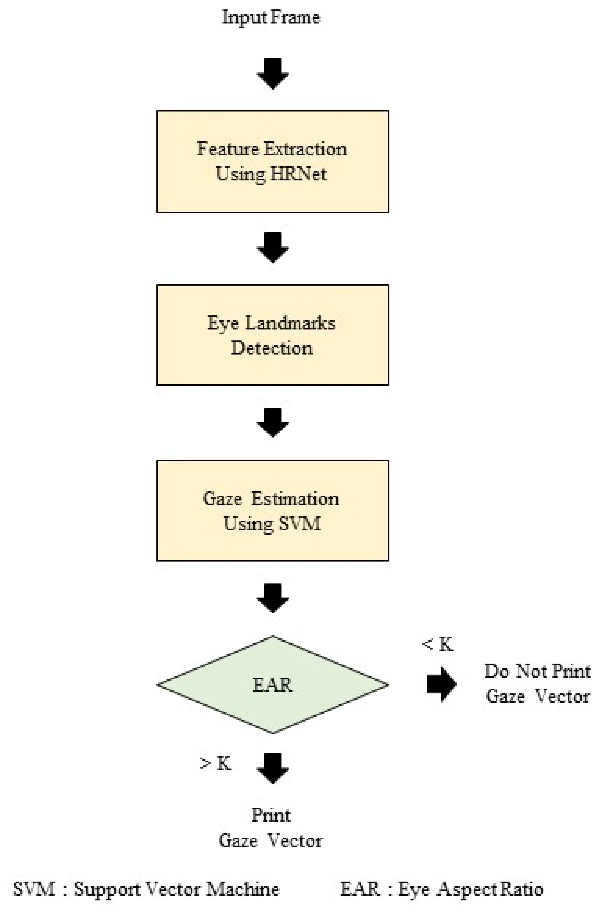
The overall flowchart of the real-time gaze-tracking method based on eye feature points is shown in Figure 4.
First, the black and white eye frames received from the IR eye camera were input to the artificial neural network HRNet. HRNet extracts feature maps for eye feature point prediction through the effective convergence of the bottom-up and top-down paths. In each channel of the extracted feature map, the positions of the eye feature points in the heat map method were predicted, and the coordinates of the predicted feature points were used to predict the gaze vector through SVR. Finally, the eye-tracking system calculates the EAR value before outputting the gaze vector and iris center coordinates, and determines whether to output the prediction result by comparing the magnitude with a preset value.

3.2. Feature Map Extraction Using HRNet

The artificial neural network plays a key role in the overall gaze-tracking method because it generates a feature map that includes information on the coordinates of the eye feature point. The performance of the eye-tracking method largely depends on the method of extraction and fusion of the feature map of an artificial neural network. In the proposed method, HRNet is adopted as an artificial neural network to increase the accuracy and improve the inference speed. Existing deep-learning-based eye-tracking methods, such as the hourglass network [23] and SimpleBaseline [24], generate the final feature map using the encoder-decoder method. These methods have the advantage of effectively using a wide range of local information by focusing on a top-down path of decoding a low-resolution into a high-resolution feature map. However, because the fusion process uses a relatively unprocessed low-resolution feature map compared with the high-resolution feature map, it has structural performance limitations.
HRNet uses a method which fuses feature maps by focusing on both the bottom-up and top-down paths, thus increasing the accuracy and reducing the number of floating-point operations (FLOPS) by half. HRNet generates low-resolution feature maps at every step, simultaneously applies a 1 × 1 convolution to the high-resolution feature maps, and maintains them to enhance features within a small area. HRNet consists of a total of four sequential steps. At each step, a feature map with 1/2 the size of the smallest resolution feature map is created, and the residual block is then applied four times. Subsequently, as the last process of each step, the exchange process is performed to converge the feature maps of all resolutions in a fully connected manner. The exchange process at each stage consists of several iterations of the exchange unit, with one iteration in the second stage, four repetitions in the third stage, and three repetitions in the fourth stage. In the exchange process, in the bottom-up path, 1 × 1 convolution and nearest-neighbor interpolation are used for upsampling, and in the top-down path, a 3 × 3 convolution with a stride of two is used for downsampling. In contrast, in the parallel path, a 1 × 1 convolution was used to maintain the resolution.
Figure 5 shows the HRNet structure applied to the gaze tracking method. The residual block is omitted in this figure. HRNet-W32, a lightweight model, was used for real-time operation, and the number of channels from the highest-resolution feature map to the low-resolution feature map was set to 32, 64, 128, and 256, respectively. The highest resolution feature map is output, and the number of channels in the final feature map is set to 48, which is the number of eye feature points.

3.3. Prediction of Eye Features

Each channel of the output feature map extracted from HRNet represents a heat map of the location of the eye feature point. Of the 48 channels, 16 correspond to the coordinates of the eye edge points, and 30 correspond to the coordinates of the iris edge points. The remaining two channels represent the coordinates of the center of the eye and the coordinates of the center of the iris. Because the point with the highest value in each heat map indicates the point with the highest probability of the presence of eye feature points, the coordinates of the corresponding points are finally output as predicted values. In the learning process of the artificial neural network, a two-dimensional (2D) Gaussian distribution with an eye feature point as the center and a standard deviation of 1 is input as the ground truth in each channel. Regarding the loss function between the output heat map and ground truth, an L2 loss mean square error, is applied. Equation (1) expresses the overall loss function for the heat map for predicting the eye feature points. In Equation (1), N   is the number of eye feature points, K is the number of pixels in the output feature map, M ^ i j is the predicted heat map, and M i j is the ground-truth heat map.
L heatmap = 1 N i = 1 N j = 1 K M ^ i j M i j 2
The equation denotes the overall loss function used for the heatmap for the prediction of eye features.

3.4. Gaze Vector Prediction Based on Eye Feature Points

There is a feature-based method which uses the distribution of eye feature point positions and a model-based method for the formation of an eye model based on these feature points as a method to predict a gaze vector. An appearance-based method can be used directly. In this study, the feature map is extracted using the artificial neural network of the appearance-based method, and the gaze was traced using the feature-based method on the predicted eye feature coordinates. Compared with other methods, the feature-based method has the advantage of being less affected by user changes and shows excellent performance even when learning uses a small amount of data. Conversely, because the positions of the eye feature points play a decisive role in the prediction of the gaze vector, there is a feature which depends considerably on the result of the eye feature point detection. The feature-based method applied in this study is as follows. First, the ocular feature point coordinates predicted through the heat map are centered on the ocular center coordinates and are normalized by the length of the ocular radius (the distance between the ocular center coordinates and the coordinates of the ocular edge point with the furthest distance). The normalized coordinates were input to an SVR model, and the model predicted a three-dimensional gaze vector. In the training process of the SVR model, the pitch of the eyeball (pitch, θ ) and yaw (yaw, φ ) are input as the ground truth.

3.5. Reduced False Positives during Eye Movements

The artificial neural network model proposed in this study predicts the positions of the set number of eye features regardless of the actual presence or absence of eye feature points in the image. Therefore, even when the user closes his/her eyes, a false positive for tracking the user’s eyes may be generated. Therefore, the eye aspect ratio (EAR) [22] was introduced to solve this problem. The EAR is used to measure the degree of eye closure as the ratio of the distance between the horizontal end points of the eye’s edge and the distance between the vertical end points. Figure 6 shows the horizontal p 1 p 3 and vertical p 2 p 4 endpoints of the eye’s edge, and the distance between the two endpoints. Equation (2) shows the formula for calculating the EAR.
EAR = p 2 p 4 p 1 p 3

EAR

When the eyes are fully opened, the EAR varies considerably from user to user. Therefore, in this eye-tracking system, after measuring the user’s EAR values for 2 s when the program is executed, 1/2 times the median value K is set as the comparison value. When the measured EAR is larger than K, the iris center coordinate and gaze vector are output. If the measured EAR is smaller than K, it is judged that the eyes are more than half closed, and the result is not output.

3.6. Dataset and Data Preprocessing

A synthetic eye image created with UnityEyes was used as a dataset for training an artificial neural network for gaze tracking. UnityEyes is a three-dimensional (3D) eye model program which uses the Unity software, and is extensively used as an alternative to real datasets which lack eye image data and high-quality ground truth. Figure 7 shows an example of a synthetic eye image created with UnityEyes and the coordinates of the eye feature points.
In total, there were 53 eye feature coordinates, including 16 eye edges, seven caruncles, and 30 iris edge coordinates. In this study, only the eye and iris edge coordinates were used, except for the caruncle coordinates. In addition, the eyeball center coordinates and iris center coordinates were calculated and added as the average of the edge coordinates. They were subsequently used as the ground truth of the heat map of the eye feature point position. The composite eye image was created with a size of 640 × 480, and were then converted to a black-and-white image, and cropped around the eye center coordinates according to the resolution of the infrared (IR) eye camera which was used. 
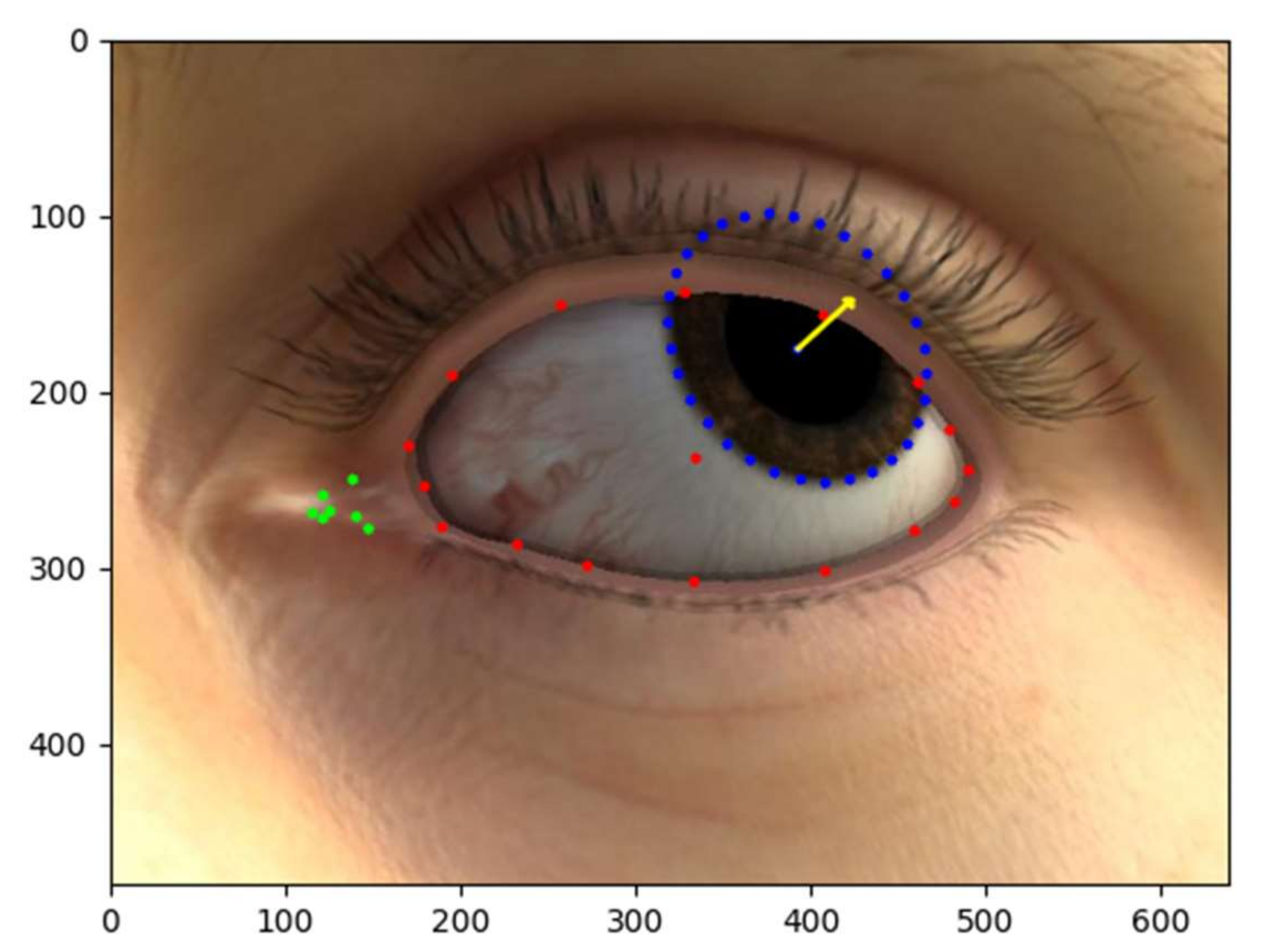
Figure 8 shows the eye feature point detection results and gaze vector prediction for the actual eye image of the eye-tracking system. The 16 eye marginal points are shown in red, the 30 iris edge points in blue, and the two central coordinates in white and green dots, respectively. In addition, the 3D gaze vector was visualized as a 2D vector projected onto the plane and is indicated in yellow.
The 3D gaze vector G x , y , z provided by UnityEyes is converted into θ and φ , and IS used as the ground truth of the SVR. Figure 9 shows the gaze vector and the angles θ and φ in the 3D coordinate system, and Equation (3) expresses mathematically the angle conversion of the gaze vector.
θ pitch = a Γ c c o s z y 2 + z 2 θ yaw = a Γ c c o s z z 2 + x 2
where Equation (3) is the transformation of the 3D gaze vector in θ and φ .

4. Materials and Methods

4.1. Participants

The examinee selected 15 subjects in their 20s and 30s (26.87 ± 3.31 years) who understood and agreed to the purpose of this study, did not have any special ophthalmic, mental, or systemic disease, and had far and near-corrected vision of 0.8 or higher. Those with restrictions on deviation or eye movement were excluded from this experiment because they judged that they could not test or track fine eye movements due to large eye movements.

4.2. Procedure

4.2.1. Case History

Before the onset of the experiment, examinees who participated in the experiment completed physical condition and history questionnaires to check for factors that could affect the experiment.
The items of the history questionnaire were divided into four categories: job, body use (excise and game), past medical history, and drive.
The sequence of the experiment included time needed to record history notes, and npc, visual fixation, and 4 Δ BO examinations; these were followed by npc, visual fixation, and 4 Δ BO examinations, which were performed using a kiosk-type examination system. To prevent order effects, kiosk-type examinations were performed by randomly setting three examinations. To manage the eye condition of the examinees, a rest period of 5 min was set before the experiment using the kiosk examination method. Figure 10 shows the examination environment of the existing method.

4.2.2. Manufacture and Composition of the Kiosk-Type Objective Visual Function Examination System

The kiosk-type objective visual function examination system used in this study was operated by implanting the gaze-tracking device described in Section 3 into a webcam camera (Logitech C922 PRO). The camera specification is 44 mm high, 95 mm wide, 71 mm deep, 1.5 m cable length, and 162 g in weight, and technical specifications include a maximum resolution of 1080p/30fps–720p/60fps, 3 megapixels, autofocus, glass lens, stereo built-in microphone (range: 1 m), 78° diagonal field of view. Figure 11 shows webcam camera (Logitech C922 PRO).
The experimental environment was set so that the micro-movement of the eyes could be detected adequately during the examination by fixing two cameras on the experimental table. During the examination, it was determined that the eyes moved when the movement of the 30 feature points on the edge of the iris was detected. Figure 12 shows the experimental environment of the kiosk-type system.

4.3. Research Data Analysis

Data analysis was performed using the paired sample t-test in SPSS (version 18.0 for Window, SPSS Inc., Chicago, IL, USA). The Wilcoxon signed-rank test was performed, and it was judged to be statistically significant when p < 0.05, with a 95% confidence interval. In general, the t-test is a statistical technique used to test the mean difference between groups. One sample-test is performed when there is one group, and independent sample-test and paired sample-test are performed when there are two groups. In this paper, the paired sample-test was conducted because the same experimental participants were measured with certain variables. In the statistical analysis of nursing care, non-parametric statistics can be used when analyzing the number of samples of less than 30 people, qualitative data, and non-continuous quantitative data due to the nature of the field. The ranking is set using the values derived from the sample data without assumption of distribution and hypothesis is tested. Among them, the Wilcoxon sign rank test used in this paper can specify information on data.

5. Results

5.1. NPC Examination Results

These are the results measured by the existing examination method and the kiosk-type examination method in comparison with npc (Table 2, Figure 13).
Regarding the comparison results of npc, the kiosk-type examination method showed a slightly shorter trend in the comparison between the existing examination method (7.92 ± 3.44 cm) and the kiosk-type examination method (7.68 ± 3.57 cm), but no statistical significance was found.

5.2. Visual Fixation Examination Results

These are the results measured by the existing examination method and the kiosk-type examination method in comparison with visual fixation (Table 3, Figure 14).
The comparison results of visual fixation showed the same results between both tests in comparison with the existing and the kiosk-type examination methods. No statistically significant differences were observed.

5.3. 4 Δ BO Examination Results

Table 4 lists the results measured by the existing examination method and the kiosk-type examination method in comparison with 4 Δ BO (Table 4).
The comparison result of 4 Δ BO showed the same results between both tests in comparison with the existing examination method and the kiosk-type examination method, and there were no statistically significant differences.

6. Discussion

When humans receive information from the external environment, these are mostly visual. To maintain good visual acuity, it is recommended to have regular visual acuity and visual function checkup once a year on average for adults over 20 years of age. To this end, clinical refraction and visual function examinations are performed, and subjective and objective examinations are performed concurrently to derive accurate examination values. In general, objective examinations require improved examination skills relative to subjective examinations.
In this study, a kiosk-type objective visual function examination system was implemented using real-time gaze-tracking equipment based on eye feature points. For the evaluation of its effectiveness, a data comparison analysis was performed between the existing examination method and the kiosk-type examination method for npc, visual fixation, and 4 Δ BO examinations.
The kiosk-type examination system is performed in the same way as the existing examination method, but the examination results are determined by detecting the movement of 30 feature points at the edge of the iris. Because it tracks the movement of the ocular input through the webcam, it is difficult to see the ocular in a relatively dark lighting environment, so the accuracy is poor. Therefore, the same lighting environment as the existing inspection method is needed. The gaze-tracking model is a model having a small resolution of 192 × 192, just an RGB camera having a resolution of 192 × 192 or more has an advantage that does not affect a driving specification. However, since the learning data have only been learned by the ocular, the performance tends to fall when the spectacles frame image is visible during the examination. In the future, it is necessary to increase the accuracy of the test results of the examinee wearing spectacles by adding image learning data including the frame of spectacles.
One of the main performances of the gaze-tracking algorithm used in this paper is micro-eye movement tracking. Examinees with deviations or abnormal eye movements were excluded from the experiment because they judged that they could not examine or trace micro-eye movements due to large eye movements. Examinees with deviations or abnormal eye movements have large eye movements when examination, making it easier to perform examinations with kiosk examinations.
In the clinical protocol, in order to measure visual function, it was measured in order by the existing examination method. Afterwards, when the examination was performed using the kiosk type examination method, the examination was performed randomly because the examinees experienced the measurement environment. An order effect was prevented by setting up a 5-min break after the existing examination and randomly performing a kiosk type examination.
In the npc examination, the kiosk-type examination method showed a slightly shorter near point compared with the existing examination method, but no statistically significant difference was observed. The npc value presented in the result is the average data value. There were also examinees whose npc was measured longer in the kiosk examination. During the npc examination, the inward adduction of the two eyes increased as the near point became closer, and one of the two eyes was abducted outward when the near point was reached. Even in the existing examination method, there were cases wherein objective examination was judged based on the use of the phenomenon of abduction, but there was a disadvantage in that it was difficult to judge because measurements involved micro-eye movements. Thus, it is generally performed as a subjective examination. In the kiosk-type examination method, it was judged that the objective examination could be performed because it was easier to check with the naked eye compared with the existing method through the detection of feature points on the edge of the iris.
In the visual fixation examination, both examinees showed the same examination results in the existing examination method and the kiosk-type examination method. Visual fixation is an essential skill for a comfortable and stable visual life. In particular, in reading ability, it is considered an important visual function along with micro-eye springing, and saccadic eye, pursuit eye, and vestibular ocular movements. If the visual fixation function falls, it is difficult to continue fixation, and subjective symptoms are reported in areas that require a start-up by focusing for a long time, including reading.
For the human eye to see clearly and comfortably for a long time, the balance between the accommodation function to make the focus clear and the disjunctive movement (vergence) to make it into one image must be well maintained. At the same time, the visual fixation function that keeps the micromovement of the eye stable is important, and there is a training program to improve and maintain this function.
In the 4 Δ BO examination, the same examination results were obtained by both the existing and the kiosk-type examination methods in the same way as the visual fixation examination. 4 Δ BO examination is an examination used to determine the presence or absence of a suppression area and microscopic central scotoma. Given that it is necessary to determine the micromovement of the eye, the proficiency of the examiner is required. If there is a suppression area or microscopic scotoma in the eye, monocular vision and stereoacuity tend to decrease, and monofixation syndrome [25,26] symptoms may appear due to small-angle strabismus [27,28]. Monocular fixation reduces stereopsis. In ocular physiology, when suppression occurs to enable binocular vision as much as possible, the non-suppression eye is weakened to make the suppression eye focus. In binocular rivalry that occurs in the brain, it increases the neural transmission power of the suppression eye.

7. Conclusions

In clinical sites, such as ophthalmic clinics and opticians, it is very important to derive accurate examination values by performing subjective and objective examinations in parallel. This means matching the symmetric characteristics between the two test examination methods. Even if the examination value is derived based on accurate objective examinations, failure may occur owing to various factors if the optical prescription is given by considering only the objective examination value without the subjective examination of the patient.
In this study, we demonstrated that npc, visual fixation, and 4 Δ BO examination can objectively examine visual function using the gaze-tracking-based kiosk-type examination method. The method’s effectiveness was verified based on comparisons with the existing examination method (derivation and proof of symmetric properties). The kiosk-type examination method proposed in this study shows that the examination can be easily conducted in parallel with the existing method. In the future, a model can be developed that can conveniently conduct examinations by the examinee at clinical centers, such as the Ophthalmic Clinic and Optician. In addition, if the clinic center establishes the kiosk-type visual function examination system proposed in this paper, users can easily and conveniently perform preliminary examination, so that they can check approximate visual function information before this test. Subjective examination and objective examination can be performed together; thus, many industrial uses are expected as the accuracy of examination results can be improved.

Author Contributions

Conceptualization, J.K. and Y.L.; clinical trial, J.K.; methodology, J.K. and S.L.; software, Y.L. and S.K. (Soonchul Kwon); project administration, S.K. (Soonchul Kwon); supervision, J.K. and S.L.; writing—original draft, J.K. and Y.L.; writing—review and editing, J.K. and S.K. (Seunghun Kim). All authors have read and agreed to the published version of the manuscript.

Funding

This research was supported in 2021 by The Ministry of Education, Republic of Korea, under the Program of University for Innovation and Transformation (2021 University for Innovation-024). This research was supported by the Basic Science Research Program through the National Research Foundation of Korea (NRF) funded by the Ministry of Science and ICT (2020R1F1A1069079). This research was supported by the MSIT (Ministry of Science and ICT), Korea, under the ITRC (Information Technology Research Center) support program (IITP-2022-2020-0-01846) supervised by the IITP (Institute for Information & communications Technology Planning & Evaluation).

Institutional Review Board Statement

According to Kwangwoon University IRB (document number: Research support-2020-01-29), this study is subject to IRB exemption as it falls under “a study that does not engage in invasive activities such as drug administration and blood collection” and “a study that uses only simple contact measurement equipment or observation equipment that does not follow physical changes”.

Informed Consent Statement

Informed consent was obtained from all subjects involved in the study.

Conflicts of Interest

The authors declare no conflict of interest.

References

  1. Jerald, J. The VR Book: Human-Centered Design for Virtual Reality. In ACM BOOKS, 1st ed.; M. Tamer Ozsu: NewYork, NY, USA, 2015; pp. 85–109. [Google Scholar]
  2. Gardner, J.J.; Sherman, A. Vision requirements in sport. In Sports Vision; Loran, D.F.C., MacEwan, C.J., Eds.; Butterworth-Heinemann: London, UK, 1995; pp. 167–170. [Google Scholar]
  3. Coffey, B.; Reichow, A.W. Optometric evaluation of the elite athletes: The pacific sports visual performance profile. Probl. Optom. 1990, 1, 32–58. [Google Scholar]
  4. Berman, A.M. Clinical evaluation of the athlete. Optom. Clin. 1993, 3, 1–26. [Google Scholar] [PubMed]
  5. Hervella, L.; Villegas, E.A.; Prieto, P.M.; Artal, P. Assessment of subjective refraction with a clinical adaptive optics visual simulator. J. Cataract Refract. Surg. 2018, 45, 87–93. [Google Scholar] [CrossRef] [PubMed]
  6. Venkataraman, A.P.; Sirak, D.; Brautaset, R.; Dominguez-Vicent, A. Evaluation of the Performance of Algorithm-Based Methods for Subjective Refraction. J. Clin. Med. 2020, 9, 3144. [Google Scholar] [CrossRef]
  7. Perches, S.; Collados, M.V.; Ares, J. Repeatability and Reproducibility of Virtual Subjective Refraction. Optom. Vis. Sci. 2016, 93, 1243–1253. [Google Scholar] [CrossRef]
  8. Anderson, H.A.; Ravikumar, A.; Benoit, J.S.; Marsack, J.D. Impact of Pupil Diameter on Objective Refraction Determination and Predicted Visual Acuity. Transl. Vis. Sci. Technol. 2019, 8, 32. [Google Scholar] [CrossRef] [PubMed] [Green Version]
  9. Navarro, R. Objective refraction from aberrometry: Theory. J. Biomed. Opt. 2009, 14, 024021. [Google Scholar] [CrossRef]
  10. Garzón, N.; Poyales, F.; Garcia-Montero, M.; Vega, F.; Millán, M.S.; Albarrán-Diego, C. Impact of Lens Material on Objective Refraction in Eyes with Trifocal Diffractive Intraocular Lenses. Curr. Eye Res. 2021, 20, 1–11. [Google Scholar] [CrossRef] [PubMed]
  11. Salchow, D.J.; Zirm, M.E.; Stieldorf, C.; Parisi, A. Comparison of objective and subjective refraction before and after laser in situ keratomileusis. J. Cataract Refract. Surg. 1999, 25, 827–835. [Google Scholar] [CrossRef]
  12. Santo, A.L.; Race, M.L.; Teel, E.F. Near Point of Convergence Deficits and Treatment Following Concussion: A Systematic Review. J. Sport Rehabil. 2020, 29, 1179–1193. [Google Scholar] [CrossRef] [Green Version]
  13. Hashemi, H.; Nabovati, P.; Khabazkhoob, M.; Yekta, A.; Emamian, M.H.; Fotouhi, A. Near Point of Convergence in Iranian Schoolchildren: Normative Values and Associated Factors. Strabismus 2018, 26, 126–132. [Google Scholar] [CrossRef] [PubMed]
  14. So, J.P. Measurement of the near point of convergence of high school students. Korean J. Vis. Sci. 2009, 11, 105–112. [Google Scholar]
  15. Apaev, A.V.; Tarutta, E.P. Comparative assessment of the parameters of visual fixation in amblyopia of different origin. Vestn. Oftalmol. 2020, 136, 26–31. [Google Scholar] [CrossRef]
  16. EAring, E.; Grönlund, M.A.; Hellström, A.; Ygge, J. Visual fixation development in children. Graefes. Arch. Clin. Exp. Ophthalmol. 2007, 245, 1659–1665. [Google Scholar] [CrossRef] [PubMed]
  17. Frantz, K.A.; Cotter, S.A.; Wick, B.R.U.C.E. Re-evaluation of the four prism diopter base-out test. Optom. Vis. Sci. 1992, 69, 777–786. [Google Scholar] [CrossRef] [PubMed] [Green Version]
  18. Ancona, C.; Stoppani, M.; Odazio, V.; La Spina, C.; Corradetti, G.; Bandello, F. Stereo tests as a screening tool for strabismus: Which is the best choice? Clin. Ophthalmol. 2014, 12, 2221–2227. [Google Scholar] [CrossRef] [Green Version]
  19. Sun, K.; Xiao, B.; Liu, D.; Wang, J. Deep high-resolution representation learning for human pose estimation. In Proceedings of the IEEE/CVF Conference on Computer Vision and Pattern Recognition, Long Beach, CA, USA, 16–20 June 2019; pp. 5686–5696. [Google Scholar]
  20. Smola, A.J.; Schölkopf, B. A tutorial on support vector regression. Stat. Comput. 2004, 14, 199–222. [Google Scholar] [CrossRef] [Green Version]
  21. Wood, E.; Baltrušaitis, T.; Morency, L.P.; Robinson, P.; Bulling, A. Learning an appearance-based gaze estimator from one million synthesised images. In Proceedings of the Ninth Biennial ACM Symposium on Eye Tracking Research & Applications, Charleston, SC, USA, 14–17 March 2016; pp. 131–138. [Google Scholar]
  22. Cech, J.; Soukupova, T. Real-time eye blink detection using facial landmarks. Cent. Mach. Percept. 2016, 21st Computer Vision Winter Workshop, 1–8. Available online: https://vision.fe.uni-lj.si/cvww2016/proceedings/papers/05.pdf (accessed on 24 February 2022).
  23. Newell, A.; Yang, K.; Deng, J. Stacked hourglass networks for human pose estimation. In Proceedings of the European Conference on Computer Vision, Amsterdam, The Netherlands, 11–14 October 2016; pp. 11–14. [Google Scholar]
  24. Xiao, B.; Wu, H.; Wei, Y. Simple baselines for human pose estimation and tracking. In Proceedings of the European Conference on Computer Vision, Munich, Germany, 8–14 September 2018; pp. 466–481. [Google Scholar]
  25. Tomac, S. Monofixation syndrome and anisometropia. Ophthalmology 2002, 109, 3–4. [Google Scholar] [CrossRef]
  26. Ing, M.R.; Roberts, K.M.; Lin, A.; Chen, J.J. The stability of the monofixation syndrome. Am. J. Ophthalmol. 2014, 157, 248–253. [Google Scholar] [CrossRef]
  27. Sanz, P.M.; Osuna, V.; Gómez de Liaño Sánchez, P.; Torres, H.E.D. Surgical treatment for small-angle vertical strabismus. Strabismus 2020, 28, 181–185. [Google Scholar] [CrossRef]
  28. Arnold, R.W. Comparative AAPOS Validation of the Birefringent Amblyopia Screener with Isolated Small-Angle Strabismus. Clin. Ophthalmol. 2020, 14, 325–329. [Google Scholar] [CrossRef] [PubMed] [Green Version]
Figure 1. Near-point-of-convergence (npc) scaler: (a) Image of scaler; (b) Schematics of scaler’s anterior and posterior sides; (c) Scaler’s vertical line chart.
Symmetry 14 00499 g001
Figure 2. Eye movement when suppression scotoma does not exist in 4 Δ BO examination: (a) Right eye prism (2 targets are shown); (b) Herring’ law; (c) Left eye returns to its original position.
Symmetry 14 00499 g002
Figure 3. Eye movement when suppression scotoma is present in the right eye in the 4 Δ BO test: (a) No eye movement due to scotoma in the right eye; (b) Left eye prism; (c) No movement to keep an eye on the right eye due to scotoma.
Symmetry 14 00499 g003
Figure 4. Flowchart of real-time gaze-tracking method.
Symmetry 14 00499 g004
Figure 5. HRNet structure applied to the gaze-tracking method.
Symmetry 14 00499 g005
Figure 6. Both horizontal and vertical endpoints of the edge of the eye and the distance between the two endpoints.
Symmetry 14 00499 g006
Figure 7. Synthetic eye image and eye feature coordinates created with UnityEyes.
Symmetry 14 00499 g007
Figure 8. Result of eye feature point detection and gaze vector prediction for real eye image.
Symmetry 14 00499 g008
Figure 9. Gaze vector and angles of θ and φ expressed in a three-dimensional coordinate system.
Symmetry 14 00499 g009
Figure 10. Existing examination environment: (a) Npc examination environment; (b) Visual fixation examination environment; (c) Four prism diopter (△) base-out examination environment.
Symmetry 14 00499 g010
Figure 11. Webcam camera (Logitech C922 PRO).
Symmetry 14 00499 g011
Figure 12. Experimental environment of the kiosk-type objective visual function system: (a) Kiosk-type examination environment; (b) Kiosk-type npc examination environment; (c) Kiosk-type visual fixation examination environment; (d) Kiosk-type 4 △ BO examination environment.
Symmetry 14 00499 g012
Figure 13. Comparison of npc examination results.
Symmetry 14 00499 g013
Figure 14. Comparison of visual fixation examination results.
Symmetry 14 00499 g014
Table 1. Evaluation outcomes based on visual fixation examination.
ResultsDescriptionRank
Steady fixation for more than 10 sVery strong5
Steady fixation for at least 10 sStrong4
Steady fixation for at least 5 sAdequate3
Steady fixation for less than 5 s or hand support neededWeak2
Unsteady fixation almost continuouslyVery weak1
(Source: John R. Griffin, J. David Grisham, and Kenneth J. Ciuffreda, “Binocular Anomalies: Diagnosis and Vision Therapy”, 4rd Ed., 2002).
Table 2. Comparison of examination near-point-of-convergence (npc) results. Units: cm.
Title 1Existing M ± SDKiosk-Type
M ± SD
tp-Value
NPC7.92 ± 3.447.68 ± 3.57−0.2750.775
SD: standard deviation.
Table 3. Comparison of visual fixation examination results. Unit: point.
Existing Rank DescriptionKiosk-Type Rank Descriptiontp-Value
visual fixationsubject 015.00 (Very strong)5.00 (Very strong)--
subject 025.00 (Very strong)5.00 (Very strong)--
subject 035.00 (Very strong)5.00 (Very strong)--
subject 045.00 (Very strong)5.00 (Very strong)--
subject 055.00 (Very strong)5.00 (Very strong)--
subject 064.00 (Strong)4.00 (Strong)--
subject 075.00 (Very strong)5.00 (Very strong)--
subject 085.00 (Very strong)5.00 (Very strong)--
subject 095.00 (Very strong)5.00 (Very strong)--
subject 105.00 (Very strong)5.00 (Very strong)--
subject 115.00 (Very strong)5.00 (Very strong)--
subject 125.00 (Very strong)5.00 (Very strong)--
subject 134.00 (Strong)4.00 (Strong)--
subject 145.00 (Very strong)5.00 (Very strong)--
subject 155.00 (Very strong)5.00 (Very strong)--
Table 4. Comparison of 4 △ BO examination results.
Existing
Description
Kiosk-Type
Description
tp-Value
4 Δ B.O testsubject 01(−): Negative No Suppression(−): Negative No Suppression--
subject 02(−): Negative No Suppression(−): Negative No Suppression--
subject 03(−): Negative No Suppression(−): Negative No Suppression--
subject 04(−): Negative No Suppression(−): Negative No Suppression--
subject 05(−): Negative No Suppression(−): Negative No Suppression--
subject 06(−): Negative No Suppression(−): Negative No Suppression--
subject 07(−): Negative No Suppression(−): Negative No Suppression--
subject 08(−): Negative No Suppression(−): Negative No Suppression--
subject 09(−): Negative No Suppression(−): Negative No Suppression--
subject 10(−): Negative No Suppression(−): Negative No Suppression--
subject 11(−): Negative No Suppression(−): Negative No Suppression--
subject 12(−): Negative No Suppression(−): Negative No Suppression--
subject 13(−): Negative No Suppression(−): Negative No Suppression--
subject 14(−): Negative No Suppression(−): Negative No Suppression--
subject 15(−): Negative No Suppression(−): Negative No Suppression--
(+): Positive, (−): Negative Suppression or No Suppression.
Publisher’s Note: MDPI stays neutral with regard to jurisdictional claims in published maps and institutional affiliations.

