Article

Symmetry between Structure–Antibacterial Effect of Polymers Functionalized with Phosphonium Salts

1
Renewable Energy Research Institute—ICER, University Politehnica of Timisoara, 300501 Timisoara, Romania
2
Faculty of Industrial Chemistry and Environmental Engineering, University Politehnica of Timişoara, 2 Piaţa Victoriei, 300006 Timisoara, Romania
3
Department 13, University of Medicine and Pharmacy Victor Babes Timisoara, 300041 Timisoara, Romania
4
Multidisciplinary Research Centre on Antimicrobial Resistance, Department of Microbiology, “Victor Babeş” University of Medicine and Pharmacy, 2 Eftimie Murgu Square, 300041 Timisoara, Romania
*
Authors to whom correspondence should be addressed.
Symmetry 2022, 14(3), 572; https://doi.org/10.3390/sym14030572
Submission received: 14 February 2022 / Revised: 10 March 2022 / Accepted: 12 March 2022 / Published: 14 March 2022
(This article belongs to the Section Life Sciences)

Abstract

:
In actual context, when the terms of biomass and bioenergy are extensively used, it becomes clear that the comparative study of some biopolymers, such as cellulose and chitosan, can offer a large usage range, based on the scientific progress obtained in the biomaterials field. Starting from the structural similarity of these two polymers, we synthesized composite materials by grafting on their surface biocide substances (phosphonium salts). After testing the biocidal effect, we can conclude that the antibacterial effect depends on the ratio of support to phosphonium salt, influenced by the interaction between the cationic component of the biocides and by the anionic component of the bacterial cellular membrane. It was also observed that for the materials obtained by cellulose functionalization with tri-n-butyl-hexadecyl phosphonium bromide, the bacterial effect on E. coli strain was much better when chitosan was used as the support material.

Graphical Abstract

1. Introduction

Cellulose and chitosan are the most abundant natural polymers [1,2,3,4], being of animal or vegetal origin [3,4,5,6,7], non-toxic and biodegradable [3]. Both of these polymers present, grafted onto their structure, different functional groups that confer them with good biocompatibility and superior properties to natural precursors [3,8,9,10]. The studied biopolymers (chitosan and cellulose) present a structural symmetry [2,11,12], being formed from the same basic structural units, linked by the same β-glyosidic bonds. The main difference between these compounds is the presence of amino groups grafted at C2 in the case of chitosan and the presence of hydroxyl groups in the case of cellulose (Figure 1).
In the native state, both biopolymers present a limited number of applications compared to the higher number of applications of modified biopolymers. By chemical modification, the most important native properties of these polymers are improved, or new properties are induced, including the substantial antimicrobial effect of modified polymers. It is well known that chitosan molecules present intrinsic antimicrobial activity due to the presence of an amino group [13], but through chitosan modification, new products are obtained with remarkable antimicrobial effects. From such products, the following can be noted: chitosan derivatives obtained by functionalization with ammonium quaternary salts [13,14,15], or derivatives containing phosphonium salts [15,16,17,18]. From the point of view of cellulose, the antimicrobial potential can be evidenced only for its derivatives [3,13,19,20], with the native product presenting only an insignificant antimicrobial effect. Based on these observations, it is justified the preparation of such advanced materials, especially due to their effective antibacterial activity against pathogen microorganisms. Experimental data proved that chitosan and its derivatives present a large antibacterial spectrum, having a higher destruction rate against Gram-positive and Gram-negative bacteria, [3,13,16,18,21,22].
Chitosan antibacterial activity depends on several factors, such as the chitosan deacetylation degree, molecular weight, medium pH, hydrophilic/hydrophobic characteristic, chelating capacity, solubility, and temperature [14,23]. An important role is played by the amino group from the chitosan structure [3,14,23,24], which can alter the bacteria surface morphology due to the modification of the bacterial membrane permeability [3,14,25,26]. The particularity of cellulose is represented by the presence of the three hydroxyl groups on the structure (one for each monomer unit); in this way, they are able to form strong intra- and inter-molecular hydrogen bonds [8].
The presence of the amino group on the chitosan molecule impresses a strong nucleophilic character [9], allowing further functionalization with a large number of pendant/active groups in order to modulate its structure for different specific applications. This structural versatility leads to a large number of applications: in the food industry, due to the formation of protective films used for product preservation [10,27,28,29]; in medicine and pharmacy as an antibacterial and antifungal agent [17,21,30]; in tissue engineering [9,31,32] and wound healing [9,33,34]; in the pharmaceutical industry [9,28,35]; in the textile industry [28,36]; in wastewater treatment technology [28,37,38], etc. The abundance of the hydroxyl groups on the cellulose surface allow its further modification with different chemical groups, leading specific properties. Such a modified material presents a large number of specific applications: high performance bio-degradable materials [39], biomedical engineered materials [5,39,40], antimicrobial agent [13,19,20,41], catalyst [42], textile industry [43], flocculant in wastewater treatment plants [37,44,45,46], and so on.
It is well known that the microbial infections represent a real challenge in many areas of modern life, determining an increased interest for the development of biocompatible materials with bacteriostatic and bactericidal activity [11,16,47,48]. The obtained experimental data demonstrate that by the functionalization of natural polymers (Cel and Ch) with active groups (containing N and P atoms), their antimicrobial activity is improved [47,48]. In this contest, we aim to prepare, characterize and compare the antimicrobial activity of materials prepared by the functionalization of two natural polymers with phosphonium salts. In present paper, we compare the antimicrobial behaviors of materials obtained by the functionalization of cellulose and chitosan with dodecyl-triphenyl phosphonium bromide (DDTPPBr) and tri-n-butyl-hexadecyl phosphonium bromide (HDTBPBr)—the structures are depicted in Figure 2.

2. Materials and Methods

In the present work, the modified biopolymers were obtained by functionalization through impregnation by using two compounds with good antimicrobial activity. This preparation method was used in order to prevent the appearance of unwanted reaction products.

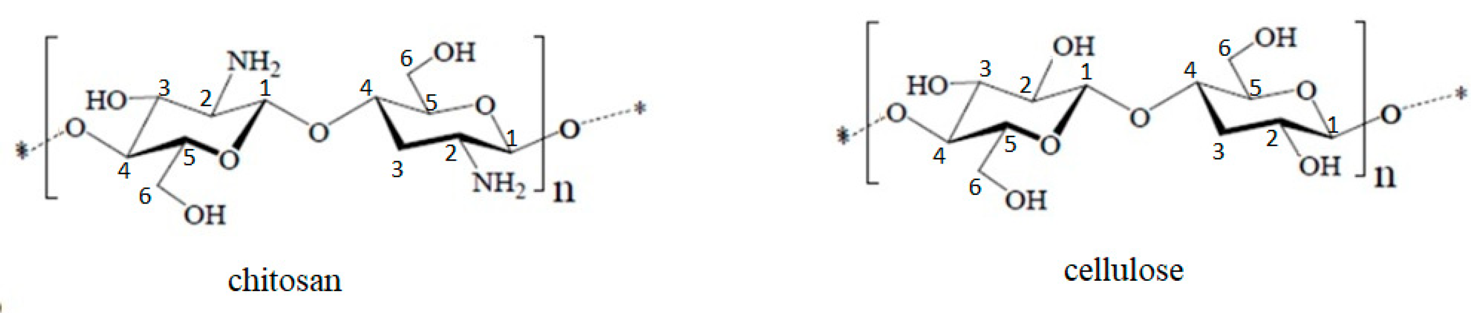
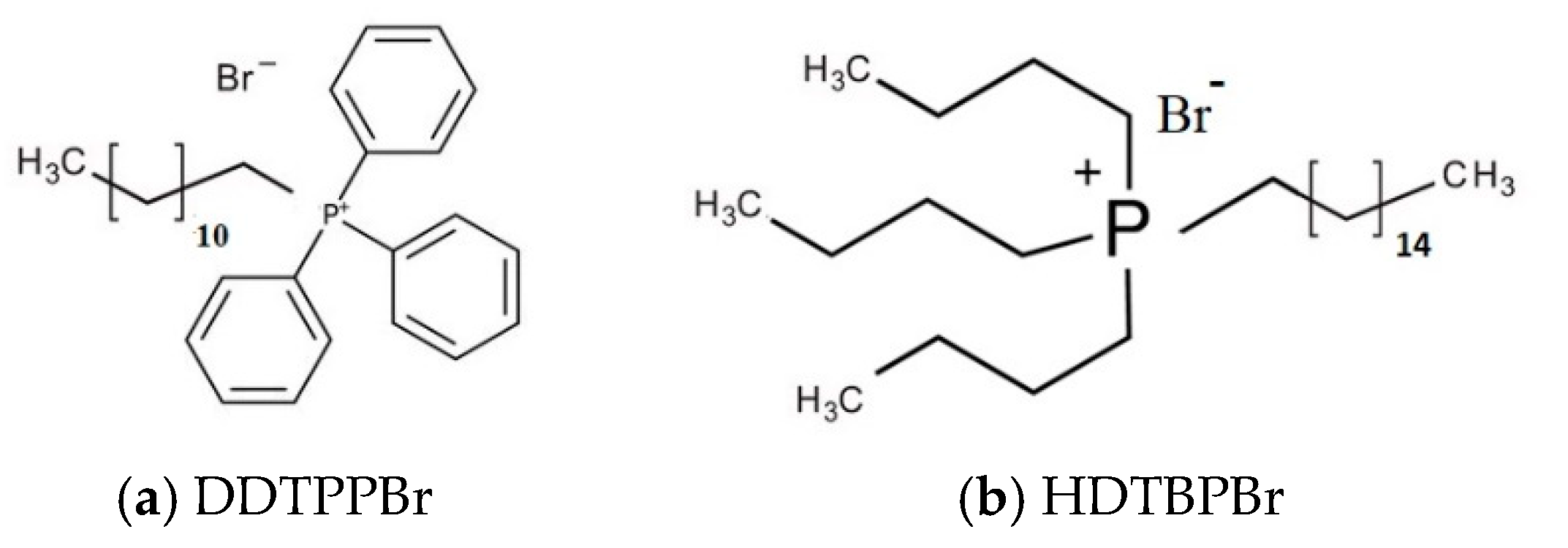
2.1. Materials Preparation and Characterization

To prepare the materials used in present study, two different support materials were used: chitosan (Ch, having a deacetilation degree of 90%, and a molecular weight of 1–3 × 10⁵, purchased from Acros Organics, Janssen Pharmaceuticalaan, Geel Belgium) and cellulose powder (Cel-powder, having the commercial name of Avicel PH-101, purchased from Sigma-Aldrich, Merck, KGaA, Darmstadt, Germany). As compounds with antimicrobial activity, two phosphonium salts were used: dodecyl-triphenyl phosphonium bromide (98% purity, purchased from Thermo Fisher, Kandel, Germany) and tri-n-butyl-hexadecyl phosphonium bromide (98% purity, purchased from ThermoFischer, Kandel, Germany). The method used for further functionalization of the used supports was the dry solvent impregnated method (SIR) [49,50,51,52]. Accordingly, with the SIR method, materials used for Cel and Ch functionalization (DDTPPBr HDTBPBr) were dissolved in distilled water. Further, the obtained solutions were mixed with solid support and kept in contact for 24 h at 298 K. After that, all samples are filtered, washed with DI water, and the obtained material was dried for 24 h at 323 K [47,48,50,51]. In this way, different materials were prepared by varying the ratio between the support and antimicrobial compound (ratios of support to antimicrobial material = 1:0.012, 1:0.025, 1:0.050, 1:0.075, 1:0.1, 1:0.2, 1:0.3, 1:0.4, 1:0.5). After preparation, all the obtained materials were characterized by using Fourier transform infrared spectroscopy (FT-IR, Bruker, Billerica, MA, U.S.A.).

2.2. Preparation of Bacterial Cultures Used during Antibacterial Tests

During antibacterial tests, we studied the dependence between exhibited antibacterial efficiency and the ratio of the support to the antimicrobial agent. After preparation, each material was transferred into the Petri dish, where the microbial culture was further added. During each test, we used a bacterial suspension with an initial concentration of 1 × 10⁸ colony-forming units (CFU). In order to highlight the antimicrobial activity against Gram-negative bacteria, Esherichia coli ATCC 25,922 and Pseudomonas aeruginosa ATCC 27,853 were used as the inocula. Antimicrobial activity against Gram-positive bacteria was determined by using the Staphylococcus aureus ATCC 25,923 strain. All tests were performed in triplicate.
Bacterial cultures were obtained by the inoculation of a solid non-selective nutritive media with 1 μL (approximatively 1 × 10⁵ CFU) bacterial suspension. This media was cast in Petri plates and incorporated with 0.2 g from the material to be tested. For all seeding processes, complete dehydrated culture media were used—Plate Count Agar purchased from Merck (peptone from casein 5.0 g L−1, yeast extract 2.5 g L−1, D (+) glucose 1.0 g L−1, agar 14.0 g L−1). For each set of experiments, a Petri plate was inoculated with a blank sample (M0—control sample), consisting only of culture media and 1 μL of the bacterial inoculum. We simultaneously inoculated two different Petri plates containing culture media, the bacterial inoculum and un-functionalized support material (M1—Ch, and, respectively, M2—Cel). All Petri plates were incubated for 48 h at 310 K, after which we counted the number of colony forming units developed on the plates surface by using YUL automatic colony counter (YUL instruments SA, Barcelona, Spain). The efficiency of the functionalized chitosan and cellulose against the reference strain was expressed as the bacterial inhibition rate [19,21,30], expressed as the ratio between colony formation units counted on the functionalized material over the number of colony formation units, as can be seen in Equation (1):
R . I . = [ 1 C F U t e s t C F U c o n t r o l ] 100
CFUcontrol = number of colony formation units on the control plate
CFUtest = number of colony formation units on the test plate.

3. Results and Discussions

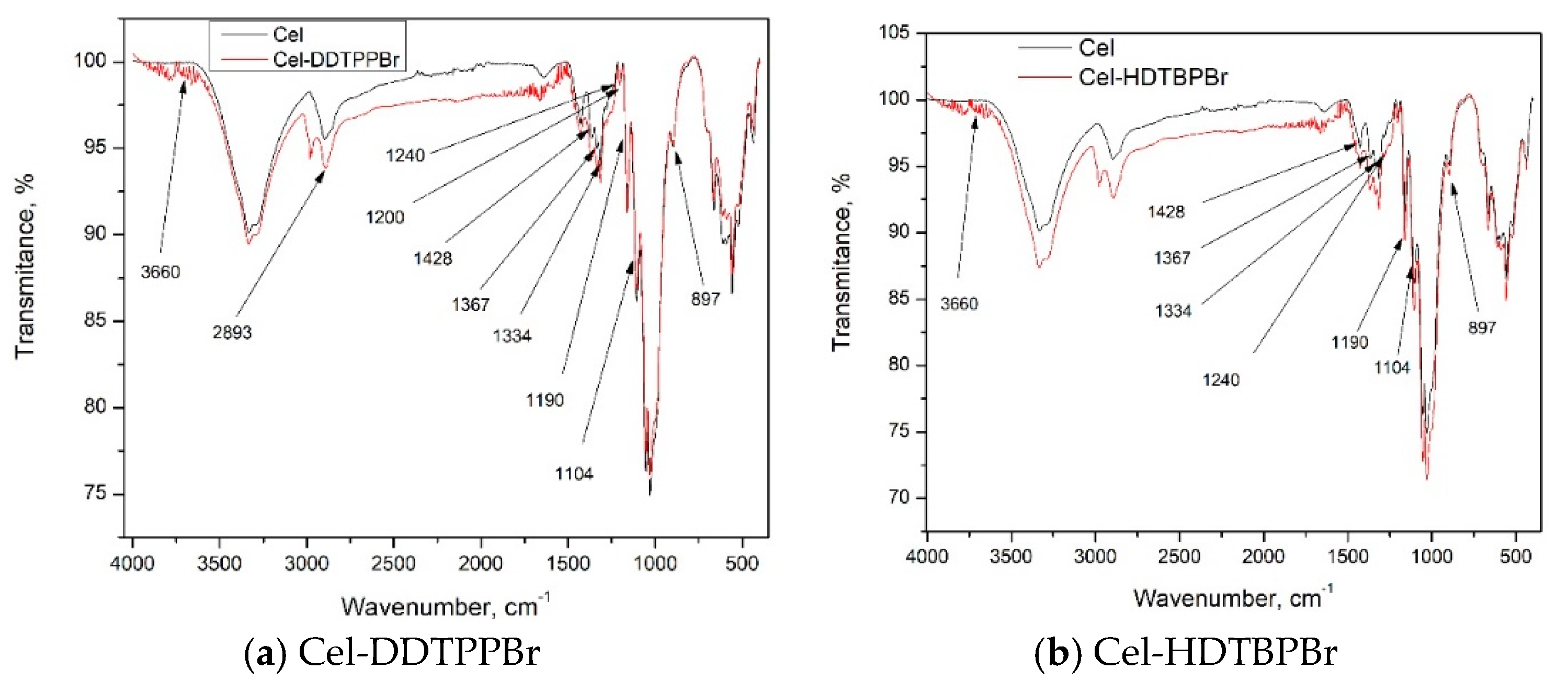
3.1. Characterization of Prepared Material by FT-IR

The obtained materials were characterized by Fourier transform infrared spectroscopy to highlight the presence of pendant groups associated with the presence of DDTPPBr and HDTBPBr, concomitant with the presence of Ch- and Cel-specific groups. Obtained spectra are depicted in Figure 3 (spectra recorded for Ch-DDTPPBr and Ch-HDTBPBr) and Figure 4 (spectra recorded for Cel-DDTPPBr and Cel-HDTBPBr).
From the data depicted in Figure 3, we can observe that the Ch-specific bands are attenuated, which can be associated with the surface functionalization with DDTPPBr or HDTBPB [47]. From Figure 3a, we can observe the presence of the P–O–Ar-specific vibrations located at wave numbers between 1240 and 1190 cm−1. We can also observe the presence of a specific peak for C–O from the DDTPPBr phenyl group located at 1200 cm−1. From the spectrum recorded for Ch-HDTBPBr (Figure 3b), we can observe the presence of the vibrations specific to the alkyl phosphate groups, located at 1180–1150 and 1080 cm−1.
Based on the spectra depicted in Figure 4, we can observe that cellulose specific bands are attenuated, which can be associated with the support functionalization with DDTPPBr or HDTBPBr [48]. In both spectra, we can observe the presence of P–O–Ar specific vibrations located between 1240 and 1190 cm−1. The peak located at 1200 cm−1 is specific for the elongation vibrations of the C–O bond from the DDTPPBr phenyl group [53]. Specific bands for Ch and Cel were explained in detail in previous published papers [47,54] and are briefly presented in Table 1.
By comparing the FT-IR spectra recorded for functionalized Ch with the control spectrum (and corroborating them with the data depicted in Table 1), we can observe a small displacement of the vibrations associated with the stretching of the chitosan hydroxilic groups. Such behavior is associated with the formation of physical intermolecular bonds (hydrogen bonds) between compounds used for functionalization and chitosan. A similar behavior is observed in the case of functionalized Cel, when the small displacements of the hydroxilic groups vibrations were associated with the formation of hydrogen bonds between the support and used antimicrobial agent. Based on this observation, we can conclude that the two used polymers were successfully functionalized with DDTPPBr and HDTBPBr.

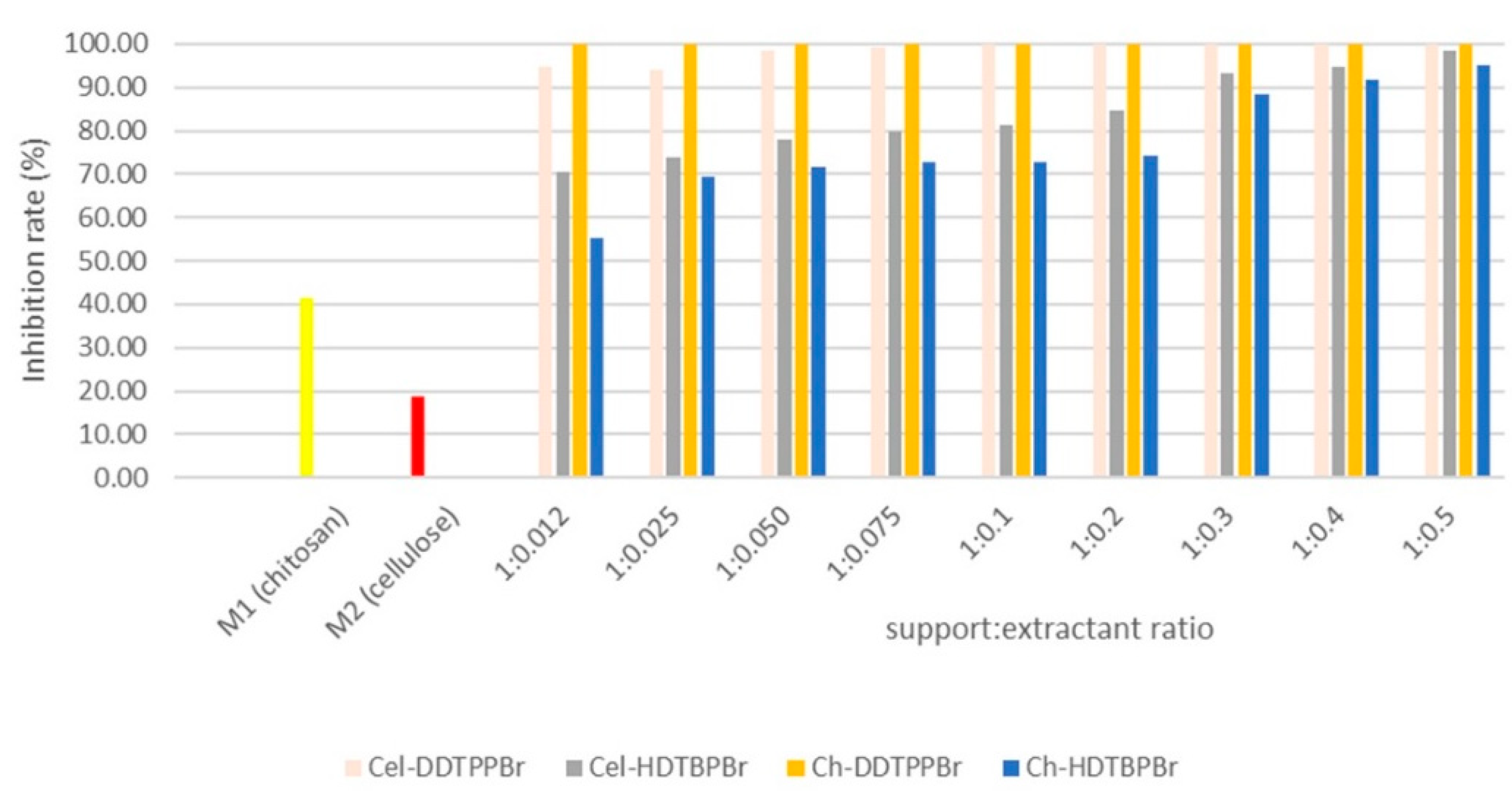
3.2. Antimicrobial Effect of Prepared Materials

3.2.1. Case of a Heterotrophic Inoculum

Phosphonium salts used for cellulose and chitosan functionalization differ both in the length of the alkyl chain (dodecyl and hexadecyl) grafted on the chitosan or cellulose base structure, as well as through the quaternizing substituent (triphenyl or tributyl).
Regardless of the phosphonium salt used for the support functionalization, we can conclude that the antimicrobial activity of the obtained materials is directly proportional to the increase in the functionalization ratio (as can be observed from Figure 5).
From the data depicted in Figure 5, we can observe a different behavior in the case of the Ch-DDTPPBr material, when the highest inhibition rate was obtained, even at the lowest functionalization ratio, meaning that is not necessary to further increase the functionalization ratio. Such behavior can be explained if we consider that in the heterotroph inoculum used during experiments, Gram-negative bacteria are predominant because such microorganisms present a higher sensibility at the antimicrobial agent’s action. The different behavior of Cel-HDTBPBr against the E.coli strain can be explained if we consider that the physical bonds established during cellulose functionalization are not so strong. So, the antimicrobial agent can be easily attached to the cellular wall, affecting the membrane permeability, leading to cell death. As well, the behavior observed in the case of Cel-HDTBPBr against the heterotroph inoculum is associated with the cumulative antibacterial effect of cellulose and the used phosponium salt.

3.2.2. Case of Reference Strains

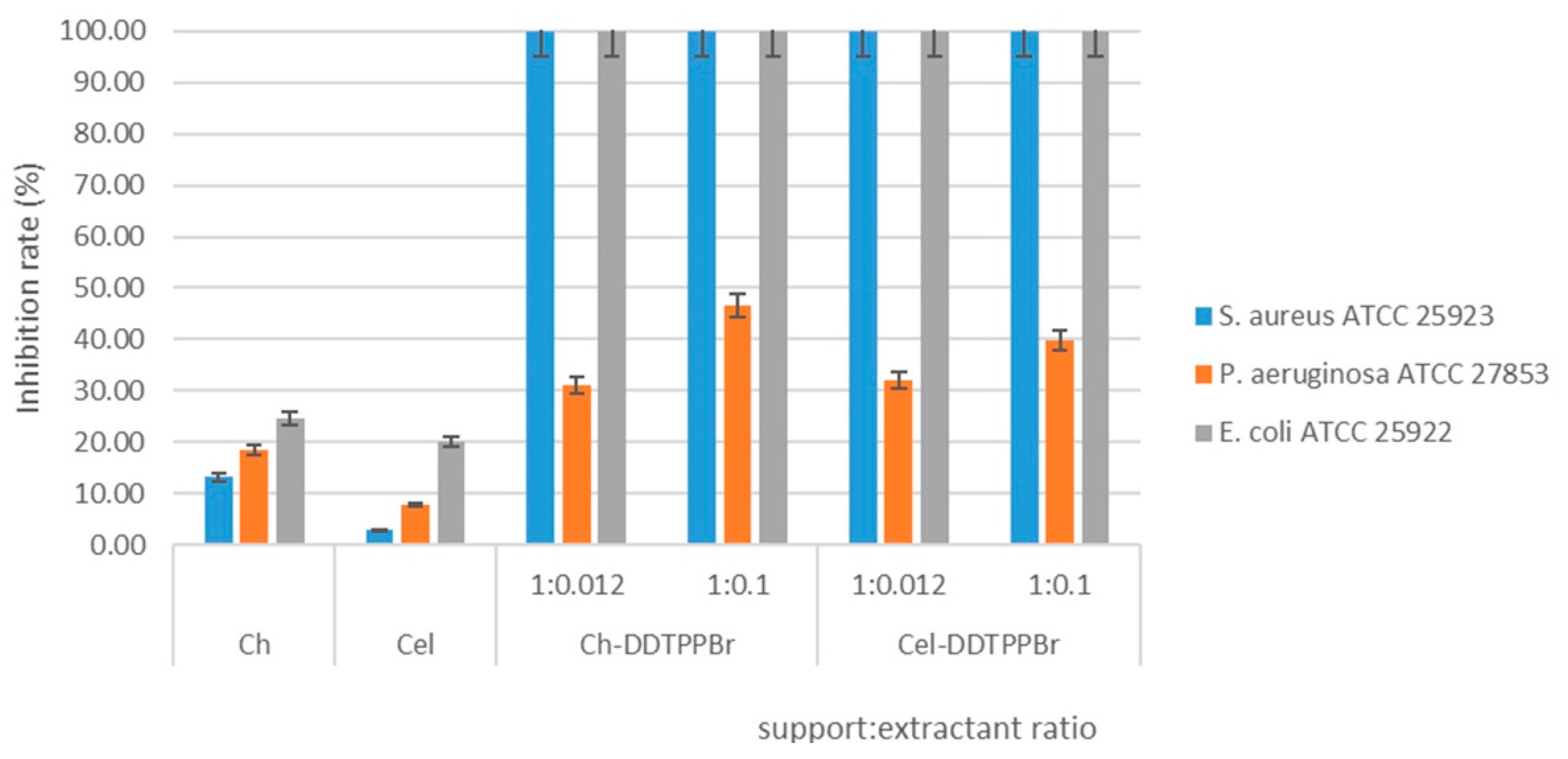
Antimicrobial Effect of Ch-DDTPPBr and Cel-DDTPPBr

From the data presented in Figure 6, we can observe an inhibition effect of 100% when Ch-DDTPPBr and Cel-DDTPPBr plates were inoculated with S. aureus ATCC 25,923 and E. coli ATCC 25,922 strains, regardless of the functionalization ratio. Likewise, we can observe a limited antimicrobial activity (maximum 46.6%) in the case of the P. aeruginosa ATCC 27,853 strain, regardless of the functionalization ratio.
In the case of Cel-DDTPPBr plates inoculated with the P. aeruginosa strain, we observed an insignificant increase in antimicrobial activity (from 32% to 39.8%) by increasing ten-fold the functionalization ratio. By contrast, in the case of Ch-DDTPPBr, a ten-fold functionalization rate increase led to an increase in antimicrobial activity from 31% to 46.6%. A possible explanation of such behavior is represented by the lower permeability of the P. aeruginosa cell wall [55] being correlated with its great adaptability to the antimicrobial agent action, which confers a greater resistance [56]. The DDTPPBr antibacterial agent used for functionalization, despite its strong hydrophobic character, was not able to penetrate the cellular wall of P. aeruginosa, explaining in this way the lower antimicrobial activity. Based on the data presented in Figure 5, we can observe that the best antimicrobial activity was exhibited by the materials obtained from chitosan functionalization. Such behavior can be explained only if we take into account the intrinsic antimicrobial activity of chitosan [13]. A significant number of researchers proved that the adsorption and interaction with the bacterial cellular membrane, followed by perturbation of the membrane selectivity and further by cell death, is favored by the presence of a positive charge on the biocide surface [3,16,18,57].

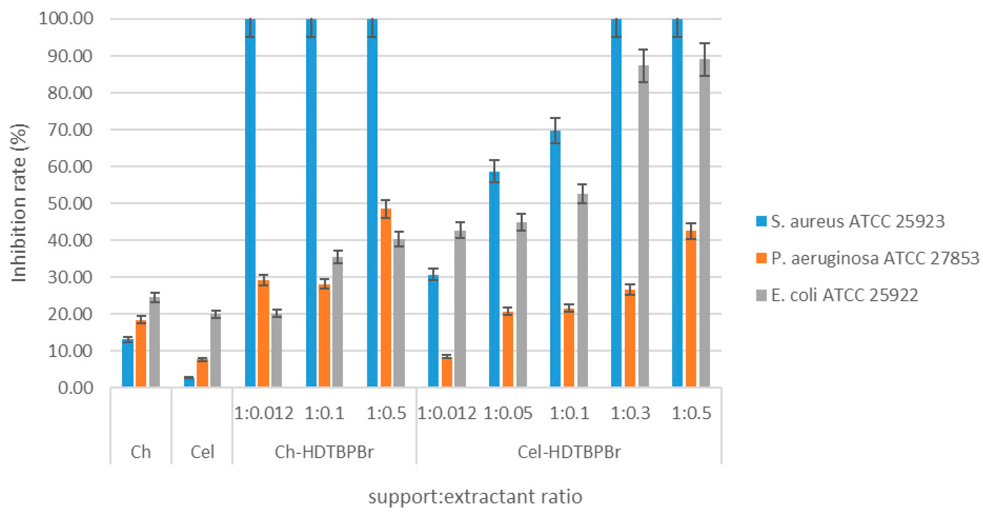
Antimicrobial Effect of Ch-HDTBPBr and Cel-HDTBPBr

In Figure 7 are presented the data regarding the antimicrobial effect of Ch-HDTBPBr and Cel-HDTBPBr against S. aureus, E. coli, and P. aeruginosa strains. Based on the obtained data, we can observe that the antimicrobial effect of Ch-based materials against S. aureus is complete, regardless of the functionalization ratio.
Gram-positive bacteria present a thick peptidoglycanic wall, enabling a strong interaction between the cationic phosphonium salt (HDTBPBr) and anionic component from the bacterial membrane [57,58]. Such interactions are possible due to the presence of teichoic acids in the cellular membrane of Gram-positive bacteria [58,59,60].
Regarding the antimicrobial activity of new produced materials against Gram-positive bacteria, based on the data presented in Figure 7, we can observe that in the case of Ch-HDTBPBr, the antibacterial activity is proportional to the functionalization ratio. However, it was not possible to reach an inhibition degree of 100%. In this case, we can say that the tested material only has a bacteriostatic effect [13,28]. It is possible that the material affected the permeability of the cellular membrane but was unable to obtain the equilibrium needed to destroy the cellular membrane and to cause bacterial death [61,62,63]. When the cellulose was functionalized with HDTBPBr, it was observed that the inhibition rate is proportional with the functionalization ratio. Total bactericidal effect was obtained against the S. aureus strain for a functionalization ratio Cel:HDTBPBr = 1:0.3. By performing the antibacterial tests against Gram-negative bacteria, it was not possible to obtain a total bactericidal effect. When the prepared cellulosic materials were tested against the P. aeruginosa strain, it was observed that the inhibition effect increased from 6% to 21.6% when the functionalization ratio increased from 1:0.01 to 1:0.1. Further, by increasing the functionalization ratio from 1:0.1 to 1:0.5, we observed an increase in the antimicrobial inhibition rate from 21.6% to 42.6%. A similar behavior was observed against the E. coli strain; when the functionalization ratio increased from 0.01 to 0.5, the inhibition rate increased from 42.7% to 89.1%. The different behavior of Cel-HDTBPBr, against the E. coli strain, can be explained if we consider that the physical bonds established during cellulose functionalization are not so strong, so the antimicrobial agent can easily attach to the cellular wall, affecting the membrane permeability. Additionally, this significant antibacterial effect can be associated with the cumulative antibacterial effect of cellulose and the used phosphonium salt.
Among the studied agents with antimicrobial activity, DDTPPBr presents the highest hydrophobicity, due to the presence of the phenyl substituent, which has a higher hydrophobicity compared with the butyl substituent from HDTBPBr. Additionally, the shortest alkyl segment (dodecyl) is equivalent to a lower distance between the support surface and the pendant group, which can lead to a considerable increase in the antimicrobial effect of the prepared materials. All these observations are consistent with the literature, which emphasizes the role of the hydrophilic–hydrophobic equilibrium against the bacteria cellular wall, and the strong relationship between the hydrophobicity of an antibacterial agent and the bactericidal effect [57,64,65,66]. In addition, an important role regarding the antimicrobial activity of different materials is played by the electrostatic attractions exerted between DDTPPBr, HDTBPBr and the bacterial cell membrane. Strong electrostatic attractions between the antimicrobial agents and bacterial cell membrane lipids lead to bacteria cells death [66,67]. Starting from this, it is possible to better understand the bactericide effect of materials obtained by cellulose functionalized with HDTBPBr against the E. coli strain. This material can achieve strong electrostatic attractions [68,69], strong intra- and intermolecular hydrogen bonds [3,5,43], which finally lead to a higher bacterial inhibition rate.

4. Conclusions

Cellulose and chitosan are two biopolymers with a higher biocompatibility, being non-toxic compounds with a large number of applications into the biomedical field. These polymers present similar structures, formed by the same type of β-glyosidic bonds between monomeric units. Regarding the chemical behavior of these polymers, cellulose is a neutral or anionic polysaccharide, while chitosan is a neutral or cationic polysaccharide. Such a difference can lead to different behavior against bacteria.
Materials obtained by the functionalization of studied polymers with DDTPPBr exhibited a total bactericidal effect against Gram-positive bacteria and against the E. coli strain. Similarly, materials obtained by the functionalization of studied natural polymers with HDTBPBr exhibited a maximal bactericidal effect only against Gram-positive bacteria. The interaction of cationic components from the polymer structures with the negatively charged components from the bacterial citoplasmatic membranes represents an important step in terms of antibacterial effect.
Based on the hydrophobicity aspect, we proved that by increasing the hydrophobicity of cationic biocides, the possibility that they interact with the citoplasmatic membrane is increased. This observation is in concordance with the experimental data from the present study, in which the best antimicrobial effects were observed in the case of materials obtained by functionalization with DDTPPBr, which present the highest hydrophobicity.
Due to the environmental regulation, and lack of by-products, the functionalization technique is the preferred one for the preparation of new materials with improved properties and large applicability, starting from synthetic or natural polymers.

Author Contributions

Conceptualization, N.S.N., A.N. and N.D.; investigation, C.A., M.C., V.F.M. and D.M.; validation, C.M.D. and P.N.; writing —original draft preparation, N.S.N., A.N. and N.D.; writing—review and editing, N.S.N., A.N. and N.D. All authors have read and agreed to the published version of the manuscript.

Funding

This research received no external funding.

Institutional Review Board Statement

Not applicable.

Informed Consent Statement

Not applicable.

Conflicts of Interest

The authors declare no conflict of interest.

References

  1. Cheba, B.A. Chitin and Chitosan: Marine Biopolymers with Unique Properties and Versatile Applications. Glob. J. Biotechnol. Biochem. 2011, 6, 149–153. [Google Scholar]
  2. Fernandes, S.C.; Freire, C.S.; Silvestre, A.J.; Neto, C.P.; Gandini, A. Novel materials based on chitosan and cellulose. Polym. Int. 2011, 60, 875–882. [Google Scholar] [CrossRef]
  3. Khattak, S.; Wahid, F.; Liu, L.-P.; Jia, S.-R.; Chu, L.-Q.; Xie, Y.-Y.; Li, Z.-X.; Zhong, C. Applications of cellulose and chitin/chitosan derivatives and composites as antibacterial materials: Current state and perspectives. Appl. Microbiol. Biotechnol. 2019, 103, 1989–2006. [Google Scholar] [CrossRef] [PubMed]
  4. Trache, D.; Hussin, M.H.; Chuin, C.T.H.; Sabar, S.; Fazita, M.R.N.; Taiwo, O.F.A.; Hassan, T.M.; Haafiz, M.K.M. Microcrystalline cellulose: Isolation, characterization and bio-composites application—A review. Int. J. Biol. Macromol. 2016, 93, 789–804. [Google Scholar] [CrossRef] [PubMed]
  5. Dutta, S.D.; Patel, D.K.; Lim, K.-T. Functional cellulose-based hydrogels as extracellular matrices for tissue engineering. J. Biol. Eng. 2019, 13, 55. [Google Scholar] [CrossRef] [PubMed]
  6. Akca, G.; Özdemir, A.; Öner, Z.G.; Şenel, S. Comparison of different types and sources of chitosan for the treatment of infections in the oral cavity. Res. Chem. Intermed. 2018, 44, 4811–4825. [Google Scholar] [CrossRef]
  7. Kaur, S.; Dhillon, G.S. The versatile biopolymer chitosan: Potential sources, evaluation of extraction methods and applications. Crit. Rev. Biotechnol. 2013, 35, 155–175. [Google Scholar] [CrossRef]
  8. Kostag, M.; El Seoud, O.A. Sustainable biomaterials based on cellulose, chitin and chitosan composites—A review. Carbohydr. Polym. Technol. Appl. 2021, 2, 100079. [Google Scholar] [CrossRef]
  9. Jiménez-Gómez, C.P.; Cecilia, J.A. Chitosan: A Natural Biopolymer with a Wide and Varied Range of Applications. Molecules 2020, 25, 3981. [Google Scholar] [CrossRef]
  10. Luo, Y.; Wang, Q. Recent Advances of Chitosan and Its Derivatives for Novel Applications in Food Science. J. Food Processing Beverages 2013, 1, 13. [Google Scholar]
  11. Ambaye, T.G.; Vaccari, M.; Prasad, S.; van Hullebusch, E.D.; Rtimi, S. Preparation and applications of chitosan and cellulose composite materials. J. Environ. Manag. 2022, 301, 113850. [Google Scholar] [CrossRef] [PubMed]
  12. Phisalaphong, M.; Jatupaiboon, N. Biosynthesis and characterization of bacteria cellulose–chitosan film. Carbohydr. Polym. 2008, 74, 482–488. [Google Scholar] [CrossRef]
  13. Coma, V.; Freire, V.; Silvestre, A. Recent advances on the development of antibacterial polysaccharide-based materials. In Polysaccharides; Ramawat, K., Mérillon, J.M., Eds.; Springer: Cham, Switzerland, 2015; pp. 1751–1803. [Google Scholar]
  14. Hosseinnejad, M.; Jafari, S.M. Evaluation of different factors affecting antimicrobial properties of chitosan. Int. J. Biol. Macromol. 2016, 85, 467–475. [Google Scholar] [CrossRef] [PubMed]
  15. Xue, Y.; Xiao, H.; Zhang, Y. Antimicrobial Polymeric Materials with Quaternary Ammonium and Phosphonium Salts. Int. J. Mol. Sci. 2015, 16, 3626–3655. [Google Scholar] [CrossRef] [PubMed] [Green Version]
  16. Kanazawa, A.; Ikeda, T.; Endo, T. Polymeric phosphonium salts as a novel class of cationic biocides. VI. Antibacterial activity of fibers surface-treated with phosphonium salts containing trimethoxysilane groups. J. Appl. Polym. Sci. 1994, 52, 641–647. [Google Scholar] [CrossRef]
  17. Tan, W.; Zhang, J.; Luan, F.; Wei, L.; Chen, Y.; Dong, F.; Li, Q.; Guo, Z. Design, synthesis of novel chitosan derivatives bearing quaternary phosphonium salts and evaluation of antifungal activity. Int. J. Biol. Macromol. 2017, 102, 704–711. [Google Scholar] [CrossRef]
  18. Xue, Y.; Pan, Y.; Xiao, H.; Zhao, Y. Novel quaternary phosphonium-type cationic polyacrylamide and elucidation of dual-functional antibacterial/antiviral activity. RSC Adv. 2014, 4, 46887–46895. [Google Scholar] [CrossRef]
  19. Csóka, L.; Božanić, D.K.; Božanić, D.K.; Nagy, V.; Dimitrijević-Branković, S.; Luyt, A.S.; Grozdits, G.; Djoković, V. Viscoelastic properties and antimicrobial activity of cellulose fiber sheets impregnated with Ag nanoparticles. Carbohydr. Polym. 2012, 90, 1139–1146. [Google Scholar] [CrossRef]
  20. Sperandeo, P.; Bosco, F.; Clerici, F.; Polissi, A.; Gelmi, M.L.; Romanelli, A. Covalent Grafting of Antimicrobial Peptides onto Microcrystalline Cellulose. ACS Appl. Bio Mater. 2020, 3, 4895–4901. [Google Scholar] [CrossRef]
  21. Belbekhouche, S.; Bousserrhine, N.; Alphonse, V.; Le Floch, F.; Mechiche, Y.C.; Menidjel, I.; Carbonnier, B. Chitosan based self-assembled nanocapsules as antibacterial agent. Colloids Surf. B-Biointerfaces 2019, 181, 158–165. [Google Scholar] [CrossRef]
  22. Kanazawa, A.; Ikeda, T.; Endo, T. Polymeric phosphonium salts as a novel class of cationic biocides. III. Immobilization of phosphonium salts by surface photografting and antibacterial activity of the surface-treated polymer films. J. Polym. Sci. Part A Polym. Chem. 1993, 31, 1467–1472. [Google Scholar] [CrossRef]
  23. Raafat, D.; Sahl, H.-G. Chitosan and its antimicrobial potential—A critical literature survey. Microb. Biotechnol. 2009, 2, 186–201. [Google Scholar] [CrossRef] [PubMed] [Green Version]
  24. Ibrahim, H.M.; El-Zairy, E.M.R. Chitosan as a Biomaterial—Structure, Properties, and Electrospun Nanofibers. In Concepts, Compounds and the Alternatives of Antibacterials; Bobbarala, V., Ed.; IntechOpen: London, UK, 2015. [Google Scholar]
  25. Goy, R.C.; de Britto, D.; Assis, O.B.G. A Review of the Antimicrobial Activity of Chitosan. Polim.-Cienc. E Tecnol. 2009, 19, 241–247. [Google Scholar] [CrossRef]
  26. Helander, I.M.; Nurmiaho-Lassila, E.L.; Ahvenainen, R.; Rhoades, J.; Roller, S. Chitosan disrupts the barrier properties of the outer membrane of Gram-negative bacteria. Int. J. Food Microbiol. 2001, 71, 235–244. [Google Scholar] [CrossRef]
  27. Kabanov, V.L.; Novinyuk, L.V. Chitosan application in food technology: A review of recent advances. Food Syst. 2020, 3, 10–15. [Google Scholar] [CrossRef] [Green Version]
  28. Morin-Crini, N.; Lichtfouse, E.; Torri, G.; Crini, G. Fundamentals and Applications of Chitosan. Sustain. Agric. Rev. 2019, 35, 49–123. [Google Scholar]
  29. Tian, B.; Liu, Y. Chitosan-based biomaterials: From discovery to food application. Polym. Adv. Technol. 2020, 31, 2408–2421. [Google Scholar] [CrossRef]
  30. Luan, F.; Wei, L.; Zhang, J.; Mi, Y.; Dong, F.; Li, Q.; Guo, Z. Antioxidant Activity and Antifungal Activity of Chitosan Derivatives with Propane Sulfonate Groups. Polymers 2018, 10, 395. [Google Scholar] [CrossRef] [Green Version]
  31. Zhang, Y.; Zhang, M. Three-dimensional macroporous calcium phosphate bioceramics with nested chitosan sponges for load-bearing bone implants. J. Biomed. Mater. Res. 2002, 61, 1–8. [Google Scholar] [CrossRef]
  32. Cheung, R.C.; Ng, T.B.; Wong, J.H.; Chan, W.Y. Chitosan: An Update on Potential Biomedical and Pharmaceutical Applications. Mar. Drugs 2015, 13, 5156–5186. [Google Scholar] [CrossRef]
  33. Patrulea, V.; Ostafe, V.; Borchard, G.; Jordan, O. Chitosan as a starting material for wound healing applications. Eur. J. Pharm. Biopharm. 2015, 97, 417–426. [Google Scholar] [CrossRef] [PubMed] [Green Version]
  34. Moeini, A.; Pedram, P.; Makvandi, P.; Malinconico, M.; Gomez d’Ayala, G. Wound healing and antimicrobial effect of active secondary metabolites in chitosan-based wound dressings: A review. Carbohydr. Polym. 2020, 233, 115839. [Google Scholar] [CrossRef] [PubMed]
  35. Mohammed, M.; Shukur, K.; Haj, N. Preparation and Bioactivity Assessment of Chitosan-1-Acetic Acid-5-Flurouracil Conjugates as Cancer Prodrugs. Molecules 2017, 22, 1629. [Google Scholar] [CrossRef] [PubMed] [Green Version]
  36. Huan, L.; Xiao, L.; Yang, G. Chitosan Application in Textile Processing. Curr. Trends Fash. Technol. Text. Eng. 2018, 4, 555635. [Google Scholar]
  37. Olivera, S.; Muralidhara, H.B.; Venkatesh, K.; Guna, V.K.; Gopalakrishna, K.; Kumar, K.Y. Potential applications of cellulose and chitosan nanoparticles/composites in wastewater treatment: A review. Carbohydr. Polym. 2016, 153, 600–618. [Google Scholar] [CrossRef]
  38. Wilts, E.M.; Herzberger, J.; Long, T.E. Addressing water scarcity: Cationic polyelectrolytes in water treatment and purification. Polym. Int. 2018, 67, 799–814. [Google Scholar] [CrossRef]
  39. Lavoine, N.; Desloges, I.; Dufresne, A.; Bras, J. Microfibrillated cellulose—Its barrier properties and applications in cellulosic materials: A review. Carbohydr. Polym. 2012, 90, 735–764. [Google Scholar] [CrossRef]
  40. Ullah, H.; Wahid, F.; Santos, H.A.; Khan, T. Advances in biomedical and pharmaceutical applications of functional bacterial cellulose-based nanocomposites. Carbohydr. Polym. 2016, 150, 330–352. [Google Scholar] [CrossRef]
  41. Song, Y.; Zhang, J.; Gan, W.; Zhou, J.; Zhang, L. Flocculation Properties and Antimicrobial Activities of Quaternized Celluloses Synthesized in NaOH/Urea Aqueous Solution. Ind. Eng. Chem. Res. 2010, 49, 1242–1246. [Google Scholar] [CrossRef]
  42. George, J.; Sabapathi, N.S. Cellulose nanocrystals: Synthesis, functional properties, and applications. Nanotechnol. Sci. Appl. 2015, 8, 45–54. [Google Scholar] [CrossRef] [Green Version]
  43. Habibi, Y.; Lucia, L.A.; Rojas, O.J. Cellulose Nanocrystals: Chemistry, Self-Assembly, and Applications. Chem. Rev. 2010, 110, 3479–3500. [Google Scholar] [CrossRef] [PubMed]
  44. Koshani, R.; Tavakolian, M.; van de Ven, T.G.M. Cellulose-based dispersants and flocculants. J. Mater. Chem. B 2020, 8, 10502–10526. [Google Scholar] [CrossRef] [PubMed]
  45. Fauzani, D.; Notodarmojo, S.; Handajani, M.; Helmy, Q.; Kardiansyah, T. Cellulose in natural flocculant applications: A review. J. Phys. Conf. Ser. 2021, 2047, 012030. [Google Scholar] [CrossRef]
  46. Terhi, S. Functionalized Nanocelluloses in Wastewater Treatment Applications. Ph.D. Thesis, Faculty of Technology, University of Oulu: Tampere, Finland, 2015. [Google Scholar]
  47. Ardean, C.; Davidescu, C.M.; Nemeş, N.S.; Negrea, A.; Ciopec, M.; Duteanu, N.; Negrea, P.; Duda-Seiman, D.; Muntean, D. Antimicrobial Activities of Chitosan Derivatives. Pharmaceutics 2021, 13, 1639. [Google Scholar] [CrossRef] [PubMed]
  48. Nemes, N.S.; Ardean, C.; Davidescu, C.M.; Negrea, A.; Ciopec, M.; Duteanu, N.; Negrea, P.; Paul, C.; Duda-Seiman, D.; Muntean, D. Antimicrobial activity of cellulose based materials. Polymers 2022, 14, 735. [Google Scholar] [CrossRef] [PubMed]
  49. Cortina, J.L.; Miralles, N.; Sastre, A.; Aguilar, M.; Profumo, A.; Pesavento, M. Solvent-impregnated resins containing di-(2,4,4-trimethylpentyl) phosphonic acid I. Comparative study of di-(2,4,4-trimethylpentyl)phosphinic acid adsorbed into Amberlite XAD-2 and dissolved in toluene. React. Polym. 1993, 21, 89–101. [Google Scholar] [CrossRef]
  50. Cortina, J.L.; Miralles, N.; Sastre, A.M.; Aguilar, M.; Profumo, A.; Pesavento, M. Solvent-impregnated resins containing di-(2,4,4-trimethylpentyl) phosphinic acid II. Study of the distribution equilibria of Zn(II), Cu(II) and Cd(II). React. Polym. 1993, 21, 103–116. [Google Scholar] [CrossRef]
  51. Cortina, J.L.; Miralles, N.; Aguilar, M.; Sastre, M. Solvent impregnated resins containing di(2-ethylhexyl)phosphoric acid. I. Preparation and study of the retention and distribution of the extractant on the resin. Solvent Extr. Ion Exch. 1994, 12, 349–369. [Google Scholar] [CrossRef]
  52. Kabay, N.; Cortina, J.L.; Trochimczuk, A.; Streat, M. Solvent-impregnated resins (SIRs)—Methods of preparation and their applications. React. Funct. Polym. 2010, 70, 484–496. [Google Scholar] [CrossRef]
  53. Hospodarova, V.; Singovszka, E.; Stevulova, N. Characterization of Cellulosic Fibers by FTIR Spectroscopy for Their Further Implementation to Building Materials. Am. J. Anal. Chem. 2018, 09, 303–310. [Google Scholar] [CrossRef] [Green Version]
  54. Negrea, A.; Gabor, A.; Davidescu, C.M.; Ciopec, M.; Negrea, P.; Duteanu, N.; Barbulescu, A. Rare Earth Elements Removal from Water Using Natural Polymers. Sci. Rep. 2018, 8, 316. [Google Scholar] [CrossRef] [PubMed] [Green Version]
  55. Lambert, P.A. Mechanisms of antibiotic resistance in Pseudomonas aeruginosa. J. R. Soc. Med. 2002, 95 (Suppl. 41), 22–26. [Google Scholar] [PubMed]
  56. Ciofu, O.; Tolker-Nielsen, T. Tolerance and Resistance of Pseudomonas aeruginosa Biofilms to Antimicrobial Agents-How P. aeruginosa Can Escape Antibiotics. Front. Microbiol. 2019, 10, 913. [Google Scholar] [CrossRef] [PubMed] [Green Version]
  57. Kumariya, R.; Sood, S.K.; Rajput, Y.S.; Saini, N.; Garsa, A.K. Increased membrane surface positive charge and altered membrane fluidity leads to cationic antimicrobial peptide resistance in Enterococcus faecalis. Biochim. Biophys. Acta (BBA)-Biomembr. 2015, 1848, 1367–1375. [Google Scholar] [CrossRef] [PubMed] [Green Version]
  58. Li, J.; Zhuang, S. Antibacterial activity of chitosan and its derivatives and their interaction mechanism with bacteria: Current state and perspectives. Eur. Polym. J. 2020, 138, 109984. [Google Scholar] [CrossRef]
  59. Rajoka, M.S.R.; Mehwish, H.M.; Wu, Y.; Zhao, L.; Arfat, Y.; Majeed, K.; Anwaar, S. Chitin/chitosan derivatives and their interactions with microorganisms: A comprehensive review and future perspectives. Crit. Rev. Biotechnol. 2020, 40, 365–379. [Google Scholar] [CrossRef]
  60. Raafat, D.; von Bargen, K.; Haas, A.; Sahl, H.-G. Insights into the Mode of Action of Chitosan as an Antibacterial Compound. Appl. Environ. Microbiol. 2008, 74, 3764–3773. [Google Scholar] [CrossRef] [Green Version]
  61. Kong, M.; Chen, X.G.; Xing, K.; Park, H.J. Antimicrobial properties of chitosan and mode of action: A state of the art review. Int. Journal Food Microbiol. 2010, 144, 51–63. [Google Scholar] [CrossRef] [PubMed]
  62. Mengatto, L.; Ferreyra, M.G.; Rubiolo, A.; Rintoul, I.; Luna, J. Hydrophilic and hydrophobic interactions in cross-linked chitosan membranes. Mater. Chem. Phys. 2013, 139, 181–186. [Google Scholar] [CrossRef]
  63. Hanpanich, O.; Wongkongkatep, P.; Pongtharangkul, T.; Wongkongkatep, J. Turning hydrophilic bacteria into biorenewable hydrophobic material with potential antimicrobial activity via interaction with chitosan. Bioresour. Technol. 2017, 230, 97–102. [Google Scholar] [CrossRef]
  64. Krasowska, A.; Sigler, K. How microorganisms use hydrophobicity and what does this mean for human needs? Front. Cell. Infect. Microbiol. 2014, 4, 112. [Google Scholar] [CrossRef] [PubMed] [Green Version]
  65. Palmer, J.; Flint, S.; Brooks, J. Bacterial cell attachment, the beginning of a biofilm. J. Ind. Microbiol. Biotechnol. 2007, 34, 577–588. [Google Scholar] [CrossRef] [PubMed]
  66. Thewes, N.; Loskill, P.; Jung, P.; Peisker, H.; Bischoff, M.; Herrmann, M.; Jacobs, K. Hydrophobic interaction governs unspecific adhesion of staphylococci: A single cell force spectroscopy study. Beilstein J. Nanotechnol. 2014, 5, 1501–1512. [Google Scholar] [CrossRef] [PubMed] [Green Version]
  67. Chen, C.; Petterson, T.; Illergård, J.; Ek, M.; Wågberg, L. Influence of Cellulose Charge on Bacteria Adhesion and Viability to PVAm/CNF/PVAm-Modified Cellulose Model Surfaces. Biomacromolecules 2019, 20, 2075–2083. [Google Scholar] [CrossRef] [PubMed]
  68. Elsabee, M.Z.; Morsi, R.E.; Al-Sabagh, A.M. Surface active properties of chitosan and its derivatives. Colloids Surf. B Biointerfaces 2009, 74, 1–16. [Google Scholar] [CrossRef] [PubMed]
  69. Sibusiso, A.; Blessing, A. Chitosan and Cellulose-Based Hydrogels for Wound Management. Int. J. Mol. Sci. 2020, 21, 9656. [Google Scholar] [CrossRef]
Figure 1. Chitosan (Ch) and cellulose (Cel) symmetrical structures. (* suggest that in picture are represented only the basic structural units)
Symmetry 14 00572 g001
Figure 2. DDTPPBr and HDTBPBr antimicrobial agents’ structures.
Symmetry 14 00572 g002
Figure 3. FT-IR spectra recorded for (a) Ch-DDTPPBr and (b) Ch-HDTBPBr materials.
Symmetry 14 00572 g003
Figure 4. FT-IR spectrum for (a) Cel-DDTPPBr and (b) Cel-HDTBPBr materials.
Symmetry 14 00572 g004
Figure 5. Inhibitory effects of chitosan (M1), cellulose (M2). Chitosan:extractants derivatives and cellulose:antimicrobial agent derivatives on growth of heterotrophic inoculum.
Symmetry 14 00572 g005
Figure 6. The antimicrobial effect observed in the case of Ch, cell, Ch-DDTPPBr and Cel-DDTPPBr materials.
Symmetry 14 00572 g006
Figure 7. The antimicrobial effect of Ch-HDTBPBr and C-HDTBPBr materials.
Symmetry 14 00572 g007
Table 1. Ch- and Cel-specific FT-IR specific vibrations bands.
GroupFT-IR Bands (cm−1)Observations
Chitosan
CH2–OH1380–1420
N–H1570
C=O1660
C–H2870; 2924
O–H3430
Cellulose (Cel)
O–H3660Large band
C–H2893Small plateau; stretching vibrations in polysaccharides
CH21428; 1367 Symmetry 14 00572 i001Vibrations specific to the crystalline structure of cellulose
C–O1334; 1158
O–C–O1104; 1027
897Amorphous region in cellulose
OH21600–900 Symmetry 14 00572 i002Water molecules vibrations
1633
Publisher’s Note: MDPI stays neutral with regard to jurisdictional claims in published maps and institutional affiliations.

