Communication

Structural and Biochemical Basis of Etoposide-Resistant Mutations in Topoisomerase IIα

by
Elizabeth G. Gibson
1,2 and
Joseph E. Deweese
1,3,4,*
1
Department of Pharmaceutical Sciences, Lipscomb University College of Pharmacy, Nashville, TN 37204, USA
2
Bristol Myers Squibb, Princeton, NJ 08453, USA
3
Department of Biological, Physical, and Human Sciences, Freed-Hardeman University, Henderson, TN 38340, USA
4
Department of Biochemistry, Vanderbilt University, Nashville, TN 37204, USA
*
Author to whom correspondence should be addressed.
Symmetry 2022, 14(7), 1309; https://doi.org/10.3390/sym14071309
Submission received: 27 May 2022 / Revised: 15 June 2022 / Accepted: 22 June 2022 / Published: 24 June 2022

Abstract

:
Etoposide is a widely used anticancer drug that targets type II topoisomerases, including topoisomerase IIα (TOP2A). TOP2A is a nuclear enzyme involved in regulating DNA topology through a double-strand passage mechanism. TOP2A is a homodimeric enzyme with two symmetrical active sites formed by residues from either half of the dimer. Both active sites cleave DNA, forming an enzyme-bound, double-stranded DNA break. Etoposide acts by binding in the active site between the ends of cleaved DNA, preventing the enzyme from ligating the DNA. In the present study, biochemical and structural data are used to examine the mechanism of etoposide resistance found with specific point mutations in TOP2A. Mutations near the active site (D463A, G534R, R487K), along with some outside of the active site (ΔA429 and P716L), are examined. We hypothesize that changes in the coordination of DNA cleavage results from mutations that impact symmetrical relationships in the active site and surrounding regions. In some cases, we report the first data on purified versions of these enzymes. Based upon our results, both local and long-distance factors can impact etoposide action and may indicate interdependent relationships in structure and function.

1. Introduction

DNA entanglements present a real threat to fundamental cellular processes such as replication, transcription, and mitosis. To overcome these challenges, cells employ a family of enzymes known as topoisomerases [1]. Type II topoisomerases are specialized enzymes that create transient double-stranded breaks in a DNA segment to pass an intact segment through the break [2]. In doing so, type II topoisomerases can detangle and relax DNA molecules. Type II topoisomerases are symmetrical enzymes, and the versions found in mammalian cells (Type IIA) are homodimers. Humans encode two Type IIA topoisomerases, known as topoisomerase IIα (TOP2A) and IIβ (TOP2B) [2].
Type IIA topoisomerases have twin active sites formed by residues from both halves of the protein [2]. For example, the active site Tyr residue from one monomer cooperates with metal-ion binding and DNA-coordinating residues from the other monomer during DNA cleavage and ligation [3,4]. Additional residues cooperate in DNA binding and in coordinating large structural movements that take place during the catalytic cycle [4]. The catalytic cycle is shown in Figure 1. Briefly, TOP2 works by binding to two segments of DNA (a DNA–DNA crossover), cutting one segment (called the gate or G-segment), passing the other segment (called the transport or T-segment) through the break in the first, and then resealing the break in the G-segment [2]. The enzyme covalently binds to the G-segment during the DNA cleavage event, which prevents the cleaved DNA from dissociating from the enzyme [2].
Due to the critical role of these enzymes in cellular growth and division, the disruption of their function has been used to kill cancer cells. Several widely used anticancer agents are known to impact the function of topoisomerase II, including etoposide and doxorubicin [5]. Etoposide has been used to examine the mechanism of the enzyme, as have etoposide-resistant mutants. Some of these mutants were discovered via mutant screens using yeast strains or human cell lines, while others were designed using structural data to examine the mechanism of the enzyme [6,7,8,9].
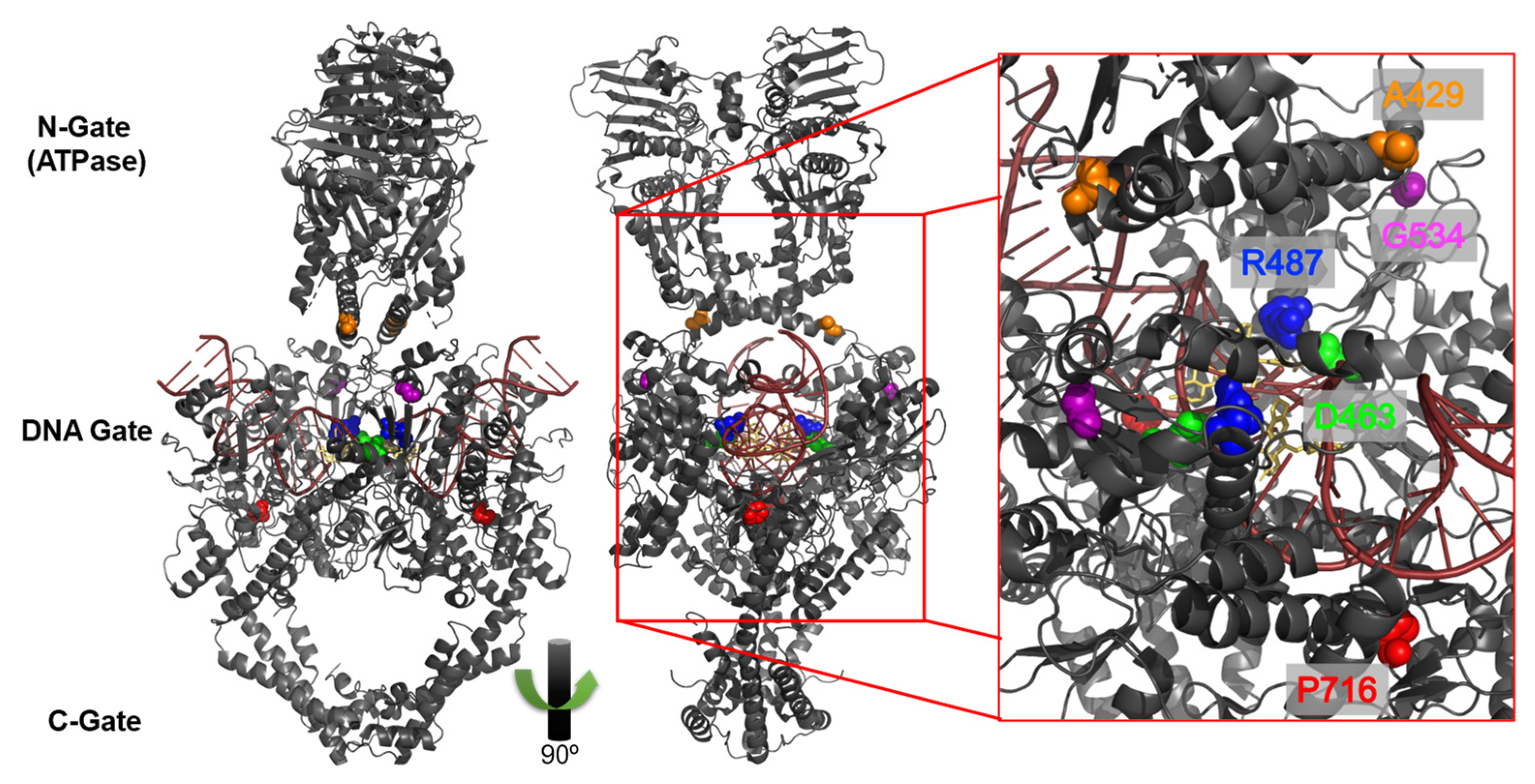
For example, Campain et al. developed two FEM-X melanoma cell lines that were highly resistant to etoposide and identified the deletion of Ala429 (ΔA429) as the key change in TOP2A [6]. This mutation is near other identified etoposide-resistant mutations, which are discussed later; however, it is not located in a highly conserved domain (Figure 1). It is located at the base of the transducer domain, which is thought to facilitate communication between the ATPase domain and the core [10]. A429 is not conserved among various groups of topoisomerase II enzymes from yeast, Drosophila, or bacteria, nor is it conserved between TOP2A and TOP2B [6]. This mutation also did not confer etoposide resistance when tested using a crude nuclear extract, which indicates that other factors are involved in the observed resistance. The mechanism of resistance was not determined and the effect this mutation had on the TOP2A catalytic cycle and enzyme coordination were not studied.
Using a yeast screen, Patel et al. identified two amsacrine-resistant TOP2A mutants that also had a threefold resistance to etoposide [7]. One of those mutants, R486K (now numbered as R487K), was specific to TOP2A as the parallel mutation in yeast TOP2 did not confer amsacrine resistance [7]. As seen in Figure 2, R487 is near the enzyme–DNA–etoposide interface. While the R487K mutant was examined for DNA cleavage, the study did not examine specific aspects of the catalytic cycle and the coordination of DNA cleavage.
In forced molecular evolution yeast screens, Leuontiou et al. identified a number of positions in human TOP2B that were mutated and conferred resistance to acridine compounds [8,9]. One position in particular, G550R, exhibited a fourfold resistance to etoposide. Interestingly, this mutation preserved DNA cleavage activity while increasing relaxation and slightly decreasing decatenation activity [9]. This position has an analogous location in TOP2A, G534. However, only the TOP2B mutation was examined. As seen in Figure 2, G534 is on an outer face away from the active site. The same lab group also identified P732L in TOP2B as resistant to both etoposide (~twofold) and mAMSA (~tenfold) [8]. While this mutant was characterized in TOP2B, the analogous P716L mutation in TOP2A was not studied. As seen in Figure 2, P716 is located below the DNA binding region and is part of the interface with the DNA gate segment (the region of DNA cleaved during the catalytic cycle).
Here, we report the purification and characterization of point mutants with altered sensitivity toward etoposide as a model for understanding the mechanism of TOP2A. The mutations we selected are based on the studies mentioned above including the following TOP2A residues: ΔA429, R487K, G534R, and P716L. In addition to these, we designed one additional mutant, D463E based upon structural evidence regarding interactions between etoposide and key residues in the active site of TOP2A (Figure 2). We examine these mutants and offer some observations regarding the catalytic mechanism and the symmetrical nature of the enzyme.

2. Materials and Methods

2.1. Enzymes and Materials

Wild-type and mutant human topoisomerase IIα (TOP2A) were expressed using the YEpWob6αHT plasmid in Saccharomyces cerevisiae JEL1Δtop1 cells and purified as described previously [12]. The enzyme was stored at −80 °C as a 1.5 mg/mL (4 µM) stock in 50 mM Tris-HCl, pH 7.7, 0.1 mM EDTA, 750 mM KCl, and 5% glycerol.
Individual point mutations in human TOP2A encoded by YEpWob6 αHT were made using the PCR-based lightning mutagenesis kit (Stratagene). R487, D463, G534, P716, and A429 were individually mutated as follows: Arg487 to Lys, Asp463 to Glu, Gly534 to Arg, Pro716 to Leu, deletion of Ala 429. Primer sequences are listed in Supplementary Table S1. In all cases, mutant TOP2A constructs were isolated and sequenced before being transformed into JEL1Δtop1 yeast cells. Mutant enzymes were expressed and purified as described above.
The substrate used in the assays was negatively supercoiled (-SC) pBR322 DNA. The pBR322 was prepared using a Plasmid Mega Kit (Qiagen) as described by the manufacturer. Etoposide (Sigma) was stored at 4 °C as a 20 mM stock solution in 100% DMSO.

2.2. Topoisomerase II-Mediated Plasmid DNA Relaxation

Relaxation assays were carried out as previously described [12]. Briefly, reaction mixtures contained 4.4 nM of the wild-type or 22–220 nM of the mutant enzymes, 5 nM-SC pBR322 DNA, and 1 mM ATP in 20 μL of 10 mM Tris-HCl, pH 7.9, 175 mM KCl, 0.1 mM NaEDTA, 5 mM MgCl2, and 2.5% glycerol. Assays were initiated upon the addition of enzyme, and reaction mixtures were incubated at 37 °C for 15 min. Reactions included 0–500 μM etoposide. Reactions were stopped using 3 μL of stop solution (77.5 mM Na2EDTA, 0.77% SDS). Samples were electrophoresed in 1% agarose gels and stained with ethidium bromide. Gels were imaged and quantified using a Bio-Rad ChemiDoc MP Imaging System and Image Lab Software (Hercules, CA, USA). Results were graphed with GraphPad Prism 9 (La Jolla, CA, USA). DNA relaxation was observed by the conversion of SC plasmid DNA to relaxed topoisomers.

2.3. Topoisomerase II-Mediated Cleavage of Plasmid DNA

Plasmid DNA cleavage reactions were carried out as described previously by Fortune and Osheroff [13]. Briefly, reaction mixtures contained 220 nM of wild-type or mutant human topoisomerase IIα and 5 nM-SC pBR322 DNA in 20 μL of 10 mM Tris-HCl, pH 7.9, 100 mM KCl, 1 mM EDTA, 5 mM MgCl2, and 2.5% glycerol. Assays were initiated by the addition of enzyme, and reaction mixtures were incubated for 6 min at 37 °C. Reactions included 0–200 μM etoposide. Reactions were stopped by the addition of 2 μL of 5% SDS followed by 2 μL of 250 mM NaEDTA, pH 8.0. Proteinase K (2 μL of a 0.8 mg/mL solution) was used to digest topoisomerase IIα. Samples were electrophoresed in 1% agarose gels in TAE buffer with ethidium bromide. Double-stranded DNA cleavage was detected by the conversion of -SC DNA to linear molecules. Single-strand breaks were assessed using the nicked band above the linear DNA band. Gels were imaged and quantified using a Bio-Rad ChemiDoc MP Imaging System and Image Lab Software (Hercules, CA, USA). Results were graphed using GraphPad Prism 9 (La Jolla, CA, USA).

3. Results

3.1. Plasmid DNA Cleavage Demonstrates Varying Levels of Etoposide-Induced Cleavage among Point Mutants

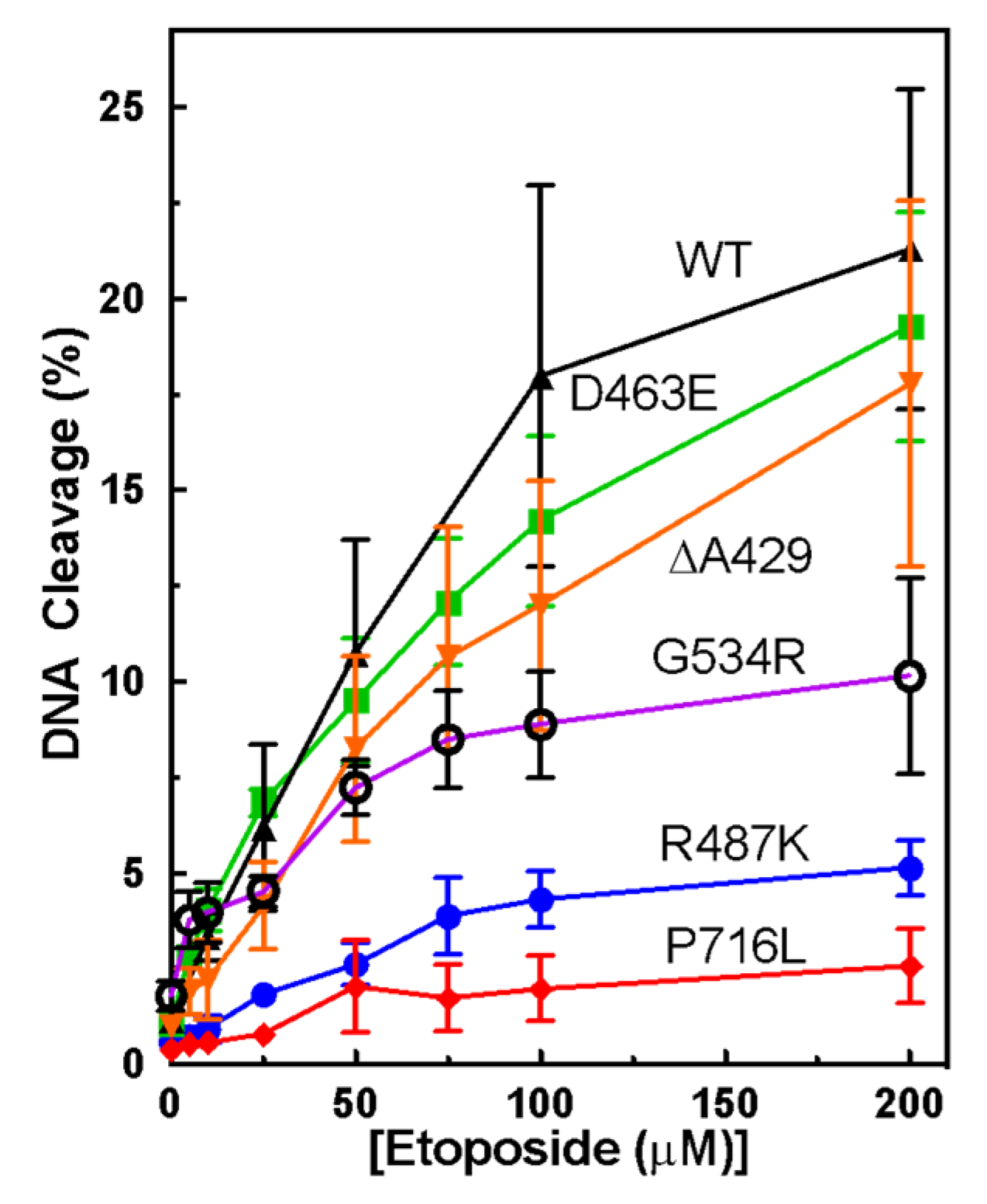
Each of the mutants was characterized using plasmid DNA cleavage assays in the presence of increasing concentrations of etoposide. As seen in Figure 3, etoposide induces around 20% DNA cleavage with WT, but each of the mutants has slightly less etoposide-enhanced DNA cleavage. While D463E and ΔA429 both remain in the range of WT, etoposide-induced DNA cleavage with G534R is about twofold less. Etoposide-induced DNA cleavage with R487K and P716L are ~fivefold and ~sevenfold less, respectively. Interestingly, high concentrations of etoposide (100–200 μM) are unable to induce increased plasmid DNA cleavage with the latter two mutants.

3.2. Measuring Etoposide-Induced Cleavage Enhancement

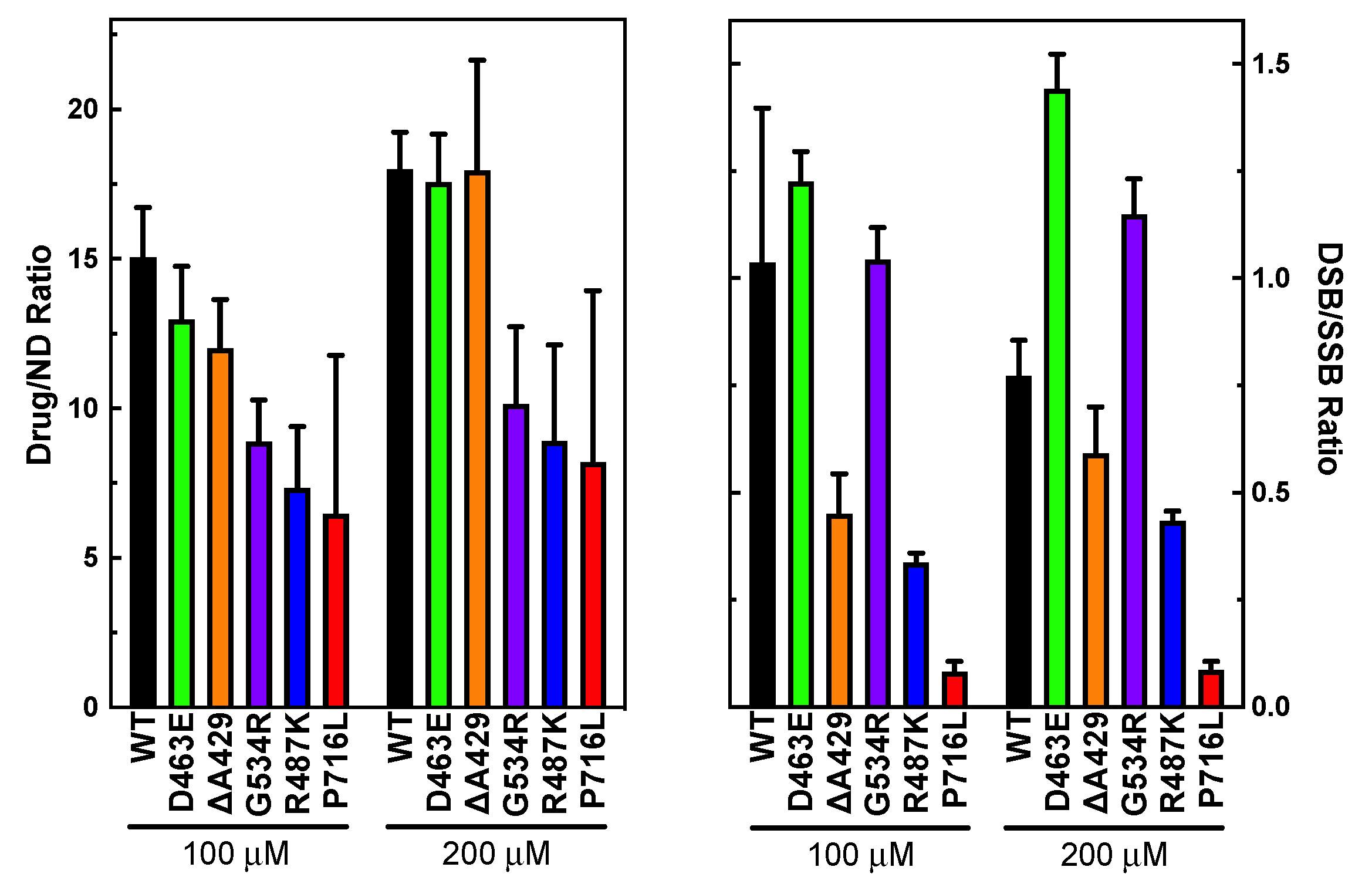
In order to determine whether the lack of enhancement was due to resistance or to a change in baseline DNA cleavage, we compared the levels of DNA cleavage in the absence and presence of 100 or 200 μM etoposide. As seen in Figure 4, the drug-to-no drug (ND) ratios are shown for WT and the mutants. Both D463E and ΔA429 are most similar to WT, which is similar to the percent DNA cleavage data. The G534R, R487K, and P716L mutants display decreased drug/ND ratios, consistent with the overall cleavage data. The resistance to etoposide seems to be ~twofold.

3.3. Examining the Level of Coordination during DNA Cleavage

Another factor we examined was whether any of the mutations increased or decreased the coordination of DNA cleavage. We measured this by taking a ratio of double-stranded to single-stranded breaks as seen in Figure 3. For WT, this ratio was near 1. D463E and G534R were both similar to WT. Interestingly, R487K, ΔA429, and P716L all displayed decreased coordination (higher level of SSB compared to DSB). P716L displayed the largest decrease in coordination.

3.4. Plasmid DNA Relaxation with Selected Mutants of TOP2A

With the mutant enzymes, we also wanted to determine if these mutations affected the overall catalytic activity of the enzyme. We utilized a DNA relaxation assay to define the time needed to fully relax a negatively supercoiled plasmid and the effect of etoposide on this activity. This assay also enables the observation of whether the enzyme is performing in a processive (fully relaxing substrates upon binding) or distributive manner (relaxing substrates in a stepwise fashion), which is a reflection of how long the enzyme remains on a substrate before dissociating. With the WT enzyme, the DNA is usually fully relaxed in 10–15 min in a highly processive manner and etoposide inhibits this process. All the mutants were able to relax DNA, although R487K and P716L were slightly slower and more distributive. R487K and G534R were less sensitive to etoposide inhibition. D463E had no change in etoposide sensitivity as compared to the WT enzyme. However, with ΔA429 and P716L, etoposide stimulated relaxation. All gels of WT and mutants plus etoposide are shown in Supplementary Figure S1 and a summary of the mutant relaxation activity is shown in Table 1.

4. Discussion

Etoposide-resistant mutations have been previously identified in biological screens, but in some cases, the mutations do not translate to etoposide resistance in a purified setting [6,7,8,9]. As seen in this current study, D463E and ΔA429 did not display major resistance in purified mutant TOP2A enzymes. On the other hand, R487K, G534R, and P716L displayed ~twofold less enhancement of cleavage in the presence of etoposide when compared to WT.
Despite the fact that D463E is in the active site near bound etoposide, the subtle change from Asp to Glu may not cause enough steric crowding to impact etoposide binding. Thus, relatively little was seen with this particular mutant. It is possible that the ability to accommodate this change may indicate flexibility in the active site region. Interestingly, D463E did display an increase in DSB at the highest etoposide concentration tested (200 μM). While the cause of this is not clear, it is possible that this mutation could influence the stability of cleavage complexes and enable cleavage to last longer, which allows for the binding of a second etoposide molecule [14].
While ΔA429 displayed near-WT levels of etoposide sensitivity, ΔA429 was less coordinated than WT, which suggests that the disruption of this region of the transducer domain may impact the coordination between the ATPase domain and the core region. Importantly, ΔA429 also displayed enhanced relaxation in the presence of etoposide, which was in contrast to the typical inhibition of relaxation seen with WT TOP2A. It is possible that the loss of A429 disturbs protomer communication and coordination. The addition of etoposide could possibly help stabilize the enzyme–DNA complex long enough for the enzyme to perform a round of catalysis. This model would require that etoposide falls out repeatedly to allow for successive rounds of relaxation. Recent cryo-EM studies have shown that the transducer α-helices where A429 is located are important for allosteric regulation and communication between the N-gate and the DNA-gate [11]. Mutation of several residues in this region, around but not including A429, demonstrated variable effects on relaxation and cleavage. The authors of the study suggested that factors that destabilize the interaction between the transducer α-helices likely impact the stability of etoposide binding [11]. This proposal is supported by the results from mutant ΔA429. Additional studies will be required to clarify this hypothesis.
The R487K mutation, identified in a yeast screen [7], displayed reduced levels of etoposide-induced cleavage and ~twofold resistance to etoposide. Given the proximity to the active site (Figure 2), it is clear that this mutation influences the ability of etoposide to poison DNA cleavage. In contrast to D463E, the R487K led to a decreased coordination of DNA cleavage compared to WT in the presence of etoposide. This opposing effect indicates that the two positions (i.e., D463E and R487K) have very different roles and likely participate in stabilizing the DNA cleavage reaction. Interestingly, R487K, similar to P716L, displayed reduced cleavage along with more distributive relaxation activities. We hypothesize that the reduction in cleavage may result from an instability of the enzyme–DNA complex and/or DNA binding in both of these mutants.
G534R was originally identified as G550R in TOP2B [9]. The mutation in TOP2A led to a ~twofold reduction in sensitivity to etoposide and a reduction in etoposide-induced DNA cleavage levels, while maintaining a similar level of coordination as WT. This effect is interesting given that this position likely interacts with the transport segment (and may influence the binding of the gate segment).
Though originally identified at P732L in TOP2B, P716L represents the analogous position in TOP2A [8]. Results with P716L demonstrated a decrease in drug-induced cleavage, a 1.5–2-fold decrease in etoposide sensitivity, and a decrease in coordination. Similar to ΔA429, P716L displayed enhanced relaxation with the addition of etoposide, although to a lesser extent than ΔA429. The location of this position outside of the active site region indicates that this position is able to influence catalytic function from a distance. While this site may interact with the transport segment after passage, the ability to affect DNA cleavage indicates a more fundamental role and interaction between P716L and the cleavage/ligation core domain. As mentioned above, the decrease in DNA cleavage combined with the more distributive relaxation profile indicates an inherent instability in the interaction between the P716L enzyme and DNA.
Taken together, the results demonstrate the variable impacts of specific point mutations in and around the DNA cleavage-ligation domain. Of particular note are the increase and decrease in coordination and the changes in processivity of relaxation. Regarding coordination, the dyad axis of symmetry between the protomers does not readily suggest mechanisms by which this enzyme can act assymetrically. Instead, assymetry in the cleavage/ligation process may be reflective of the binding modes of the DNA and the orientation of the DNA molecules. For instance, the binding of positive vs. negative supercoils clearly have different three-dimensional shapes and thus different interaction points with the enzyme [15]. This recognition of DNA topology is the basis of substrate selectivity observed between TOP2A and TOP2B [16,17,18,19,20,21]. Additionally, crossovers formed by separate DNA molecules may also display a wide range of conformations [15,22]. Thus, some of the assymetry observed in cleavage and ligation may result from the binding modes of the substrate(s) under study.

5. Conclusions

Point mutations in TOP2A were analyzed for impact on cleavage, relaxation, and coordination in the presence of etoposide. Results indicated that mutations can significantly impact these enzymatic functions. The exploration of these effects suggested that the stability of the enzyme–DNA and/or the enzyme–drug–DNA interactions were affected by some of the mutations. While it is still unclear exactly how TOP2A acts in an assymetrical manner, the level of coordination is influenced by mutations. Additional studies are warranted to further examine these questions.

Supplementary Materials

The following are available online at https://www.mdpi.com/article/10.3390/sym14071309/s1, Figure S1: Plasmid DNA relaxation gels and Table S1: Primers used for mutagenesis.

Author Contributions

Conceptualization, methodology, experimentation, analysis of results, writing and draft preparation: E.G.G. and J.E.D. All authors have read and agreed to the published version of the manuscript.

Funding

This research received no external funding.

Institutional Review Board Statement

Not applicable.

Informed Consent Statement

Not applicable.

Data Availability Statement

Not applicable.

Acknowledgments

We would like to thank Anni Andersen for providing the TOP2A construct used to make the enzyme in this study. We thank Alexandria Oviatt, Kellie Regal, and J. Myles Keck for helpful feedback on a draft of this manuscript.

Conflicts of Interest

This work was completed while EGG was a trainee at Lipscomb University College of Pharmacy.

References

  1. Pommier, Y.; Sun, Y.; Huang, S.N.; Nitiss, J.L. Roles of eukaryotic topoisomerases in transcription, replication and genomic stability. Nat. Rev. Mol. Cell Biol. 2016, 17, 703–721. [Google Scholar] [CrossRef] [PubMed]
  2. Nitiss, J.L. DNA topoisomerase II and its growing repertoire of biological functions. Nat. Rev. Cancer 2009, 9, 327–337. [Google Scholar] [CrossRef] [PubMed] [Green Version]
  3. Deweese, J.E.; Osheroff, N. The Use of Divalent Metal Ions by Type II Topoisomerases. Metallomics 2010, 2, 450–459. [Google Scholar] [CrossRef] [PubMed] [Green Version]
  4. Wendorff, T.J.; Schmidt, B.H.; Heslop, P.; Austin, C.A.; Berger, J.M. The structure of DNA-bound human topoisomerase IIα: Conformational mechanisms for coordinating inter-subunit interactions with DNA cleavage. J. Mol. Biol. 2012, 424, 109–124. [Google Scholar] [CrossRef] [Green Version]
  5. Murphy, M.B.; Mercer, S.L.; Deweese, J.E. Inhibitors and Poisons of Mammalian Type II Topoisomerases. In Advances in Molecular Toxicology; Fishbein, J.C., Heilman, J., Eds.; Academic Press: Cambridge, MA, USA, 2017; Volume 11, pp. 203–240. [Google Scholar] [CrossRef]
  6. Campain, J.A.; Gottesman, M.M.; Pastan, I. A novel mutant topoisomerase II alpha present in VP-16-resistant human melanoma cell lines has a deletion of alanine 429. Biochemistry 1994, 33, 11327–11332. [Google Scholar] [CrossRef]
  7. Patel, S.; Keller, B.A.; Fisher, L.M. Mutations at arg486 and glu571 in human topoisomerase IIalpha confer resistance to amsacrine: Relevance for antitumor drug resistance in human cells. Mol. Pharmacol. 2000, 57, 784–791. [Google Scholar] [CrossRef] [Green Version]
  8. Leontiou, C.; Lakey, J.H.; Lightowlers, R.; Turnbull, R.M.; Austin, C.A. Mutation P732L in human DNA topoisomerase IIβ abolishes DNA cleavage in the presence of calcium and confers drug resistance. Mol. Pharmacol. 2006, 69, 130–139. [Google Scholar] [CrossRef]
  9. Leontiou, C.; Watters, G.P.; Gilroy, K.L.; Heslop, P.; Cowell, I.G.; Craig, K.; Lightowlers, R.N.; Lakey, J.H.; Austin, C.A. Differential selection of acridine resistance mutations in human DNA topoisomerase IIbeta is dependent on the acridine structure. Mol. Pharmacol. 2007, 71, 1006–1014. [Google Scholar] [CrossRef] [Green Version]
  10. Oestergaard, V.H.; Bjergbaek, L.; Skouboe, C.; Giangiacomo, L.; Knudsen, B.R.; Andersen, A.H. The transducer domain is important for clamp operation in human DNA topoisomerase IIalpha. J. Biol. Chem. 2004, 279, 1684–1691. [Google Scholar] [CrossRef] [Green Version]
  11. Vanden Broeck, A.; Lotz, C.; Drillien, R.; Haas, L.; Bedez, C.; Lamour, V. Structural basis for allosteric regulation of Human Topoisomerase IIα. Nat. Commun. 2021, 12, 2962. [Google Scholar] [CrossRef]
  12. Regal, K.M.; Mercer, S.L.; Deweese, J.E. HU-331 is a catalytic inhibitor of topoisomerase IIα. Chem. Res. Toxicol. 2014, 27, 2044–2051. [Google Scholar] [CrossRef] [PubMed]
  13. Fortune, J.M.; Osheroff, N. Merbarone inhibits the catalytic activity of human topoisomerase IIα by blocking DNA cleavage. J. Biol. Chem. 1998, 273, 17643–17650. [Google Scholar] [CrossRef] [Green Version]
  14. Bromberg, K.D.; Burgin, A.B.; Osheroff, N. A two-drug model for etoposide action against human topoisomerase IIα. J. Biol. Chem. 2003, 278, 7406–7412. [Google Scholar] [CrossRef] [PubMed] [Green Version]
  15. Deweese, J.E.; Osheroff, M.A.; Osheroff, N. DNA topology and topoisomerases: Teaching a “knotty” subject. Biochem. Mol. Biol. Educ. 2009, 37, 2–10. [Google Scholar] [CrossRef] [PubMed] [Green Version]
  16. McClendon, A.K.; Dickey, J.S.; Osheroff, N. Ability of viral topoisomerase II to discern the handedness of supercoiled DNA: Bimodal recognition of DNA geometry by type II enzymes. Biochemistry 2006, 45, 11674–11680. [Google Scholar] [CrossRef] [Green Version]
  17. Dickey, J.S.; Osheroff, N. Impact of the C-terminal domain of topoisomerase IIα on the DNA cleavage activity of the human enzyme. Biochemistry 2005, 44, 11546–11554. [Google Scholar] [CrossRef] [PubMed] [Green Version]
  18. McClendon, A.K.; Osheroff, N. The geometry of DNA supercoils modulates topoisomerase-mediated DNA cleavage and enzyme response to anticancer drugs. Biochemistry 2006, 45, 3040–3050. [Google Scholar] [CrossRef] [PubMed] [Green Version]
  19. McClendon, A.K.; Gentry, A.C.; Dickey, J.S.; Brinch, M.; Bendsen, S.; Andersen, A.H.; Osheroff, N. Bimodal recognition of DNA geometry by human topoisomerase II alpha: Preferential relaxation of positively supercoiled DNA requires elements in the C-terminal domain. Biochemistry 2008, 47, 13169–13178. [Google Scholar] [CrossRef] [Green Version]
  20. Gilroy, K.L.; Austin, C.A. The impact of the C-terminal domain on the interaction of human DNA topoisomerase II alpha and beta with DNA. PLoS ONE 2011, 6, e14693. [Google Scholar] [CrossRef] [Green Version]
  21. Meczes, E.L.; Gilroy, K.L.; West, K.L.; Austin, C.A. The impact of the human DNA topoisomerase II C-terminal domain on activity. PLoS ONE 2008, 3, e1754. [Google Scholar] [CrossRef] [Green Version]
  22. Zechiedrich, E.L.; Osheroff, N. Eukaryotic topoisomerases recognize nucleic acid topology by preferentially interacting with DNA crossovers. EMBO J. 1990, 9, 4555–4562. [Google Scholar] [CrossRef] [PubMed]
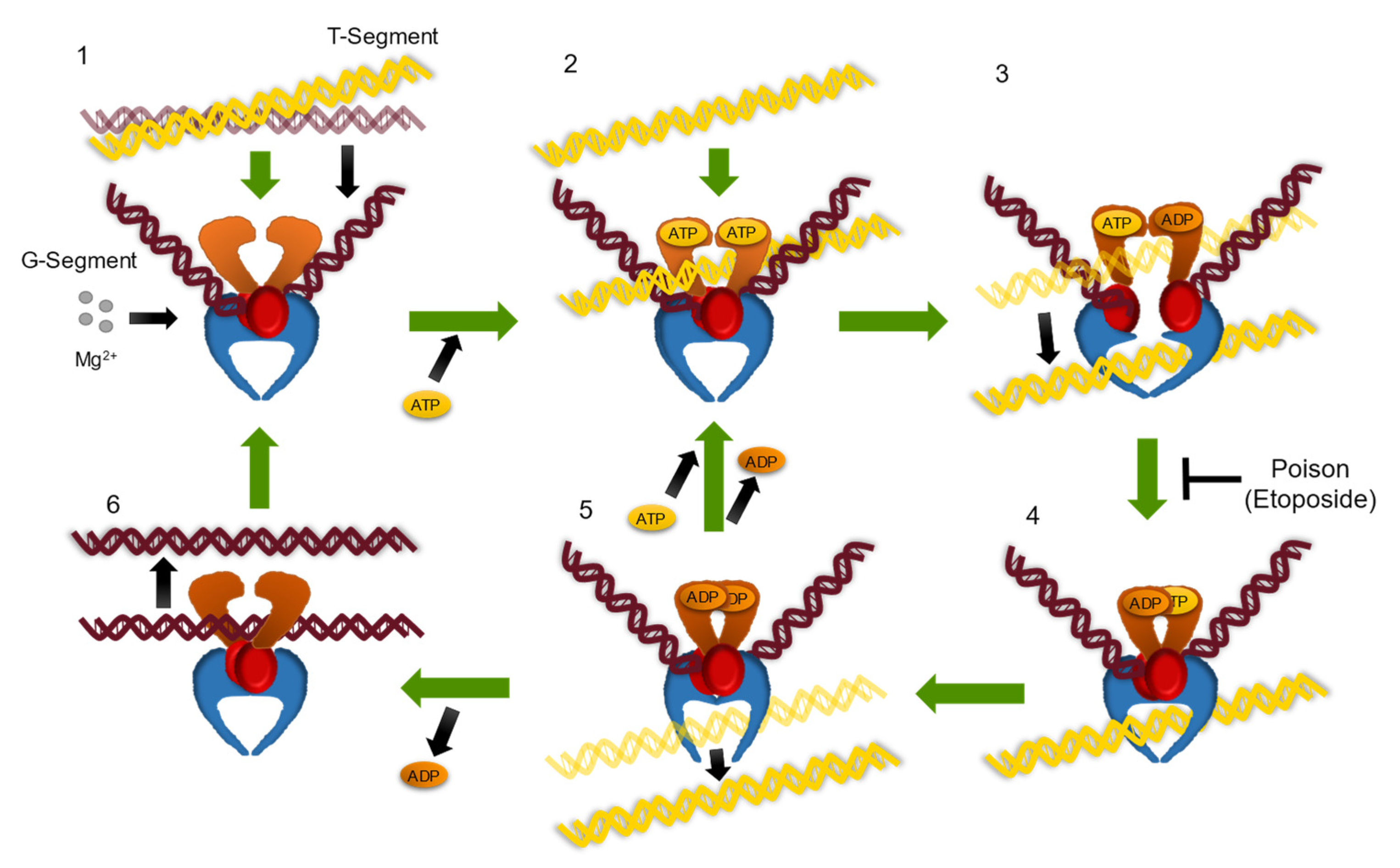
Figure 1. Catalytic cycle of TOP2. (1) The TOP2 enzyme captures a helix–helix crossover. (2) The gate DNA segment (G-segment, maroon) is bent and a double-strand break is formed in the presence of Mg(II). The transport DNA segment (T-segment, yellow) is retained by the closing of the N-terminal domain (orange) in the presence of ATP. (3) The DNA-gate opens (red), and the T-segment moves through the break. (4) Once strand passage is completed, the DNA-gate closes and the G-segment DNA is ligated together. (5) Next, the C-gate opens (lower blue region) releasing the T-segment. (6) TOP2 resets and can now perform another round of catalysis. Etoposide is a TOP2 poison and blocks ligation of the cleaved DNA.
Symmetry 14 01309 g001
Figure 2. Locations of residues examined in this study mapped onto human topoisomerase IIα. Human TOP2A homodimer is modeled in grey (domains noted on the left) with DNA in maroon. Etoposide is shown in yellow and key positions examined in this work are denoted by colored spheres (A429, orange; D463, green; R487, blue; G534, magenta; P716, red). Structure modeled using PDB ID 6ZY8 and generated using Pymol 2.5 [11].
Symmetry 14 01309 g002
Figure 3. Mutants of human topoisomerase IIα display variable levels of etoposide-induced plasmid DNA cleavage. Percent double-stranded DNA cleavage is shown for WT and mutant human topoisomerase IIα across a range of etoposide concentrations (0–200 μM). Error bars represent the standard deviation of the mean of three or more independent experiments.
Symmetry 14 01309 g003
Figure 4. Cleavage enhancement and strand break ratios for WT and mutant human topoisomerase IIα. Left panel, ratios of plasmid double-stranded DNA cleavage in the presence (drug) and absence (no drug, ND) of 100 or 200 μM etoposide are calculated for WT and mutant enzymes. Right panel, ratios of double-stranded (DSB) to single-stranded breaks (SSB) are shown for reactions with 100 or 200 μM etoposide in the presence of WT and mutant topoisomerase IIα.
Symmetry 14 01309 g004
Table 1. Plasmid DNA relaxation of mutants of human topoisomerase IIα compared to WT enzyme. The equal sign (=) indicates similarity to wild type. Processive or distributive characteristics are indicated by a P or D, respectively.
Enzyme Relaxation Activity
No Drug
Relaxation Activity
+Etoposide
Processive (P) or
Distributive (D)
WT----P
D463E==P
ΔA429=StimulatedP
G534R=Less sensitiveP
R487KSlowerLess sensitiveD
P716LSlowerStimulatedD
Publisher’s Note: MDPI stays neutral with regard to jurisdictional claims in published maps and institutional affiliations.