Opinion

Organ Patterning at the Shoot Apical Meristem (SAM): The Potential Role of the Vascular System

Department of Plant Developmental Biology, Faculty of Biological Sciences, University of Wrocław, 50-137 Wrocław, Poland
*
Author to whom correspondence should be addressed.
Symmetry 2023, 15(2), 364; https://doi.org/10.3390/sym15020364
Submission received: 27 December 2022 / Revised: 23 January 2023 / Accepted: 25 January 2023 / Published: 30 January 2023
(This article belongs to the Section Life Sciences)

Abstract

:
Auxin, which is transported in the outermost cell layer, is one of the major players involved in plant organ initiation and positioning at the shoot apical meristem (SAM). However, recent studies have recognized the role of putative internal signals as an important factor collaborating with the well-described superficial pathway of organogenesis regulation. Different internal signals have been proposed; however, their nature and transport route have not been precisely determined. Therefore, in this mini-review, we aimed to summarize the current knowledge regarding the auxin-dependent regulation of organ positioning at the SAM and to discuss the vascular system as a potential route for internal signals. In addition, as regular organ patterning is a universal phenomenon, we focus on the role of the vasculature in this process in the major lineages of land plants, i.e., bryophytes, lycophytes, ferns, gymnosperms, and angiosperms.

1. Introduction

The indeterminate growth and development of land plants are related to continuous postembryonic organogenesis. Subsequent lateral organs, such as leaves, are initiated by the precisely regulated activity of shoot apical meristems (SAM) located at the tip of each stem. SAM is a group of dividing cells, which contain stem (initial) cells and whose perpetual proliferation is the source of cells for all shoot tissues and organs. Lateral organs are initiated in the peripheral (organogenic) zone of the SAM, which encircles the central part of the apex, including the initial zone with stem cells and the organizing center (Figure 1) [1,2]. This region contains a few layers of cells, and the entire circumference is equally competent in forming new organs [3,4,5]. However, organogenesis is induced locally when the cells in the sub-superficial layers (L2 and L3 in Arabidopsis) begin to divide periclinally (parallel to the surface) instead of anticlinally (perpendicularly to the surface). The subsequent divisions of neighboring cells result in the bulging of a new organ primordium in an established position in reference to existing (older) organs at the apex [4,6,7,8,9,10]. Consequently, the resultant organ arrangement upon initiation creates regular symmetric spatial patterns called phyllotaxis [7,10,11,12,13]. Phyllotaxis is a wider term that is also used to describe the arrangement of leaf-like flower elements that are initiated due to the activity of the flower meristem or distribution of flowers within inflorescences (e.g., in capitula). There are different types of phyllotactic patterns, such as distichous, whorled, and helical (spiral). Although interest in phyllotaxis dates to ancient times and has flourished since the 19th century, the mechanisms underlying phyllotactic pattern formation remain incompletely elucidated [12,13,14].
Various internal signals have been proposed to contribute to organogenesis regulation; however, the precise pathways remain unclear. This review elaborates on the role of the vascular system in providing such signals. First, we discuss the two major historical groups of theories regarding organogenesis regulation at the SAM, following which we present the possible links between the vascular system and organogenesis, specifically involving polar auxin transport (PAT).

2. Two Main Historical Views on the Regulation of Phyllotactic Pattern Formation

How the site of new organ initiation at the SAM is defined is one of the main questions. Historically, two major groups of theories have emerged [15]. According to the first group, the place in which a new primordium is initiated is determined in the meristem by mutual interactions between the already existing organ primordia. These interactions can differ in nature, involving, e.g., biophysical and biochemical factors, e.g., [8,9,10,16,17,18,19,20,21,22,23]. One of the most widely accepted theories is that older primordia produce a morphogen (of unknown nature) that acts as an inhibitor for the initiation of new primordia. Morphogens diffusing from the existing organs form inhibitory fields; consequently, the new primordium is initiated at the most distant position to the older primordia [10,20,21,24].
The second group of theories assumes that the site of the new primordium initiation is determined by internal signals that originate from already differentiated regions. These signals can be transported to the meristem via the vascular system [22,25,26,27,28,29,30,31], which in seed plants is functionally interrelated to the organs, and its spatial structure corresponds directly to phyllotactic patterns (Figure 1) [32,33]. The support for these theories came from observations that the pattern of organ initiation at the apex is consistent with that existing in the differentiated region below and that the vascular strands supplying subsequently initiated organs can develop prior to any morphological signs of organ initiation [25,26,28,29]. However, the surgical isolation of the SAM from vascular tissues neither prevented organogenesis nor affected the phyllotactic pattern [19]; therefore, the regulatory role of the vascular system in phyllotaxis is not commonly accepted [34,35].
These two major groups of theories evolved with the technical progress of the tools used, and the current understanding of some aspects of phyllotactic pattern formation and underlying mechanisms are presented and discussed below.

3. Potential Role of the Interactions between Organ Primordia in Phyllotactic Pattern Formation

To explain the mechanism of leaf initiation, researchers have focused on interactions between incipient and existing primordia (the first group of theories mentioned above). Early experiments showed that the application of exogenous auxin at the intact SAM or replacement of the primordium with auxin applied in lanolin affected the initiation and distribution of subsequently formed primordia [18,36,37]. This suggested that this hormone is important for leaf positioning.
A breakthrough in phyllotaxis research occurred at the turn of the 20th and 21st centuries. The discovery of PIN1 (PINFORMED1) proteins, which are responsible for PAT in Arabidopsis shoots, and the finding that their mutation in the PIN1 gene or chemical treatment with the PAT-specific inhibitor NPA (N-1-naphthylphthalamic acid) impedes organ formation showed that PIN1-dependent PAT is involved in the regulation of organogenesis [38,39]. Furthermore, auxin was shown to be capable of recovering organogenesis in plants with PAT damage when applied to the peripheral zone of the SAM [3,4]. At that time, only basipetal PAT was known to occur in the vascular strands of the stem, whereas successive organs at the SAM were initiated acropetally; thus, linking these processes was difficult [3,40]. Soon after, the use of immunodetection techniques showed that PIN1 proteins were localized not only in vascular strands but also in the epidermis and L1 layer of the meristem. Their polar apical localization in the plasma membrane of the outermost layer of cells provided evidence that auxin, which is responsible for organogenesis, can be transported acropetally in L1 towards the peripheral zone of the SAM, where the maxima of auxin concentration are formed [4]. Then, in a developing primordium, auxin is transported using PIN1 proteins from the primordium tip to the internal tissues and basipetally towards the differentiated vasculature, thus inducing the development of a new vascular strand [4,6]. Consequently, a model of the auxin-dependent regulation of phyllotaxis was proposed, which stated that changes in PIN1 polarization resulted in the formation of regular patterns in the local maxima of auxin concentration, inducing organ initiation. Newly initiated primordia drain auxin from the surrounding cells, forming areas of auxin depletion and preventing organogenesis in their neighborhood; consequently, the distribution of successive organs is precisely regulated. This model is conceptually in line with an inhibitory field theory (the first group of historical theories mentioned above), although the areas of inhibition are formed due to auxin drainage from neighboring tissues [4,6] and not by the diffusion of the inhibitor from the primordium. However, it has recently been shown that additional inhibitory fields are generated by the transport of the cytokinin signaling inhibitor AHP6 (Arabidopsis Histidine Phosphotransfer Protein 6) from the leaf primordium into the surrounding cells. It has further been suggested that cytokinin signaling regulates the rhythmicity of organ initiation but not phyllotaxis [41].
The auxin-dependent model of phyllotaxis regulation has subsequently been confirmed in numerous experimental and in silico studies [42,43,44] and is now generally accepted as the regulatory mechanism of organ patterning and the development of the shoot vascular system. Recently, a new model has been developed that includes temporal changes in the pattern of auxin distribution within the SAM. This model emphasizes that the central part of the SAM is a site of high auxin concentration, and auxin maxima are formed at the SAM periphery in a finger-like pattern. Changes in auxin concentration trigger a transcriptional response via ARFs (Auxin Response Factors) [45]. In addition, recent studies on the specific plant model, the flower head of Gerbera, have shown that, in this case, the local interactions between older existing but not subsequently initiated organs are decisive for defining the site of a new primordium [46,47]. Such local interactions between neighboring organ primordia may also possibly explain the emergence of irregular phyllotaxis in non-circular flower meristems [48]. Importantly, in all the mentioned above models, the role of the L1 layer and positive feedback between auxin and its carrier, PIN1, are stressed as key players.
Although the involvement of PIN1 proteins and PAT in the phyllotactic pattern formation have been elegantly explained by auxin-based models, some aspects of organogenesis have not been fully explained; for instance, there is the question of why organogenesis is not completely blocked in plants with PAT damage (due to the PIN1 mutation or PAT-inhibitor treatment). In addition, unsolved questions remain regarding the cellular mechanism of PIN1 protein polarization in the plasma membrane, which is crucial for directional PAT and the dynamics of this process during phyllotactic pattern formation [43,44,49,50]. Despite the discovery of the PIN protein phosphorylation as a factor responsible for its polarity [51] and other factors possibly involved in regulating PIN1 polarization, such as mechanical stress, changes in the cell wall structure, and cytoskeleton organization (MT), this process remains unexplained [14,52,53,54]. Numerous models have been formulated in attempts to explain the mechanisms of PIN1 polarization during organogenesis and vascularization, focusing on two options: polarization “up-the-gradient” and “with-the-flux” [49,54,55,56,57]. The “dual-polarization” model, in which both options cooperate, seems to have the best experimental support [49,58]. In the search for the underlying molecular mechanism, the biochemical reaction network was proposed, which is based on a separate measurement of the auxin influx and efflux; auxin concentration in the cell can potentially control the change between both polarization modes [50,58,59]. The molecular components involved in auxin sensing within the cell require, however, identification.
The role of auxin biosynthesis in SAM for organ positioning, pattern establishment early in ontogeny, the formation of naturally occurring irregular phyllotaxes, and the mechanism of pattern formation in organisms in which PAT is not found remain unclear. Furthermore, an increasing amount of data show that PIN1-dependent PAT in L1 is not the only mechanism involved in these processes and that the L1 pathway can cooperate with internal signals [2,50,60,61,62].

4. Possibility of Vascular System as an Additional Source of the Signals Interacting with the Superficial PAT

Undoubtedly, organogenesis is related to vascularization. A detailed analysis of the best-characterized model plant Arabidopsis showed that both processes are: (1) regulated by PIN1-mediated PAT; (2) induced in the peripheral (organogenic) zone of the SAM; and (3) spatiotemporally co-regulated. While there is some evidence pointing to the involvement of the vascular system in organogenesis, its actual role is unclear [63,64,65,66,67,68]. Theoretically, different relations can occur between phyllotaxis and the vascular system.
The first possibility stresses SAM autonomy in the regulation of organ positioning, which fully depends on acropetal auxin transport in L1 and changes in PIN1 polarization within the meristem. The formation of vascular strands is a further consequence of basipetal auxin transport from the primordia. This hypothesis is supported by the localization of PIN1 proteins and the direction of PAT [4,49]. The significance of the L1 layer for organogenesis and vascularization was proven in experiments which showed that the expression of PIN1 exclusively in the outermost cell layer was sufficient for regular phyllotaxis formation and for vascular development. It raises, however, a question of why the vascular system developed normally despite the lack of PIN1 in incipient procambial strands [62]. The second possibility is that the vascular system determines the organ position at the meristem. This possibility was already suggested in the early works of Larson [28,29] based on histological observations that, in Populus, procambial strands, which supply incipient leaf primordia, are present in the stems of a few plastochrones prior to organ development. Interestingly, in other species, organ initiation was observed earlier than the initiation of the procambium strands supplying them [26]. The set of methods available at that time allowed the detection of organogenesis and vascularization only when the morphological signs of both processes were visible. The current studies with the use of molecular markers or transgenic lines show that the site of organ initiation is determined prior to any morphological signs of organogenesis, e.g., [4,49,54,62]. As well as this, the vascular system is determined before divisions, which precede procambium emergence and become visible [62,63,69]. Importantly, although early analyses were conducted only at the morphological level, speciesspecific differences in the coordination of both systems, i.e., organ positioning and the vascular system development, cannot be neglected.
The third possibility is that organ positioning is independent of the vascular system (as in the first option), but further development of the primordium is directed at vasculature. Auxin drainage from the primordium is an important condition for proper organ development [49,62,63]. Recent laser ablation experiments have shown that damage to the developing vascular strand leads to dysfunction in auxin drainage from primordia and to increased auxin accumulation in the site of primordium initiation. In turn, it results in transient changes in primordia size and phyllotaxis, proving the role of the vascular system in organ development. Additionally, organogenesis and the development of the supplying vascular strand were suggested as two parallel and non-sequential processes linked by auxin [63].
Importantly, basipetal auxin flow from the primordium (drainage) is precisely directed towards already differentiated and existing vascular strands, which points to their role as sinks for auxin [49]. This strongly suggests that the mature vascular strand is a source of a signal that communicates with the organ primordium and directs basipetal auxin flow [49,70,71]. On the other hand, computational models show that the basipetal differentiation of the vascular strand towards the existing mature vasculature is possible without such a signal [58,59,70]. The model assumes the existence of two different variants of the PIN1 protein that differ in auxin transport efficiency. The presence of such variants was confirmed in Brachypodium [72] but many plant species, including Arabidopsis, have only one variant of PIN1. Thus, to explain the differences in auxin transport efficiency, additional factors were proposed to be involved. Recent studies have shown that auxin can be transported via plasmodesmata (PD) [73,74,75] and regulates the PD size [76,77]. Computer simulations showed that auxin diffusion through PD can affect PAT and a new vascular strand development without any additional signals from the mature vasculature. The cooperation of facilitated diffusion with PAT can have the same effect [59]. Importantly, simulations were conducted for the venation pattern of leaves but not for stems, and even in leaves, they do not assure the reconstruction of the entire reticulate venation. Therefore, in stems with different vascularization, it cannot be excluded that the signal from the mature vasculature affects the PAT direction and development of a vascular connection to a newly formed organ.
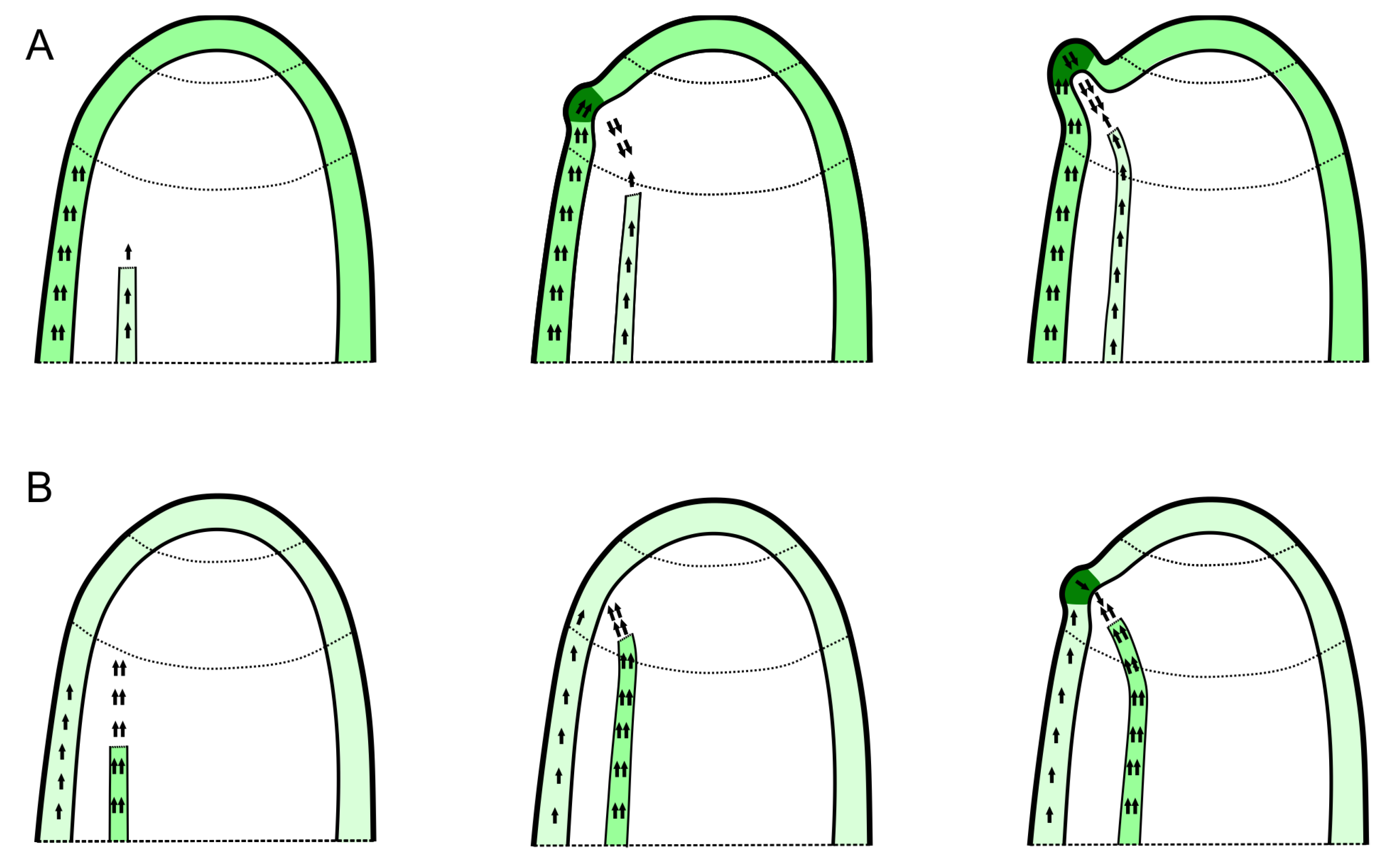
Studies of the pin1 mutant development in Arabidopsis have shown that organ presence is not obligatory for stem vasculature formation. In the young inflorescence stems of the mutant, despite the damage of the PAT and blockage of organogenesis, the stem’s vascular system was formed, although it showed differences in spatial organization compared to the wild-type plants [38,39,64,65]. However, the blockage of organogenesis in pin1 mutants occurs only when the acropetally differentiating protoxylem elements of the vascular strand are distant from the peripheral zone of the SAM. When they are close to the peripheral zone, as in vegetative rosettes or older inflorescence stems of the mutant, their presence is always correlated with organ formation. Based on these observations, a dual acropetal auxin transport pathway has been proposed [64]. According to this hypothesis, auxin-inducing organogenesis is transported towards the SAM in two independent pathways: in L1 with PIN1 proteins and internally by the vascular system. Organogenesis is completely blocked only when both pathways are dysfunctional. This implies that bidirectional communication and mature vascular strands are necessary for the formation of functional organs in the SAM (Figure 2). In addition, it can explain why organ initiation can occur prior to the development of the procambial strand supplying the organ (if the pathway in L1 prevails, as shown in Figure 2A) or why the procambium develops first (if the internal pathway is dominant, as shown in Figure 2B). However, whether it also affects organ localization remains unknown.
It is worth stressing here, however, that the majority of studies were conducted on model angiosperm plants that were characterized mostly by helical phyllotaxis. Little is known about the mutual regulation of both processes in species that have patterns different than helical, including whorled and even irregular patterns and different construction of the vascular system. In this context, especially interesting are species in which the number of vascular strands connecting to the vascular cylinder (so-called trunk bundles) is smaller than the number of elements in a whorl. Elements of one whorl can differ in their identity (e.g., phyllodes, stipules, leaflets of a compound leaf) and have their own shorter vascular strands, which connect to the trunk bundles directly or indirectly via a girdling (ring) bundle in a node [48,78]. It is difficult to interpret the formation of such a vascular network in the context of the auxin-based model. Supposedly, the formation of smaller vascular strands supplying organs in a whorl is related to the basipetal PAT from primordia, whereas the development of the trunk bundles and/or girdling bundle can be regulated by different mechanisms. The analysis of such unusual examples at the molecular level may shed light on general questions about the auxin-related coordination of organogenesis and vascularization processes.

5. Relationship between Vascular System and Organ Positioning in Other Major Groups of Land Plants

The mechanisms underlying regular organ positioning at the SAM are mostly studied in the model plant Arabidopsis, which is a representative of angiosperms, with the well-described layered structure of the SAM (Figure 1). The vasculature in this group of plants (in dicots) is usually composed of discrete vascular strands running axially in the stem and branching regularly to give rise to a vascular supply (vascular traces) for lateral organs initiated at the SAM (Figure 1) [5]. The involvement of auxin and its polar transport in the regulation of organ positioning and stem vascularization, as described above in Arabidopsis, seems to be universal in other angiosperms [72,79]. Whether a similar mechanism can occur in gymnosperms and other groups of land plants, which differ in SAM organization and vasculature structure, requires further research. Furthermore, similar lateral organ arrangements refer to different generations (gametophytic vs. sporophytic) and organs of different evolutionary origins (phyllids in bryophytes, microphylls in lycophytes, and true leaves (megaphylls) in ferns and seed plants).

5.1. Gymnosperms

The role of auxin in gymnosperm organogenesis and its impact on phyllotaxis has hardly been examined. The presence of the PIN protein PaPIN1 has been revealed in Picea abies [80]. Interestingly, although this protein clusters with Arabidopsis AtPIN3, AtPIN4, and AtPIN7, its expression pattern is similar to that of AtPIN1. During the early stages of somatic embryogenesis, PaPIN1 expression is detectable in developing cotyledons, whereas, in mature embryos, it is present at the cotyledon tips and in procambial cells, resembling the expression pattern of PIN1 in Arabidopsis embryos [80]. The analysis also showed that the treatment of developing somatic embryos of Picea abies with the PAT-inhibitor NPA results in the fusion of cotyledons and enlargement of the procambial domain [81,82]. The presence of PIN proteins was also confirmed in Pinus taeda: PtPINE, PtPINH. They complement the AtPIN1 function in the Arabidopsis mutant pin1, restoring normal organogenesis and the phenotype of the inflorescence stem [83]. Taken together, these results suggest that organogenesis and vascularization in gymnosperms are PAT-dependent and are similarly regulated as in angiosperms.
The spatial organization of the vascular system in gymnosperms resembles that of angiosperms and forms regular vascular traces connecting lateral organs and axial strands [32,33]. SAM is non-layered and contains a group of superficial initials [5]. At the meristem, the type of initiated organs changes during a season, forming, alternately, leaves (needles) and scales [84,85]. Despite changes in organ identity, in most conifers, such as Abies and Picea, phyllotactic patterns are relatively stable and usually continue along one axis [85,86]. In contrast, in the coniferous genus Torreya, the phyllotactic pattern changes every vegetative season along the same axis, which suggests that it is established de novo at the SAM [87]. The analysis of the internal structure of Torreya shoots shows that the probable reason for such changes in phyllotaxis could be the temporal and repetitive isolation of the meristem from differentiated vascular tissues by a thick collenchymatous plate (crown), which probably forms a barrier for the acropetal propagation of signals. In contrast, in Abies and Picea, where the phyllotactic pattern is continued, such an isolation of the SAM does not occur, probably due to the different structure of the collenchymatous plate. Taken together, these results suggest that organ positioning at the meristem can be affected by internal signals transported via the vascular system, or alternatively, SAM can be autonomous in its pattern formation [87]. However, the link between the vascular system and phyllotactic pattern formation, and PAT involvement in both processes, requires further study.

5.2. Ferns

SAM in ferns usually contains a single tetrahedral apical cell (AC), which divides along the sidewalls, forming subsequent segments (merophytes). In fern species that are characterized by relatively wide and flat meristems, derivatives of merophytes can form a distinct layer of elongated cells (prismatic cells, prismatic layer), which resemble, to some extent, the L1 layer of the SAM of angiosperms. Divisions within merophytes give rise to the initial cells for leaf primordia; however, in most species, a segment and leaf are not distinctly related [5,88]. In general, only a limited number of patterns are present in ferns, which is likely related to the geometry of the apex [89].
Surprisingly, little is known about auxin involvement in organogenesis regulation at the SAM in ferns. The analyses of leaf development in Adiantum peltatum [90], Osmunda cinnamomea [91,92,93,94], and Dryopteris [95] showed that auxin is present in the pinnae (leaf lamina) during all stages of development. Furthermore, the leaves were proposed to be the site of auxin biosynthesis, as was suggested by auxin basipetal transport within the rachis (leaf petiole) [96]. In ferns, the presence of canonical PIN proteins has been confirmed, and their structures have been shown to be similar to those in other vascular plants [97,98]. The link between auxin and leaf initiation and positioning at the SAM was inferred from experimental studies on the microsurgical isolation of leaf primordia. They showed that the artificial application of exogenous auxin in the leaf position recovers normal histogenesis within the apex [99,100]. The vasculature within the fern apices is usually of the protostele (young sporophytes) or siphonostele type, with parenchymatous leaf gaps at the site of leaf trace departure from the vascular cylinder. The removal of the leaf primordia from the apex results in the formation of a solid cylinder of vascular tissues (without leaf gaps), whereas the application of exogenous auxin causes the parenchymatization of the stele and recovery of the normal structure. Based on such experiments, which were performed in a few fern species, leaf primordia were shown to be the source of auxin for the proper development of the vascular system (as in seed plants) [100,101].

5.3. Lycophytes

In lycophytes, the structure of SAM and phyllotaxis differ in relation to their taxonomic position.
In Selaginella, a single AC [102] or two transient initial cells [103] function in the SAM. Most analyses were performed for the dorsiventral shoots of S. kraussiana, in which two transient AC divided to give rise to alternate segments, and the leaves were initiated according to the decussate pattern [103]. Recently, the AC has been shown to be a site of auxin accumulation in the SAM [104], and damage to PAT led to a decrease in auxin in the AC and SAM termination [105]. This suggests that the PAT can be acropetal towards the apex. However, the analysis of radioactively labeled auxin showed that the transport of auxin within the shoots of Selaginella is polar and basipetal [105]. Further experiments using the PAT inhibitor NPA showed aberrations in the phyllotaxis, which were rescued by supplementation with the exogenous auxin NAA (1-naphthaleneacetic acid). Interestingly, culturing plants on an NAA-supplemented medium did not affect phyllotaxis. Based on these results, Sanders and Langdale [105] concluded that PAT is required for proper leaf positioning but not for leaf initiation, showing that organogenesis at the SAM in Selaginella is regulated differently from that in seed plants.
The recent transcriptomic analyses of the model lycophyte Selaginella moellendorffii revealed the presence of genes whose homologues in Arabidopsis are involved in auxin biosynthesis, transport, and signaling. Among them, homologues of PIN genes have been detected, including homologues of PIN3 and PIN7, which in Arabidopsis localize to the plasma membrane [106] and are homologues of PIN 5/6/8 [107] which are mostly related to the endoplasmic reticulum. Interestingly, although the presence of the AtPIN1 homologue, which is one of the most important proteins for PAT in Arabidopsis shoots, has not been identified thus far, the SmPINR protein in complementation tests was found to rescue the AtPIN1 function in Arabidopsis inflorescence development [83]. This poses the question of SmPINR protein function in Selaginella and whether and to what extent it is involved in organ positioning. Furthermore, although elements of the auxin machinery are present, their mutual interactions and involvement in leaf positioning at the SAM in Selaginella remain unknown.
The vascular system of Selaginella is of the protostelic type with a species-dependent, variable in number, and dynamically changing arrangement of meristeles [5,108]. The incipient procambium (stele) is determined just below the AC in the derived segments (merophytes), whereas the leaf primordium is initiated by a division in the superficial cells of an older segment. Interestingly, the leaf trace procambium is initiated in an isolated position in the leaf primordium [109] and connects with the stelar system during further development without leaving a leaf gap. However, in Selaginella shoots, communication between the vascular system and developing organs has not yet been studied.
The representatives of Lycopodiales are characterized by the presence of a single group of superficial initials, resembling the SAM structure in gymnosperms [5]. Little is known about the mechanisms underlying the pattern of organ distribution in this group of plants and the factors responsible for their diversity. A comprehensive analysis has shown that canonical PIN proteins are present in a lycopod Huperzia squarrosa [97]. However, no data are available on the occurrence of PAT in lycopods. Thus, it is difficult to infer whether the mechanism of phyllotactic pattern formation is PAT-dependent and similar to that in other vascular plants.
The lycopod vascular system is composed of a differently partitioned stele with vascular connections to microphylls that depart from the protoxylem poles of the stele. In Lycopodium clavatum and Spinulum annotinum, the arrangement of microphyll traces is not as regular as in seed plants; the lengths of the traces are variable, and they connect with the unpredictable protoxylem pole in the stele. This suggests that the shoot vasculature is not so closely or developmentally related to organ arrangement, and this may account for the diversity of phyllotaxis [110]. SAM could be, to some extent, autonomous in the regulation of the new organ positioning. On the other hand, the role of the vasculature as a route for transporting internal signals for organogenesis cannot be ruled out as the stelar procambium reaches the group of superficial initials in the SAM. Likely, the adjustment of both systems, phyllotaxis, and vasculature, could happen during evolution to integrate the growth and development of lycopods [110,111].

5.4. Bryophytes

In bryophytes, only one initial cell (apical cell, AC) functions in SAM. The AC is mostly tetrahedral and cuts off segments that are parallel to the sidewalls. In each segment, a new gametophytic leaf (phyllid) is initiated, resulting in a regular arrangement of leaves. In SAM with a wedge-shaped AC, only two merophytes are alternately cut off, resulting in a distichous leaf arrangement, whereas in meristems with a tetrahedral AC, three merophytes with a divergence of 120° are produced, and the leaves are arranged in three vertical lines (ortostichies) [89,112,113,114,115,116]. In some mosses, however, these lines are not vertical but helically winding along the gametophore, resembling the typical Fibonacci phyllotaxis, with a divergence angle close to 137°. Such an increase in the divergence angle is proposed to be a consequence of the slightly oblique insertion of the newly formed cell wall during asymmetric AC division [116]. The different orientations of the inserted cell wall (rotation angle), as shown through measurements of both the merophyte and phyllid positions, may also explain the diversity of helical patterning in mosses [117].
The molecular mechanisms governing the AC division plane and the presence of other factors involved in leaf positioning in bryophytes remain elusive. Most studies have focused on the model moss Physcomitrium patens (previously Physcomitrella patens), trying to correlate the phenomenon of leaf formation with the AC divisional pattern. Recently, defects in leaf patterning have been shown to be related to the aberrant orientation of the cytokinetic spindle (phragmoplast) in mutants with a dysfunctional cytoskeleton [115,118]. AC divisions are also related to the maintenance of SAM identity. Therefore, the dysfunction of genes involved in SAM self-renewal may lead to aberrant divisions and leaf arrangements [114].
The presence of auxin [119,120,121,122,123] and auxin efflux carriers, including PIN proteins [97,98,114,122], has been confirmed in bryophytes. Recently, vegetative and generative apical cells have been found to be responsible for auxin biosynthesis [124]. Interestingly, the authors proposed that the lack of sensing and responsiveness to this hormone may account for the maintenance of AC meristematic characteristics. Thus, AC may potentially be a source of auxin for leaf positioning in the model Physcomitrium SAM. Despite the presence of canonical PINA and PINB proteins in Physcomitrium [97,98,114,125,126], their bipolar localization has only been confirmed close to the leaf tips [114,122,126], whereas, in the leaf base and gametophore cells, they are more uniformly distributed in the plasma membrane [97,114]. Therefore, PAT has not yet been confirmed in bryophytes. Auxin has been proposed to be transported by a diffusion-based mechanism that is mediated by plasmodesmata [122,127]. Taken together, regardless of the progress in understanding auxin patterning and biosynthesis in mosses, the actual role of auxin (re)distribution within the SAM in leaf positioning requires further study.
Little is known about the internal factors that may be involved in leaf positioning. The model moss Physcomitrium has a central strand of conducting cells [112], which has been shown to be functional for long-distance acropetal transport in experiments with fluorescent tracers [128,129]. Potentially, this single central strand could be a route for transporting signaling molecules toward the meristem; however, it does not form connections to leaves or points to the leaf position. Such connections (leaf traces) are present only in some bryophytes (Polytrichaceae), although their actual role in conducting water vs. assimilates has been discussed [112]. Interestingly, they are reduced in terms of the number of elements close to the central strand. Furthermore, in other mosses (Mniaceae and Splachnaceae), false leaf traces can occur that extend from the leaf toward the central strand but do not connect with it [112]. It would be interesting to analyze whether there is a communication between the leaf and central strand in these bryophyte species in the context of the evolution of interrelated patterns of organ distribution and vascular structure.

6. Summary

Currently, the most commonly accepted model of organ positioning at the SAM is based on the formation of the local maxima of auxin concentration and the area of auxin depletion (inhibitory fields) around the primordia. This process is regulated by PAT and changes in PIN1 polarization. However, recent studies have shown that this model requires supplementation with other factors, including internal signals, which cooperate with the superficial pathway of organogenesis regulation. The nature of the signals and their modes of action have not yet been fully recognized.
In this review, we focused on the vascular system as a potential source of internal signals. The involvement of vasculature in organ arrangement has been postulated several times. This system has been shown to be an important player in organ positioning; however, its actual role is not fully understood. To date, studies on the mechanisms underlying phyllotactic pattern formation have focused on Arabidopsis. However, in recent years, the process of organogenesis has been analyzed in the evolutionary context in other major groups of land plants differing in SAM structure and functioning. Perhaps this direction of research will provide answers about the developmental and functional integration of the superficial regular patterns at the SAM and the internal vascular system. This, in turn, will help us to better understand the mechanisms of organ positioning and the role of the vascular system in this process.

Author Contributions

Concept of the work, literature research, and writing A.B. and E.M.G. All authors have read and agreed to the published version of the manuscript.

Funding

This research was funded by the University of Wrocław, Poland (2023/501/73 MPK 2599150000) and the National Science Centre (NCN), Poland, Grant No. 2014/15/B/NZ3/00858.

Data Availability Statement

Not applicable.

Acknowledgments

We thank to our colleagues from the Department of Plant Developmental Biology, Katarzyna Sokołowska, Alicja Dolzblasz, and Elżbieta Myśkow for their critical reading of the manuscript. We are also grateful to the Reviewers for suggestions that helped to improve the manuscript.

Conflicts of Interest

The authors declare no conflict of interest.

References

  1. Soyars, C.L.; James, S.R.; Nimchuk, Z.L. Ready, aim, shoot: Stem cell regulation of the shoot apical meristem. Curr. Opin. Plant Biol. 2016, 29, 163–168. [Google Scholar] [CrossRef] [PubMed] [Green Version]
  2. Shi, B.; Vernoux, T. Patterning at the shoot apical meristem and phyllotaxis. Curr. Top. Dev. Biol. 2019, 131, 81–107. [Google Scholar] [CrossRef] [PubMed]
  3. Reinhardt, D.; Mandel, T.; Kuhlemeier, C. Auxin regulates the initiation and radial position of plant lateral organs. Plant Cell 2000, 12, 507–518. [Google Scholar] [CrossRef] [Green Version]
  4. Reinhardt, D.; Pesce, E.R.; Stieger, P.; Mandel, T.; Baltensperger, K.; Bennett, M.; Traas, J.; Friml, J.; Kuhlemeier, C. Regulation of phyllotaxis by polar auxin transport. Nature 2003, 426, 255–260. [Google Scholar] [CrossRef] [PubMed]
  5. Evert, R.F. Esau’s Plant Anatomy: Meristems, Cells, and Tissues of the Plant Body: Their Structure, Function, and Development, 3rd ed.; John Wiley & Sons: Hoboken, NJ, USA, 2007. [Google Scholar] [CrossRef]
  6. Reinhardt, D. Phyllotaxis—A new chapter in an old tale about beauty and magic numbers. Curr. Opin. Plant Biol. 2005, 8, 487–493. [Google Scholar] [CrossRef]
  7. Kuhlemeier, C. Phyllotaxis. Trends Plant Sci. 2007, 12, 143–150. [Google Scholar] [CrossRef]
  8. Snow, M.; Snow, R. Experiments on phyllotaxis. I. The effect of isolating a primordium. Philos. Trans. R. Soc. Lond. Ser. B 1931, 222, 353–400. [Google Scholar]
  9. Snow, M.; Snow, R. Experiments on phyllotaxis. II. The effect of displacing a primordium. Philos. Trans. R. Soc. Lond. Ser. B 1933, 220, 1–43. [Google Scholar]
  10. Schwabe, W.W. Phyllotaxis. In Positional Controls in Plant Development; Barlow, P.W., Carr, D.J., Eds.; Cambridge University Press: Cambridge, UK, 1984; pp. 403–440. [Google Scholar]
  11. Jean, R.V. Phyllotaxis: A Systemic Study in Plants Morphogenesis; Cambridge University Press: Cambridge, UK, 1994. [Google Scholar]
  12. Adler, I.; Barabé, D.; Jean, R.V. A history of the study of phyllotaxis. Ann. Bot. 1997, 80, 231–244. [Google Scholar] [CrossRef] [Green Version]
  13. Reinhardt, D.; Gola, E.M. Law and order in plants—The origin and functional relevance of phyllotaxis. Trends Plant Sci. 2022, 27, 1017–1032. [Google Scholar] [CrossRef]
  14. Yin, X. Phyllotaxis: From classical knowledge to molecular genetics. J. Plant Res. 2021, 134, 373–401. [Google Scholar] [CrossRef] [PubMed]
  15. Dengler, N.G. The shoot apical meristem and development of vascular architecture. Can. J. Bot. 2006, 84, 1660–1671. [Google Scholar] [CrossRef]
  16. Hofmeister, W. Allgemeine Morphologie der Gewachse. In Handbuch der Physiologischen Botanik; Bary, A., Irmisch, T.H., Sachs, J., Eds.; Engelmann: Leipzig, Germany, 1868; pp. 405–664. [Google Scholar]
  17. Snow, M.; Snow, R. Experiments on phyllotaxis. III. Diagonal split through decussate apices. Philos. Trans. R. Soc. Lond. Ser. B 1935, 225, 63–94. [Google Scholar]
  18. Snow, M.; Snow, R. Auxin and leaf formation. New Phytol. 1937, 36, 1–18. [Google Scholar] [CrossRef]
  19. Snow, M.; Snow, R. On the determination of leaves. New Phytol. 1947, 46, 5–19. [Google Scholar] [CrossRef]
  20. Wardlaw, C.W. Further experimental observations on the shoot apex of Dryopteris aristata Druce. Philos. Trans. R. Soc. Lond. Ser. B 1949, 233, 415–451. [Google Scholar]
  21. Mitchison, G.J. Phyllotaxis and the Fibonacci series. Science 1977, 196, 270–275. [Google Scholar] [CrossRef] [Green Version]
  22. Williams, R.F.; Brittain, E.G. A geometrical model of phyllotaxis. Austr. J. Bot. 1984, 32, 43–72. [Google Scholar] [CrossRef]
  23. Green, P.B.; Steele, C.; Rennich, S.C. Phyllotactic patterns: A biophysical mechanism for their origin. Ann. Bot. 1996, 77, 515–527. [Google Scholar] [CrossRef] [Green Version]
  24. Young, D.A. On the diffusion theory of phyllotaxis. J. Theor. Biol. 1978, 71, 421–432. [Google Scholar] [CrossRef]
  25. Esau, K. Plant Anatomy, 2nd ed.; John Wiley: New York, NY, USA, 1965. [Google Scholar]
  26. Esau, K. Vascular Differentiation in Plants; Holt, Rinehart, Winston: New York, NY, USA, 1965. [Google Scholar]
  27. Girolami, G. Relation between phyllotaxis and primary vascular organization in Linum. Amer. J. Bot. 1953, 40, 618–625. [Google Scholar] [CrossRef]
  28. Larson, P.R. Development and organization of the primary vascular system in Populus deltoides according to phyllotaxy. Am. J. Bot. 1975, 62, 1084–1099. [Google Scholar] [CrossRef]
  29. Larson, P.R. Phyllotactic transitions in the vascular system of Populus deltoides Bartr. as determined by 14C labeling. Planta 1977, 134, 241–249. [Google Scholar] [CrossRef] [PubMed]
  30. Larson, P.R. Establishment of the vascular system in seedlings of Populus deltoides Bartr. Am. J. Bot. 1979, 66, 452–462. [Google Scholar] [CrossRef]
  31. Larson, P.R. Interrelations between phyllotaxis, leaf development and the primary-secondary vascular transition in Populus deltoides. Ann. Bot. 1980, 46, 757–769. [Google Scholar] [CrossRef]
  32. Namboodiri, K.K.; Beck, C.B. A comparative study of the primary vascular system of conifers. I. Genera with helical phyllotaxis. Am. J. Bot. 1968, 55, 447–457. [Google Scholar] [CrossRef] [Green Version]
  33. Namboodiri, K.K.; Beck, C.B. A comparative study of the primary vascular system of conifers. II. Genera with opposite and whorled phyllotaxis. Am. J. Bot. 1968, 55, 458–463. [Google Scholar] [CrossRef] [Green Version]
  34. Wardlaw, C.W. The organization of the shoot apex. In Encyclopedia of Plant Physiology; Springer: Berlin/Heidelberg, Germany, 1965; Volume 15, pp. 966–1076. [Google Scholar]
  35. Steeves, T.A.; Sussex, I.A. Patterns in Plant Development; Cambridge University Press: Cambridge, UK, 1989. [Google Scholar]
  36. Ball, E. The effects of synthetic growth substances on the shoot apex of Tropaeolum majus L. Am. J. Bot. 1944, 31, 316–327. [Google Scholar] [CrossRef]
  37. Meicenheimer, R.D. Changes in Epilobium phyllotaxy induced by N-1-naphthylphthalamic acid and α-4-chlorophenoxyisobutyric acid. Am. J. Bot. 1981, 68, 1139–1154. [Google Scholar] [CrossRef]
  38. Okada, K.; Ueda, J.; Komaki, M.K.; Bell, C.J.; Shimura, Y. Requirement of the auxin polar transport system in early stages of Arabidopsis floral bud formation. Plant Cell 1991, 3, 677–684. [Google Scholar] [CrossRef] [Green Version]
  39. Mattsson, J.; Sung, Z.R.; Berleth, T. Responses of plant vascular systems to auxin transport inhibition. Development 1999, 126, 2979–2991. [Google Scholar] [CrossRef] [PubMed]
  40. Gälweiler, L.; Guan, C.; Müller, A.; Wisman, E.; Mendgen, K.; Yephremov, A.; Palme, K. Regulation of polar auxin transport by AtPIN1 in Arabidopsis vascular tissue. Science 1998, 282, 2226–2230. [Google Scholar] [CrossRef] [PubMed] [Green Version]
  41. Besnard, F.; Refahi, Y.; Morin, V.; Marteaux, B.; Brunoud, G.; Chambrier, P.; Rozier, F.; Mirabet, V.; Legrand, J.; Lainé, S.; et al. Cytokinin signalling inhibitory fields provide robustness to phyllotaxis. Nature 2014, 505, 417–421. [Google Scholar] [CrossRef] [PubMed]
  42. Heisler, M.G.; Ohno, C.; Das, P.; Sieber, P.; Reddy, G.V.; Long, J.A.; Meyerowitz, E.M. Patterns of auxin transport and gene expression during primordium development revealed by live imaging of the Arabidopsis inflorescence meristem. Curr. Biol. 2005, 15, 1899–1911. [Google Scholar] [CrossRef] [PubMed] [Green Version]
  43. Smith, R.; Guyomarch, S.; Mandel, T.; Reinhardt, D.; Kuhlemeier, C.; Prusinkiewicz, P. A plausible model of phyllotaxis. Proc. Natl. Acad. Sci. USA 2006, 103, 1301–1306. [Google Scholar] [CrossRef] [Green Version]
  44. Jönsson, H.; Heisler, M.G.; Shapiro, B.E.; Meyerowitz, E.M.; Mjolsness, E. An auxin-driven polarized transport model for phyllotaxis Proc. Natl. Acad. Sci. USA 2006, 103, 1633–1638. [Google Scholar] [CrossRef] [Green Version]
  45. Galvan-Ampudia, C.S.; Cerutti, G.; Legrand, J.; Brunoud, G.; Martin-Arevalillo, R.; Azais, R.; Bayle, V.; Moussu, S.; Wenzl, C.; Jaillais, Y.; et al. Temporal integration of auxin information for the regulation of patterning. eLife 2020, 9, e55832. [Google Scholar] [CrossRef] [PubMed]
  46. Zhang, T.; Cieslak, M.; Owens, A.; Wang, F.; Broholm, S.K.; Teeri, T.H.; Elomaa, P.; Prusinkiewicz, P. Phyllotactic patterning of gerbera flower heads. Proc. Natl. Acad. Sci. USA 2021, 118, e2016304118. [Google Scholar] [CrossRef]
  47. Prusinkiewicz, P.; Zhang, T.; Owens, A.; Cieslak, M.; Elomaa, P. Phyllotaxis without symmetry: What can we learn from flower heads? J. Exp. Bot. 2022, 73, 3319–3329. [Google Scholar] [CrossRef]
  48. Rutishauser, R. Acacia (wattle) and Cananga (ylang-ylang): From spiral to whorled and irregular (chaotic) phyllotactic patterns—A pictorial report. Acta Soc. Bot. Pol. 2016, 85, 3531. [Google Scholar] [CrossRef] [Green Version]
  49. Bayer, E.M.; Smith, R.S.; Mandel, T.; Nakayama, N.; Sauer, M.; Prusinkiewicz, P.; Kuhlemeier, C. Integration of transport-based models for phyllotaxis and midvein formation. Genes Dev. 2009, 23, 373–384. [Google Scholar] [CrossRef] [PubMed] [Green Version]
  50. Hartmann, F.P.; Barbier de Reuille, P.; Kuhlemeier, C. Toward a 3D model of phyllotaxis based on a biochemically plausible auxin transport mechanism. PLoS Comput. Biol. 2019, 15, e1006896. [Google Scholar] [CrossRef] [PubMed] [Green Version]
  51. Ki, D.; Sasayama, D.; Cho, H.T. The M3 phosphorylation site is required for trafficking and biological roles of PIN-FORMED1, 2, and 7 in Arabidopsis. Front. Plant Sci. 2016, 7, 1479. [Google Scholar] [CrossRef]
  52. Jesuthasan, S.; Green, P.B. On the mechanism of decussate phyllotaxis: Biophysical studies on the tunica layer of Vinca major. Amer. J. Bot. 1989, 76, 1152–1166. [Google Scholar] [CrossRef]
  53. Hamant, O.; Heisler, M.G.; Jönsson, H.; Krupinski, P.; Uyttewaal, M.; Bokov, P.; Corson, F.; Sahlin, P.; Boudaoud, A.; Meyerowitz, E.M.; et al. Developmental patterning by mechanical signals in Arabidopsis. Science 2008, 322, 1650–1655. [Google Scholar] [CrossRef] [PubMed] [Green Version]
  54. Heisler, M.G.; Hamant, O.; Krupinski, P.; Uyttewaal, M.; Ohno, C.; Jönsson, H.; Traas, J.; Meyerowitz, E.M. Alignment between PIN1 polarity and microtubule orientation in the shoot apical meristem reveals a tight coupling between morphogenesis and auxin transport. PLoS Biol. 2010, 8, e1000516. [Google Scholar] [CrossRef] [Green Version]
  55. Stoma, S.; Lucas, M.; Chopard, J.; Schaedel, M.; Traas, J.; Godin, C. Flux-based transport enhancement as a plausible unifying mechanism for auxin transport in meristem development. PLoS Comput. Biol. 2008, 4, e1000207. [Google Scholar] [CrossRef]
  56. Merks, R.M.; Van de Peer, Y.; Inzé, D.; Beemster, G.T. Canalization without flux sensors: A traveling-wave hypothesis. Trends Plant Sci. 2007, 12, 384–390. [Google Scholar] [CrossRef]
  57. Wabnik, K.; Kleine-Vehn, J.; Balla, J.; Sauer, M.; Naramoto, S.; Reinöhl, V.; Merks, R.M.; Govaerts, W.; Friml, J. Emergence of tissue polarization from synergy of intracellular and extracellular auxin signaling. Mol Syst. Biol. 2010, 6, 447. [Google Scholar] [CrossRef] [Green Version]
  58. Cieslak, M.; Runions, A.; Prusinkiewicz, P. Auxin-driven patterning with unidirectional fluxes. J. Exp. Bot. 2015, 66, 5083–5102. [Google Scholar] [CrossRef] [Green Version]
  59. Cieslak, M.; Owens, A.; Prusinkiewicz, P. Computational Models of Auxin-Driven Patterning in Shoots. Cold Spring Harb. Perspect. Biol. 2021, 14, a040097. [Google Scholar] [CrossRef] [PubMed]
  60. Cheng, Y.; Dai, X.; Zhao, Y. Auxin synthesized by the YUCCA flavin monooxygenases is essential for embryogenesis and leaf formation in Arabidopsis. Plant Cell 2007, 19, 2430–2439. [Google Scholar] [CrossRef] [PubMed] [Green Version]
  61. Bhatia, N.; Bozorg, B.; Larsson, A.; Ohno, C.; Jönsson, H.; Heisler, M.G. Auxin acts through MONOPTEROS to regulate plant cell polarity and pattern phyllotaxis. Curr. Biol. 2016, 26, 3202–3208. [Google Scholar] [CrossRef]
  62. Kierzkowski, D.; Lenhard, M.; Smith, R.; Kuhlemeier, C. Interaction between meristem tissue layers controls phyllotaxis. Dev. Cell 2013, 26, 616–628. [Google Scholar] [CrossRef] [PubMed] [Green Version]
  63. Deb, Y.; Marti, D.; Frenz, M.; Kuhlemeier, C.; Reinhardt, D. Phyllotaxis involves auxin drainage through leaf primordia. Development 2015, 142, 1992–2001. [Google Scholar] [CrossRef] [Green Version]
  64. Banasiak, A. Putative dual pathway of auxin transport in organogenesis of Arabidopsis. Planta 2011, 233, 49–61. [Google Scholar] [CrossRef]
  65. Banasiak, A.; Biedroń, M.; Dolzblasz, A.; Berezowski, M.A. Ontogenetic changes in auxin biosynthesis and distribution determine the organogenic activity of the shoot apical meristem in pin1 mutants. Int. J. Mol. Sci. 2019, 20, 180. [Google Scholar] [CrossRef] [Green Version]
  66. Avsian-Kretchmer, O.; Cheng, J.C.; Chen, L.; Moctezuma, E.; Renee Sung, Z. Indole acetic acid distribution coincides with vascular differentiation pattern during Arabidopsis leaf ontogeny. Plant Physiol. 2002, 130, 199–209. [Google Scholar] [CrossRef] [Green Version]
  67. Kwiatkowska, D. The relationships between the primary vascular system and phyllotactic patterns of Anagallis arvensis (Primulaceae). Amer. J. Bot. 1992, 79, 904–913. [Google Scholar] [CrossRef]
  68. Kwiatkowska, D. Ontogenetic changes in the shoot primary vasculature of Anagallis arvensis L. Acta Soc. Bot. Pol. 1995, 64, 213–222. [Google Scholar] [CrossRef] [Green Version]
  69. Kang, J.; Tang, J.; Donnelly, P.; Dengler, N. Primary vascular pattern and expression of ATHB-8 in shoots of Arabidopsis. New Phytol. 2003, 158, 443–454. [Google Scholar] [CrossRef] [PubMed] [Green Version]
  70. Lavania, D.; Linh, N.M.; Scarpella, E. Of Cells, Strands, and Networks: Auxin and the Patterned Formation of the Vascular System. Cold Spring Harb. Perspect. Biol. 2021, 13, a039958. [Google Scholar] [CrossRef] [PubMed]
  71. Smith, R.S.; Bayer, E.M. Auxin transport-feedback models of patterning in plants. Plant Cell Environ. 2009, 32, 1258–1271. [Google Scholar] [CrossRef] [PubMed]
  72. O’Connor, D.L.; Runions, A.; Sluis, A.; Bragg, J.; Vogel, J.P.; Prusinkiewicz, P.; Hake, S. A division in PIN-mediated auxin patterning during organ initiation in grasses. PLoS Comput. Biol. 2014, 10, e1003447. [Google Scholar] [CrossRef] [Green Version]
  73. Verna, C.; Ravichandran, S.J.; Sawchuk, M.G.; Linh, N.M.; Scarpella, E. Coordination of tissue cell polarity by auxin transport and signaling. eLife 2019, 8, e51061. [Google Scholar] [CrossRef] [PubMed]
  74. Gao, C.; Liu, X.; De Storme, N.; Jensen, K.H.; Xu, Q.; Yang, J.; Liu, X.; Chen, S.; Martens, H.J.; Schulz, A.; et al. Directionality of plasmodesmata-mediated transport in Arabidopsis leaves supports auxin channeling. Curr. Biol. 2020, 30, 1970–1977.e4. [Google Scholar] [CrossRef]
  75. Band, L.R. Auxin fluxes through plasmodesmata. New Phytol. 2021, 231, 1686–1692. [Google Scholar] [CrossRef]
  76. Han, X.; Hyun, T.K.; Zhang, M.; Kumar, R.; Koh, E.J.; Kang, B.H.; Lucas, W.J.; Kim, J.Y. Auxin-callose-mediated plasmodesmalgating is essential for tropic auxin gradient formation and signaling. Dev. Cell 2014, 28, 132–146. [Google Scholar] [CrossRef] [Green Version]
  77. Sager, R.; Wang, X.; Hill, K.; Yoo, B.C.; Caplan, J.; Nedo, A.; Tran, T.; Bennett, M.J.; Lee, J.Y. Auxin-dependent control of a plasmodesmal regulator creates a negative feedback loop modulating lateral root emergence. Nat. Commun. 2020, 11, 364. [Google Scholar] [CrossRef] [Green Version]
  78. Rutishauser, R. Polymerous leaf whorls in vascular plants: Developmental morphology and fuzziness or organ identities. Int. J. Plant Sci. 1999, 160 (Suppl. S6), S81–S103. [Google Scholar] [CrossRef] [Green Version]
  79. O’Connor, D.L.; Elton, S.; Ticchiarelli, F.; Hsia, M.M.; Vogel, J.P.; Leyser, O. Cross-species functional diversity within the PIN auxin efflux protein family. eLife 2017, 6, e31804. [Google Scholar] [CrossRef] [PubMed]
  80. Palovaara, J.; Hallberg, H.; Stasolla, C.; Luit, B.; Hakman, I. Expression of a gymnosperm PIN homologous gene correlates with auxin immunolocalization pattern at cotyledon formation and in demarcation of the procambium during Picea abies somatic embryo development and in seedling tissues. Tree Phys. 2010, 30, 479–489. [Google Scholar] [CrossRef] [PubMed] [Green Version]
  81. Hakman, I.; Hallberg, H.; Palovaraa, J. The polar auxin transport inhibitor NPA impairs embryo morphology and increases expression of an auxin efflux facilitator protein PIN during Picea abies somatic embryo development. Tree Phys. 2009, 29, 483–496. [Google Scholar] [CrossRef] [PubMed]
  82. Holloway, D.M.; Rozada, I.; Bray, J.J.H. Two-stage patterning dynamics in conifer cotyledon whorl morphogenesis. Ann. Bot. 2018, 121, 525–534. [Google Scholar] [CrossRef] [Green Version]
  83. Zhang, Y.; Rodriguez, L.; Li, L.; Zhang, X.; Friml, J. Functional innovations of PIN auxin transporters mark crucial evolutionary transitions during rise of flowering plants. Sci. Adv. 2020, 6, eabc8895. [Google Scholar] [CrossRef]
  84. Parke, R.V. Growth periodicity and the shoot tip of Abies concolor. Am. J. Bot. 1959, 46, 110–119. [Google Scholar] [CrossRef]
  85. Zagórska-Marek, B. Phyllotactic patterns and transitions in Abies balsamea. Can. J. Bot. 1985, 63, 1844–1854. [Google Scholar] [CrossRef]
  86. Zagórska-Marek, B.; Banasiak, A. Related to phyllotaxis interlocked systems of vascular sympodia and cortical resin canals in Abies and Picea shoots. Acta Soc. Bot. Pol. 2000, 69, 165–172. [Google Scholar] [CrossRef] [Green Version]
  87. Banasiak, A.; Zagórska-Marek, B. Signals flowing from mature tissues to SAM determine the phyllotactic continuity in successive annual increments of the conifer shoot. Acta Soc. Bot. Pol. 2006, 75, 113–121. [Google Scholar] [CrossRef] [Green Version]
  88. Bierhorst, D.W. On the stem apex, leaf initiation and early leaf ontogeny in filicalean ferns. Amer. J. Bot. 1977, 64, 125–152. [Google Scholar] [CrossRef]
  89. Gola, E.; Banasiak, A. Diversity of phyllotaxis in land plants in reference to the shoot apical meristem structure. Acta Soc. Bot. Pol. 2016, 85, 3529. [Google Scholar] [CrossRef] [Green Version]
  90. Wetmore, R.H.; Pratt, C. The growth and auxin relations of leaves of the maidenhair fern, Adiantum pedatum L. Am. J. Bot. 1949, 36, 806. [Google Scholar]
  91. Steeves, T.A.; Briggs, W.R. Morphogenetic studies on Osmunda cinnamomea L.—The origin and early development of vegetative fronds. Phytomorphology 1958, 8, 60–72. [Google Scholar]
  92. Steeves, T.A.; Briggs, W.R. Morphogenetic studies on Osmunda cinnamomea L. The auxin relationships of expanding fronds. J. Exp. Bot. 1960, 11, 45–67. [Google Scholar] [CrossRef]
  93. Steeves, T.A. On the determination of leaf primordia in ferns. In Trends in Plant Morphogenesis; Cutter, E.G., Ed.; Longmans, Green and Co. Ltd.: London, UK, 1967; pp. 200–219. [Google Scholar]
  94. Steeves, T.; Hicks, G.; Steeves, M.; Retalla, K.B. Leaf determination in the fern Osmunda cinnamomea—A reinvestigation. Ann. Bot. 1993, 71, 511–517. [Google Scholar] [CrossRef]
  95. Wardlaw, C.W. The comparative investigation of apices of vascular plants by experimental methods. Philos. Trans. R. Soc. Lond. Ser. B 1950, 234, 583–604. [Google Scholar]
  96. Voeller, B.R. Regulation of “fiddlehead” uncoiling in ferns. Naturwissenschaften 1960, 47, 70–71. [Google Scholar] [CrossRef]
  97. Bennett, T.A.; Liu, M.M.; Aoyama, T.; Bierfreund, N.M.; Braun, M.; Coudert, Y.; Dennis, R.J.; O’Connor, D.; Wang, X.Y.; White, C.D.; et al. Plasma membrane-targeted PIN proteins drive shoot development in a moss. Curr. Biol. 2014, 24, 2776–2785. [Google Scholar] [CrossRef] [Green Version]
  98. Bennett, T.A. PIN proteins and the evolution of plant development. Trends Plant Sci. 2015, 20, 498–507. [Google Scholar] [CrossRef]
  99. Wardlaw, C.W.; Cutter, E.G. Experimental and analytical studies of Pteridophytes. XXXI. The effect of shallow incisions on organogenesis in Dryopteris aristata Druce. Ann. Bot. 1948, 20, 39–56. [Google Scholar] [CrossRef]
  100. Ma, Y.; Steeves, T.A. Auxin effects on vascular differentiation in Ostrich fern. Ann. Bot. 1992, 70, 277–282. [Google Scholar] [CrossRef]
  101. Ma, Y.; Steeves, T.A. Vascular differentiation in the shoot apex of Matteuccia struthiopteris. Ann. Bot. 1994, 74, 573–585. [Google Scholar] [CrossRef]
  102. Imaichi, R. Meristem organization and organ diversity. In Biology and Evolution of Ferns and Lycophytes; Ranker, T.A., Haufler, C.H., Eds.; Cambridge University Press: Cambridge, UK, 2008; pp. 75–106. [Google Scholar]
  103. Harrison, C.J.; Rezvani, M.; Langdale, J.A. Growth from two transient apical initials in the meristem of Selaginella kraussiana. Development 2007, 134, 881–889. [Google Scholar] [CrossRef] [Green Version]
  104. Frank, M.H.; Edwards, M.B.; Schultz, E.R.; McKain, M.R.; Fei, Z.; Sørensen, I.; Rose, J.K.C.; Scanlon, M.J. Dissecting the molecular signatures of apical cell-type shoot meristems from two ancient land plant lineages. New Phytol. 2015, 207, 893–904. [Google Scholar] [CrossRef] [PubMed]
  105. Sanders, H.L.; Langdale, J.A. Conserved transport mechanisms but distinct auxin responses govern shoot patterning in Selaginella kraussiana. New Phytol. 2013, 198, 419–428. [Google Scholar] [CrossRef]
  106. Zhu, Y.; Chen, L.; Zhang, C.; Hao, P.; Jing, X.; Li, X. Global transcriptome analysis reveals extensive gene remodeling, alternative splicing and differential transcription profiles in non-seed vascular plant Selaginella moellendorffii. BMC Genom. 2017, 18 (Suppl. 1), e1042. [Google Scholar] [CrossRef] [PubMed]
  107. Mello, A.; Efroni, I.; Rahni, R.; Birnbaum, K.D. The Selaginella rhizophore has a unique transcriptional identity compared with root and shoot meristems. New Phytol. 2019, 222, 882–894. [Google Scholar] [CrossRef] [PubMed]
  108. Gifford, E.M.; Foster, A.S. Morphology and Evolution of Vascular Plants, 3rd ed.; Freeman: New York, NY, USA, 1989. [Google Scholar]
  109. Jacobs, W.P. Development of procambium, xylem, and phloem in the shoot apex of Selaginella. Bot. Gaz. 1988, 149, 64–70. [Google Scholar] [CrossRef]
  110. Gola, E.M.; Jernstedt, J.A.; Zagórska-Marek, B. Vascular architecture in shoots of early divergent vascular plants, Lycopodium clavatum L. and Lycopodium annotinum L. New Phytol. 2007, 107, 774–786. [Google Scholar] [CrossRef]
  111. Tomescu, A.M.F. The stele—A developmental perspective on the diversity and evolution of primary vascular architecture. Biol. Rev. 2021, 96, 1263–1283. [Google Scholar] [CrossRef]
  112. Glime, J. Bryophyte Ecology. 2021. Available online: https://digitalcommons.mtu.edu/oabooks/4 (accessed on 5 December 2022).
  113. Crum, H. Structural Diversity of Bryophytes; University of Michigan Herbarium: Ann Arbor, MI, USA, 2001. [Google Scholar]
  114. Lin, W.; Wang, Y.; Coudert, Y.; Kierzkowski, D. Leaf morphogenesis: Insights from the moss Physcomitrium patens. Front. Plant Sci. 2021, 12, 736212. [Google Scholar] [CrossRef]
  115. Véron, E.; Vernoux, T.; Coudert, Y. Phyllotaxis from a single apical cell. Trends Plant Sci. 2021, 26, 124–131. [Google Scholar] [CrossRef] [PubMed]
  116. Zagórska-Marek, B.; Sokołowska, K.; Turzańska, M. Chiral events in developing gametophores of Physcomitrella patens and other moss species are driven by an unknown, universal direction-sensing mechanism. Am. J. Bot. 2018, 105, 1986–1994. [Google Scholar] [CrossRef] [PubMed] [Green Version]
  117. Kamamoto, N.; Tano, T.; Fujimoto, K.; Shimamura, M. Rotation angle of stem cell division plane controls spiral phyllotaxis in mosses. J. Plant Res. 2021, 134, 457–473. [Google Scholar] [CrossRef] [PubMed]
  118. Kozgunova, E.; Yoshida, M.W.; Reski, R.; Goshima, G. Spindle motility skews division site determination during asymmetric cell division in Physcomitrella. Nat. Commun. 2022, 13, 2488. [Google Scholar] [CrossRef] [PubMed]
  119. Sztein, A.E.; Cohen, J.D.; de la Fuente, I.G.; Cooke, T.J. Auxin metabolism in mosses and liverworts. Am. J. Bot. 1999, 86, 1544–1555. [Google Scholar] [CrossRef]
  120. Sztein, A.E.; Cohen, J.D.; Cooke, T.J. Evolutionary patterns in the auxin metabolism in green plants. Int. J. Plant Sci. 2000, 161, 849–885. [Google Scholar] [CrossRef]
  121. Harrison, C.J.; Roeder, A.H.K.; Meyerowitz, E.M.; Langdale, J.A. Local cues and asymmetric cell divisions underpin body plan transitions in the moss Physcomitrella patens. Curr. Biol. 2009, 19, 461–471. [Google Scholar] [CrossRef] [PubMed] [Green Version]
  122. Coudert, Y.; Palubicki, W.; Ljung, K.; Novak, O.; Leyser, O.; Harrison, C.J. Three ancient hormonal cues co-ordinate shoot branching in a moss. eLife 2015, 4, e06808. [Google Scholar] [CrossRef] [PubMed]
  123. Eklund, M.D.; Thelander, M.; Landberg, K.; Staldal, V.; Nilsson, A.; Valsecchi, J.M.; Pederson, E.R.; Kowalczyk, M.; Ljung, K.; Ronne, H.; et al. Homologues of the Arabidopsis thaliana SHI/STY/LRP1 genes control auxin biosynthesis and affect growth and development in the moss Physcomitrella patens. Development 2010, 137, 1275–1284. [Google Scholar] [CrossRef] [Green Version]
  124. Landberg, K.; Simura, J.; Ljung, K.; Sundberg, E.; Thelander, M. Studies of moss reproductive development indicate that auxin biosynthesis in apical stem cells may constitute an ancestral function for focal growth control. New Phytol. 2021, 229, 845–860. [Google Scholar] [CrossRef]
  125. Viaene, T.; Delwiche, C.F.; Rensing, S.A.; Friml, J. Origin and evolution of PIN auxin transporters in the green lineage. Trends Plant Sci. 2013, 18, 5–10. [Google Scholar] [CrossRef]
  126. Viaene, T.; Landberg, K.; Thelander, M.; Medvecka, E.; Pederson, E.; Feraru, E.; Cooper, E.D.; Karimi, M.; Delwiche, C.F.; Ljung, K.; et al. Directional auxin transport mechanisms in early diverging land plant. Curr. Biol. 2014, 24, 2786–2791. [Google Scholar] [CrossRef] [Green Version]
  127. Mohanasundaram, B.; Bhide, A.J.; Palit, S.; Chaturvedi, G.; Lingwan, M.; Masakapalli, S.K.; Banerjee, A.K. The unique bryophyte-specific repeat-containing protein SHORT-LEAF regulates gametophore development in moss. Plant Physiol. 2021, 187, 203–217. [Google Scholar] [CrossRef]
  128. Sokołowska, K.; Turzańska, M.; Nilsson, M.-C. Symplasmic and apoplasmic transport inside feather moss stems of Pleurozium schreberi and Hylocomium splendens. Ann. Bot. 2017, 120, 805–817. [Google Scholar] [CrossRef] [Green Version]
  129. Regmi, K.C.; Li, L.; Gaxiola, R.A. Alternate modes of photosynthate transport in the alternating generations of Physcomitrella patens. Front. Plant Sci. 2017, 8, 1956. [Google Scholar] [CrossRef]
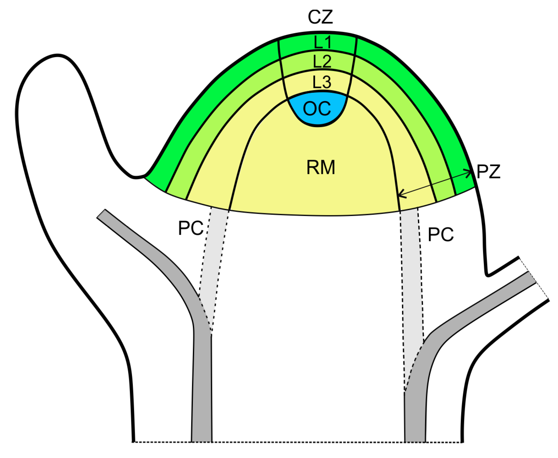
Figure 1. Diagram of the shoot apical meristem (SAM) in the model plant Arabidopsis, showing structural and functional zonation. L1 and L2 refer to the layers of the initial cells giving rise to two tunica layers (shown in green) and L3 to the internal layer of the initial cells for the corpus (shown in yellow). Vascular strands are shown in grey; procambium reaches the internal parts of the peripheral zone of the meristem is shown in light grey, differentiating vascular traces are in dark grey. Abbreviations: CZ—central zone; OC—organizing center; PC—procambium; PZ—peripheral (organogenic) zone; RM—rib meristem.
Symmetry 15 00364 g001
Figure 2. The dual pathway of auxin transport. (A) Transport in L1 (green) prevails, leading to formation of auxin maximum (dark green) in the peripheral zone of the SAM. Then, basipetal auxin transport from the primordium results in the development of the vascular strand (light green). (B) Transport in developing vascular strand (green) is dominant; vascular strand (green) reaches the peripheral zone of the SAM prior to formation of the primordium (dark green); L1 is shown in light green. Acropetally transported auxin is marked by arrows.
Symmetry 15 00364 g002
Disclaimer/Publisher’s Note: The statements, opinions and data contained in all publications are solely those of the individual author(s) and contributor(s) and not of MDPI and/or the editor(s). MDPI and/or the editor(s) disclaim responsibility for any injury to people or property resulting from any ideas, methods, instructions or products referred to in the content.