Review

Symmetry Breaking and Establishment of Dorsal/Ventral Polarity in the Early Sea Urchin Embryo

by
Vincenzo Cavalieri
1,2,* and
Giovanni Spinelli
1,*
1
Department of Biological, Chemical and Pharmaceutical Sciences and Technologies (STEBICEF), University of Palermo, Viale delle Scienze Edificio 16, Palermo 90128, Italy
2
Mediterranean Center for Human Health Advanced Biotechnologies (CHAB), University of Palermo, Viale delle Scienze Edificio 16, Palermo 90128, Italy
*
Authors to whom correspondence should be addressed.
Symmetry 2015, 7(4), 1721-1733; https://doi.org/10.3390/sym7041721
Submission received: 27 July 2015 / Revised: 28 August 2015 / Accepted: 22 September 2015 / Published: 28 September 2015
(This article belongs to the Special Issue Symmetry and Asymmetry in Biology)

Abstract

:
The mechanisms imposing the Dorsal/Ventral (DV) polarity of the early sea urchin embryo consist of a combination of inherited maternal information and inductive interactions among blastomeres. Old and recent studies suggest that a key molecular landmark of DV polarization is the expression of nodal on the future ventral side, in apparent contrast with other metazoan embryos, where nodal is expressed dorsally. A subtle maternally-inherited redox anisotropy, plus some maternal factors such as SoxB1, Univin, and p38-MAPK have been identified as inputs driving the spatially asymmetric transcription of nodal. However, all the mentioned factors are broadly distributed in the embryo as early as nodal transcription occurs, suggesting that repression of the gene in non-ventral territories depends upon negative regulators. Among these, the Hbox12 homeodomain-containing repressor is expressed by prospective dorsal cells, where it acts as a dorsal-specific negative modulator of the p38-MAPK activity. This review provides an overview of the molecular mechanisms governing the establishment of DV polarity in sea urchins, focusing on events taking place in the early embryo. Altogether, these findings provide a framework for future studies aimed to unravel the inceptive mechanisms involved in the DV symmetry breaking.

1. Introduction

During development of bilaterian embryos symmetry breaking is imposed through establishing of distinct polarities, which are precursors of the larval axes. Identifying the molecular mechanisms underlying the initial symmetry breaking is still now an overriding goal in understanding how nature is able to produce organisms with consistent and distinct anatomies.
In indirectly developing sea urchins, embryonic patterning along the dorsal/ventral (DV) axis, also known as oral/aboral axis, has been extensively studied in various species. In contrast to vertebrates, there is no developmental relationship between the embryonic DV axis and the structures of the body plan formed along the adult DV axis, which arises within the imaginal rudiment during the post-embryonic larval metamorphosis [1,2,3].
Irreversible establishment of the embryonic DV axis does certainly not occur prior to fertilization, nor does it display any consistent relation to the point of sperm entry [4,5,6]. Classical experimental embryology investigations surveying several sea urchin species indicated that cleavage per se does not play a causal role in the establishment of the DV axis. In fact, although there is a predominant association between the plane of the first cleavage division and the future DV axis in Strongylocentrotus purpuratus [7], the orientation of the first cleavage furrow and DV axis differs widely between species, and even amongst the embryos of a single individual [8,9,10,11,12].
More than one century ago, the seminal bisection experiments of Driesch testifying the totipotency of two- and four-cell stage sea urchin blastomeres [13], clearly indicated that the DV axis is not firmly established in the unfertilized egg. Rather, it is progressively specified during early cleavage through a conditional process that relies on a combination of inherited maternal information and inductive interactions among blastomeres [14,15,16]. Nodal signaling provides a fundamental driver input for specifying DV asymmetries [17,18], although, differently with respect to other metazoan embryos, in the sea urchin Nodal operates on the ventral rather than on the dorsal side.
DV polarity becomes morphologically recognizable from the onset of gastrulation, when the embryo begins to flatten on the ventral side, and two bilaterally symmetric thickenings of the ectoderm form on ventrolateral regions (Figure 1). In the intervening time, subpopulations of primary mesenchyme cells migrate within the blastocoel cavity guided by signals emitted by these regions, settle into two ventrolateral aggregates adjacent to the ectoderm thickenings, and eventually mineralize two skeletal primordia [19,20,21]. At the end of gastrulation, the opening of the larval mouth occurs by bending of the archenteron towards the ventral ectoderm and subsequent fusion of the two epithelia. At the pluteus stage (Figure 1), the ectoderm is noticeably partitioned into four main domains along the DV axial coordinates: (1) the ventral/oral ectoderm, a thickened epithelium surrounding the mouth; (2) the dorsal/aboral ectoderm, a squamous epithelium that covers most of the rest of the larval body; (3) the ciliary band, a belt of ciliated cells positioned at the border between ventral and dorsal ectoderm; and (4) the apical neurogenic domain. Furthermore, the dark-red pigment cells, derived from secondary mesenchyme precursors, can be easily recognized by their dispersed disposition embedded within the dorsal, but not the ventral, ectoderm layer (Figure 1).
With careful examination, one can detect many earlier signs of DV asymmetries and there are now a number of early molecular indicators of these properties.
Figure 1. Simplified scheme depicting key developmental stages and early molecular activities regulating morphogenesis along the DV axis of the sea urchin embryo. See text for details.
Symmetry 07 01721 g001

2. Symmetry Breaking: Subtle Redox Anisotropies Prefiguring the DV Axis

The first molecular manifestation that foreshadows DV polarity occurs during the initial cleavage stages, consisting in a redox gradient sustained by mitochondrial cytochrome oxidase activity [22,23]. More precisely, high redox potential, and the resulting oxidizing environment, significantly correlates with ventral fate, although this juxtaposing association is not statistically observed in 100% of cases [24,25,26].
Pioneering evidence highlighted that DV polarity can be biased by exposing embryos to hypoxic condition or respiratory inhibitors [22,27]. More recent studies in S. purpuratus also indicated that DV polarity is similarly assigned if embryos are cultured in immobilized tight clusters, wherein a redox gradient forms across the inside-outside axis of the cluster [24]. Additional pieces of information highlighted that such a redox gradient relies upon the maternally-derived uneven distribution of mitochondria (Figure 1), and that disruption of such an anisotropy, by either centrifugation of eggs (which displaces mitochondria toward the centrifugal pole) or injection of purified mitochondria, can entrain DV polarization [25]. In support of these findings, it has been shown that treatment with compounds, such as Cobalt (Co(II)), that in other systems stimulate the generation of uniform high levels of reactive oxygen species (ROS) perturbs DV patterning of the resulting embryos, most likely altering a necessary redox asymmetry [28,29,30].
Indeed, the asymmetrical mitochondrial distribution in the S. purpuratus embryo correlates with both differential intracellular ROS levels, mainly in the form of H2O2, and nodal expression [26]. In fact, quenching mitochondrial H2O2 emissions by clonal overexpression of mitochondrially-targeted catalase entrains DV polarity by under-expression of nodal [26]. Similarly, embryos cultured under hypoxic conditions through early cleavage exhibit significantly decreased levels of H2O2 and develop as radialized larvae lacking DV polarity [25,31]. Worth mentioning, nodal expression is not abolished in these larvae, but rather its spatial localization to one side of the embryo is prevented by hypoxia, revealing that redox anisotropies are not required to activate nodal transcription by itself, but rather to provide an initial spatial bias in the rate of nodal transcription [32].
A possible link between redox gradient and nodal expression has been postulated by the known function of the p38 MAP kinase signaling pathway in the transcriptional responses downstream of oxidative stress in several metazoans [33,34,35,36]. Formerly identified in Lytechinus variegatus as a kinase whose activity is required for nodal expression [37], p38 has been subsequently demonstrated to be responsive to ROS in S. purpuratus embryos [26]. Further downstream, p38 is thought to activate the maternal redox-sensitive bZIP and Oct1/2 factors only on the ventral side of the early embryo, reflecting the initial respiratory anisotropy into a polarized transcriptional regulatory state [37,38,39,40]. Importantly, binding sites for the mentioned factors have been identified in the cis-regulatory module controlling the initiation of nodal gene transcription [38,39], strongly suggesting that redox signaling influence DV patterning through nodal.

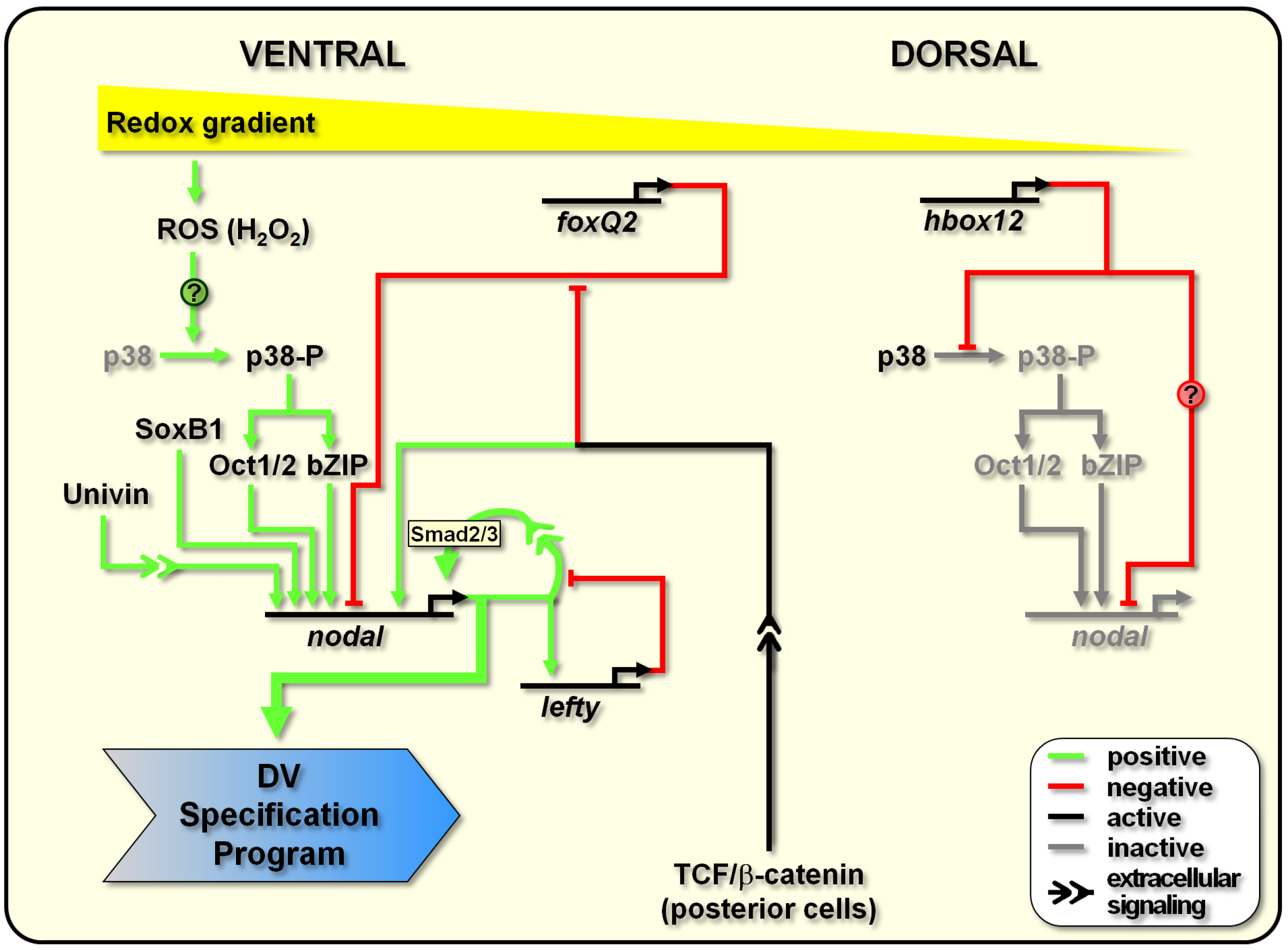
3. DV Polarity Establishment: Early Positive and Negative Activities Shaping the Nodal-Expressing Organizing Centre

The genetic landmark of polarization along the secondary axis is the zygotic expression of Nodal, a member of the TGF-β superfamily. Previous studies in Paracentrotus lividus and S. purpuratus species shown that nodal is first transcribed broadly, at an extremely low level, all around the prospective ectoderm of the 32/60-cell stage embryo [17,18,40]. Afterwards, nodal expression is rapidly downregulated in the presumptive dorsal blastomeres, exclusively marking the prospective ventral ectoderm of the early blastula [17,18,40]. Not only nodal expression is required for specification of the latter territory, but the small group of cells that specifically express nodal at the early blastula stage, thereupon behaves as an organizing centre imposing DV polarity in all three germ layers of the embryo [17,18,41,42,43].
As mentioned in the previous section, the p38 signaling pathway could bridge the redox gradient to nodal expression. Although being globally active during embryogenesis of the sea urchin, p38 becomes transiently inactivated in dorsal blastomeres, viz at the low end of the respiratory gradient, just before the onset of nodal expression [37]. Moreover, inhibition of the p38 function abrogates nodal transcription and, in turn, disrupts DV polarization [37]. According to the mentioned model, at the early blastula stage the maternally related anisotropy in redox gradient would transiently inactivate the p38 kinase in the future dorsal ectoderm [26,37], somehow leading to the activation of predicted redox-sensitive transcription factors on the ventral side [38,39,40]. The cis-regulatory apparatus of nodal responds to these factors, as well as to the maternal positive inputs of SoxB1 and Univin (another TGF-β family member), directing the expression of the gene within a discrete sector of the ectoderm that exactly corresponds to the presumptive ventral ectoderm [17,18,38,39,44].
DV axis formation and nodal expression are also dependent on functional Wnt signaling emanating from the posterior, or vegetal, pole of the cleaving embryo [17]. Nuclearization of β-catenin in posterior blastomeres allows the interaction with TCF/Lef transcription factor to regulate downstream gene expression and, in turn, expands the β-catenin signal to adjacent cell layers in a posterior to anterior direction [45,46,47,48].
Dissection of the cis-regulatory apparatus of the nodal gene identified a TCF/Lef binding site that is required for proper expression of a promoter-reporter transgene [39]. However, direct association of TCF/β-catenin heterodimer to this site has not been mapped so far, probably because the nuclearization of β-catenin is not detected in cells of the anterior, or animal, hemisphere [46,49]. Most probably, β-catenin regulates nodal transcription not directly but, rather, through an unidentified signal originating from posterior cells. In addition, although blocking the nuclearization of β-catenin represses nodal expression at the blastula stage [17], the initial transcription of nodal is not affected [50], indicating that such a vegetal signal acts on the maintenance of nodal expression rather than the initial activation.
Although all of the known positive inputs activating nodal transcription are present maternally and very broadly distributed in the early embryo, ectopic expression of nodal beyond the ventral ectoderm founder cells is specifically hampered by tissue-specific negative regulators. In this regard, we have shown in P. lividus that the Hbox12 homeodomain repressor is expressed by prospective dorsal ectoderm cells, spatially facing and preceding the onset of nodal transcription, where it acts preventing the ectopic activation of nodal expression [51,52,53,54]. Functional analysis revealed that expression of hbox12 and nodal genes are mutually exclusive and both required for DV polarization. In fact, overexpression of Hbox12 specifically attenuates nodal transcription, while loss of Hbox12 function allows broad ectopic expression of nodal, and in both the experimental assays the resulting embryos do not acquire DV polarity [53,54]. Intriguingly, Hbox12 is functionally upstream of p38, being specifically involved in the transient inactivation of the kinase in dorsal blastomeres [53]. Therefore, not only hbox12 is the earliest known zygotic gene differentially expressed along DV axis, but it also represents the foremost negative regulator allowing competence for spatial positioning of the DV organizer.
FoxQ2, another transcription repressor, also contributes to prevent the spatial spreading of nodal expression in the animal hemisphere. Expression of foxQ2 begins slightly earlier than that of nodal, initially in all animal blastomeres, driven by the Six3 transcription activator [50,55,56]. Soon thereafter, posterior Wnt1 and Wnt8 signals act in parallel to progressively restrict foxQ2 transcription in the apical neurogenic ectoderm [50,57,58]. This temporally coordinating mechanism prevents nodal expression from prematurely reaching high levels. However, although overexpression of foxQ2 efficiently abrogates nodal transcription, the opposite effect on nodal in the animal plate is obtained only when the functions of FoxQ2 and Lefty (see below) are doubly knocked down, suggesting a functional cooperation between the two negative regulators of Nodal signaling [50].
A further attractive regulator potentially involved in the spatial control of nodal expression is the Myb transcription factor. Although the spatial distribution of Myb in the early embryo has not yet been determined, Myb is a redox-sensitive factor [59,60] known to act as a repressor in ventral territories of the sea urchin embryo [61]. This evidence has been provided by the cis-regulatory analysis of the CyIIIa and hbox12 genes, both beginning to be specifically transcribed in the presumptive dorsal ectoderm during early embryogenesis [52,61]. Surprisingly, the cis-regulatory apparatus of the nodal gene also contains a Myb-like consensus binding sequence that, when mutated in gene transfer assays, leads to increased reporter expression [39]. This opens the possibility that the Myb repressor might work to fine-tune the transcriptional output of the nodal gene in ventral cells.

4. DV Polarity Maintenance: How the Organizing Centre Works

Once the expression of nodal is initiated by early inputs, the peculiar ventral localization of the nodal-expressing domain is probably consolidated by a reaction-diffusion system, although direct evidence have to be provided in the sea urchin. In fact, the best direct evidence for it comes from a recent study in zebrafish, where the effective diffusion coefficients of GFP-tagged TGF-β proteins were measured during embryogenesis [62]. The reaction-diffusion system would involve a positive feedback loop related to the short-range Nodal signal transduction system, and a concurrent Nodal-dependent mechanism given by the production of the Nodal antagonist Lefty [17,38,63,64]. With respect to the former facet, experimental confirmation comes from cis-regulatory studies, since mutation, in the promoter of nodal, of sequences postulated to be targeted by Smad2/3, a transducer of Nodal signaling [26,65], reduces the reporter expression in transgenic embryos [38,39].
Lefty and Nodal are produced by the same cells, albeit the former is thought to diffuse more rapidly, thus acting as a long-range Nodal inhibitor [17,63,64]. In accordance with this hypothesis, DV axis and nodal expression are both lost in embryos in which Lefty is overexpressed. By contrast, impairing Lefty function converts most of the embryonic territories toward a ventral fate through ectopic expression of nodal [41,63].
Direct targets of Nodal signaling within the ventral ectoderm also include genes encoding the TGF-β pathway extracellular components BMP2/4 and Chordin [17,66,67,68,69,70]. The BMP2/4 ligand acts as a diffusible relay molecule to specify the dorsal ectoderm, to which its signaling activity is confined, due to inhibition of BMP2/4 reception by Chordin within the ventral ectoderm [17,67,69,71]. This model, which is also supported by computational simulation [72], explains why in the absence of nodal expression, not only the specification of ventral structures is abolished, but the differentiation of dorsal territories is suppressed as well [17].
It must to be also taken into account that signaling events require differential migration of all the mentioned cytokines across the fibrous mesh of the extracellular matrix that surrounds and support cells. In such a milieu, proteoglycans has been suggested to play fundamental roles in the differential localization and stability of TGF-β ligands during axial patterning of various metazoans, including sea urchins [69,73,74,75,76]. Likewise, a recent study indicates that dynamin-mediated endocytosis limits the range of Nodal diffusion in S. purpuratus embryos [77].
The amount of details available on molecular circuits that govern DV patterning of the embryonic territories downstream of nodal expression is growing rapidly and, so far, more than fifty genes are known to be regulated, either directly or indirectly, by Nodal signaling [44,70,71,78,79,80]. Large scale studies began to describe the epistatic relationships among these genes [44,81], highlighting that sequential interplay between inductive and suppressive events controlling Nodal signaling is critical for DV patterning. In this regard, wnt1 signaling from the posterior pole suppresses nodal transcription in posterior ventral cells during gastrulation, contributing to sculpt the spatial expression pattern of nodal even at later stages [82]. In wnt1 morphants the nodal expressing territory is expanded posteriorly in the ventral side, provoking DV patterning defects that include a ventral to dorsal shift in the position of the posterior ciliary band [82]. This finding reveals that there is a continuing requirement to restrict Nodal signaling throughout development.

5. Conclusions and Future Perspectives

While DV polarization is morphologically not apparent in the sea urchin zygote, axial specification is accomplished at the molecular level in the early embryo. Deciphering the molecular hierarchies that govern the establishment of DV axis provides explanation of how the spatially asymmetric expression of regulatory genes is gradually translated into the anatomically distinct structures of the embryo. The critical molecular events discussed in the previous sections are diagrammatically summarized in Figure 2. Although the substantial progress earned in the last decade of experiments, since the role of nodal began to be characterized in sea urchins, a number of fuzzy points still need to be clarified.
How the labile redox anisotropies in the egg and early embryo are reflected into polarized gene expression patterns remains a central but largely enigmatic issue. Functional analysis revealed that Hbox12 is required for the transient inactivation of the p38 kinase in dorsal blastomeres, raising the question of whether hbox12 gene expression and/or function are directly regulated by the redox state. Being hbox12 expression achieved in the reducing environment specifically associated to the dorsal side of the early embryo, a likely candidate involved in the activation of hbox12 transcription could be HIF1α. This hypothesis deserves further investigation, being supported by recent studies showing that the HIF1α transcription activator is stabilized in a reducing environment [83,84], and by the fact that knock-down of HIF1α function strongly reduces the expression of dorsal-specific markers in the very early sea urchin embryo [85].
In an alternative scenario, inter-specific differences could exist in the initial mechanisms used to specify DV polarity. In fact, while in recent years the role of the mitochondrial gradient and redox signaling have been firmly established in S. purpuratus, they have not been rigorously investigated in any other species. However, threaded between the lines of a recent paper, it is mentioned that experimental manipulations that perturb the redox gradient have very modest effects on the spatial expression of nodal in P. lividus [44]. In light of this, it remains possible that redox polarization represents a species-specific mechanism.
Actually, given the robustness of the reaction-diffusion device, all that is needed for the Nodal-Lefty genetic circuit to break DV symmetry is any of a number of alternative stochastic means that generate an initial spatial bias in nodal transcription along the secondary axis. Thus, S. purpuratus may use mitochondrial signaling, while other species may utilize, for instance, localization of a transcription factor.
In this regard, the requirement for Hbox12 in DV axis specification has thus far only been established in the P. lividus species [53,54]. Available evidence indeed indicates that there is no hbox12 orthologue in S. purpuratus, and that the closest homolog of hbox12 in the latter species is pmar1 [51,53], which is instead involved in micromere lineage specification [86]. This strongly suggests that DV axis specification in S. purpuratus cannot depend on hbox12, since the gene probably does not exist in that species. Hence, there is the very captivating possibility that the mechanism of initial axis specification is completely different between S. purpuratus and P. lividus species. In other words, the former utilizes redox polarization to achieve the initial localization of nodal activity, while the latter requires localized transcriptional activation of Hbox12, independently of redox signaling.
This hypothesis is advocated by the well-fitting clue that the two sea urchin species actually differ in some aspects of DV axis determination. In particular, in S. purpuratus the DV axis passes through a plane about 45° clockwise from the first cleavage furrow [7], indicating that secondary axis specification is initiated between fertilization and first cleavage, which is consistent with the asymmetric distribution of mitochondria within eggs and early embryos of this species [25]. Classical studies in P. lividus embryos instead demonstrate that DV axis is randomly oriented with respect to the first cleavage plane, and that it is established between the fifth and eighth cleavage [8], which broadly corresponds to the peak of hbox12 transcription [51,53].
Whatever is the mechanism providing the initial bias of nodal activity in the early embryo, it most likely reverberates on the differential modulation of p38 activity. Therefore, it will be important to determine not only whether p38 directly phosphorylates the predicted bZIP and Oct1/2 downstream targets, but also whether or not the phosphorylation status influences the activity of the mentioned transcription factors in the sea urchin embryo. Albeit investigation on Oct1/2 recently began [40], the exact identity of the bZIP transcription factor, among those conserved in the echinoderm genomes, supposed to be involved in the redox transduction pathway is currently not known. No less important will be the mapping of the physical binding of the redox-sensitive transcription factors, including Myb, to the cognate cis-regulatory sequences predicted in the nodal promoter.
Figure 2. Diagram illustrating the early molecular events regulating the establishment of the DV organizing centre in the sea urchin embryo. See text for details.
Symmetry 07 01721 g002

Acknowledgments

We apologize to those authors whose work has not been cited. We are grateful to the three anonymous reviewers for their stimulating suggestions and comments. Research in our lab is funded by grants from Università degli Studi di Palermo (STEMBIO Award and ex60% to Vincenzo Cavalieri) and Assessorato Regionale della Salute, Regione Siciliana (PO FESR 4.1.1.1 RIMEDRI to Vincenzo Cavalieri and Giovanni Spinelli).

Conflicts of Interest

The authors declare no conflict of interest.

References

  1. Pearse, J.S.; Cameron, R.A. Echinodermata: Echinoidea. In Reproduction of Marine Invertebrates; Giese, A.C., Pearse, J.S., Pears, V.B., Eds.; Boxwood: Pacific Grove, CA, USA, 1991; pp. 513–622. [Google Scholar]
  2. Peterson, K.J.; Cameron, R.A.; Davidson, E.H. Set-aside cells in maximal indirect development: Evolutionary and developmental significance. BioEssays 1997, 19, 623–631. [Google Scholar] [CrossRef] [PubMed]
  3. Smith, M.M.; Cruz Smith, L.; Cameron, R.A.; Urry, L.A. The larval stages of the sea urchin, Strongylocentrotus purpuratus. J. Morphol. 2008, 269, 713–733. [Google Scholar] [CrossRef] [PubMed]
  4. Horstadius, S. Experimental Embryology of Echinoderms; Clarendon Press: Oxford, UK, 1973. [Google Scholar]
  5. Schroeder, T. Expressions of the prefertilization polar axis in sea urchin eggs. Dev. Biol. 1980, 79, 428–443. [Google Scholar] [CrossRef]
  6. Henry, J.J. The development of dorsoventral and bilateral axial properties in sea urchin embryos. Semin. Cell Dev. Biol. 1998, 9, 43–52. [Google Scholar] [CrossRef] [PubMed]
  7. Cameron, R.A.; Fraser, S.E.; Britten, R.J.; Davidson, E.H. The oral-aboral axis of a sea urchin embryo is specified by first cleavage. Development 1989, 106, 641–647. [Google Scholar] [PubMed]
  8. Horstadius, S.; Wolski, A. Studien uber die Determination der Bilateralsymmetrie des jungen Seeigelkeims. Wilhelm Roux Arch. Entwicklungsmech. Org. 1936, 135, 69–113. [Google Scholar] [CrossRef]
  9. Kominami, T. Determination of dorsoventral axis in early embryos of the sea urchin, Hemicentrotus pulcherrimus. Dev. Biol. 1988, 127, 187–196. [Google Scholar] [CrossRef]
  10. Henry, J.J.; Klueg, K.M.; Raff, R.A. Evolutionary dissociation between cleavage, cell lineage and embryonic axes in sea urchin embryos. Development 1992, 114, 931–938. [Google Scholar] [PubMed]
  11. Jeffery, W.R. Axis determination in sea urchin embryos: From confusion to evolution. Trends Genet. 1992, 8, 223–225. [Google Scholar] [CrossRef]
  12. Summers, R.G.; Piston, D.W.; Harris, K.M.; Morrill, J.B. The orientation of first cleavage in the sea urchin embryo, Lytechinus variegatus, does not specify the axes of bilateral symmetry. Dev. Biol. 1996, 175, 177–183. [Google Scholar] [CrossRef] [PubMed]
  13. Driesch, H. The potency of the first two cleavage cells in echinoderm development: Experimental production of partial and double formations. In Foundations of Experimental Embryology; Willier, B.H., Oppenheimer, J.M., Eds.; Hafner: New York, NY, USA, 1892; pp. 39–50. [Google Scholar]
  14. Brandhorst, B.P.; Klein, W.H. Molecular patterning along the sea urchin animal-vegetal axis. Int. Rev. Cytol. 2002, 213, 183–232. [Google Scholar] [PubMed]
  15. Angerer, L.M.; Angerer, R.C. Patterning the sea urchin embryo: Gene regulatory networks, signaling pathways, and cellular interactions. Curr. Top. Dev. Biol. 2003, 53, 159–198. [Google Scholar] [PubMed]
  16. Molina, M.D.; de Crozé, N.; Haillot, E.; Lepage, T. Nodal: Master and commander of the dorsal-ventral and left-right axes in the sea urchin embryo. Curr. Opin. Genet. Dev. 2013, 23, 445–453. [Google Scholar] [CrossRef] [PubMed]
  17. Duboc, V.; Röttinger, E.; Besnardeau, L.; Lepage, T. Nodal and BMP2/4 signaling organizes the oral-aboral axis of the sea urchin embryo. Dev. Cell 2004, 6, 397–410. [Google Scholar] [CrossRef]
  18. Flowers, V.L.; Courteau, G.R.; Poustka, A.J.; Weng, W.; Venuti, J.M. Nodal/activin signaling establishes oral-aboral polarity in the early sea urchin embryo. Dev. Dyn. 2004, 231, 727–740. [Google Scholar] [CrossRef] [PubMed]
  19. Duloquin, L.; Lhomond, G.; Gache, C. Localized VEGF signaling from ectoderm to mesenchyme cells controls morphogenesis of the sea urchin embryo skeleton. Development 2007, 134, 2293–2302. [Google Scholar] [CrossRef] [PubMed]
  20. Röttinger, E.; Saudemont, A.; Duboc, V.; Besnardeau, L.; McClay, D.; Lepage, T. FGF signals guide migration of mesenchymal cells, control skeletal morphogenesis and regulate gastrulation during sea urchin development. Development 2008, 135, 353–365. [Google Scholar] [CrossRef] [PubMed]
  21. Cavalieri, V.; Guarcello, R.; Spinelli, G. Specific expression of a TRIM-containing factor in ectoderm cells affects the skeletal morphogenetic program of the sea urchin embryo. Development 2011, 138, 4279–4290. [Google Scholar] [CrossRef] [PubMed]
  22. Child, C.M. Exogastrulation by sodium azide and other inhibiting conditions in Strongylocentrotus purpuratus. J. Exp. Zool. 1948, 107, 1–38. [Google Scholar] [CrossRef] [PubMed]
  23. Czihak, G. Investigation of developmental physiology in echinoids (distribution and role of cytochrome oxidase). Roux Arch. Entwick Mech. Org. 1963, 154, 272–292. [Google Scholar] [CrossRef]
  24. Coffman, J.A.; Davidson, E.H. Oral-aboral axis specification in the sea urchin embryo. I. Axis entrainment by respiratory asymmetry. Dev. Biol. 2001, 230, 18–28. [Google Scholar] [CrossRef]
  25. Coffman, J.A.; McCarthy, J.J.; Dickey-Sims, C.; Robertson, A.J. Oral-aboral axis specification in the sea urchin embryo II. Mitochondrial distribution and redox state contribute to establishing polarity in Strongylocentrotus purpuratus. Dev. Biol. 2004, 273, 160–171. [Google Scholar] [CrossRef] [PubMed]
  26. Coffman, J.A.; Coluccio, A.; Planchart, A.; Robertson, A.J. Oral-aboral axis specification in the sea urchin embryo III. Role of mitochondrial redox signaling via H2O2. Dev. Biol. 2009, 330, 123–130. [Google Scholar] [CrossRef] [PubMed]
  27. Pease, D.C. Echinoderm bilateral determination in chemical concentration gradients. I. The effects of cyanide, ferricyanide, iodoacetate, picrate, dinitrophenol, urethane, iodine, malonate, etc. J. Exp. Zool. 1941, 86, 381–404. [Google Scholar]
  28. Chandel, N.S.; Maltepe, E.; Goldwasser, E.; Mathieu, C.E.; Simon, M.C.; Schumacker, P.T. Mitochondrial reactive oxygen species trigger hypoxia-induced transcription. Proc. Natl. Acad. Sci. USA 1998, 95, 11715–11720. [Google Scholar] [CrossRef] [PubMed]
  29. Pourahmad, J.; O’Brien, P.J.; Jokar, F.; Daraei, B. Carcinogenic metal induced sites of reactive oxygen species formation in hepatocytes. Toxicol. In Vitro 2003, 17, 803–810. [Google Scholar] [CrossRef]
  30. Agca, C.; Klein, W.H.; Venuti, J.M. Respecification of ectoderm and altered Nodal expression in sea urchin embryos after cobalt and nickel treatment. Mech. Dev. 2009, 126, 430–442. [Google Scholar] [CrossRef] [PubMed]
  31. Coluccio, A.E.; LaCasse, T.J.; Coffman, J.A. Oxygen, pH, and oral-aboral axis specification in the sea urchin embryo. Mol. Reprod. Dev. 2011, 78. [Google Scholar] [CrossRef] [PubMed]
  32. Coffman, J.A.; Wessels, A.; DeSchiffart, C.; Rydlizky, K. Oral-aboral axis specification in the sea urchin embryo, IV: Hypoxia radializes embryos by preventing the initial spatialization of nodal activity. Dev. Biol. 2014, 386, 302–307. [Google Scholar] [CrossRef] [PubMed]
  33. Clerk, A.; Fuller, S.J.; Michael, A.; Sugden, P.H. Stimulation of “stress-regulated” mitogen-activated protein kinases (stress-activated protein kinases/c-Jun N-terminal kinases and p38-mitogen-activated protein kinases) in perfused rat hearts by oxidative and other stresses. J. Biol. Chem. 1998, 273, 7228–7234. [Google Scholar] [CrossRef] [PubMed]
  34. Torres, M.; Forman, H.J. Redox signaling and the MAP kinase pathways. Biofactors 2003, 17, 287–296. [Google Scholar] [CrossRef] [PubMed]
  35. Inoue, H.; Hisamoto, N.; An, J.H.; Oliveira, R.P.; Nishida, E.; Blackwell, T.K.; Matsumoto, K. The C. elegansp38 MAPK pathway regulates nuclear localization of the transcription factor SKN-1 in oxidative stress response. Genes Dev. 2005, 19, 2278–2283. [Google Scholar] [CrossRef]
  36. Son, Y.; Kim, S.; Chung, H.T.; Pae, H.O. Reactive oxygen species in the activation of MAP kinases. Methods Enzymol. 2013, 528, 27–48. [Google Scholar] [PubMed]
  37. Bradham, C.A.; McClay, D.R. p38 MAPK is essential for secondary axis specification and patterning in sea urchin embryos. Development 2006, 133, 21–32. [Google Scholar] [CrossRef] [PubMed]
  38. Nam, J.; Su, Y.H.; Lee, P.Y.; Robertson, A.J.; Coffman, J.A.; Davidson, E.H. Cis-regulatory control of the nodal gene, initiator of the sea urchin oral ectoderm gene network. Dev. Biol. 2007, 306, 860–869. [Google Scholar] [CrossRef] [PubMed]
  39. Range, R.; Lapraz, F.; Quirin, M.; Marro, S.; Besnardeau, L.; Lepage, T. Cis-regulatory analysis of nodal and maternal control of dorsal-ventral axis formation by Univin, a TGF-b related to Vg1. Development 2007, 134, 3649–3664. [Google Scholar] [CrossRef] [PubMed]
  40. Range, R.; Lepage, T. Maternal Oct1/2 is required for Nodal and Univin/Vg1 expression during dorsal-ventral axis specification in the sea urchin embryo. Dev. Biol. 2011, 357, 440–449. [Google Scholar] [CrossRef] [PubMed]
  41. Duboc, V.; Lapraz, F.; Saudemont, A.; Bessodes, N.; Mekpoh, F.; Haillot, E.; Quirin, M.; Lepage, T. Nodal and BMP2/4 pattern the mesoderm and endoderm during development of the sea urchin embryo. Development 2010, 137, 223–235. [Google Scholar] [CrossRef] [PubMed]
  42. Ohguro, Y.; Takata, H.; Kominami, T. Involvement of Delta and Nodal signals in the specification process of five types of secondary mesenchyme cells in embryo of the sea urchin, Hemicentrotus pulcherrimus. Dev. Growth Differ. 2011, 53, 110–123. [Google Scholar] [CrossRef] [PubMed]
  43. Materna, S.C.; Ransick, A.; Li, E.; Davidson, E.H. Diversification of oral and aboral mesodermal regulatory states in pregastrularsea urchinembryos. Dev. Biol. 2013, 375, 92–104. [Google Scholar] [CrossRef]
  44. Saudemont, A.; Haillot, E.; Mekpoh, F.; Bessodes, N.; Quirin, M.; Lapraz, F.; Duboc, V.; Röttinger, E.; Range, R.; Oisel, A.; et al. Ancestral regulatory circuits governing ectoderm patterning downstream of Nodal and BMP2/4 revealed by gene regulatory network analysis in an echinoderm. PLoS Genet. 2010, 6. [Google Scholar] [CrossRef] [PubMed]
  45. Wikramanayake, A.H.; Huang, L.; Klein, W.H. beta-Catenin is essential for patterning the maternally specified animal-vegetal axis in the sea urchin embryo. Proc. Natl. Acad. Sci. USA 1998, 95, 9343–9348. [Google Scholar] [CrossRef] [PubMed]
  46. Logan, C.Y.; Miller, J.R.; Ferkowicz, M.J.; McClay, D.R. Nuclear beta-catenin is required to specify vegetal cell fates in the sea urchin embryo. Development 1999, 126, 345–357. [Google Scholar] [PubMed]
  47. Davidson, E.H.; Rast, J.P.; Oliveri, P.; Ransick, A.; Calestani, C.; Yuh, C.H.; Minokawa, T.; Amore, G.; Hinman, V.; Arenas-Mena, C.; et al. A provisional regulatory gene network for specification of endomesoderm in the sea urchin embryo. Dev. Biol. 2002, 246, 162–190. [Google Scholar] [CrossRef] [PubMed]
  48. Sethi, A.J.; Wikramanayake, R.M.; Angerer, R.C.; Range, R.C.; Angerer, L.M. Sequential signaling crosstalk regulates endomesoderm segregation in sea urchin embryos. Science 2012, 335, 590–593. [Google Scholar] [CrossRef] [PubMed]
  49. Yazaki, I.; Tsurugaya, T.; Santella, L.; Chun, J.T.; Amore, G.; Kusunoki, S.; Asada, A.; Togo, T.; Akasaka, K. Ca2+ influx-linked protein kinase C activity regulates the β-catenin localization, micromere induction signalling and the oral-aboral axis formation in early sea urchin embryos. Zygote 2015, 23, 426–446. [Google Scholar] [CrossRef] [PubMed]
  50. Yaguchi, S.; Yaguchi, J.; Angerer, R.C.; Angerer, L.M. A Wnt-FoxQ2-nodal pathway links primary and secondary axis specification in sea urchin embryos. Dev. Cell 2008, 14, 97–107. [Google Scholar] [CrossRef] [PubMed]
  51. Di Bernardo, M.; Russo, R.; Oliveri, P.; Melfi, R.; Spinelli, G. Homeobox-containing gene transiently expressed in a spatially restricted pattern in the early sea urchin embryo. Proc. Natl. Acad. Sci. USA 1995, 92, 8180–8184. [Google Scholar] [CrossRef] [PubMed]
  52. Cavalieri, V.; Di Bernardo, M.; Anello, L.; Spinelli, G. cis-Regulatory sequences driving the expression of the Hbox12 homeobox-containing gene in the presumptive aboral ectoderm territory of the Paracentrotus lividus sea urchin embryo. Dev. Biol. 2008, 321, 455–469. [Google Scholar] [CrossRef] [PubMed]
  53. Cavalieri, V.; Spinelli, G. Early asymmetric cues triggering the dorsal/ventral gene regulatory network of the sea urchin embryo. Elife 2014, 3. [Google Scholar] [CrossRef] [PubMed] [Green Version]
  54. Cavalieri, V.; Spinelli, G. Ectopic hbox12 expression evoked by histone deacetylase inhibition disrupts axial specification of the sea urchin embryo. PLoS ONE 2015. submitted for publication. [Google Scholar]
  55. Tu, Q.; Brown, C.T.; Davidson, E.H.; Oliveri, P. Sea urchin Forkhead gene family: Phylogeny and embryonic expression. Dev. Biol. 2006, 300, 49–62. [Google Scholar] [CrossRef] [PubMed]
  56. Wei, Z.; Yaguchi, J.; Yaguchi, S.; Angerer, R.C.; Angerer, L.M. The sea urchin animal pole domain is a Six3-dependent neurogenic patterning center. Development 2009, 136, 1179–1189. [Google Scholar] [CrossRef] [PubMed]
  57. Range, R.C.; Angerer, R.C.; Angerer, L.M. Integration of canonical and noncanonical Wnt signaling pathways patterns the neuroectoderm along the anterior-posterior axis of sea urchin embryos. PLoS Biol. 2013, 11. [Google Scholar] [CrossRef] [PubMed]
  58. Range, R. Specification and positioning of the anterior neuroectoderm in deuterostome embryos. Genesis 2014, 52, 222–234. [Google Scholar] [CrossRef] [PubMed]
  59. Guehmann, S.; Vorbrueggen, G.; Kalkbrenner, F.; Moelling, K. Reduction of a conserved Cys is essential for Myb DNA-binding. Nucleic Acids Res. 1992, 20, 2279–2286. [Google Scholar] [CrossRef] [PubMed]
  60. Myrset, A.H.; Bostad, A.; Jamin, N.; Lirsac, P.N.; Toma, F.; Gabrielsen, O.S. DNA and redox state induced conformational changes in the DNA-binding domain of the Myb oncoprotein. EMBO J. 1993, 12, 4625–4233. [Google Scholar] [PubMed]
  61. Coffman, J.A.; Kirchhamer, C.V.; Harrington, M.G.; Davidson, E.H. SpMyb functions as an intramodular repressor to regulate spatial expression of CyIIIa in sea urchin embryos. Development 1997, 124, 4717–4727. [Google Scholar] [PubMed]
  62. Müller, P.; Rogers, K.W.; Jordan, B.M.; Lee, J.S.; Robson, D.; Ramanathan, S.; Schier, A.F. Differential diffusivity of Nodal and Lefty underlies a reaction-diffusion patterning system. Science 2012, 336, 721–724. [Google Scholar] [CrossRef] [PubMed]
  63. Duboc, V.; Lapraz, F.; Besnardeau, L.; Lepage, T. Lefty acts as an essential modulator of Nodal activity during sea urchin oral-aboral axis formation. Dev. Biol. 2008, 320, 49–59. [Google Scholar] [CrossRef] [PubMed]
  64. Bolouri, H.; Davidson, E.H. The gene regulatory network basis of the “community effect”, and analysis of a sea urchin embryo example. Dev. Biol. 2010, 340, 170–178. [Google Scholar] [CrossRef] [PubMed]
  65. Yaguchi, S.; Yaguchi, J.; Burke, R.D. Sp-Smad2/3 mediates patterning of neurogenic ectoderm by nodal in the sea urchin embryo. Dev. Biol. 2007, 302, 494–503. [Google Scholar] [CrossRef] [PubMed]
  66. Hwang, S.L.; Chen, C.A.; Chen, C. Sea urchin TgBMP2/4 gene encoding a bone morphogenetic protein closely related to vertebrate BMP2 and BMP4 with maximal expression at the later stages of embryonic development. Biochem. Biophys. Res. Commun. 1999, 258, 457–463. [Google Scholar] [CrossRef] [PubMed]
  67. Angerer, L.M.; Oleksyn, D.W.; Logan, C.Y.; McClay, D.R.; Dale, L.; Angerer, R.C. A BMP pathway regulates cell fate allocation along the sea urchin animal-vegetal embryonic axis. Development 2000, 127, 1105–1114. [Google Scholar] [PubMed]
  68. Bradham, C.A.; Oikonomou, C.; Kühn, A.; Core, A.B.; Modell, J.W.; McClay, D.R.; Poustka, A.J. Chordin is required for neural but not axial development in sea urchinembryos. Dev. Biol. 2009, 328, 221–233. [Google Scholar]
  69. Lapraz, F.; Besnardeau, L.; Lepage, T. Patterning of the dorsal-ventral axis in echinoderms: Insights into the evolution of the BMP-chordin signaling network. PLoS Biol. 2009, 7. [Google Scholar] [CrossRef] [PubMed]
  70. Yaguchi, S.; Yaguchi, J.; Angerer, R.C.; Angerer, L.M.; Burke, R.D. TGF-beta signaling positions the ciliary band and patterns neurons in the sea urchin embryo. Dev. Biol. 2010, 347, 71–81. [Google Scholar] [CrossRef] [PubMed] [Green Version]
  71. Chen, J.H.; Luo, Y.J.; Su, Y.H. The dynamic gene expression patterns of transcription factors constituting the sea urchin aboral ectoderm gene regulatory network. Dev. Dyn. 2011, 240, 250–260. [Google Scholar] [CrossRef] [PubMed]
  72. Van Heijster, P.; Hardway, H.; Kaper, T.J.; Bradham, C.A. A computational model for BMP movement in sea urchin embryos. J. Theor. Biol. 2014, 363, 277–289. [Google Scholar] [CrossRef] [PubMed]
  73. Fujise, M.; Takeo, S.; Kamimura, K.; Matsuo, T.; Aigaki, T.; Izumi, S.; Nakato, H. Dally regulates Dpp morphogen gradient formation in the Drosophila wing. Development 2003, 130, 1515–1522. [Google Scholar] [CrossRef] [PubMed]
  74. Ohta, K.; Lupo, G.; Kuriyama, S.; Keynes, R.; Holt, C.E.; Harris, W.A.; Tanaka, H.; Ohnuma, S. Tsukushi functions as an organizer inducer by inhibition of BMP activity in cooperation with chordin. Dev. Cell 2004, 7, 347–358. [Google Scholar] [CrossRef] [PubMed]
  75. Marjoram, L.; Wright, C. Rapid differential transport of Nodal and Lefty on sulphated proteoglycan rich extracellular matrix regulates left-right asymmetry in Xenopus. Development 2011, 138, 475–485. [Google Scholar] [CrossRef] [PubMed]
  76. Bergeron, K.F.; Xu, X.; Brandhorst, B.P. Oral-aboral patterning and gastrulation of sea urchin embryos depend on sulfated glycosaminoglycans. Mech. Dev. 2011, 128, 71–89. [Google Scholar] [CrossRef] [PubMed]
  77. Ertl, R.P.; Robertson, A.J.; Saunders, D.; Coffman, J.A. Nodal-mediated epigenesis requires dynamin-mediated endocytosis. Dev. Dyn. 2011, 240, 704–711. [Google Scholar] [CrossRef] [PubMed]
  78. Su, Y.H. Gene regulatory networks for ectoderm specification in sea urchin embryos. Biochem. Biophys. Acta 2009, 1789, 261–267. [Google Scholar] [CrossRef]
  79. Li, E.; Materna, S.C.; Davidson, E.H. Direct and indirect control of oral ectoderm regulatory gene expression by Nodal signaling in the sea urchin embryo. Dev. Biol. 2012, 369, 377–385. [Google Scholar] [CrossRef] [PubMed]
  80. Li, E.; Materna, S.C.; Davidson, E.H. New regulatory circuit controlling spatial and temporal gene expression in the sea urchin embryo oral ectoderm GRN. Dev. Biol. 2013, 382, 268–279. [Google Scholar] [CrossRef] [PubMed]
  81. Su, Y.H.; Li, E.; Geiss, G.K.; Longabaugh, W.J.; Krämer, A.; Davidson, E.H. A perturbation model of the gene regulatory network for oral and aboral ectoderm specification in the sea urchin embryo. Dev. Biol. 2009, 329, 410–421. [Google Scholar] [CrossRef] [PubMed]
  82. Wei, Z.; Range, R.; Angerer, R.; Angerer, L. Axial patterning interactions in the sea urchin embryo: Suppression of nodal by Wnt1 signaling. Development 2012, 139, 1662–1669. [Google Scholar] [CrossRef] [PubMed]
  83. Chen, H.; Shi, H. A reducing environment stabilizes HIF-2alpha in SH-SY5Y cells under hypoxic conditions. FEBS Lett. 2008, 582, 3899–3902. [Google Scholar] [CrossRef] [PubMed]
  84. Guo, S.; Bragina, O.; Xu, Y.; Cao, Z.; Chen, H.; Zhou, B.; Morgan, M.; Lin, Y.; Jiang, B.H.; Liu, K.J.; et al. Glucose up-regulates HIF-1 alpha expression in primary cortical neurons in response to hypoxia through maintaining cellular redox status. J. Neurochem. 2008, 105, 1849–1860. [Google Scholar] [CrossRef] [PubMed]
  85. Ben-Tabou de-Leon, S.; Su, Y.H.; Lin, K.T.; Li, E.; Davidson, E.H. Gene regulatory control in the sea urchin aboral ectoderm: Spatial initiation, signaling inputs, and cell fate lockdown. Dev. Biol. 2013, 374, 245–254. [Google Scholar] [CrossRef] [PubMed]
  86. Oliveri, P.; Carrick, D.M.; Davidson, E.H. A regulatory gene network that directs micromere specification in the sea urchin embryo. Dev. Biol. 2002, 246, 209–228. [Google Scholar] [CrossRef] [PubMed]

