Article

A Flow-Through Reaction Cell for Studying Minerals Leaching by In-Situ Synchrotron Powder X-ray Diffraction

1
Chemistry and Physics, Murdoch University, Perth, WA 6150, Australia
2
Australian Synchrotron, Clayton, VIC 3168, Australia
*
Author to whom correspondence should be addressed.
Minerals 2020, 10(11), 990; https://doi.org/10.3390/min10110990
Submission received: 25 September 2020 / Revised: 30 October 2020 / Accepted: 6 November 2020 / Published: 8 November 2020
(This article belongs to the Section Mineral Processing and Extractive Metallurgy)

Abstract

:
A flow-through reaction cell has been developed for studying minerals leaching by in-situ time-resolved powder X-ray diffraction, allowing for a better understanding of the leaching mechanisms and kinetics. The cell has the capability of independent control of temperature (up to 95 °C) and flow rate (>0.5 mL min−1) for atmospheric pressure leaching. It was successfully tested at the powder diffraction beamline at the Australian Synchrotron. Galena powder was leached in a citrate solution under flow-through condition at a flow rate of 0.5 mL min−1, while diffraction patterns were collected during the entire leaching process, showing rapid galena dissolution without the formation of secondary mineral phases. The flow-through cell can be used to study leaching processes of other ore minerals.

1. Introduction

A clear understanding of the mechanism and kinetics of mineral leaching is important for further optimization of leaching parameters [1,2,3,4]. Leaching is commonly studied by ex-situ techniques, which involve the recovery of leached products by cooling and filtration and subsequent characterization [3,4,5,6,7]. Ex-situ techniques provide valuable information, but the results may not reflect the true mechanism of leaching because samples may have changed during recovery. On the other hand, in-situ techniques characterize reaction products under leaching conditions [8,9,10,11], avoiding the problems experienced by ex-situ techniques. Hence, in-situ techniques are important complementary tools to ex-situ techniques for a complete understanding of the leaching reactions.
There are a number of in-situ techniques, capable of studying the solution phase by X-ray absorption spectroscopy [12] and Raman spectroscopy [13], and solid phase by transmission electron microscopy [14], atomic force microscopy [15], neutron diffraction [16,17,18], and powder X-ray diffraction (PXRD) [19,20,21,22]. In-situ PXRD in the presence of an aqueous solution can reveal the phase evolution of the crystalline materials and has been applied extensively in materials chemistry for elucidating crystallization mechanisms of hydrothermal materials syntheses [23,24,25,26,27,28]. This technique has also been applied in studying mineral leaching, but mostly under static conditions using glass capillary tubes [8,9,10,11].
For in-situ PXRD studies of mineral leaching, flow-through reaction cells are particularly useful for achieving a wider range of solid/solution ratios to mimic various types of leaching conditions. For this purpose, a number of flow-through cells have been reported [8,29,30,31]. Some of them were specifically designed for electrochemical reactions at room temperatures [32,33], and have been demonstrated in studying electrochemical reaction in ion-selective electrodes [29,34], and electrochemical oxidation of copper [32]. Some cells were designed to flow the solution through a flat substrate [8,35], and have been used for studying scale formation on a steel substrate mimicking Bayer processing condition at 70 °C [8], and the evolution of PbO2/PbSO4 surface layers during copper electrowinning on a Pb anode substrate at 45 °C [35]. For higher temperatures, a reflux flow-through cell using polytetrafluoroethylene (PTFE) membrane as the sample holder was developed and demonstrated by crystallization of titanium phosphate at up to 175 °C [30]; however, the cell was not designed to control flow rate or to hold pressure, so the solid materials may react with vapor instead of aqueous solution above the boiling point of the solvent. Compared with reflection mode flow-through cells, transmission mode cells can avoid signals from the substrate. For this, a flow-through cell using a glass capillary tube has been developed [31], and has been used to study Cu isotope fractionation during bornite dissolution [36], Cs exchange into hexagonal H-birnessite [37], chalcopyrite dissolution [10], and oxidative dissolution of chalcocite and covellite [31], but all at room temperatures.
Many minerals leaching processes are conducted at non-ambient temperatures and controllable flow rates. To meet these conditions, we have designed a flow-through cell setup and demonstrated its in-situ PXRD capability for minerals leaching at the Australian Synchrotron, Melbourne, Australia.

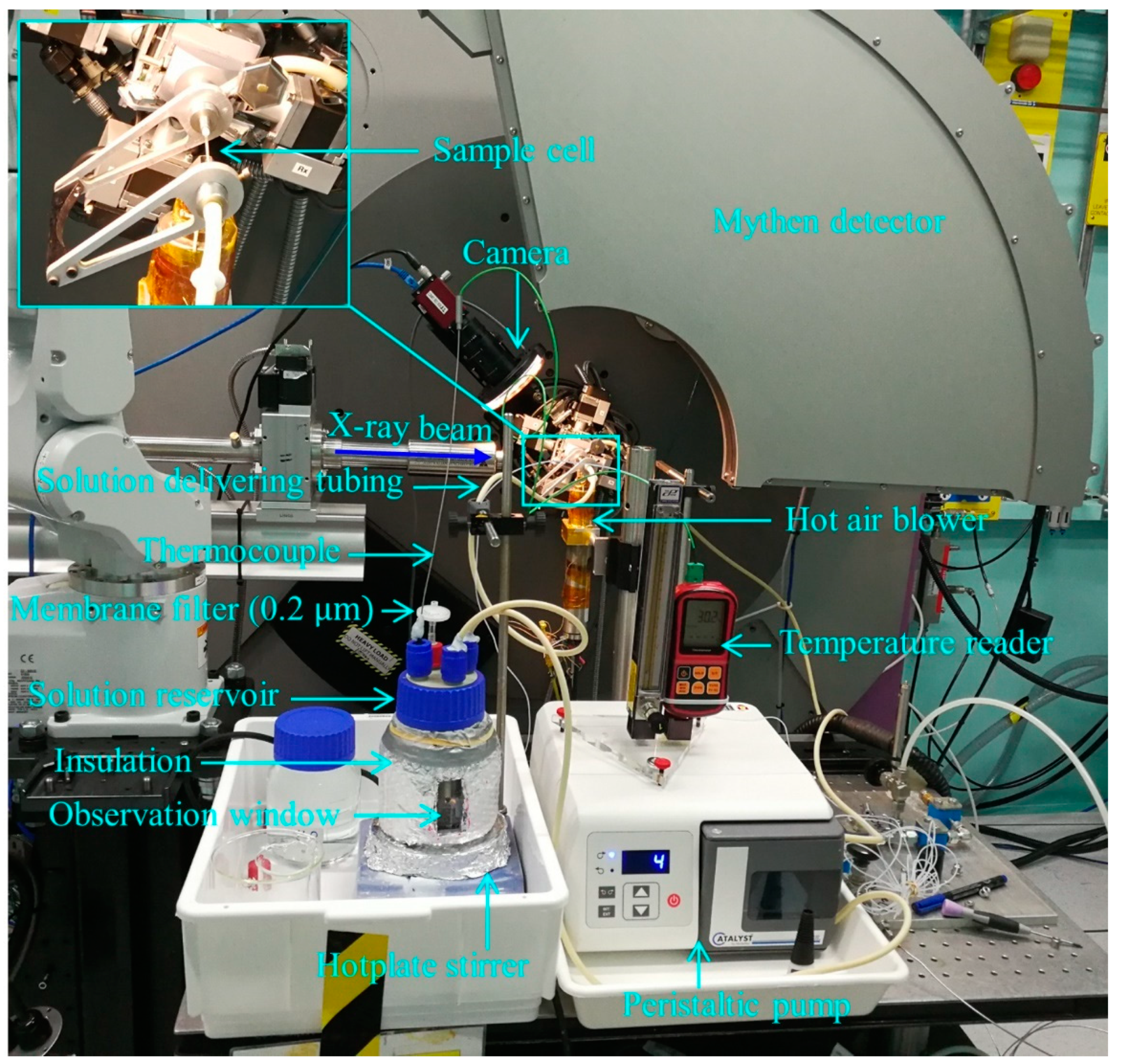
2. The Flow-Through Cell Setup

The design of the flow-through cell setup is shown in Figure 1 and a photograph of the set-up is shown in Figure 2. The leaching solution is pre-heated in a solution reservoir by a hot plate under stirring and pumped by a peristaltic pump through flexible tubing and flow through the mineral sample in the sample cell. The temperature of the sample cell is maintained by a hot air blower underneath the sample section. After reacting with the mineral sample and flowing out of the sample cell, the leaching solution returns to the solution reservoir, completing a closed loop. PXRD patterns are continuously collected during solution-mineral reactions in the sample cell, achieving in-situ monitoring of dissolution/crystallization processes. The specifications of the sample cell, the solution reservoir, and the pump are described in the following paragraphs.
The design of the sample cell is shown in Figure 3. It consists of an open-end capillary tube (Figure 3b), two 316 stainless steel fittings (one T-piece and one straight piece), and an aluminum frame to hold the two fittings. The capillary tube is made of either silica glass or sapphire and is 50 mm long, 1.0–1.3 mm in internal diameter, and 0.1 mm in wall thickness. Silica glass is stable under acidic to mild alkaline conditions (e.g., pH < 9), while sapphire is inert under higher pH conditions. On each end of the tube, a PTFE ferrule is glued onto the tube using superglue. The powder sample (~3 mm long) is loaded into the middle section of the tube, and the sample position is fixed by adding a silica glass wool plug on both sides of the sample. The capillary tube is fixed to the two 316 stainless-steel fittings. The sealing on each side is by cone/cone seal between the cone surface of the ferrule and the cone surface of the ports of the fittings. Both fittings are fixed to an aluminum frame. The T-piece fitting is for solution inlet and the straight fitting is for solution outlet. The assembled sample cell can be fixed horizontally to the goniometer head spinner of the X-ray diffractometer (Figure 2 inset). A second set of T-piece and straight piece fittings were made by Hastelloy for more corrosive leaching conditions that are not manageable by 316 stainless steel.
The solution reservoir is a 500 mL borosilicate glass bottle with four ports on the lid, one for solution delivering tubing inlet, one for solution delivering tubing outlet, one for a K-type thermocouple, and one for pressure equalization using a 0.2 μm membrane filter. The reservoir was insulated using a silver foil air bubble wrap, in which a small window (20 × 80 mm) was cut to allow flow observation during in-situ PXRD experiments. The solution in the reservoir was pre-heated under magnetic stirring using a hotplate stirrer (IKA RCT basic).
The peristaltic pump (Thermo Scientific FH 100) has a flow capacity of 0.5–300 mL min−1. For the in-situ PXRD setup, a low flow rate (e.g., 0.5–2 mL min−1) is required for the small diameter of the capillary tube. The solution was circulated through the reaction system using Norprene tubing, a polypropylene-based tubing that is appropriate for the acidic solution and has a temperature rating between −59 °C to 135 °C. Viton tubing can also be used for higher temperature applications up to 230 °C.

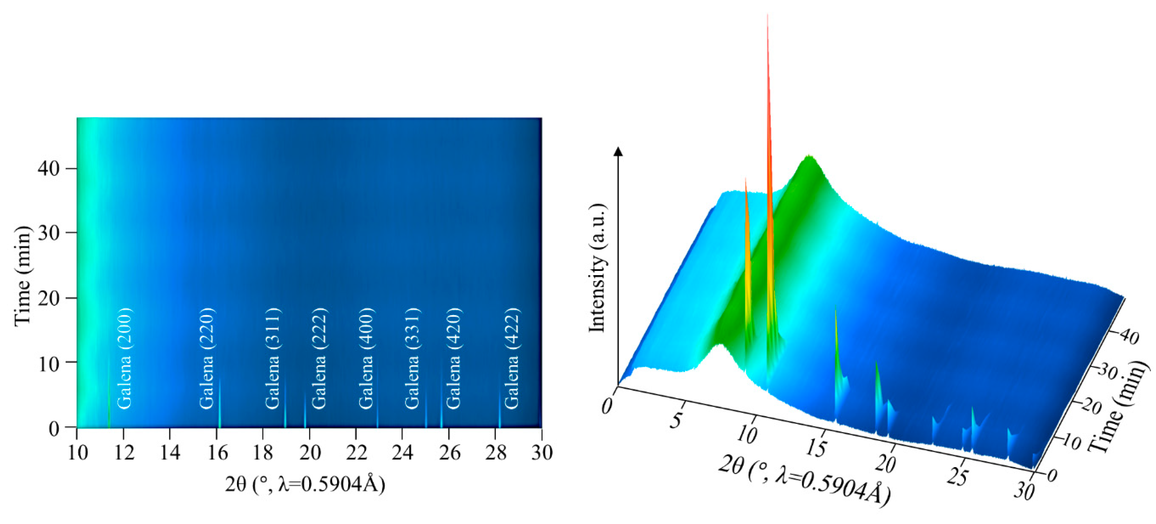
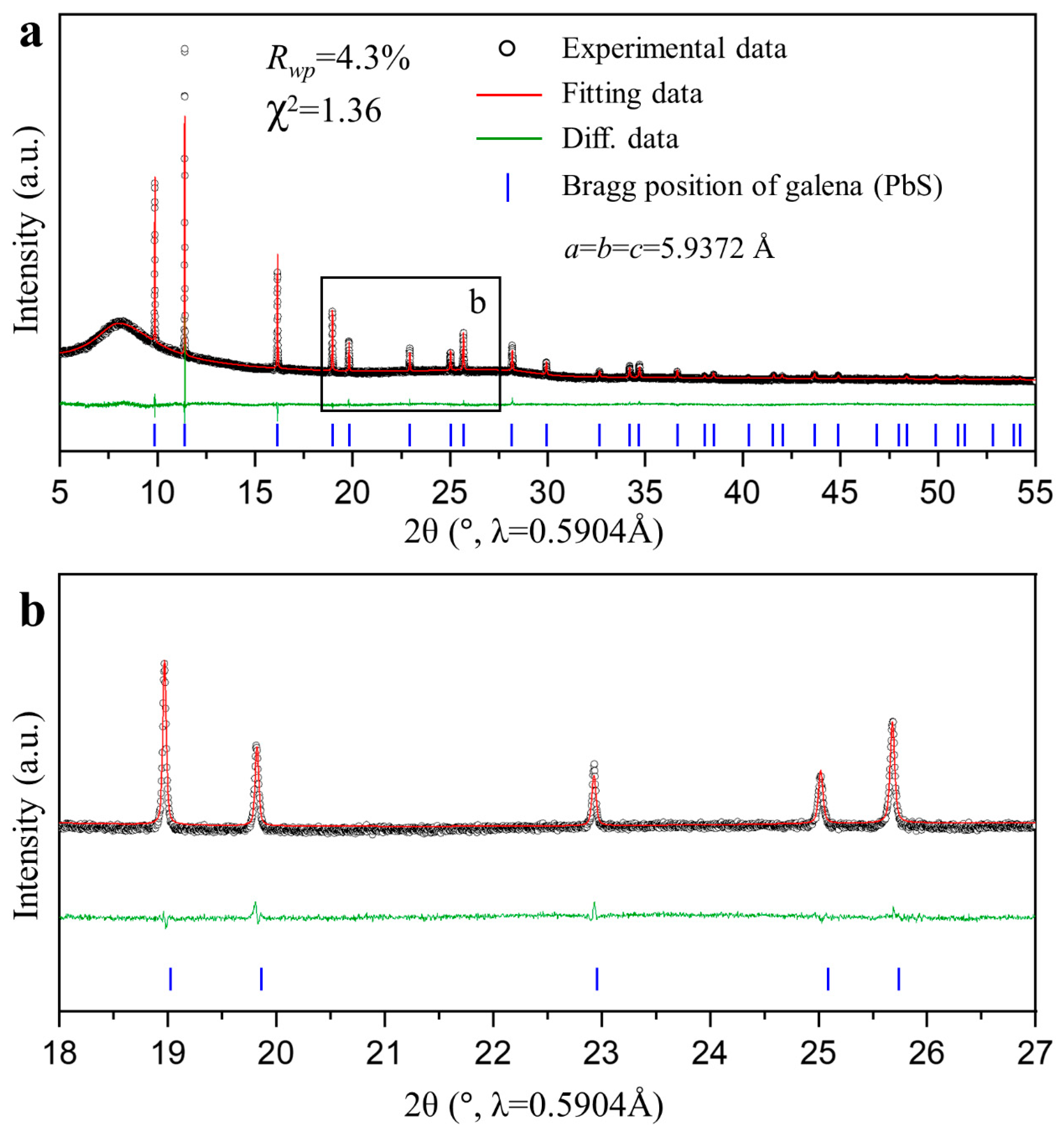
3. In-Situ PXRD of Galena Leaching

The flow-through-cell was tested at the powder X-ray diffraction beamline at the Australian Synchrotron using an X-ray beam energy of 21 keV (λ = 0.5904 Å). An in-situ PXRD experiment on galena leaching was carried out under the condition summarized in Table 1. A high-purity galena specimen from Quiruvilca Mine, Santiago de Chuco Province, La Libertad, Peru, was used [6,38]. The galena was ground to less than 38 μm, controlled by a 38 μm sieve. To reduce X-ray micro-absorption by Pb, galena was diluted by mixing with inert silica glass powder (also < 38 μm). The solid packing ratio is 20% (determined using a 10 mL bottle) in the mineral sample position of the capillary (Figure 3b), and the volume ratio between galena and silica powder is 1:9. This means only 2% volume of the sample position of the capillary was occupied by galena, resulting in a low total X-ray microabsorption (mμR = 0.91).
The lixiviant was prepared using Milli-Q water and reagent-grade chemicals, including H2O2 (30%, Rowe Scientific), citric acid anhydrous (≥99.5%, Sigma-Aldrich), and trisodium citrate dihydrate (≥98%, Sigma-Aldrich).
Before the in-situ PXRD experiment, the temperature of the hot air blower was calibrated based on the polymorphic phase transition from orthorhombic to trigonal KNO3 at 128 °C. For the in-situ PXRD experiment, as soon as the solution was pumped through the sample at 35 °C, data acquisition started Each PXRD pattern was collected for 2 min, using a Mythen position-sensitive detector in the 2θ range of 1–81°, under the Debye-Scherrer geometry. The sample cell was set to oscillate over 45° with a frequency of 0.07 Hz (Video S1 in Supplementary Materials), for uniform heating, minimizing the preferred orientation effect, and improving particle statistics. During the in-situ PXRD experiment, temperature of the solution reservoir, solution level in the reservoir, and flow rate (Video S2 in Supplementary Materials) were monitored using a web camera, while temperature of the hot air blower (e.g., the sample section) was recorded in the diffraction files.
Figure 4 shows the PXRD patterns during the in-situ leaching of galena. A systematic reduction in galena peak intensity with time suggests progressive dissolution of galena. Galena peaks disappeared after 14 min, suggesting rapid leaching under the studied condition. This agrees with rapid leaching of galena reported in an ex-situ study under similar conditions [38].
The data quality was good, suggesting effective minimization of microabsorption by dilution. As an example, the first dataset is shown in Figure 5. Rietveld-based analysis was conducted on this dataset using Topas Academic v6.0. The detector zero shift was determined by refining a Lab6 standard (NIST SRM 660b). The background was modeled using a 5th order polynomial function, and the peak shape was modeled using a Pseudo-Voigt function. The broad peak centered at about 8° is from the amorphous silica tube, silica powder, and leaching solution, and was treated as background. The unit cell parameters of galena were refined, starting from the crystal structure of galena, #9008694, obtained from the Crystallography Open Database (COD) [39]. The value a = b = c = 5.9372 Å is similar to the reported value, a = b = c = 5.9362 Å [39]. Preferred orientation was observed as the intensity of the (200) peak at 11.4° is higher than expected (Figure 5). This is due to the perfect cleavage of galena on {100} lattice planes.

4. Conclusions

In this study, a flow-through cell was designed for in-situ PXRD studies of minerals leaching. The cell was successfully tested at the Australian Synchrotron using galena leaching at 35 °C by 0.75 M H2O2 in a pH 6 citrate buffer solution, under a flow rate of 0.5 mL min−1. The results showed rapid galena dissolution. The test demonstrated the ability of the flow-through cell for performing leaching experiments in which the solid is kept in the sample cell as the stationary phase, and the desired amount of solution circulates through the sample. The flow-through cell can be used to study leaching of other ore minerals by in-situ PXRD.

Supplementary Materials

The following are available online at https://www.mdpi.com/2075-163X/10/11/990/s1, Video S1: cell oscillation, Video S2: flow monitoring.

Author Contributions

Conceptualization, F.X.; Methodology, F.N., F.X., Q.G., and J.A.K.; Formal Analysis, F.N. and F.X.; Investigation, F.N., F.X., X.Y., and I.A.A.; Resources, F.X., Q.G., and J.A.K.; Writing—Original Draft Preparation, F.N.; Writing—Review & Editing, F.X.; Project Administration, F.X.; Funding Acquisition, F.X. and F.N. All authors have read and agreed to the published version of the manuscript.

Funding

This research was funded by the Australian Research Council grant number DP170101893 and Australian Synchrotron beam time award numbers AS173/PD/12455 and AS173/PD/12540.

Acknowledgments

F. Nikkhou and I.A. Adegoke acknowledge Murdoch University for the Murdoch International Postgraduate Scholarship (MIPS). X. Yao acknowledges Murdoch University for a Murdoch University Strategic Scholarship (MUSS). We appreciate three anonymous reviewers whose comments helped improve the paper.

Conflicts of Interest

The authors declare no conflict of interest.

References

  1. Ruiz, M.; Vera, M.; Padilla, R. Mechanism of enargite pressure leaching in the presence of pyrite. Hydrometallurgy 2011, 105, 290–295. [Google Scholar] [CrossRef]
  2. Zhao, J.; Xia, F.; Pring, A.; Brugger, J.; Grundler, P.V.; Chen, G. A novel pre-treatment of calaverite by hydrothermal mineral replacement reactions. Miner. Eng. 2010, 23, 451–453. [Google Scholar] [CrossRef]
  3. Nikkhou, F.; Xia, F.; Deditius, A.P. Variable surface passivation during direct leaching of sphalerite by ferric sulfate, ferric chloride, and ferric nitrate in a citrate medium. Hydrometallurgy 2019, 188, 201–215. [Google Scholar] [CrossRef]
  4. Kartal, M.; Xia, F.; Ralph, D.; Rickard, W.D.; Renard, F.; Li, W. Enhancing chalcopyrite leaching by tetrachloroethylene-assisted removal of sulphur passivation and the mechanism of jarosite formation. Hydrometallurgy 2019, 191, 105192. [Google Scholar] [CrossRef]
  5. Dutrizac, J. The dissolution of galena in ferric chloride media. Metall. Trans. B 1986, 17, 5–17. [Google Scholar] [CrossRef]
  6. Nikkhou, F.; Xia, F.; Deditius, A.P.; Yao, X. Formation mechanisms of surface passivating phases and their impact on the kinetics of galena leaching in ferric chloride, ferric perchlorate, and ferric nitrate solutions. Hydrometallurgy 2020, 197, 105468. [Google Scholar] [CrossRef]
  7. Zárate-Gutiérrez, R.; Lapidus, G.; Morales, R. Pressure leaching of a lead–zinc–silver concentrate with nitric acid at moderate temperatures between 130 and 170 °C. Hydrometallurgy 2010, 104, 8–13. [Google Scholar] [CrossRef]
  8. Webster, N.A.; Madsen, I.C.; Loan, M.J.; Scarlett, N.V.; Wallwork, K.S. A flow cell for in situ synchrotron x-ray diffraction studies of scale formation under Bayer processing conditions. Rev. Sci. Instrum. 2009, 80, 084102. [Google Scholar] [CrossRef]
  9. Scarlett, N.V.; Grey, I.E.; Brand, H.E. In situ synchrotron diffraction studies on the formation kinetics of jarosites. J. Synchrotron Radiat. 2013, 20, 366–375. [Google Scholar] [CrossRef]
  10. Majuste, D.; Ciminelli, V.; Eng, P.; Osseo-Asare, K. Applications of in situ synchrotron XRD in hydrometallurgy: Literature review and investigation of chalcopyrite dissolution. Hydrometallurgy 2013, 131, 54–66. [Google Scholar] [CrossRef]
  11. Madsen, I.C.; Scarlett, N.V.; Whittington, B.I. Pressure acid leaching of nickel laterite ores: An in situ diffraction study of the mechanism and rate of reaction. J. Appl. Crystallogr. 2005, 38, 927–933. [Google Scholar] [CrossRef]
  12. Testemale, D.; Argoud, R.; Geaymond, O.; Hazemann, J.-L. High pressure/high temperature cell for x-ray absorption and scattering techniques. Rev. Sci. Instrum. 2005, 76, 043905. [Google Scholar] [CrossRef]
  13. Geisler, T.; Dohmen, L.; Lenting, C.; Fritzsche, M.B. Real-time in situ observations of reaction and transport phenomena during silicate glass corrosion by fluid-cell Raman spectroscopy. Nat. Mater. 2019, 18, 342–348. [Google Scholar] [CrossRef] [PubMed]
  14. Jung, J.H.; Chen, C.-Y.; Wu, W.-W.; Hong, J.-I.; Yun, B.K.; Zhou, Y.; Lee, N.; Jo, W.; Chen, L.-J.; Chou, L.-J. In situ observation of dehydration-induced phase transformation from Na2Nb2O6–H2O to NaNbO3. J. Phys. Chem. C 2012, 116, 22261–22265. [Google Scholar] [CrossRef] [Green Version]
  15. Fu, W.; Vaughan, J.; Gillespie, A. In situ AFM investigation of gibbsite growth in high ionic strength, highly alkaline, aqueous media. Hydrometallurgy 2016, 161, 71–76. [Google Scholar] [CrossRef] [Green Version]
  16. Xia, F.; Brugger, J.; Qian, G.; Ngothai, Y.; O’Neill, B.; Zhao, J.; Pullen, S.; Olsen, S.; Pring, A. Single-pass flow-through reaction cell for high-temperature and high-pressure in situ neutron diffraction studies of hydrothermal crystallization processes. J. Appl. Cryst. 2012, 45, 166–173. [Google Scholar] [CrossRef]
  17. Xia, F.; Qian, G.; Brugger, J.; Studer, A.; Olsen, S.; Pring, A. A large volume cell for in situ neutron diffraction studies of hydrothermal crystallizations. Rev. Sci. Instrum. 2010, 81, 105107. [Google Scholar] [CrossRef]
  18. Xia, F.; O’Neill, B.; Ngothai, Y.; Peak, J.; Tenailleau, C.; Etschmann, B.; Qian, G.; Brugger, J.; Studer, A.; Olsen, S. A thermosyphon-driven hydrothermal flow-through cell for in situ and time-resolved neutron diffraction studies. J. Appl. Cryst. 2010, 43, 511–519. [Google Scholar] [CrossRef]
  19. Norby, P. In-situ XRD as a tool to understanding zeolite crystallization. Curr. Opin. Colloid Interface Sci. 2006, 11, 118–125. [Google Scholar] [CrossRef]
  20. Chupas, P.J.; Chapman, K.W.; Kurtz, C.; Hanson, J.C.; Lee, P.L.; Grey, C.P. A versatile sample-environment cell for non-ambient X-ray scattering experiments. J. Appl. Crystallogr. 2008, 41, 822–824. [Google Scholar] [CrossRef]
  21. Bösenberg, U.; Pistidda, C.; Tolkiehn, M.; Busch, N.; Saldan, I.; Suarez-Alcantara, K.; Arendarska, A.; Klassen, T.; Dornheim, M. Characterization of metal hydrides by in-situ XRD. Int. J. Hydrog. Energy 2014, 39, 9899–9903. [Google Scholar] [CrossRef] [Green Version]
  22. Clausen, B.S.; Steffensen, G.; Fabius, B.; Villadsen, J.; Feidenhans, R.; Topsøe, H. In situ cell for combined XRD and on-line catalysis tests: Studies of Cu-based water gas shift and methanol catalysts. J. Catal. 1991, 132, 524–535. [Google Scholar] [CrossRef]
  23. Xia, F.; Chen, D.; Scarlett, N.V.; Madsen, I.C.; Lau, D.; Leoni, M.; Ilavsky, J.; Brand, H.E.; Caruso, R.A. Understanding solvothermal crystallization of mesoporous anatase beads by in situ synchrotron PXRD and SAXS. Chem. Mater. 2014, 26, 4563–4571. [Google Scholar] [CrossRef]
  24. Zhang, L.; Xia, F.; Song, Z.; Webster, N.A.; Luo, H.; Gao, Y. Synthesis and formation mechanism of VO2(A) nanoplates with intrinsic peroxidase-like activity. RSC Adv. 2015, 5, 61371–61379. [Google Scholar] [CrossRef] [Green Version]
  25. Song, J.; Yuan, J.; Xia, F.; Liu, J.; Zhang, Y.; Zhong, Y.L.; Zheng, J.; Liu, Y.; Li, S.; Zhao, M. Large-scale production of bismuth chalcogenide and graphene heterostructure and its application for flexible broadband photodetector. Adv. Electron. Mater. 2016, 2, 1–9. [Google Scholar] [CrossRef]
  26. Rodriguez, E.F.; Xia, F.; Chen, D.; Hollenkamp, A.F.; Caruso, R.A. N-doped Li₄Ti₅O₁₂ nanoflakes derived from 2D protonated titanate for high performing anodes in lithium ion batteries. J. Mater. Chem. A 2016, 4, 7772–7780. [Google Scholar] [CrossRef] [Green Version]
  27. Tan, J.Z.; Nursam, N.M.; Xia, F.; Sani, M.-A.; Li, W.; Wang, X.; Caruso, R.A. High-performance coral reef-like carbon nitrides: Synthesis and application in photocatalysis and heavy metal ion adsorption. ACS Appl. Mater. Interfaces 2017, 9, 4540–4547. [Google Scholar] [CrossRef]
  28. Mi, J.-L.; Nørby, P.; Bremholm, M.; Iversen, B.B. Insight into nucleation and growth of Bi2−xSbxTe3 (x = 0−2) nanoplatelets in hydrothermal synthesis. Chem. Mater. 2017, 29, 5070–5079. [Google Scholar] [CrossRef] [Green Version]
  29. De Marco, R.; Pejcic, B.; Prince, K.; Van Riessen, A. A multi-technique surface study of the mercury (II) chalcogenide ion-selective electrode in saline media. Analyst 2003, 128, 742–749. [Google Scholar] [CrossRef]
  30. Ferrer, P.; da Silva, I.; Rubio-Zuazo, J.; Alfonso, B.F.; Trobajo, C.; Khainakov, S.; Garcia, J.R.; Garcia-Granda, S.; Castro, G.R. A flow-through reaction cell for in situ X-ray diffraction and absorption studies of heterogeneous powder–liquid reactions and phase transformations. J. Synchrotron Radiat. 2012, 19, 93–100. [Google Scholar] [CrossRef] [Green Version]
  31. Wall, A.J.; Heaney, P.J.; Mathur, R.; Post, J.E.; Hanson, J.C.; Eng, P.J. A flow-through reaction cell that couples time-resolved X-ray diffraction with stable isotope analysis. J. Appl. Cryst. 2011, 44, 429–432. [Google Scholar] [CrossRef]
  32. Kautek, W.; Mirwald, S.; Sahre, M.; Nauer, G.E. In-situ grazing incidence X-ray diffractometry of polycrystalline copper in alkaline chloride and sulphate electrolytes. Electrochim. Acta 1998, 43, 2979–2984. [Google Scholar] [CrossRef]
  33. Nagy, Z.; You, H.; Yonco, R. Cell design for in situ x-ray scattering studies of metal/solution interfaces under electrochemical control. Rev. Sci. Instrum. 1994, 65, 2199–2205. [Google Scholar] [CrossRef]
  34. De Marco, R.; Jiang, Z.-T.; Pejcic, B.; Van Riessen, A. In situ synchrotron radiation grazing incidence X-ray diffraction—A powerful technique for the characterization of solid-state ion-selective electrode surfaces. Electrochim. Acta 2006, 51, 4886–4891. [Google Scholar] [CrossRef]
  35. Clancy, M.; Styles, M.J.; Bettles, C.J.; Birbilis, N.; Chen, M.; Zhang, Y.; Gu, Q.; Kimpton, J.A.; Webster, N.A. In situ synchrotron X-ray diffraction investigation of the evolution of a PbO2/PbSO4 surface layer on a copper electrowinning Pb anode in a novel electrochemical flow cell. J. Synchrotron Radiat. 2015, 22, 366–375. [Google Scholar] [CrossRef]
  36. Wall, A.J.; Mathur, R.; Post, J.E.; Heaney, P.J. Cu isotope fractionation during bornite dissolution: An in situ X-ray diffraction analysis. Ore Geol. Rev. 2011, 42, 62–70. [Google Scholar] [CrossRef]
  37. Fleeger, C.R.; Heaney, P.J.; Post, J.E. A time-resolved X-ray diffraction study of Cs exchange into hexagonal H-birnessite. Am. Mineral. 2013, 98, 671–679. [Google Scholar] [CrossRef]
  38. Nikkhou, F.; Xia, F.; Knorsch, M.; Deditius, A.P. Mechanisms of surface passivation during galena leaching by hydrogen peroxide in acetate and citrate solutions at 25–50 °C. ACS Sustain. Chem. Eng. 2020, 8, 14407–14416. [Google Scholar] [CrossRef]
  39. Margulieux, G.W.; Weidemann, N.; Lacy, D.C.; Moore, C.E.; Rheingold, A.L.; Figueroa, J.S. Isocyano analogues of [Co(CO)4]n: A tetraisocyanide of cobalt isolated in three states of charge. J. Am. Chem. Soc. 2010, 132, 5033–5035. [Google Scholar] [CrossRef]
Figure 1. Schematic diagram of the flow-through cell experimental apparatus. Blue arrows show the flow direction.
Minerals 10 00990 g001
Figure 2. Photo of the flow-through cell setup at the Powder Diffraction beamline at the Australian Synchrotron.
Minerals 10 00990 g002
Figure 3. Photos of (a) the sample cell, and (b) capillary tube.
Minerals 10 00990 g003
Figure 4. In-situ PXRD patterns showing rapid galena leaching at 35 °C in a pH 6 solution.
Minerals 10 00990 g004
Figure 5. Rietveld refinement of the first dataset from the in-situ PXRD experiment.
Minerals 10 00990 g005
Table 1. Summary of leaching condition.
ParameterValue
MineralGalena (PbS)
Particle size<38 μm
Lixiviant0.08 M citric acid + 0.92 M trisodium citrate + 0.75 M H2O2
Solution pH6
Flow rate0.5 mL min−1
Temperature35 °C
Leaching time48 min
Publisher’s Note: MDPI stays neutral with regard to jurisdictional claims in published maps and institutional affiliations.

