A Mineral X-ray Linear Attenuation Coefficient Tool (MXLAC) to Assess Mineralogical Differentiation for X-ray Computed Tomography Scanning


Abstract
:1. Introduction
2. Methodology
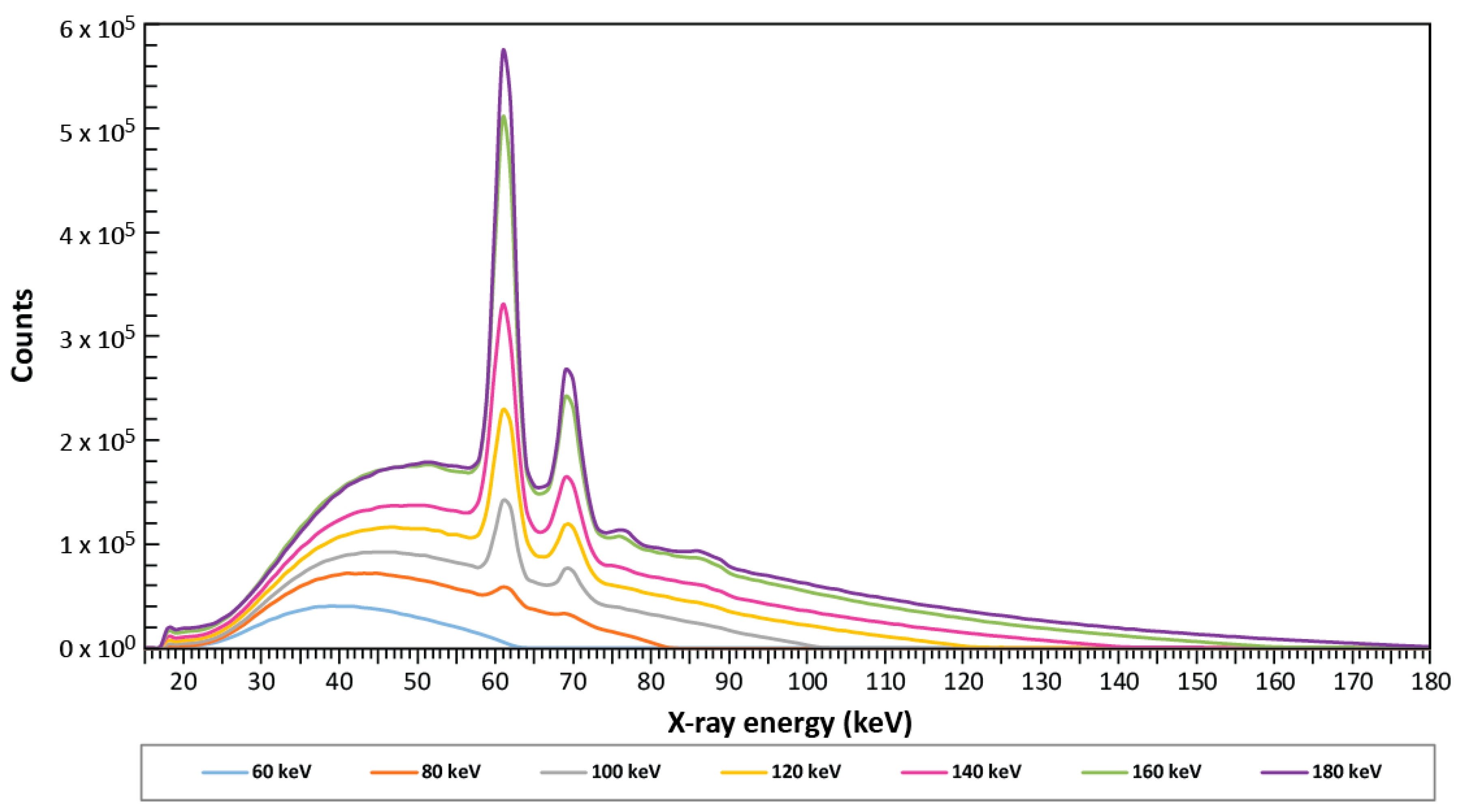
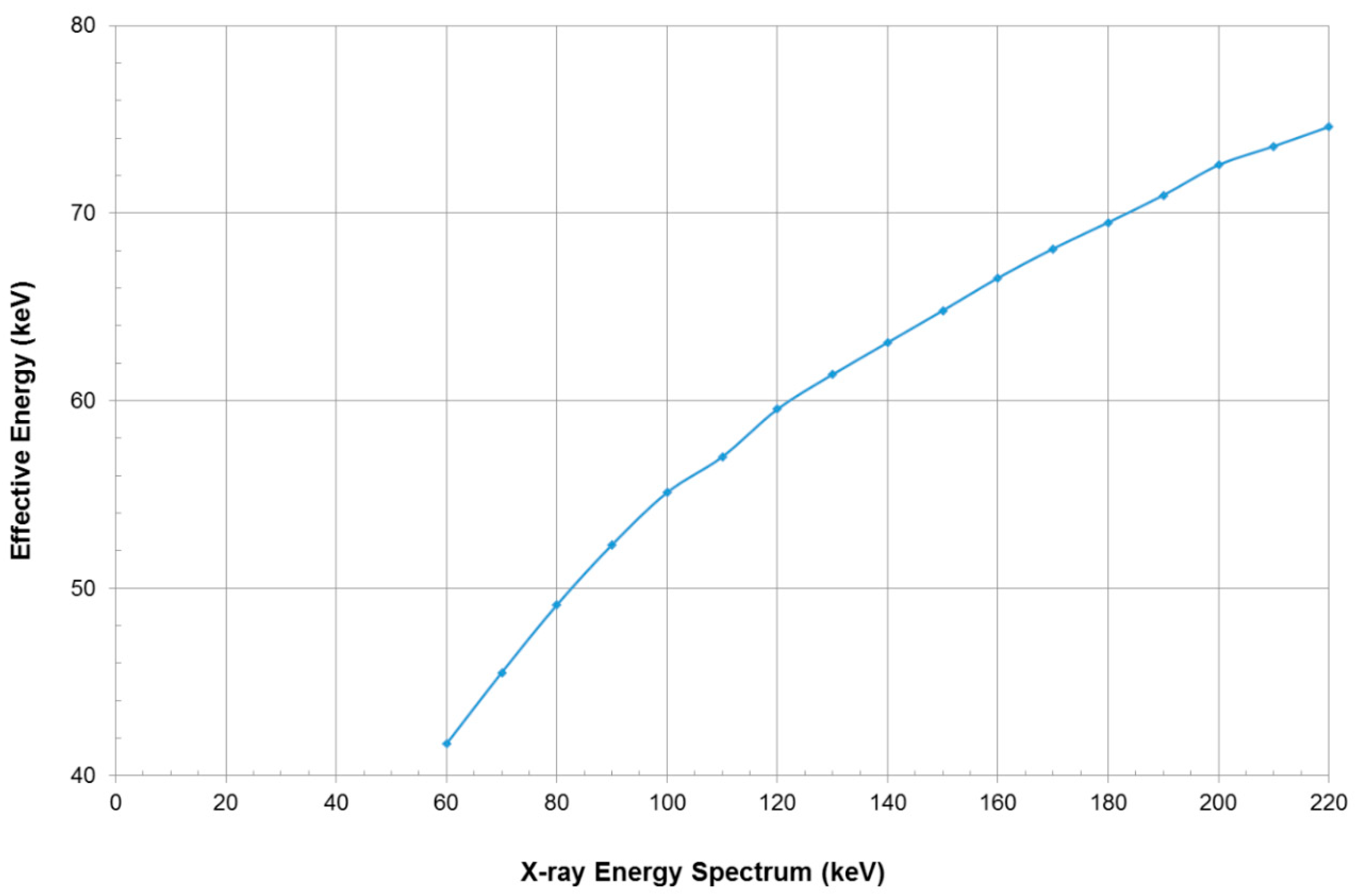
2.1. Tungsten Energy Spectrum
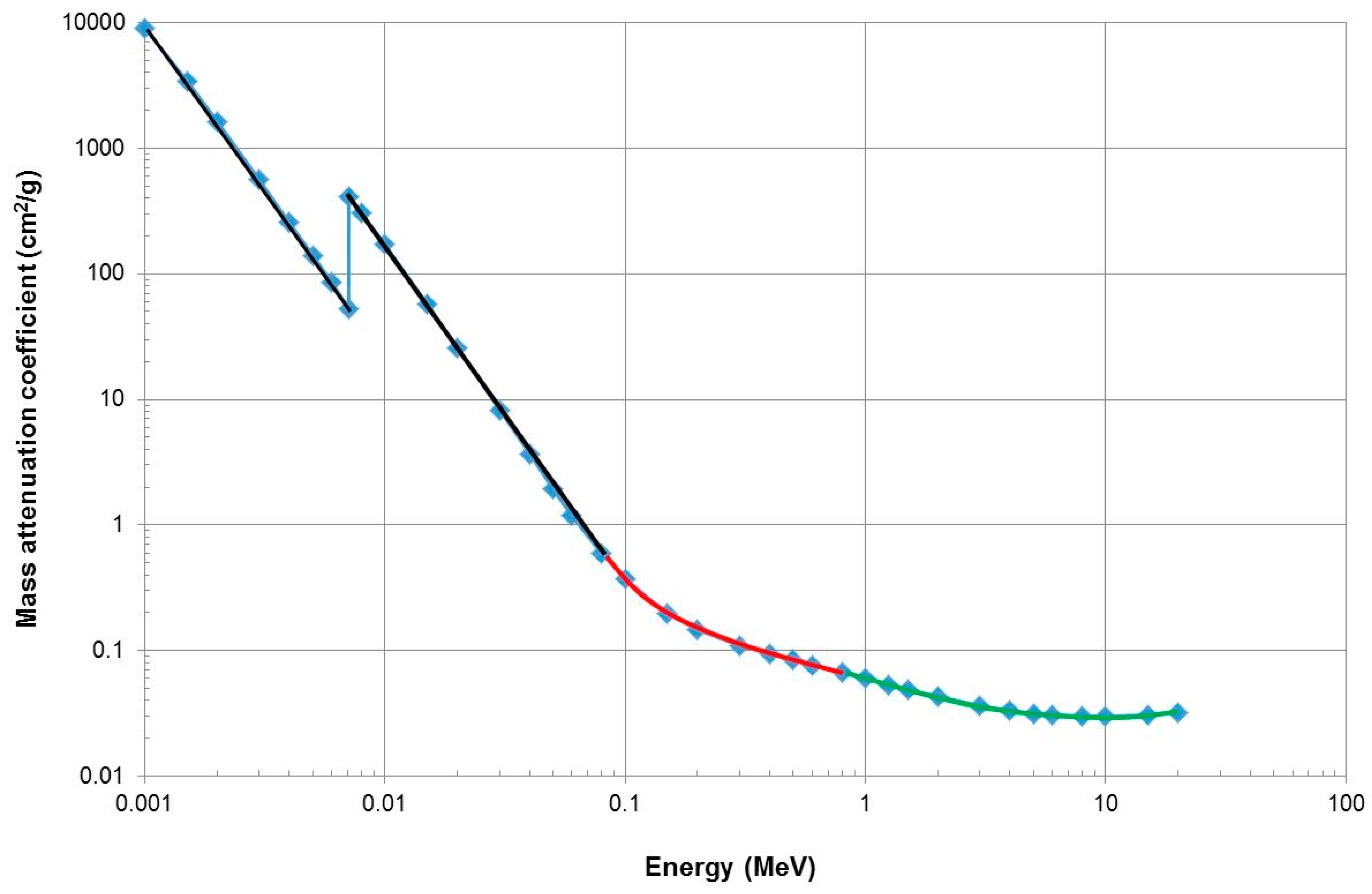
2.2. Development of the Attenuation Coefficient Data Bank
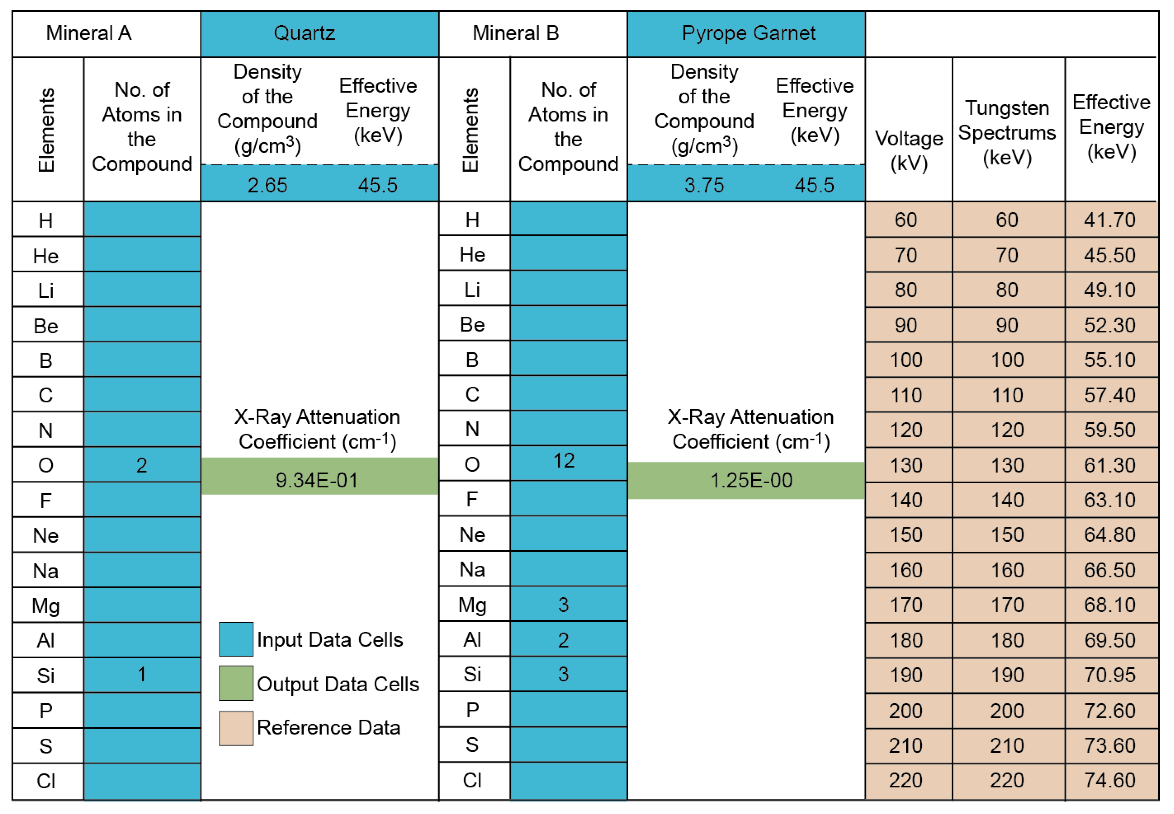
2.3. Development of User Spreadsheet
2.4. Validation of Linear Attenuation Coefficients
3. Results
3.1. Calculated Linear Attenuation Coefficients
3.2. Minimum Attenuation Coefficient Difference to Determine Discrimination
4. Discussion
4.1. Mineral Composition and Linear Attenuation Coefficient
4.2. Mineral Density and Attenuation
4.3. Influence of Mineral Composition vs. Density on Attenuation Coefficient
5. Conclusions
Supplementary Materials
Author Contributions
Funding
Acknowledgments
Conflicts of Interest
References
- Momose, A.; Keiichi, H. The possibility of phase-contrast X-ray microtomography. Jpn. J. Appl. Phys. 1999, 38, 625. [Google Scholar] [CrossRef] [Green Version]
- Mees, F.; Swennen, R.; Van Geet, M.; Jacobs, P. (Eds.) Applications of X-ray Computed Tomography in the Geosciences, 215th ed.; The Geological Society London: London, UK, 2003; ISBN 1862391394. [Google Scholar]
- Ketcham, R.A.; Carlson, W.D. Acquisition, optimization and interpretation of X-ray computed tomographic imagery: Applications to the geosciences. Comput. Geosci. 2001, 27, 381–400. [Google Scholar] [CrossRef]
- Kalender, W.A. X-ray computed tomography. Phys. Med. Biol. 2006, 51, R29–R43. [Google Scholar] [CrossRef] [PubMed] [Green Version]
- Cnudde, V.; Masschaele, B.; Dierick, M.; Vlassenbroeck, J.; Van Hoorebeke, L.; Jacobs, P. Recent progress in X-ray CT as a geosciences tool. Appl. Geochem. 2006, 21, 826–832. [Google Scholar] [CrossRef]
- Hamdani, A.H. X-ray computed tomography analysis of sajau coal, Berau Basin, Indonesia: 3D imaging of cleat and microcleat characteristics. Int. J. Geophys. 2015. [Google Scholar] [CrossRef] [Green Version]
- Kyle, J.R.; Ketcham, R.A. Application of high resolution X-ray computed tomography to mineral deposit origin, evaluation, and processing. Ore Geol. Rev. 2015. [Google Scholar] [CrossRef]
- Siddiqui, I.; Solangi, S.H.; Soomro, A.A.; Warar, M.A.; Samoon, M.K.; Ajmal, S. Application of X-ray computed tomography for analyzing cleats and pores for coalbed methane in coal from thar coalfield. Can. J. Pure Appl. Sci. 2014, 8, 2743–2749. [Google Scholar]
- Panahi, H.; Kobchenko, M.; Renard, F.; Mazzini, A.; Scheibert, J.; Dysthe, D.; Jamtveit, B.; Malthe-Sorensses, A.; Meakin, P. A 4D synchrotron X-ray-tomography study of the formation of hydrocarbon-migration pathways in heated organic-rich shale. Soc. Pet. Eng. J. 2012, 18, 366–377. [Google Scholar] [CrossRef]
- Miller, J.A.; Faber, C.; Rowe, C.D.; Macey, P.H.; du Plessis, A. Eastward transport of the Monapo Klippe, Mozambique determined from field kinematics and computed tomography and implications for late tectonics in central Gondwana. Precambrian Res. 2013, 237, 101–115. [Google Scholar] [CrossRef]
- Kaufhold, A.; Zacher, G.; Halisch, M.; Kaufhold, S. X-ray computed tomography investigation of structures in opalinus clay from large scale to small scale after mechanical testing. Solid Earth 2016, 7, 1–19. [Google Scholar] [CrossRef] [Green Version]
- Backeberg, N.R.; Iacoviello, F.; Rittner, M.; Mitchell, T.M.; Jones, A.P.; Day, R.; Wheeler, J.; Shearing, P.R.; Vermeesch, P.; Striolo, A. Quantifying the anisotropy and tortuosity of permeable pathways in clay-rich mudstones using models based on X-ray tomography. Sci. Rep. 2017, 7, 1–12. [Google Scholar] [CrossRef] [PubMed]
- Iacoviello, F.; Lu, X.; Mitchell, T.M.; Brett, D.J.L.; Shearing, P.R. The imaging resolution and Knudsen effect on the mass transport of shale gas assisted by multi-length scale X-ray computed tomography. Sci. Rep. 2019, 9, 1–10. [Google Scholar] [CrossRef] [PubMed]
- Ma, Y.; Zhong, N.; Cheng, L.; Pan, Z.; Dai, N.; Zhang, Y.; Yang, L. Pore structure of the graptolite-derived OM in the Longmaxi Shale, southeastern Upper Yangtze Region, China. Mar. Pet. Geol. 2016, 72, 1–11. [Google Scholar] [CrossRef]
- Ashi, J. Computed tomography scan image analysis of sediments. In Ocean Drilling Program, Scientific Results; Shipley, T.H., Ogawa, Y., Blum, P., Bahr, J.M., Eds.; Integrated Ocean Drilling Program: College Station, TX, USA, 1997; Volume 156, pp. 151–159. [Google Scholar]
- Tanaka, A.; Nakano, T.; Ikehara, K. X-ray computerized tomography analysis and density estimation using a sediment core from the Challenger Mound area in the Porcupine Seabight, off Western Ireland. Earth Planets Space 2011, 63, 103–110. [Google Scholar] [CrossRef] [Green Version]
- Le Roux, S.G.; Du Plessis, A.; Rozendaal, A. The quantitative analysis of tungsten ore using X-ray microCT: Case study. Comput. Geosci. 2015, 85, 75–80. [Google Scholar] [CrossRef]
- Jardine, M.A.; Miller, J.A.; Becker, M. Coupled X-ray computed tomography and grey level co-occurrence matrices as a method for quantification of mineralogy and texture in 3D. Comput. Geosci. 2018, 111, 105–117. [Google Scholar] [CrossRef]
- Sprawls, P. Physical Principles of Medical Imaging, 2nd ed.; Aspen Publishers: Gaithersburg, MD, USA, 1993; ISBN 1523-1739. [Google Scholar]
- Bam, L.C.; Miller, J.A.; Becker, M.; De Beer, F.C.; Basson, I. X-ray computed tomography—Determination of rapid scanning parameters for geometallurgical analysis of iron ore. In Proceedings of the third AusIMM International Geometallurgy Conference, Perth, Australia, 15–17 June 2016; pp. 209–219. [Google Scholar]
- Mccullough, E.C. Photon attenuation in computed tomography. Med. Phys. 1975, 2, 307–320. [Google Scholar] [CrossRef]
- Matsubara, K.; Ichikawa, K.; Murasaki, Y.; Hirosawa, A.; Koshida, K. Accuracy of measuring half- and quarter-value layers and appropriate aperture width of a convenient method using a lead-covered case in X-ray computed tomography. J. Appl. Clin. Med. Phys. 2014, 15, 309–316. [Google Scholar] [CrossRef]
- Yada, N.; Onishi, H. Validation of computed tomography-based attenuation correction of deviation between theoretical and actual values in four computed tomography scanners. Asia Ocean. J. Nucl. Med. Biol. 2016, 4, 81–819. [Google Scholar] [CrossRef]
- Olarinoye, I. Variation of effective atomic numbers of some thermoluminescence and phantom materials with photon energies. Res. J. Chem. Sci. 2011, 1, 64–69. [Google Scholar]
- Akça, B.; Erzeneoʇlu, S.Z. The mass attenuation coefficients, electronic, atomic, and molecular cross sections, effective atomic numbers, and electron densities for compounds of some biomedically important elements at 59.5 kev. Sci. Technol. Nucl. Install. 2014, 2014, 1–9. [Google Scholar] [CrossRef] [Green Version]
- McCullough, E.C.; Baker, H.L.; Wayne Houser, O.; Reese, D.F. An evaluation of the quantitative and radiation features of a scanning X-ray transverse axial tomograph: The EMI scanner. Radiology 1974, 111, 709–715. [Google Scholar] [CrossRef] [PubMed]
- Tsuchiyama, A.; Hanamoto, T.; Nakashima, Y.; Nakano, T. Quantitative evaluation of attenuation contrast of minerals Tsuchiyama 2000.pdf. J. Miner. Pet. Sci. 2000, 95, 125–137. [Google Scholar] [CrossRef] [Green Version]
- Deer, W.A.; Howie, R.A.; Zussman, J. An Introduction to the Rock-Forming Minerals, 2nd ed.; Pearson Prentice-Hall: London, UK, 1992. [Google Scholar]


| Exposure Time (sec) | No. of Projections | Voltage (kV)/ Effective Energy (keV) | Filter Material |
|---|---|---|---|
| 4 | 3000 | 70/45.5 | No filter |
| 4 | 3000 | 70/45.5 | 0.25 mm Cu |
| 4 | 3000 | 70/45.5 | 1 mm Al + 1 mm Cu |
| Mineral. | Chemical Formula | Density (g/cm3) | NIST | MXLAC | %Error | NIST | MXLAC | %Error |
|---|---|---|---|---|---|---|---|---|
| Attenuation Coefficient (cm−1), at 44.79 keV | Attenuation Coefficient (cm−1), at 62.53 keV | |||||||
| Acanthite | Ag2S | 7.24 | 79.68 | 80.60 | 1.14 | 32.43 | 32.70 | 0.83 |
| Almandine | Fe3Al2Si3O12 | 4.32 | 4.77 | 4.55 | 4.56 | 2.21 | 2.19 | 0.53 |
| Andradite | Ca3Fe3+2Si3O12 | 3.86 | 4.09 | 3.91 | 4.44 | 1.91 | 1.90 | 0.35 |
| Ankerite | CaFe(CO3)2 | 3.20 | 3.33 | 3.19 | 4.20 | 1.56 | 1.56 | 0.00 |
| Apatite | Ca5(PO4)3OH | 3.19 | 2.36 | 2.28 | 3.39 | 1.19 | 1.19 | 0.00 |
| Arsenopyrite | FeAsS | 6.18 | 20.23 | 20.40 | 0.83 | 8.26 | 8.51 | 2.94 |
| Barite | BaSO4 | 4.48 | 48.67 | 48.10 | 1.17 | 20.31 | 21.00 | 3.29 |
| Borax | Na2B4O5(OH)4·8H2O | 1.70 | 0.42 | 0.41 | 3.02 | 0.34 | 0.33 | 2.94 |
| Calcite | CaCO3 | 2.71 | 1.80 | 1.73 | 4.09 | 0.94 | 0.94 | 0.54 |
| Carnotite | K2(UO2)2(VO4)2·3H2O | 4.91 | 38.90 | 40.60 | 4.19 | 17.83 | 17.20 | 3.53 |
| Chalcocite | Cu2S | 5.60 | 16.48 | 17.20 | 4.19 | 6.72 | 6.49 | 3.42 |
| Chalcopyrite | CuFeS2 | 4.20 | 9.48 | 9.58 | 1.04 | 3.95 | 3.89 | 1.52 |
| Chlorite | (Mg)5Al2Si3O10(OH)8 | 3.20 | 1.09 | 1.04 | 4.44 | 0.73 | 0.72 | 1.30 |
| Chromite | FeCr2O4 | 4.79 | 7.93 | 8.03 | 1.21 | 3.42 | 3.40 | 0.55 |
| Corundum | Al2O3 | 4.02 | 1.41 | 1.34 | 4.96 | 0.92 | 0.91 | 1.09 |
| Dolomite | CaMg(CO3)2 | 2.85 | 1.39 | 1.33 | 4.32 | 0.80 | 0.80 | 0.00 |
| Fluorite | CaF2 | 3.13 | 2.50 | 2.41 | 3.76 | 1.23 | 1.23 | 0.38 |
| Gibbsite | Al(OH)3 | 2.34 | 0.74 | 0.70 | 4.89 | 0.52 | 0.51 | 2.16 |
| Goethite | FeO(OH) | 4.28 | 7.34 | 7.01 | 4.49 | 3.17 | 3.16 | 0.22 |
| Grossular | Ca3Al2Si3O12 | 3.65 | 2.22 | 2.12 | 4.45 | 1.19 | 1.18 | 0.77 |
| Hematite | Fe2O3 | 5.26 | 9.89 | 9.45 | 4.48 | 4.21 | 4.20 | 0.16 |
| Ilmenite | FeTiO3 | 4.76 | 7.26 | 6.95 | 4.34 | 3.17 | 3.13 | 1.05 |
| Kaolinite | Al2Si2O5(OH)4 | 2.60 | 0.91 | 0.87 | 4.90 | 0.60 | 0.59 | 1.95 |
| K-feldspar | KAlSi3O8 | 2.56 | 1.22 | 1.16 | 4.65 | 0.71 | 0.71 | 0.00 |
| Lepidolite | KLi2AlSi4O10(OH)2 | 2.83 | 1.21 | 1.16 | 4.45 | 0.74 | 0.73 | 1.35 |
| Magnetite | Fe3O4 | 5.18 | 10.03 | 9.58 | 4.48 | 4.25 | 4.25 | 0.14 |
| Molybdenite | MoS2 | 5.00 | 29.65 | 30.50 | 2.79 | 12.03 | 12.50 | 3.76 |
| Olivine | Fe2SiO4 | 3.32 | 5.20 | 4.97 | 4.42 | 2.27 | 2.26 | 0.44 |
| Pecoraite | Ni3S2O5(OH4) | 3.47 | 6.65 | 6.92 | 3.87 | 2.57 | 2.70 | 4.81 |
| Pyrite | FeS2 | 5.01 | 8.02 | 7.81 | 2.52 | 3.48 | 3.50 | 0.63 |
| Pyrope | Mg3Al2Si3O12 | 3.75 | 1.34 | 1.28 | 4.51 | 0.87 | 0.86 | 1.30 |
| Quartz | SiO2 | 2.65 | 1.01 | 0.96 | 4.95 | 0.64 | 0.63 | 1.68 |
| Rynersonite | CaTa2O6 | 6.39 | 35.64 | 36.00 | 0.99 | 15.13 | 15.20 | 0.46 |
| Safflorite | CoAs2 | 7.47 | 32.11 | 31.10 | 3.13 | 12.91 | 13.40 | 3.66 |
| Siderite | FeCO3 | 3.96 | 5.41 | 5.17 | 4.47 | 2.41 | 2.40 | 0.35 |
| Spessartine | Mn3Al2Si3O12 | 4.29 | 4.22 | 4.33 | 2.51 | 2.00 | 1.98 | 0.94 |
| Sphalerite | ZnS | 4.10 | 11.68 | 11.90 | 1.82 | 4.78 | 4.80 | 0.42 |
| Talc | Mg3Si4O10(OH)2 | 2.75 | 0.99 | 0.94 | 4.85 | 0.64 | 0.64 | 0.00 |
| Uvarovite | Ca3Cr2Si3O12 | 3.85 | 3.55 | 3.55 | 0.15 | 1.70 | 1.69 | 0.69 |
| Wolframite | FeWO4 | 7.30 | 38.12 | 38.70 | 1.50 | 16.16 | 16.30 | 0.86 |
| Zircon | ZrSiO4 | 4.71 | 20.06 | 21.10 | 4.94 | 8.26 | 8.40 | 1.67 |
| Mineral | Chemical Formula | Density g/cm3 | Attenuation Coefficient cm−1 | Mean Grey Value |
|---|---|---|---|---|
| Almandine | Fe3Al2Si3O12 | 4.32 | 4.36 | 28,084.5 |
| Andradite | Ca3Fe3+2Si3O12 | 3.86 | 3.74 | 34,569.4 |
| Grossular | Ca3Al2Si3O12 | 3.65 | 2.05 | 22,971.9 |
| Quartz | SiO2 | 2.65 | 0.93 | 10,540.6 |
| Kaolinite | Al2Si2O5(OH)4 | 2.60 | 0.85 | 11,014.9 |
| Dolomite | CaMg(CO3)2 | 2.85 | 1.29 | 15,047.8 |
| Calcite | CaCO3 | 2.71 | 1.66 | 18,285.0 |
| Fluorite | CaF2 | 3.13 | 2.31 | 23,956.0 |
| Apatite | Ca5(PO4)3OH | 3.15 | 2.15 | 21,737.9 |
| Goethite | FeO(OH) | 4.28 | 6.70 | 36,097.6 |
| Chromite | FeCr2O4 | 4.79 | 7.70 | 38,856.9 |
| Magnetite | Fe3O4 | 5.18 | 9.15 | 54,895.8 |
| Hematite | Fe2O3 | 5.26 | 9.02 | 57,344.8 |
| Mineral Comparison | Filter Material/BH Correction Factor | % Grey Value Difference | % Attenuation Coefficient Difference | % Density Difference | Discrimination |
|---|---|---|---|---|---|
| Almandine vs. Andradite | 0.25 mm Cu/2 | 18.8 | 14.2 | 10.7 | Yes |
| Almandine vs. Grossular | 18.2 | 53.0 | 15.5 | Yes | |
| Grossular vs. Andradite | 33.6 | 45.2 | 5.44 | Yes | |
| Quartz vs. Kaolinite | No Filter/2 | 4.31 | 9.42 | 1.89 | Partial |
| Quartz vs. Dolomite | 30.0 | 27.6 | 7.02 | Yes | |
| Quartz vs. Calcite | 42.4 | 43.7 | 2.21 | Yes | |
| Kaolinite vs. Dolomite | 26.8 | 34.4 | 8.77 | Yes | |
| Kaolinite vs. Calcite | 39.8 | 49.0 | 4.06 | Yes | |
| Dolomite vs. Calcite | 17.7 | 22.3 | 4.91 | Yes | |
| Fluorite vs. Apatite | 0.25 mm Cu/1 | 9.26 | 6.06 | 0.63 | Yes |
| Goethite vs. Chromite | 0.25 mm Cu/2 | 7.10 | 13.0 | 10.7 | Partial |
| Gothite vs. Magnetite | 1 mm Cu + 1 mm Al/3 | 34.2 | 26.8 | 17.4 | Yes |
| Goethite vs. Hematite | 37.1 | 25.7 | 18.6 | Yes | |
| Chromite vs. Magnetite | 29.2 | 15.9 | 7.53 | Yes | |
| Chromite vs. Hematite | 32.2 | 14.6 | 8.94 | Yes | |
| Magnetite vs. Hematite | 4.27 | 1.42 | 1.52 | No |
| Iron Mineral | Formula | Density g/cm3 | Gangue Mineral | Formula | Density g/cm3 |
|---|---|---|---|---|---|
| Hematite | Fe2O3 | 5.26 | Quartz | SiO2 | 2.65 |
| Magnetite | Fe3O4 | 5.18 | Kaolinite | Al2Si2O5(OH)4 | 2.60 |
| Goethite | FeO(OH) | 4.28 | Fluorite | CaF2 | 3.13 |
| Siderite | FeCO3 | 3.96 | Barite | BaSO4 | 4.48 |
| Chlorite | (Mg)5Al2Si3O10(OH)8 | 3.20 | Apatite | Ca5(PO4)3OH | 3.19 |
| Pyrite | FeS2 | 5.01 | Gibbsite | Al(OH)3 | 2.34 |
| Ilmenite | FeTiO3 | 4.76 | Ankerite | CaFe(CO3)2 | 3.20 |
© 2020 by the authors. Licensee MDPI, Basel, Switzerland. This article is an open access article distributed under the terms and conditions of the Creative Commons Attribution (CC BY) license (http://creativecommons.org/licenses/by/4.0/).
Share and Cite
Bam, L.C.; Miller, J.A.; Becker, M. A Mineral X-ray Linear Attenuation Coefficient Tool (MXLAC) to Assess Mineralogical Differentiation for X-ray Computed Tomography Scanning. Minerals 2020, 10, 441. https://doi.org/10.3390/min10050441
Bam LC, Miller JA, Becker M. A Mineral X-ray Linear Attenuation Coefficient Tool (MXLAC) to Assess Mineralogical Differentiation for X-ray Computed Tomography Scanning. Minerals. 2020; 10(5):441. https://doi.org/10.3390/min10050441
Chicago/Turabian StyleBam, Lunga C., Jodie A. Miller, and Megan Becker. 2020. "A Mineral X-ray Linear Attenuation Coefficient Tool (MXLAC) to Assess Mineralogical Differentiation for X-ray Computed Tomography Scanning" Minerals 10, no. 5: 441. https://doi.org/10.3390/min10050441
APA StyleBam, L. C., Miller, J. A., & Becker, M. (2020). A Mineral X-ray Linear Attenuation Coefficient Tool (MXLAC) to Assess Mineralogical Differentiation for X-ray Computed Tomography Scanning. Minerals, 10(5), 441. https://doi.org/10.3390/min10050441