Biostratinomy and Diagenetic Impact on Exceptional Preservation of Coccospheres from Lower Oligocene Coccolith Limestones


Abstract
:1. Introduction
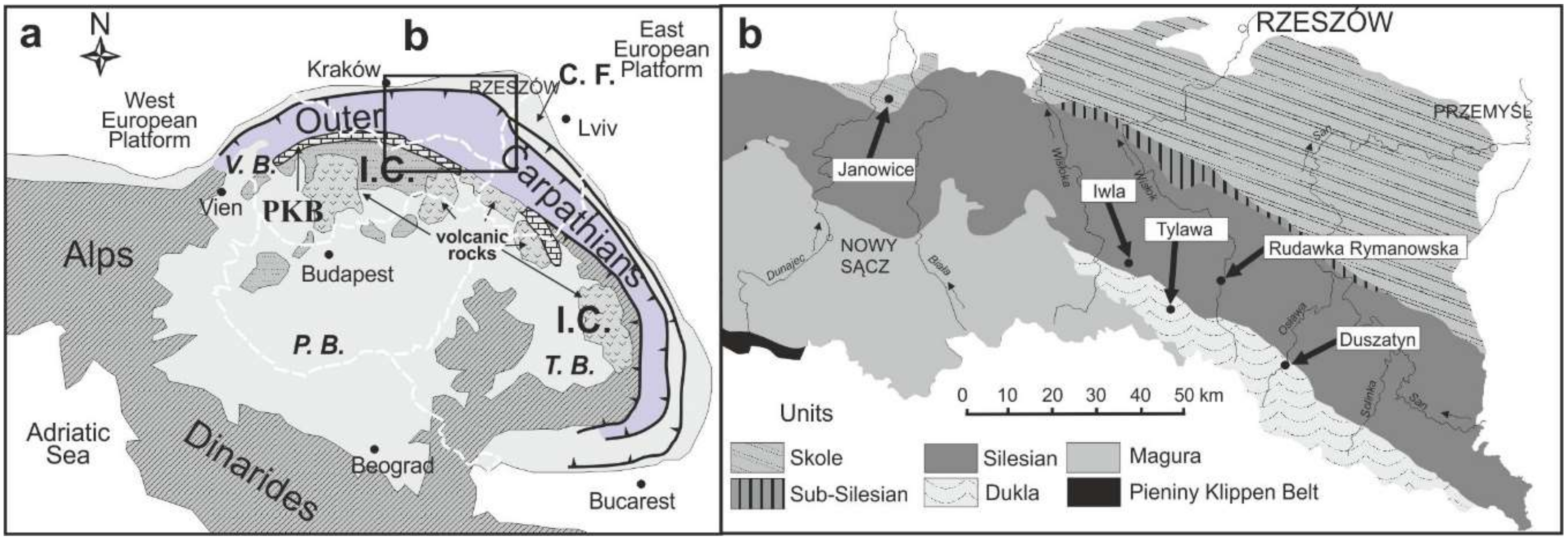
2. Geological Setting
2.1. General Outline
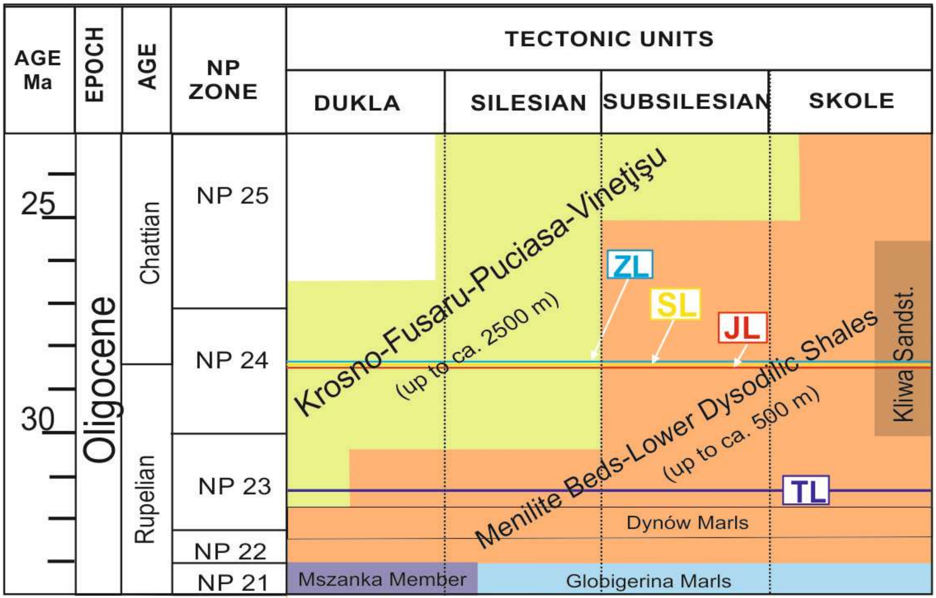
2.2. Lithostratigraphy and Biostratigraphy
3. Materials and Methods
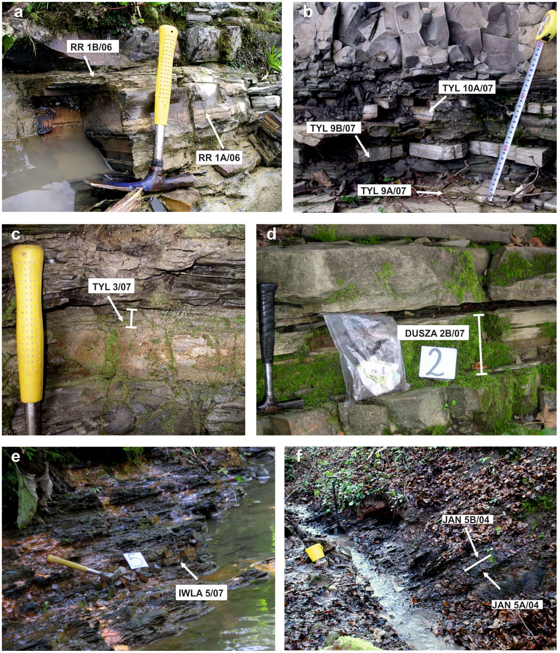
3.1. Sampling
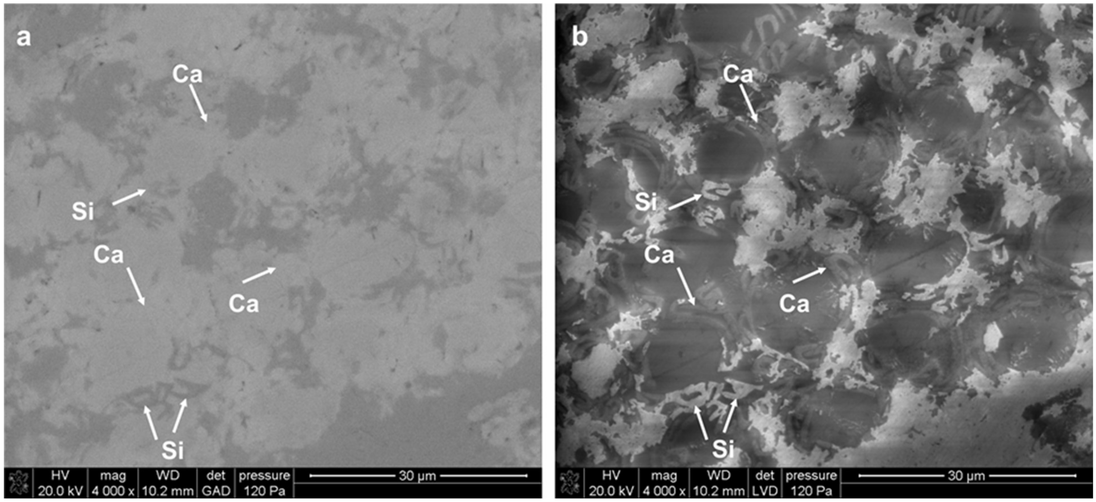
3.2. Transmitted Light Microscopy (TLM) and Scanning Electron Microscopy (SEM)
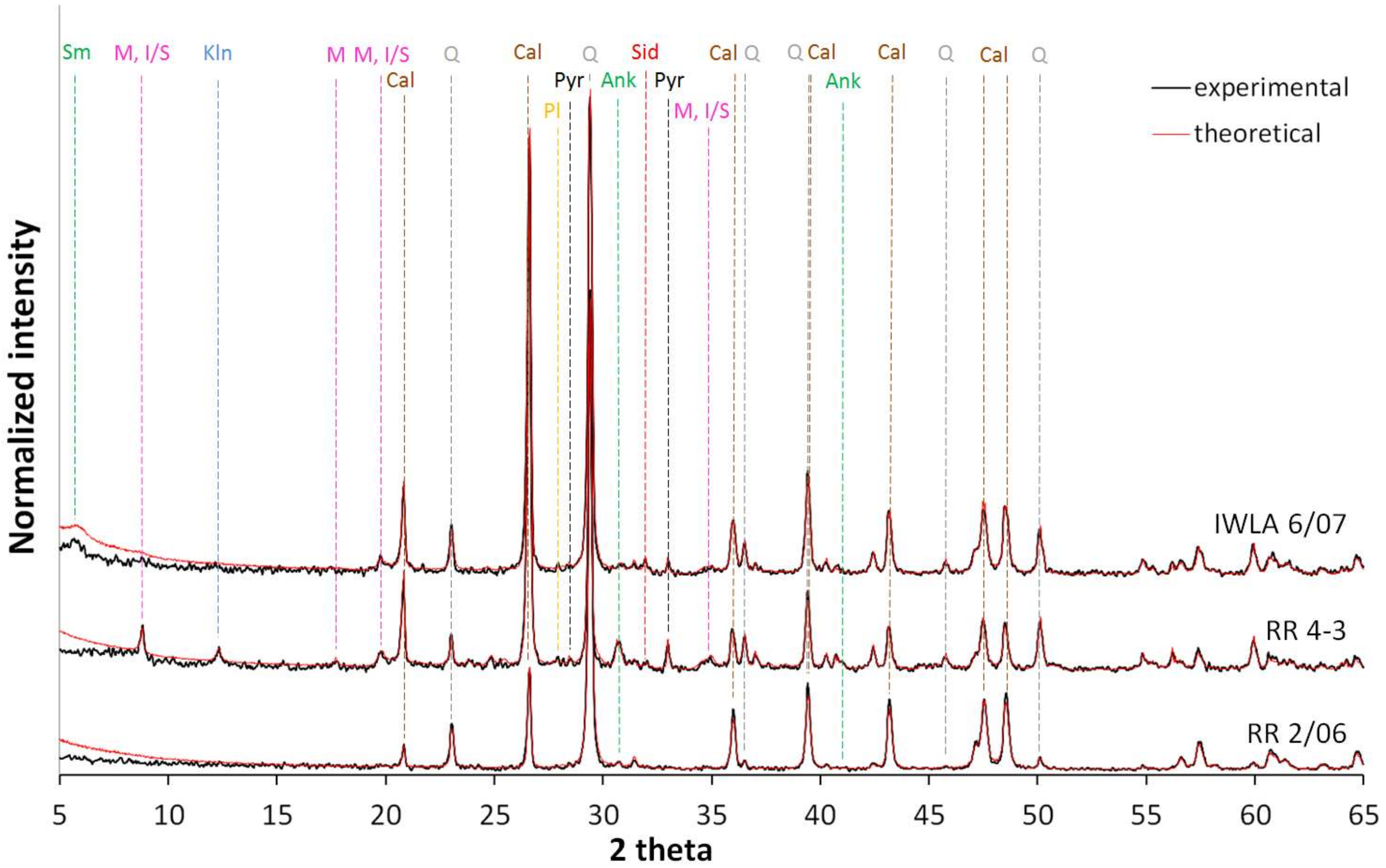
3.3. X-ray Powder Diffraction
4. Results
4.1. General Sedimentary Features of the Tylawa Limestones
4.2. Microtexture of the Tylawa Limestones
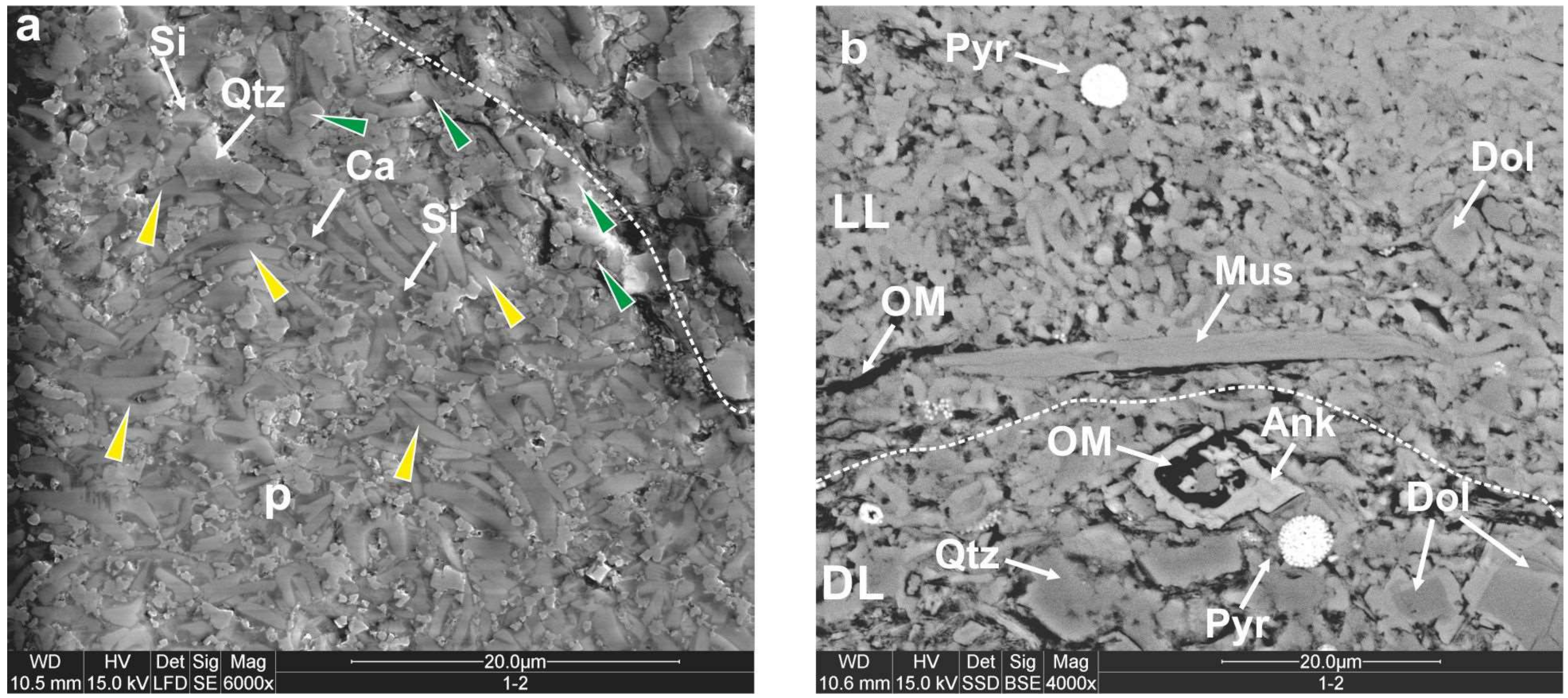
4.2.1. Biogenic Fraction
4.2.2. Abiogenic Micritic Fraction
4.3. Mineralogical Composition
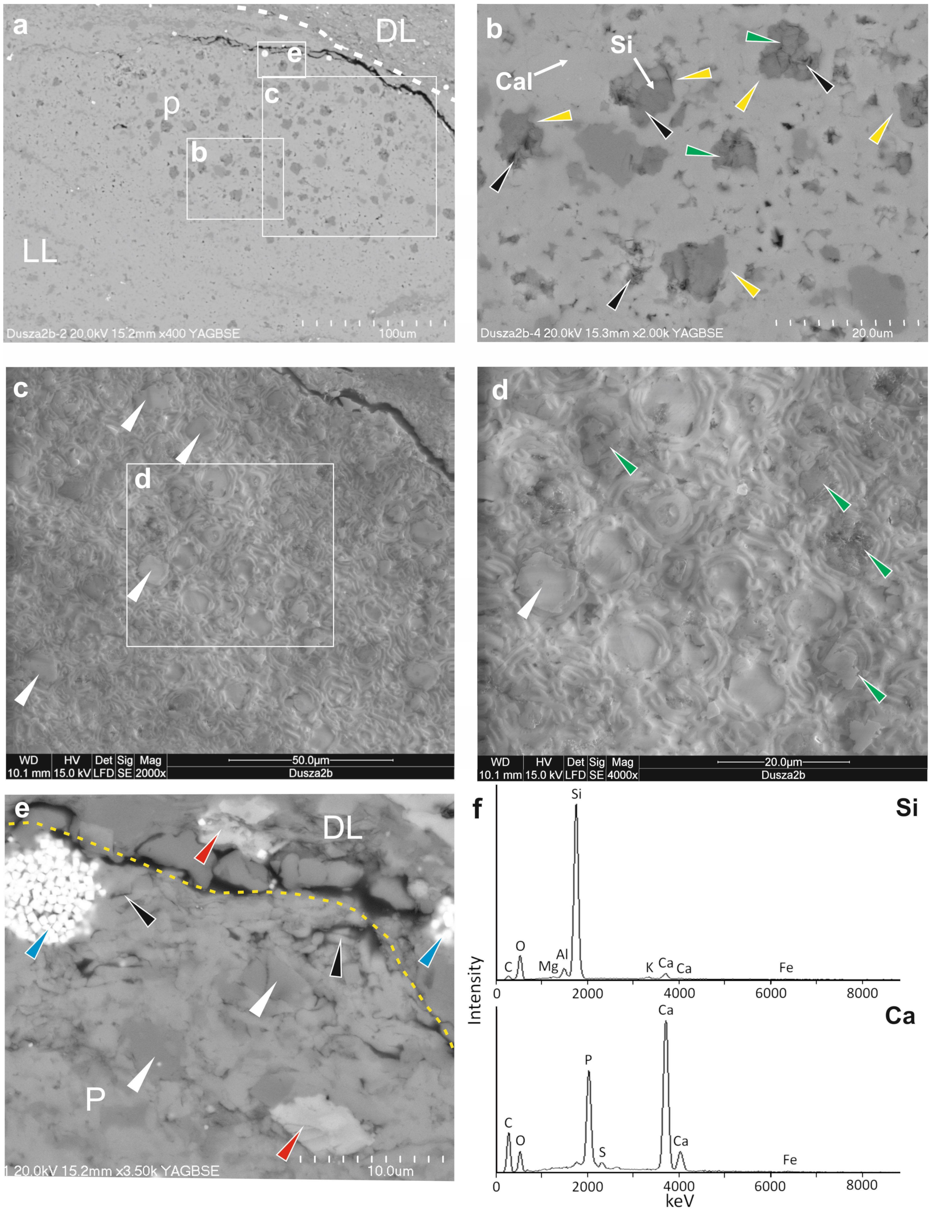
4.4. Cement Types
Cementation inside Pellets with Coccospheres
5. Discussion
5.1. Species Taxonomic Affiliation in Relation to Coccospheres Preservation
5.2. Factors Affecting the Coccolithophore Remains before Settling on the Bottom
5.2.1. Role of the Zooplankton Grazing
5.2.2. Contribution of Oxygen Minimum Zone in the Water Column
5.3. Factors Affecting the Remains after Settling on the Bottom but before Burial
5.4. Mineralization of Voids and Pores inside and outside of Pellets
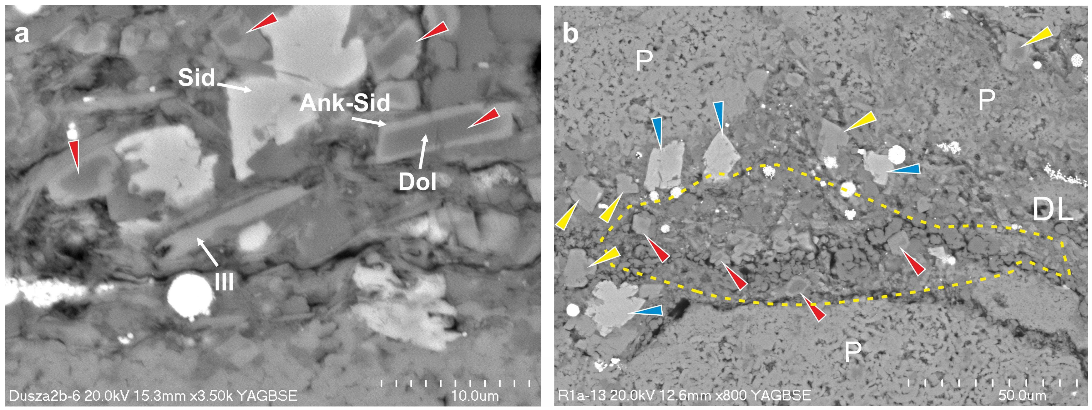
5.4.1. Calcite
5.4.2. Dolomite
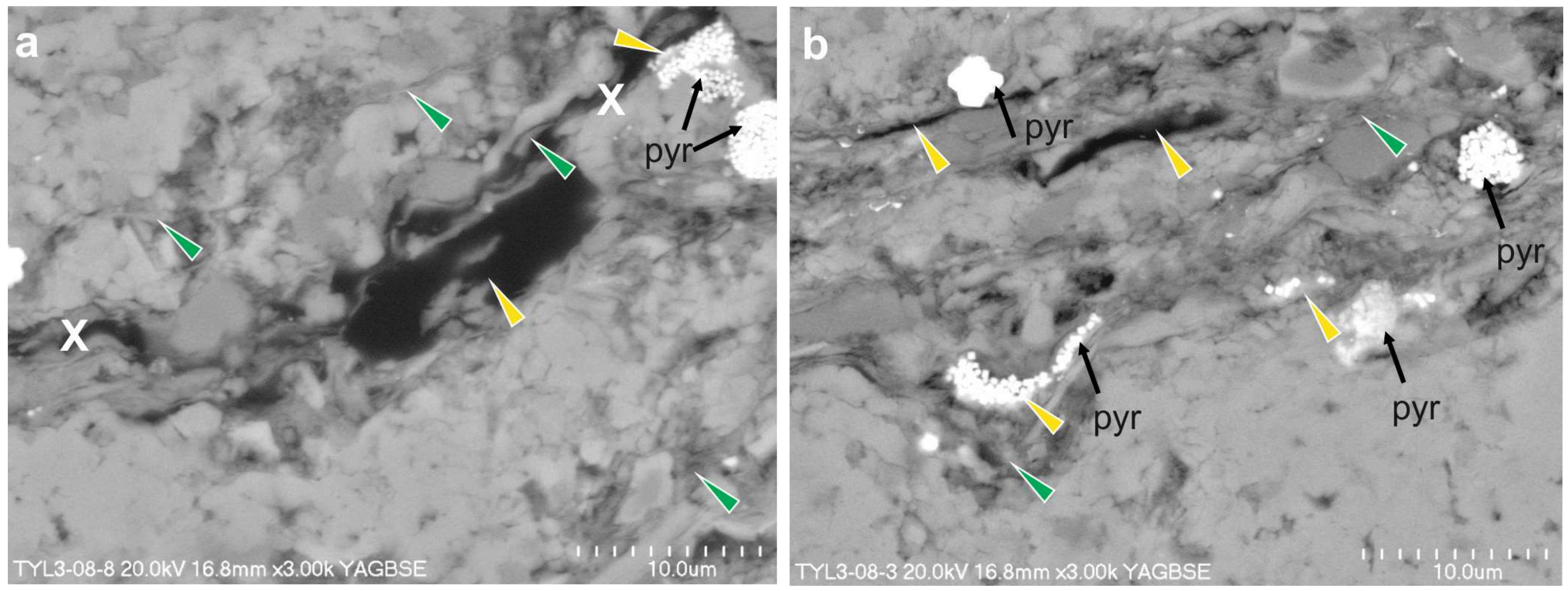
5.4.3. Pyrite
5.4.4. Siderite
5.4.5. Quartz
5.5. Timing and Depth of Diagenetic Processes Protecting Coccospheres
6. Conclusions
Supplementary Materials
Author Contributions
Funding
Acknowledgments
Conflicts of Interest
References
- Henriksen, K.; Stipp, S.L.S.; Young, J.R.; Marsh, M.E. Biological control on calcite crystallization: AFM investigation of coccolith polysaccharide function. Am. Mineral. 2004, 89, 1709–1716. [Google Scholar] [CrossRef]
- McIntyre, A.; McIntyre, R. Coccolith Concentrations and Differential Solution in Oceanic Sediments. In The Micropalaeontology of Oceans; Funnell, B.M., Reidel, W.R., Eds.; Cambridge University Press: Cambridge, UK, 1971; pp. 253–261. [Google Scholar]
- Müller, G.; Blaschke, R. Coccoliths: Important rock-forming elements in bituminous shales of central Europe. Sedimentology 1971, 17, 119–124. [Google Scholar] [CrossRef]
- Pożaryska, K. The sedimentological problems of Upper Maastrichtian and Danian of the Pulawy Environment (Middle Vistula). Biul. Państw. Instyt. Geol. 1952, 81, 1–104. [Google Scholar]
- Bąk, K. Forainiferal biostratigraphy of the Egerian flysch sediments in the Silesian Nappe, Outer Carpathians, Polish part of the Bieszczady Mountains. Anna. Soc. Geol. Pol. 2005, 75, 71–93. [Google Scholar]
- Olszewska-Nejbert, D.; Świerczewska-Gładysz, E. Campanian (Late Cretaceous) hexactinellid sponges from the white chalk of Mielnik (Eastern Poland). Acta Geol. Pol. 2011, 61, 383–417. [Google Scholar]
- Leszczyński, K. The internal geometry and lithofacies pattern of the Upper Cretaceous-Danian sequence in the Polish Lowlands. Geol. Q. 2012, 56, 363–386. [Google Scholar] [CrossRef] [Green Version]
- Jurkowska, A.; Świerczewska-Gładysz, E.; Bąk, M.; Kowalik, S. The role of biogenic silica in the formation of Upper Cretaceous pelagic carbonates and its palaeoecological implications. Cretac. Res. 2019, 93, 170–187. [Google Scholar] [CrossRef]
- Covington, J.M. New morphologic information on Cretaceous nannofossils from the Niobrara Formation (Upper Cretaceous) of Kansas. Geology 1985, 13, 683–686. [Google Scholar] [CrossRef]
- Mai, H.; von Salis Perch-Nielsen, K.; Willems, H.; Romein, A.J.T. Fossil coccospheres from the K/T boundary section from Geulhemmerberg, The Netherlands. Micropaleontology 1997, 43, 281–302. [Google Scholar] [CrossRef]
- Mai, H. Paleocene coccoliths and coccospheres in deposits of the Maastrichtian stage at the ‘type locality’ and type area in SE Limburg, The Netherlands. Mar. Micropaleontol. 1999, 36, 1–12. [Google Scholar] [CrossRef]
- Mai, H. New Coccolithophorid Taxa from Geulhemmerberg Airshaft, Lower Paleocene, the Netherlands. Micropaleontology 2001, 47, 144–154. [Google Scholar] [CrossRef]
- Dunkley Jones, T.; Bown, P.R.; Pearson, P.N. Exceptionally well preserved upper Eocene to lower Oligocene calcareous nannofossils (Prymnesiophycidae) from the Pande Formation (Kilwa Group), Tanzania. J. Syst. Palaeontol. 2009, 7, 359–411. [Google Scholar] [CrossRef]
- Bown, P.; Gibbs, S.; Sheward, R.; O’Dea, S.; Higgins, D. Searching for cells: The potential of fossil coccospheres in coccolithophore research. J. Nannoplankton Res. 2014, 34, 5–21. [Google Scholar]
- Goy, G. Nannofossils calcaires des schistes carton (Toarcien Inferieur) du bassin de Paris. In Documents de la RCP 459; BRGM: Orléans, France, 1981; p. 86. [Google Scholar]
- Gallois, R.; Medd, A.W. Coccolith-rich marker-bands in the English Kimmeridge Clay. Geol. Mag. 1979, 116, 247–334. [Google Scholar] [CrossRef]
- Lees, J.A.; Bown, P.R.; Young, J.R.; Riding, J.B. Evidence for annual records of phytoplankton productivity in the Kimmeridge Clay Formation coccolith stone bands (Upper Jurassic, Dorset, UK). Mar. Micropaleontol. 2004, 52, 29–49. [Google Scholar] [CrossRef]
- Bown, P.R.; Cooper, M.K.E. Jurassic. In Calcareous Nannofossil Biostratigraphy; Bown, P.R., Ed.; British Micropalaeontological Society Series; Chapman & Hall; The University Press: Cambridge, UK, 1998; pp. 34–85. [Google Scholar]
- Bown, P.R.; Dunkley-Jones, T.; Lees, J.A.; Randell, R.D.; Mizzi, J.A.; Pearson, P.N.; Coxall, H.K.; Young, J.R.; Nicholas, C.J.; Karega, A.; et al. A Paleogene Calcareous microfossil Konservat-Lagerstatte from the Kilwa Group of coastal Tanzania. GSA Bull. 2008, 120, 3–12. [Google Scholar] [CrossRef]
- Gibbs, S.J.; Poulton, A.J.; Bown, P.R.; Daniels, C.; Hopkins, J.; Young, J.R.; O’Dea, S.A.; Newsam, C. A cellular perspective on plankton sensitivity to past global climate change. Nat. Geosci. 2013, 6, 218–222. [Google Scholar] [CrossRef]
- Ślączka, A. Stratygrafia fałdów dukielskich okolic Komańczy—Wisłoka Wielkiego. Stratigraphy of the Dukla folds in the Komańcza—Wisłok Wielki region (Carpathians). Kwart. Geol. 1959, 3, 583–604. [Google Scholar]
- Kotlarczyk, J. Łupki tylawskie w jednostce skolskiej Karpat polskich. Spraw. Pos. Komisji Nauk. PAN Oddział w Krakowie 1977, 20, 182–183. [Google Scholar]
- Haczewski, G. Poziomy wapieni kokkolitowych w serii menilitowo-krośnieńskiej—Rozróżnianie, korelacja i geneza. Anna. Soc. Geol. Pol. 1989, 59, 435–523. [Google Scholar]
- Ciurej, A.; Haczewski, G. The Tylawa Limestones—A regional marker horizon in the Lower Oligocene of the Paratethys: Diagnostic characteristics from the type area. Geol. Q. 2012, 56, 833–844. [Google Scholar] [CrossRef] [Green Version]
- Bojanowski, M.J.; Ciurej, A.; Haczewski, G.; Jokubauskas, P.; Schouten, S.; Tyszka, J.; Bijl, P.K. The Central Paratethys during Oligocene as an ancient counterpart of the present-day Black Sea: Unique records from the coccolith limestones. Mar. Geol. 2018, 403, 301–328. [Google Scholar] [CrossRef]
- Nagymarosy, A.; Voronina, A. Calcareous nannoplankton from the Lower Maikopian beds (Early Oligocene, Union of Independent States). In Nannoplankton Research, Proc. of the Fourth INA Conference, Prague 1991; Hamršmíd, B., Young, J.R., Eds.; Knihovnička Zemnyho Plynu a Nafty; Moravské Naftové Doly: Hodonjin, Czech Republic, 1992; Volume 14b, pp. 187–221. [Google Scholar]
- Melinte, M. Oligocene palaeoenvironmental changes in the Romanian Carpathians, revealed by calcareous nannofossils. Stud Geol. Pol. 2005, 124, 341–352. [Google Scholar]
- Kotlarczyk, J.; Jerzmańska, A.; Świdnicka, E.; Wiszniowska, T. A framework of ichnofaunal, ecostratigraphy of the Polish Outer Carpathians Basin. Anna. Soc. Geol. Pol. 2006, 76, 1–111. [Google Scholar]
- Ciurej, A. Procesy i Warunki Sedymentacji Wapieni Tylawskich w Oligocenie Karpat Zewnętrznych (ze Szczególnym Uwzględnieniem Polskiej Części Karpat). Ph.D. Thesis, Akademia Górniczo-Hutnicza, Kraków, Poland, 2009; p. 266. [Google Scholar]
- Oszczypko, N.; Malata, E.; Bąk, K.; Kędzierski, M.; Oszczypko-Clowes, M. Lithostratigraphy and biostratigraphy of the Upper Albian–Lower/Middle Eocene flysch deposits in the Bystrica and Rača subunits of the Magura Nappe (Beskid Wyspowy and Gorce Ranges; Poland. Anna. Soc. Geol. Pol. 2005, 75, 27–69. [Google Scholar]
- Garecka, M.; Szydło, A. Calcareous nannofossils and foraminifera from the youngest deposits of the Siary Subunit (Oligocene, Magura Nappe, Polish Outer Carpathians). Geol. Q. 2015, 85, 205–219. [Google Scholar] [CrossRef] [Green Version]
- Bieńkowska, M. Taphonomy of ichthyofauna from an Oligocene sequence (Tylawa Limestones horizon) of the Outer Carpathians, Poland. Geol. Q. 2004, 48, 181–192. [Google Scholar]
- Barthel, K.W.; Swinburne, N.H.M.; Conway-Morris, S. Solnhofen: A Study in Mesozoic Palaeontology; Cambridge University Press: Cambridge, UK, 1990; p. 236. [Google Scholar]
- Von Meyer, H. Archaeopteryx lithographica aus dem lithographischen Schiefer von Solenhofen. Palaeontographica 1862, 10, 53–56. [Google Scholar]
- Ebert, M.; Kölbl-Ebert, M.; Lane, J.A. Fauna and Predator-Prey Relationships of Ettling, an Actinopterygian Fish-Dominated Konservat-Lagerstätte from the Late Jurassic of Southern Germany. PLoS ONE 2015, 10, e0116140. [Google Scholar] [CrossRef]
- Thomsen, E. Seasonal variability in the production of Lower Cretaceous calcareous nannoplankton. Geology 1989, 17, 715–717. [Google Scholar] [CrossRef]
- Książkiewicz, M. The Tectonics of the Carpathians. In Geology of Poland, Tectonics, 4; Pożaryski, W., Ed.; Wydawnictwa Geologiczne: Warszawa, Poland, 1977; pp. 476–620. [Google Scholar]
- Oszczypko, N. The structural position and tektonosedimentary evolution of the Polish Outer Carpathians. Prz. Geol. 2004, 52, 780–791. [Google Scholar]
- van der Boon, A.; Beniest, A.; Ciurej, A.; Gaździcka, E.; Grothe, A.; Sachsenhofer, R.F.; Langereis, C.G.; Krijgsman, W. The Eocene-Oligocene transition in the North Alpine Foreland Basin and subsequent closure of a Paratethys gateway. Glob. Planet. Change 2018, 162, 101–119. [Google Scholar] [CrossRef]
- Jucha, S.; Kotlarczyk, J. Seria menilitowo-krośnieńska w Karpatach fliszowych. Prace Geologiczne PAN Oddz. Krakowie 1961, 4, 1–115. [Google Scholar]
- Bąk, K.; Rubinkiewicz, J.; Garecka, M.; Machaniec, E.; Dziubińska, B. Exotics-bearing layer in the Oligocene flysch of the Krosno Beds in the Fore-Dukla Zone (Silesian Nappe, Outer Carpathians), Poland. Geol. Carpath. 2001, 52, 159–171. [Google Scholar]
- Bąk, K.; Wolska, A. Exotic orthogneiss pebbles from Paleocene flysch of the Dukla Nappe (Outer Eastern Carpathians, Poland). Geol. Carpath. 2005, 56, 205–221. [Google Scholar]
- Bąk, K.; Wolska, A.; Zielińska, M.; Bąk, M. Coal-bearing submarine slump sediments from Oligocene–Miocene transition of the Eastern Carpathians (Bieszczady Mountains, SE Poland). Geol. Q. 2015, 59, 300–315. [Google Scholar]
- Siemińska, A. Starzec, K. Godlewski, P. Wendorff, M. Sedimentary response to tectonic uplift of the Dukla basin margin recorded at Skrzydlna—The Menilite Beds (Oligocene), Outer Carpathians, S Poland. Geol. Geophys. Environ. 2018, 44, 231–244. [Google Scholar] [CrossRef]
- Leszczyński, S. Origin of the Sub-Menilite Globigerina Marl (Eocene—Oligocene transition), in the Polish Outer Carpathians). Anna. Soc. Geol. Pol. 1997, 67, 367–424. [Google Scholar]
- Rögl, F. Palaeogeographic Consideration for Mediterranean and Paratethys Seaways (Oligocene to Miocene). Anna. Naturhistorischen Mus. Wien. 1998, 99A, 279–310. [Google Scholar]
- Kováč, M.; Plašienka, D.; Soták, J.; Vojtko, R.; Oszczypko, N.; Less, G.; Ćosović, V.; Fügenschuh, B.; Králiková, S. Paleogene palaeogeography and basin evolution of the Western Carpathians, Northern Pannonian domain and adjoining areas. Glob. Planet. Change 2016, 140, 9–27. [Google Scholar] [CrossRef]
- Jones, R.W.; Simmons, M.D. A review of the stratigraphy of Eastern Paratethys (Oligocene-Holocene). Bull. Natur. Hist. Mus. London. Geol. 1996, 52, 25–49. [Google Scholar]
- Olszewska, B. Foraminiferal biostratigraphy of the Polish Outer Carpathians: A record of basin geohistory. Anna. Soc. Geol. Pol. 1997, 67, 325–337. [Google Scholar]
- Martini, E. Standard Tertiary and Quaternary Calcareous Nannoplankton Zonation. In Proceedings of the II Planktonic Conference, Rome, Italy, 1970; Farinacci, A., Ed.; Tecnoscienza: Rome, Italy, 1971; pp. 739–785. [Google Scholar]
- Ciurej, A.; Haczewski, G. The Sokoliska Limestone—A new regional marker horizon of coccolith laminites in the Oligocene of the Outer Carpathians: Diagnostic features and stratigraphic position. Ann. Soc. Geol. Pol. 2016, 86, 415–427. [Google Scholar] [CrossRef]
- Krhovský, J. Mikrobiostratigrafické korelace vnějších jednotek flyšového pasma a vliv eustatických změn na jejich paleogeografický vývoj. Microbiostratigraphic correlations in the outer flysch units of the Southern Moravia and influence of the eustacy on their paleo-geographical development. Zemni Plyn a Nafta 1981, 26, 665–688. [Google Scholar]
- Bąk, K. Late Oligocene Foraminifera from the Krosno Beds in the San valley section (Bieszczady Mountains); Silesian Unit, Polish Outer Carpathians. Anna. Soc. Geol. Pol. 1999, 69, 195–217. [Google Scholar]
- Švábenická, L.; Bubík, M.; Stráník, Z. Biostratigraphy and paleoenvironmental changes on the transition from the Menilite to Krosno lithofacies (Western Carpathians, Czech Republic). Geol. Carpath. 2007, 58, 237–262. [Google Scholar]
- Gradstein, F.M.; Ogg, J.G.; Schmitz, M.; Ogg, G. The Geologic Time Scale; Elsevier Publ. Co.: Amsterdam, The Netherlands, 2012; p. 1176. [Google Scholar]
- Ciurej, A. Procedures for obtaining optimal SEM images of coccolithophore debris in coccolith limestones. Acta Palaeontol. Pol. 2010, 55, 169–171. [Google Scholar] [CrossRef] [Green Version]
- Saïag, J.; Collin, P.-Y.; Sizun, J.-P.; Herbst, F.; Faÿ-Gomord, O.; Chateau Smith, C.; Caline, B.; Lasseur, É. Classifying chalk microtextures: Sedimentary versus diagenetic origin (Cenomanian–Santonian, Paris Basin, France). Sedimentology 2019, 66, 2976–3007. [Google Scholar]
- Xu, K.; Hutchins, D.; Gao, K. Coccolith arrangement follows Eulerian mathematics in the coccolithophore Emiliania huxleyi. Peer J. 2018, 6, e4608. [Google Scholar] [CrossRef] [Green Version]
- Weigelt, J. Über Biostratonomie. Geologe 1927, 42, 1069–1076. [Google Scholar]
- Seilacher, A. Biostratinomy: The Sedimentology of Biologically Standardized Particles. In Evolving Concepts in Sedimentology; Ginsburg, R.N., Ed.; Johns Hopkins University Press: Baltimore, MD, USA, 1973; pp. 159–177. [Google Scholar]
- Honjo, S. Coccoliths: Production, transportation and sedimentation. Mar. Micropaleontol. 1976, 1, 65–79. [Google Scholar] [CrossRef]
- Honjo, S.; Okada, H. Community structure of coccolithophores in the photic layer of the mid-Pacific. Micropaleontology 1974, 29, 209–230. [Google Scholar] [CrossRef]
- Kinkel, H.; Baumann, K.-H.; Cepek, M. Coccolithophores in the equatorial Atlantic Ocean: Response to seasonal and Late Quaternary surface water variability. Mar. Micropaleontol. 2000, 39, 87–112. [Google Scholar] [CrossRef]
- McCave, I.N. Vertical flux of particles in the ocean. Deep-Sea Res. 1975, 22, 491–502. [Google Scholar] [CrossRef]
- Hattin, D.E. Petrology and origin of faecal pellets in Upper Cretaceous strata of Kansas and Saskatchewan. J. Sediment. Petrol. 1975, 45, 686–696. [Google Scholar]
- Small, L.F.; Fowler, S.W.; Unlu, M.Y. Sinking rates of natural copepod fecal pellets. Mar. Biol. 1979, 51, 233–241. [Google Scholar] [CrossRef]
- Schrader, H. Faecal pellets: Their role in the sedimentation of pelagic diatoms. Science 1971, 174, 55–57. [Google Scholar] [CrossRef]
- Bayliss, P.; Syvitski, J.P.M. Clay diagenesis in recent marine fecal pellets. Geo-Marine Lett. 1982, 2, 83–88. [Google Scholar] [CrossRef]
- Bąk, K.; Bąk, M.; Górny, Z.; Wolska, A. Environmental conditions in a Carpathian deep sea basin during the period preceding Oceanic Anoxic Event 2—A case study from the Skole Nappe. Geol. Carpath. 2014, 65, 433–450. [Google Scholar] [CrossRef] [Green Version]
- Bąk, M. Tethyan radiolarians at the Cenomanian–Turonian Anoxic Event from the Apennines (Umbria-Marche) and the Outer Carpathians: Palaeoecological and Palaeoenvironmental implications. Methods and Applications in Micropalaeontology. Part II. Stud. Geol. Pol. 2011, 134, 1–279. [Google Scholar]
- Pomeroy, L.R.; Hanson, R.B.; McGillivary, P.A.; Sherr, B.F.; Kirchman, D.; Deibel, D. Microbiology and chemistry of fecal products of pelagic tunicates: rate and fates. Bull. Mar. Sci. 1984, 35, 426–439. [Google Scholar]
- Bąk, M.; Bąk, K.; Ciurej, A. Palaeoenvironmental signal from the microfossils record in the Mikuszowice Cherts of the Silesian Nappe, Polish Outer Carpathians. In Integrating Microfossil Records from the Oceans and Epicontinental Seas; Bąk, M., Kaminski, M.A., Waśkowska, A., Eds.; The Grzybowski Foundation: Krakow, Poland, 2011; Volume 17, pp. 15–25. [Google Scholar]
- Báldi, T. Tethys and Paratethys through Oligocene times. Remarks and comment. Geol. Carpath. 1989, 40, 85–99. [Google Scholar]
- Popov, S.V.; Rögl, F.; Rozanov, A.Y.; Steininger, F.F.; Shcherba, I.G.; Kovač, M. Lithological-Paleogeographic maps of Paratethys—10 Maps Late Eocene to Pliocene. Courier Forschungsinstitut Senckenberg 2004, 250, 1–46. [Google Scholar]
- Schulz, H.M.; Bechtel, A.; Sachsenhofer, R.F. The birth of the Paratethys during the Early Oligocene: From Tethys to an ancient Black Sea analogue? Glob. Planet. Change 2005, 49, 163–176. [Google Scholar] [CrossRef]
- Bąk, K.; Fabiańska, M.; Bąk, M.; Misz-Kennan, M.; Zielińska, M.; Dulemba, P.; Bryndal, T.; Naglik, B. Organic matter in upper Albian marine sediments in the High-Tatric units, central western Carpathians related to Oceanic Anoxic Event 1d—Geochemistry, microfacies and palynology. Palaeogeogr. Palaeoclimatol. Palaeoecol. 2016, 454, 212–227. [Google Scholar] [CrossRef]
- Bąk, K. Environmental changes during the Cenomanian—Turonian boundary event in the Outer Carpathian basins: A synthesis of data from various tectonic-facies units. Anna. Soc. Geol. Pol. 2007, 77, 171–191. [Google Scholar]
- Bąk, K.; Bąk, M.; Dulemba, P.; Okoński, S. Late Cenomanian environmental conditions at the submerged Tatric Ridge, Central Western Carpathians during the period preceding Oceanic Anoxic Event 2—A palaeontological and isotopic approach. Cretac. Res. 2016, 63, 95–112. [Google Scholar] [CrossRef]
- Lampitt, R.S.; Noji, T.; von Bodungen, B. What happens to zooplankton faecal pellets? Implications for material flux. Mar. Biol. 1990, 104, 15–23. [Google Scholar] [CrossRef]
- Jin, X.; Liu, C. Ecological and taphonomical influences on coccoliths in surface sediments in the shelf of the Yellow and East China Seas. Cont. Shelf Res. 2017, 140, 27–36. [Google Scholar] [CrossRef]
- Bąk, M.; Bąk, K.; Michalik, M. Decadal to millennial variations in water column parameters in pelagic marine environments of the Western Tethys (Carpathian realm) during Middle–Late Jurassic—Evidence from the radiolarian record. Glob. Planet. Change 2018, 162, 148–162. [Google Scholar] [CrossRef]
- Andruleit, H.; Rogalla, U. Coccolithophores in surface sediments of the Arabian Sea in relation to environmental gradients in surface waters. Mar. Geol. 2002, 186, 505–526. [Google Scholar] [CrossRef]
- Harris, R.P. Zooplankton grazing on the coccolithophore Emiliania huxleyi and its role in inorganic carbon flux. Mar. Biol. 1994, 119, 431–439. [Google Scholar] [CrossRef]
- Zijlstra, H.J.P. Early diagenetic silica precipitation, in relation to redox boundaries and bacterial metabolism, in late Cretaceous chalk of the Maastrichtian type locality. Geologie Mijnbouw 1987, 66, 343–355. [Google Scholar]
- Lundegard, P.D.; Land, L.S. Carbonate equilibria and pH buffering by organic acids response to changes in pCO2. Chem. Geol. 1989, 74, 277–287. [Google Scholar] [CrossRef]
- Compton, J.S.; Siever, R. Diffusion and mass balance of Mg during early dolomite formation, Monterey Formation. Geochim. Cosmochim. Acta 1986, 50, 125–135. [Google Scholar] [CrossRef]
- Raiswell, R.; Buckley, F.; Berner, R.A.; Anderson, T.F. Degree of pyritisation as a paleoenvironmental indicator of bottom water oxygenation. J. Sediment. Petrol. 1988, 58, 812–819. [Google Scholar]
- Burdige, D.J. The biogeochemistry of manganese and iron reduction in marine sediments. Earth Sci. Rev. 1993, 35, 249–284. [Google Scholar] [CrossRef]
- Van der Weijden, C.H.; Middelburg, J.J.; van Gaans, P.F.M. Comment and Reply: Early diagenetic silica precipitation, in relation to redox boundaries and bacterial metabolism, in late Cretaceous chalk of the Maastrichtian type locality. Geol. Mijnbouw 1989, 68, 263–270. [Google Scholar]
- Ellwood, B.B.; Chrzanowski, T.H.; Hrouda, F.; Long, G.J.; Buhl, M.L. Siderite formation in anoxic deep-sea sediments: A synergetic bacterially controlled process with important implications in paleomagnetism. Geology 1988, 16, 980–982. [Google Scholar] [CrossRef]
- Haese, R.R.; Wallmann, K.; Kretzmann, U.; Müller, P.J.; Schulz, H.D. Iron species determination to investigate the early diagenetic reactivity in marine sediments. Geochim. Cosmochim. Acta 1997, 61, 63–72. [Google Scholar] [CrossRef]
- Berner, R.A. Principles of Chemical Sedimentology; McGraw-Hill: New York, NY, USA, 1971; p. 240. [Google Scholar]
- Jørgensen, B.B. Bacterial sulfate reduction within reduced microniches of oxidized marine sediments. Mar. Biol. 1977, 41, 7–17. [Google Scholar] [CrossRef]
- Haese, R.R. The Biogeochemistry of Iron. In Marine Geochemistry; Schulz, H.D., Zabel, M., Eds.; Springer: Berlin, Germany, 2006; pp. 207–240. [Google Scholar]
- Bąk, K.; Bąk, M.; Błachowski, A.; Gatlik, J. Oscillating redox conditions in the Middle–Late Jurassic Alpine Tethys: Insights from selected geochemical indices and 57Fe Mössbauer spectroscopy. Palaeogeogr. Palaeoclimatol. Palaeoecol. 2020, 537, 109440. [Google Scholar] [CrossRef]
- El Albani, A.; Cloutier, R.; Candilier, A.M. Early diagenesis of the Upper Devonian Escuminac Formation in the Gaspe Peninsula, Quebec: Sedimentological and geochemical evidence. Sediment. Geol. 2002, 146, 209–223. [Google Scholar] [CrossRef]
- Wei, H.; Tang, Z.; Qiu, Z.; Yan, D.; Bai, M. Formation of large carbonate concretions in black cherts in the Gufeng Formation (Guadalupian) at Enshi, South China. Geobiology 2020, 18, 14–30. [Google Scholar] [CrossRef] [PubMed]
- Loyd, S.J.; Berelson, W.M.; Lyons, T.W.; Hammond, D.E.; Corsetti, F.A. Constraining pathways of microbial mediation for carbonate concretions of the Miocene Monterey Formation using carbonate-associated sulfate. Geochim. Cosmochim. Acta 2012, 78, 77–98. [Google Scholar] [CrossRef]
- Abdel-Wahab, A.; McBride, E.F. Origin of giant calcite-cemented concretions, Temple member, Qasr El Sagha Formation (Eocene), Faiyum Depression, Egypt. J. Sediment. Res. 2001, 71, 70–81. [Google Scholar] [CrossRef]
- Coleman, M.L.; Raiswell, R. Source of carbonate and origin of zonation in pyritiferous carbonate concretions: Evaluation of a dynamic model. Am. J. Sci. 1995, 295, 282–308. [Google Scholar] [CrossRef]
- Dong, J.; Zhang, S.; Jiang, G.; Li, H.; Gao, R. Greigite from carbonate concretions of the Ediacaran Doushantuo Formation in South China and its environmental implications. Precambrian Res. 2013, 225, 77–85. [Google Scholar] [CrossRef]
- Berner, R.A. A new geochemical classification of sedimentary environments. J. Sediment. Petrol. 1981, 51, 359–365. [Google Scholar]
- Maynard, J.B. Extension of Berner’s ‘New Geochemical Classification of Sedimentary Environments’ to ancient sediments. J. Sediment. Petrol. 1982, 52, 1325–1331. [Google Scholar] [CrossRef]
- Hendry, J.P. Calcite cementation during bacterial manganese, iron and sulfate reduction in Jurassic shallow marine carbonates. Sedimentology 1993, 40, 87–106. [Google Scholar] [CrossRef]


| Sample | Lithology | Quartz | Plagioclase | Calcite | Pyrite | Siderite | Ankerite | Kaolinite | Muscovite | Illite-Smectite | Smectite | SUM |
|---|---|---|---|---|---|---|---|---|---|---|---|---|
| RR 2/06 | Laminated limestone | 10.3 (0.192°) | 85.6 | 0.5 | 3.6 | 100.0 | ||||||
| RR 6/06 | Laminated limestone | 24.7 (0.198°) | 62.0 | 0.8 | 0.6 | 11.9 | 100.0 | |||||
| IWLA 5/07 | Laminated limestone | 30.9 (0.181°) | 67.9 | 0.3 | 0.3 | 0.6 | 100.0 | |||||
| IWLA 6/07 | Laminated limestone | 28.2 (0.193°) | 1.1 | 53.4 | 1.2 | 1.0 | 0.7 | 0.6 | 8.4 | 5.4 | 100.0 | |
| DUSZA 2B/07 | Laminated limestone | 25.5 (0.187°) | 52.1 | 1.1 | 0.5 | 6.3 | 9.7 | 4.8 | 100.0 | |||
| DUSZA 6/07 | Laminated limestone | 13.9 (0.173°) | 82.4 | 2.0 | 1.7 | 100.0 | ||||||
| TYL 3/07 | Dark lamina | 10.5 (0.171°) | 74.8 | 0.5 | 1.8 | 4.8 | 7.6 | 100.0 | ||||
| RR 4-3 | Dark lamina | 33.3 (0.185°) | 1.2 | 35.0 | 2.5 | 0.4 | 3.5 | 6.7 | 8.3 | 9.1 | 100.0 |
© 2020 by the authors. Licensee MDPI, Basel, Switzerland. This article is an open access article distributed under the terms and conditions of the Creative Commons Attribution (CC BY) license (http://creativecommons.org/licenses/by/4.0/).
Share and Cite
Ciurej, A.; Bąk, M.; Szczerba, M. Biostratinomy and Diagenetic Impact on Exceptional Preservation of Coccospheres from Lower Oligocene Coccolith Limestones. Minerals 2020, 10, 616. https://doi.org/10.3390/min10070616
Ciurej A, Bąk M, Szczerba M. Biostratinomy and Diagenetic Impact on Exceptional Preservation of Coccospheres from Lower Oligocene Coccolith Limestones. Minerals. 2020; 10(7):616. https://doi.org/10.3390/min10070616
Chicago/Turabian StyleCiurej, Agnieszka, Marta Bąk, and Marek Szczerba. 2020. "Biostratinomy and Diagenetic Impact on Exceptional Preservation of Coccospheres from Lower Oligocene Coccolith Limestones" Minerals 10, no. 7: 616. https://doi.org/10.3390/min10070616