Article

Characterization of Kazakhstan’s Clays by Mössbauer Spectroscopy and X-ray Diffraction

by
Adilkhan Shokanov
1,
Irina Manakova
2,*,
Mikhail Vereshchak
2 and
Anastassiya Migunova
2,3
1
Institute of Mathematics, Physics and Informatics, Abai Kazakh National Pedagogical University, Dostyk Av. 13, Almaty 050010, Kazakhstan
2
Institute of Nuclear Physics, Ibragimov St. 1, Almaty 050032, Kazakhstan
3
Department of Physics and Technology, Al-Farabi Kazakh National University, Al-Farabi Av. 71, Almaty 050040, Kazakhstan
*
Author to whom correspondence should be addressed.
Minerals 2024, 14(7), 713; https://doi.org/10.3390/min14070713
Submission received: 4 June 2024 / Revised: 4 July 2024 / Accepted: 6 July 2024 / Published: 13 July 2024

Abstract

:
Studies of the mineralogical composition were carried out, and the features of the clays from the deposits of Kazakhstan were established using Mössbauer spectroscopy (MS) and X-ray diffraction analysis (XRD). According to the XRD results, all the samples were mixed-layer clays of the kaolinite–illite type. The lattice parameters of the kaolinite were determined, and it was shown that its structure was disordered and contained a certain amount of impurity in some of the clay samples. A special feature of two of the samples was the additionally identified muscovite polytype 2M1. The spectra of the iron-containing clays were amenable to being resolved into separate components, with similar Mössbauer parameters of the kaolinite, muscovite, illite, and glauconite. The oxidation state of the iron was determined using MS. The predominant part of paramagnetic iron in most samples was in the trivalent state. The primary minerals contributing to Fe2+ were illite and muscovite. The results obtained during the study of the clay samples with complex mineralogical compositions using MS and XRD methods both complemented one another and were found to be in good agreement.

1. Introduction

Clay is a complex mineral raw material that is increasingly being used in various applications. Clays form in different types of rocks and soils. More specifically, granites and gneiss break down into individual components (feldspars, quartz, and mica) as a result of weathering [1,2,3]. The chemical weathering of feldspars produces clay mineral formations [2]. This process takes hundreds of thousands of years.
Feldspars are the solid solutions of the triple system of the isomorphic series K[AISi3O8]-Na[AISi3O8]-Ca[AI2Si2O8], the end members of which are orthoclase, albite, and anorthite, respectively. One of the most common potassium feldspars is microcline, which has a triclinic symmetry. One of the most common sodium feldspars is albite with a monoclinic lattice.
The majority of the mass of clays is formed by the following clay minerals: secondary hydrous silicates, aluminosilicates, and ferrosilicates [4,5,6]. These minerals are formed as a result of feldspar alteration under the influence of water, temperature, and pressure. Organic substances can act as catalysts for chemical reactions that lead to the formation of clay minerals. The above factors interact with each other and determine the type and properties of clay minerals formed under specific conditions. The main components of these minerals are SiO2, A12O3, Fe2O3, CaO, K2O, TiO2, MgO, and Na2O; however, MnO and P2O5 may also be present in small amounts. In addition, some clay deposits contain significant quantities of CaCO3 and MgCO3 [7]. The particle sizes of the clay materials in clay mostly do not exceed 0.01 mm. In reference to their crystal-like structures, clay materials belong to either layered or pseudo-layered silicates. These minerals include layers consisting of silicate tetrahedra and aluminum- or magnesium-hydroxyl octahedra. There are several groups of clay minerals depending on the number and arrangement of tetrahedral and octahedral sheets in their basic structure [8,9]. The most common groups are the kaolinite group, fine-grained micas, smectite, vermiculite, and chlorite. Minerals of the kaolinite group (e.g., kaolinite and halloysite) have one tetrahedral and one octahedral sheet in their layer structure (1:1 layer type). The layers are both firmly connected and located close to one another. Therefore, water molecules and metal cations are not able to penetrate the interlayer space. The groups of smectites (montmorillonite, nontronite, beidellite, etc.) and vermiculites are characterized by a three-sheet layer of the tetrahedron–octahedron–tetrahedron type (2:1 layer type). The minerals of the fine-grained mica (e.g., illite and glauconite) group also have a three-sheet layer structure but with a strong connection between the layers. They are characterized by a high content of potassium, which enters the voids of the mineral structure. The structure of the chlorite group minerals (2:1:1 layer type) is formed with 2:1 layers, between which there are sheets of octahedrons. It is also possible that there is a group of mixed-layer minerals with alternating layers of different types.
Clay minerals have a unique set of physical and chemical properties that make them useful in a variety of applications [4,10,11]. The most important properties include the following: small particle size, large surface area, cation exchange capacity, plasticity, and chemical reactivity. The small particle size makes clay minerals incredibly light and capable of forming fine-grained rocks. The mineralogical composition and dispersibility of clays determine their plasticity, which allows such materials to be used as raw materials for the production of ceramics and bricks [12,13,14]. A large surface area, cation exchange capacity, and chemical reactivity are all important characteristics for the use of clays in water treatment, as well as for storage of household or nuclear waste [15,16,17].
A more specific application of clay minerals is their use in the production of proppants for hydraulic fracturing in the oil and gas industry. The hydraulic fracturing method is used in the development of hard-to-reach oil wells [18,19,20]. Its purpose lies in the artificial creation of new cracks in the formation and the expansion of existing ones. Granular material (proppant) is transported into the cracks that are formed by fracturing fluids. It is then used to secure the cracks open after the excess pressure is relieved. This leads to an increase in oil recovery by expanding the area of the productive zone. The choice of proppants depends on several factors: the deposit characteristics, well depth, and the method of hydraulic fracturing. A well-reasoned and detailed answer to the question of which proppants are more profitable to use is given in [21]. Currently, quartz sand and ceramic proppants are widely used as proppants [22]. Quartz sands have a high density (up to 2.65 g/cm3) and low strength; as such, they are used for hydraulic fracturing at depths of up to 2500 m [23]. For use at greater depths (up to 3500 m and higher), stronger ceramic proppants are developed [21,23]. Ceramic proppants have a density of 2.7–3.8 g/cm3 with grain sizes of 0.2–1.0 mm [23]. Ceramic proppants vary in composition, such as high-alumina, aluminosilicate, magnesia-silicate, etc. In addition to possessing a higher strength compared to quartz sand, ceramic proppants are also more resistant to crushing and have high thermal and chemical stability [21,23]. These properties contribute to its higher conductivity inside fractures. Ceramic proppants can be produced from almost any natural and industrial raw material [23,24], including clay raw materials [25,26].
The natural raw materials necessary for proppant production are abundantly available in Kazakhstan. However, their physical, mechanical, and chemical properties, as well as phase composition, have not been sufficiently studied. The most commonly used analytical methods for characterizing clay minerals include X-ray diffraction, energy-dispersive X-ray fluorescence analysis, scanning electron microscopy, and thermogravimetric analysis [27,28,29,30]. Since many clays contain iron (1–10 wt.%), Mössbauer spectroscopy is also an effective tool for studying clays and ceramics [31,32,33,34,35,36]. While X-ray diffraction provides extensive information about the type of clay mineral, Mössbauer spectroscopy investigates the iron-containing compounds that are often difficult to detect using X-rays. Thus, these two methods complement each other quite well.
Clays consist of one or more clay minerals, but they may also contain sand and carbonate particles. In addition, clays may contain iron oxides or oxyhydroxides. Iron may also be present in clay minerals in the form of Fe2+ and Fe3+, replacing Al3+. The mineralogical, physical, and chemical aspects of using Mössbauer spectroscopy in the study of clays were described in [37]. Distinguishing several of the clay minerals that are present in clays based on Mössbauer spectroscopy is not always easy since the spectra of clay minerals differ slightly. However, it is possible to determine the ratio of the divalent and trivalent irons contained in any mixture of clay minerals quite well. X-ray diffraction [38] is certainly a useful method for identifying individual clay minerals.
The iron oxides and oxyhydroxides found in clays and their Mössbauer spectra have been described in detail in many papers [31,39,40]. They are mainly represented by hematite (α-Fe2O3), goethite (α-FeOOH), and (less commonly) ferrihydrite (Fe5O8·4H2O). The Mössbauer spectra of individual clay minerals were studied in [41,42,43,44,45]. It was established in [40,41,42] that Fe3+ dominates in the spectra of most Al2Si2O5(OH)4 kaolinites. At the same time, the isomer shift relative to metallic Fe is ~0.35 mm/s, and the quadrupole splitting for kaolinites that do not contain Fe2+ is ~0.5 mm/s. However, certain kaolinites contain Fe2+ with a quadrupole splitting of ~2.5 mm/s and an isomer shift of ~1.1 mm/s [41,42,43,44]. Halloysite Al2Si2O5(OH)4 is a polymorphic form of kaolinite, i.e., it has the same chemical composition but contains additional interlayer water and differences in its structure. The authors of [44] noted the presence of only Fe3+ in their detected Mössbauer spectrum, while ~10 at.% of Fe2+ was detected in [45]. The Mössbauer spectra of glauconite (K,Na)(Fe3+,Al,Mg)2(Si,Al)4O10(OH)2 have been described by different numbers of doublets. Since iron atoms can occupy four non-equivalent positions (trans and cis site populations) in the structure of glauconite, the authors of [39,46,47,48,49] proposed Fe3+ doublets with an isomer shift of 0.24–0.49 mm/s, as well as Fe2+ doublets with an isomer shift of 1.0–1.3 mm/s. Another common component of clays is illite (K,H3O)(Al,Mg,Fe)2(Si,Al)4O10[(OH)2,(H2O)]. The octahedrally coordinated Fe3+ in illite has a quadrupole splitting of ≥0.6 mm/s [40,50,51], i.e., more than kaolinite. In [52,53], gray clay was studied, of which the parameters of hyperfine interactions almost completely coincided with the parameters of the monomineral illite’s spectrum. Various authors have attempted to determine the cis and trans site populations of iron atoms in minerals. However, this is only possible if there are well-resolved unique contributions. Indeed, this was the subject of study in the muscovite samples of KAl2[AlSi3O10](OH)2 in [54,55].
Knowledge of the elemental and phase composition of clays from various deposits determines the scope of their use. Therefore, determining the general physical-chemical properties and features of specific minerals from various clay deposits is a pressing and urgent task. Solving this problem will make it possible to control the efficiency of use and quality of natural raw materials processing, particularly the production of proppants for the oil and gas industry. Mössbauer spectroscopy combined with X-ray diffraction [38,45,56] is a widely used approach in the study of clay minerals. Meanwhile, there is currently not enough information available on clays of complex mineralogical composition. Thus, the purpose of this research was to obtain reliable experimental data on the phase composition of clays from various deposits in Kazakhstan, as well as the features of the minerals they contain.

2. Materials and Methods

Clay samples from six deposits in Kazakhstan were sampled in this study. A comparative analysis was carried out for the clay from the Aktash deposit in Uzbekistan. The sample designations and geographic coordinates of the studied samples are provided in Table 1 (These tables are illustrated on the geological map in the Supplementary Materials Figure S1). The deposits of the No. 1, 2, and 6 samples are located in the Central Kazakhstan Paleozoic massif, which was formed by differently oriented block-folded structures of different ages [57,58]. The deposit of the No. 4 sample is located on the territory of the Turan platform, which is a vast Paleozoic folded area [59,60]. The No. 5 sample was taken from the deposits of the Mugojar folded system, which is part of the Ural folded region. The No. 7 sample was taken from the deposits of the East European Platform, specifically the Caspian syneclise. The Mugojar folded system belongs to the Hercynian structure [61]. The Caspian syneclise is characterized by lower (sub-salt), salt Kungurian and upper (supra-salt) complexes [62,63]. The deposit of the No. 3 sample is located on the border of the Central Kazakhstan Paleozoic massif and the Southern Tien Shan [64,65].
The chemical composition of powder samples was determined on an RLP-21 X-ray fluorescence energy dispersive spectrometer, which is designed for analyses of the powder samples of rocks, minerals, ores, and concentrates. The X-ray fluorescence of the samples was excited via an air-cooled X-ray tube with a power of 50 W, a W anode, and a number of intermediate targets. The spectrum was recorded with a semiconductor detector made of ultrapure Si and cooled using the Peltier effect. The spectrum processing algorithm took into account the matrix effect based on a highly efficient version of the fundamental coefficient method (which does not require measurements of standard samples for content calculations). The work was carried out in accordance with the procedure “Determination of elemental composition of powder samples” [66,67].
X-ray diffraction (XRD) analysis made it possible to identify the mineralogical composition of the studied powder samples, specifically clay minerals, and helped to determine their quantities. Measurements were carried out on a D8 ADVANCE diffractometer (Bruker, Germany) (Cu anode, operating parameters 40 kV, 40 mA, increment 0.02°, scan speed 1.0 s/s) with double accumulation to improve the signal-to-noise ratio. Qualitative phase analysis was carried out in the EVA program with an integrated ICDD card database. Quantitative phase analysis was carried out in the Profex program (BGMN) via the Rietveld full-pattern-fitting method [68]. At the same time, the features of the object and research method described in [69,70] were taken into account. The structural model of disordered kaolinite, Kaolinitedis.str, which best described the profiles of the diffraction patterns, was used to refine the kaolinite phase in the samples. We used a flexible structural model of muscovite 1Md, Musc1md.str, for the final identification of the illite phase in the diffraction patterns, and this significantly improved the degree of fit of the measured scans to the expected results. We found a similar approach in [71], and J. Coevas et al. also showed that this structural model could be considered illite. The discrepancy values (χ2) were 1.1–1.4.
Mössbauer spectroscopy (MS) was used to study the chemical state of the iron in the crystalline and magnetic structures of the iron-containing minerals that were present in the clays. The Mössbauer measurements were carried out on an MS-1104Em spectrometer at room temperature in transmission geometry with a moving absorber. The spectra were recorded in constant acceleration mode with a triangular law of velocity change. The 57Co in a chromium matrix with an activity of 25 mCi served as a source of γ-quanta. A resonant scintillation detector was used to record the Mössbauer spectra (a detailed technique for recording spectra using a resonant detector is given in [35,72,73]). The α-Fe foil was the reference absorber. The studied sample had the shape of a disk with a diameter of 20 mm. For preparation, ~300 mg of clay was mixed with paraffin and pressed into a tablet. Then, the Mössbauer spectra were processed using the SpectrRelax program [74]. In addition, the pseudo-Voigt function, which is a superposition of the Lorentz and Gaussian functions, was chosen to describe the line shape. For each spectrum, the following parameters were determined: isomer shift relative to the metallic iron δ, quadrupole splitting Δ, quadrupole shift for magnetically split spectra ε, the magnetic hyperfine field H, the line width Γ, and the relative spectral area A. The assessment of the paramagnetic component of the Mössbauer structures was reduced to fitting them under the superposition of several quadrupole doublets, with different quadrupole splittings for each oxidation state. The Mössbauer Mineral Handbook [75] provided great assistance for the spectra interpretation.

3. Results and Discussion

3.1. X-ray Fluorescence Analysis

Twenty-four chemical elements were discovered via the X-ray fluorescence (XRF) method in our study of the clay samples. The results of the chemical composition analysis for the studied samples are summarized in Table 2. The basic components were SiO2 and Al2O3, and the content of SiO2 and Al2O3 ranged from 44 to 62 wt.% and 9 to 21 wt.%, respectively. The Fe2O3 content did not exceed 7.46 wt.%, and it was even less than 1 wt.% in the No. 2 and 6 samples. The maximum amount of CaO was found in the No. 4 sample, and the maximum amount of K2O was found in the No. 3 sample. Furthermore, the TiO2 content was ~1 wt.% in all samples.

3.2. X-ray Diffraction

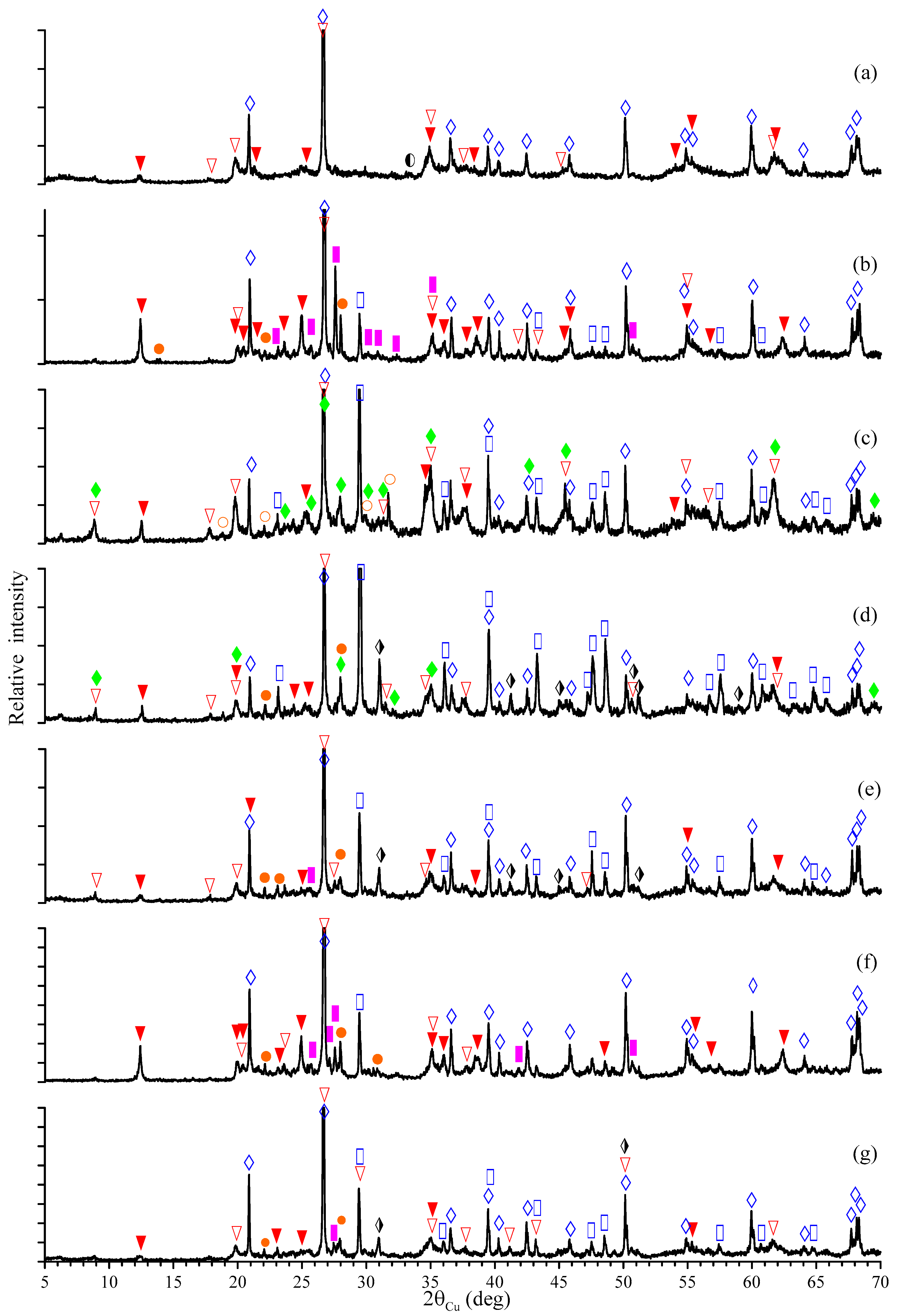
Figure 1 shows the diffraction patterns of the studied samples. The results of the quantitative phase analysis are shown in Table 3. All of the samples were found to contain quartz (the strongest peak was 26.64° 2θ, and the other peaks were 20.86, 36.54, and 50.13°), as well as a well-crystallized hexagonal crystal lattice and the kaolinite of the triclinic syngony (12.36° 2θ, and 20.36, 21.17, 24.86, 39.26°). An illite–muscovite mixture with disordered layers (henceforth referred to as illite) was another significant component of all of the samples.
The No. 1 sample was the most enriched in the clay minerals. In addition to kaolinite and illite (19.81° 2θ, as well as 8.77, 24.41, 26.52, 29.02, and 34.93°), the No. 1 sample also contained a small amount of glauconite (24.35° 2θ; 8.84, 19.54, 28.89, 34.66, and 55.05°) and hematite (33.16° 2θ; 24.15, 35.64, 40.87, 49.47, and 54,08°). This clay also contained the largest amount of kaolinite compared to all of the other samples—almost 56 wt.%.
The No. 2 sample contained feldspars in microcline (27.63° 2θ; 20.86, 23.67, 41.89, and 50.77°) and albite (where its strongest peaks were 22.02 and 28.05° 2θ, as well as 23.88, 27.79, 36.10, and 52.60°). Calcite CaCO3 (29.40° 2θ; 23.05, 35.96, 39.40, 48.50°) was also found to be present in small quantities.
The No. 3 sample was distinguished via its large amount of the illite–muscovite phase (~37.8 wt.%). The content of the kaolinite and quartz was approximately the same (11–12 wt.%). In addition, a polytype of muscovite with a parameter of c = 20 Å (8.85° 2θ; 19.94, 25.54, 26.76, 29.90, 35.11, 42.48, and 55.27°) was discovered in an amount of ~10 wt.%. The muscovite differed from the illite due to a greater degree of substitution of Si4+ for Al3+ and, therefore, higher potassium content. Thus, the theoretical K2O content in the muscovite was 11.8 wt.% [76], whereas it ranged from 3–4 to 8 wt.% in the illites in most cases [38]. In the No. 3 sample, according to the XRD results, the maximum content of illite and muscovite, which correlated with the potassium content according to the XRD data (see Table 2), was determined. A feature of this sample was a significant amount of dellaite Ca6Si3O11(OH)2 (31.66° 2θ; 13.22, 19.26, 25.97, 27.43, 29.14, 35.15, and 39.39°) at 19.5 wt.%.
In the No. 4 sample, the predominant phases were illite (~27.2 wt.%) and calcite (~25.1 wt.%). The kaolinite and muscovite with a parameter of c = 20 Å were present in approximately equal proportions (10–11 wt.%). However, the content of albite and dolomite CaMg(CO3)2 (30.98° 2θ; 41.17, 44.97, 50.58°) was found to be significantly lower (6–7 wt.%).
The No. 5 and No. 7 samples were identical in phase composition and contained kaolinite, glauconite, and illite. The kaolinite content in the No. 7 sample was noticeably lower compared to the No. 5 sample; however, albite, dolomite, and microcline were also present.
The No. 6 sample was found to be similar to the No. 2 sample in color and mineralogical composition. The main phases were kaolinite, quartz, and illite.
The calculation of the kaolinite crystal lattice parameters (Table 4) showed that, in all samples (except No. 2 and 6), the values of a and b were overestimated relative to those given in card 80-0885 of the ICDD/PDF2 database (a = 5.155 Å, b = 8.943 Å, and c = 7.405 Å). This may indicate not only that the structure of kaolinite was disordered but also that it contained a certain amount of impurity. These can be iron ions that replace Al3+ ions in octahedral positions. Since the Fe3+ ion was larger than the Al3+ ion, the average unit cell size of kaolinite increased when replacing the aluminum with iron. In the No. 2 and 6 samples, the lattice parameters turned out to be close to the values for pure triclinic kaolinite, thus meaning that kaolinite in these clays did not have impurity ions in its structure.
Thus, according to the results of the XRD studies, 11 minerals were identified in the composition of the studied clays, and all samples were mixed-layer clays of the kaolinite–illite type. A special feature of the No. 3 and 4 samples was the additionally presented polytype of muscovite 2M1.

3.3. Mössbauer Spectroscopy

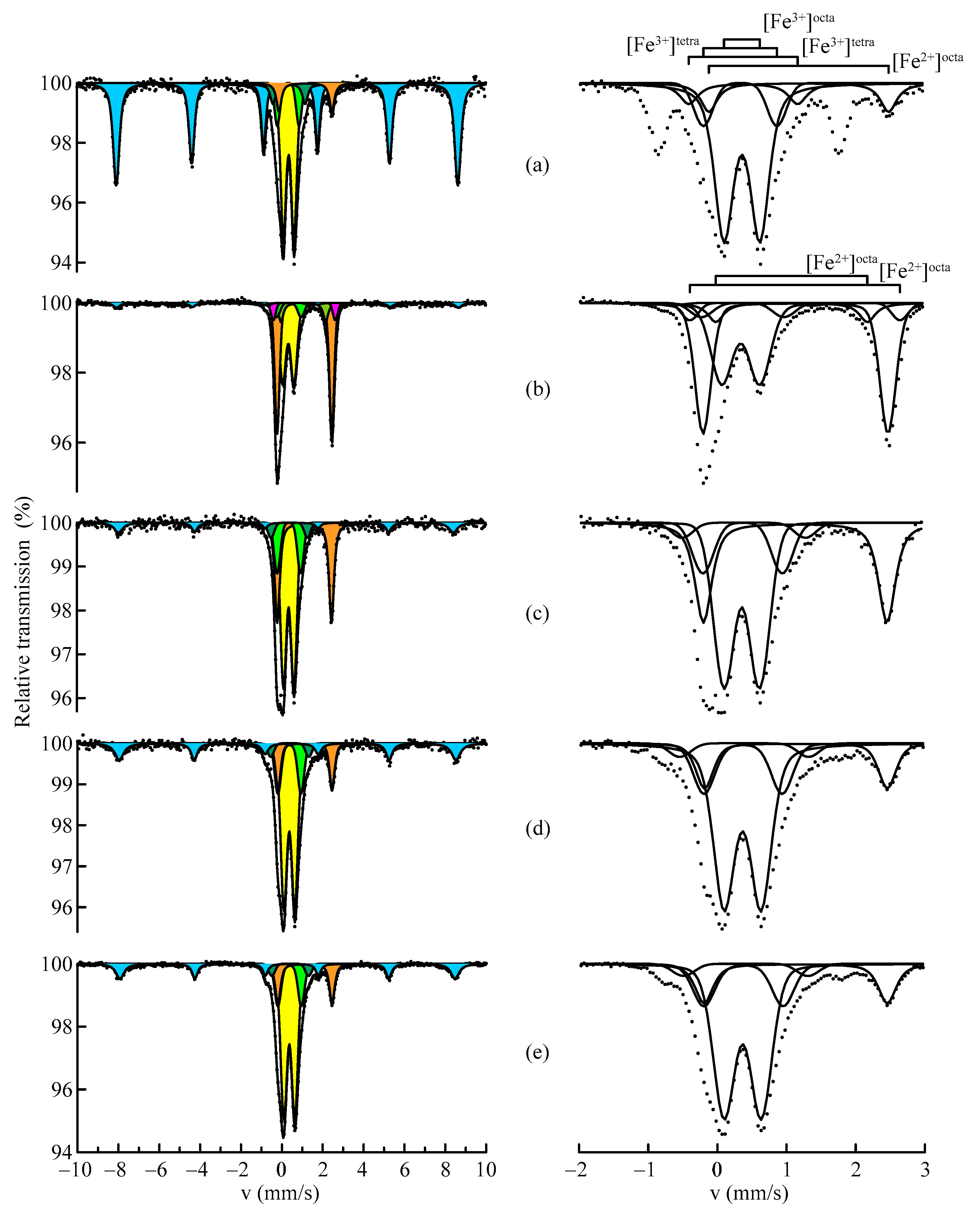
As is widely known, many of the minerals contained in clays include iron. This makes it possible to use MS extensively in the study of clays. Figure 2 shows the MS spectra of the No. 1, 3, 4, 5, and 7 samples, where the left panel exhibits the Mössbauer spectra that were in the velocity range of ±10 mm/s, and the right panel shows the enlarged central parts of the corresponding spectra in the velocity range of −2–+3 mm/s. The MS spectra of the No. 2 and 6 samples were of poor quality due to the low iron content in these clays (see Table 1). Therefore, these samples were excluded from the analysis of the MS results. Table 5 shows the hyperfine parameters of the MS spectra. The error in estimating the values of the hyperfine structure parameters was determined as the largest when comparing the instrumental and calculated errors.
As can be seen from Figure 2, it is clear that the iron in the samples of the studied clays was in magnetically ordered and paramagnetic states. The magnetic component of the MS spectra was represented by hematite, and its content was 6–16 at.%. The hyperfine magnetic fields of Fe2O3 turned out to be somewhat lower than the reference field [31,39]. This was due to the presence of aluminum in the hematite crystal lattice, which reduced the effective magnetic fields on the 57Fe nuclei [39,40]. The Al concentration in the Fe2O3 was determined according to the method described in [34], and this amounted to ~4 at.%. The maximum Fe2O3 content was observed in the No. 1 sample, which also correlated with the XRD data.
The paramagnetic part of the spectra was a superposition of the Fe3+ and Fe2+ doublets. A model-dependent method was used to fit the paramagnetic components of the Mössbauer spectra. This method is based on the use of individual components that can be associated with different compounds or local microenvironments. In the proposed model, the isomer shifts and line widths were assumed to be equal for all the doublets of each valency.
The predominant paramagnetic part of the iron in all samples, except No. 3, was in the trivalent state. As already mentioned, Fe3+ can occur in both tetrahedral IVFe3+ and octahedral VIFe3+ positions. The two species can be identified by slightly different isomer shifts. Tetrahedrally coordinated iron has a higher electron density at the nucleus than octahedral iron; therefore, it has a smaller isomer shift [37]. However, in our study, it was not possible to separate the tetrahedral and octahedral positions since the studied clays had complex mineralogical compositions. For the same reason, when fitting the paramagnetic components, it was possible to overlap several partial spectra. The isomer shift of the Fe3+ doublets was δ = 0.33–0.36 mm/s, and the quadrupole splitting of the D1 doublet was Δ = 0.40–0.50 mm/s. Kaolinite [42,43] and glauconite [46,47,48] have similar parameters, and glauconite may also be characterized by a D3 doublet with Δ = 1.05–1.23 mm/s (according to [49]). Doublet D2 had Δ = 0.58–0.71 mm/s. These values were close to the quadrupole splitting of illite [50,51] and muscovite 2M1 [54,55]. According to the authors of [52,53], the difference in the quadrupole splittings of doublets D2–D4 with similar isomer shifts may be due to variations in the local environment of the 57Fe atoms in the illite structure.
Iron in the divalent state was characterized by D5–D7 doublets, with the main share accounted for by the D5 doublet with δ = 1.12–1.14 mm/s and Δ = 2.63–2.67 mm/s. The D5 doublet included Fe2+ contributions in octahedral coordination from the kaolinite, glauconite, and illite [42,43,45,46,47,48,49,50,51], though illite can also be manifested in the D6 doublet [52,53]. The values of the quadrupole splitting Δ = 1.81–2.03 mm/s in the D6 doublet and Δ = 2.97 mm/s in the D7 doublet corresponded to muscovite 2M1, and they were also in good agreement with the data previously obtained in [54,55].
The No. 1, 5, and 7 samples had similar MS spectra. This provided a reason to assume that the mineral composition of their iron-containing phases was identical in the kaolinite, illite, and glauconite. The Fe2+/Fe3+ ratio in the spectra of these samples did not exceed 0.2. Moreover, in the MS spectrum of the No. 4 sample, the Fe2+/Fe3+ ratio was found to be higher due to the contribution of the D5 doublet in the total Fe2+. In this case, the contribution of the trivalent doublet D2 was also considerable. Based on the above, we can assume a high illite content in the No. 4 sample. Furthermore, no muscovite 2M1 was found in this sample, and this can be explained either by the absence of impurity iron ions in it or by the presence of a mixed-layer muscovite–illite structure at the final stage of muscovite hydration. The MS spectrum of the No. 3 sample was also characterized by a high contribution of divalent iron—Fe2+/Fe3+ > 1. The large contribution of the D2 doublet to the MS spectrum of this sample may indicate a significant content of illite and/or muscovite. In addition, the presence of the D6 and D7 doublets was observed, the parameters of which confirmed the presence of muscovite 2M1. According to the MS data, glauconite was also present in the No. 3 and 4 samples. However, it was not possible to detect it in these samples using XRD. This may be due to its low content, which would be beyond XRD detection, or it could be due to the presence of amorphous phases.
To check the presence of superparamagnetic fractions, the MS spectra of the No. 1, 3, and 4 samples were measured at a temperature of T = 85 K. No change in the ratio of the magnetic to paramagnetic components was observed in the obtained spectra, which indicates the absence of superparamagnetism in the clays.
We then detailed the expected phase composition of the samples by summarizing the results of the Mössbauer studies and taking into account the characteristics of the materials studied, as shown in Table 6.
Thus, using the MS method, the degree of iron oxidation was determined in all the samples. Kaolinite, illite, and glauconite were found in the composition of the studied clays according to the results of the MS studies. The Fe2+/Fe3+ ratio in the spectra of all the samples, except for the No. 3 sample, did not exceed 1. The primary minerals that contributed to Fe2+ were illite and muscovite. The spectrum of the No. 3 sample additionally contained doublets corresponding to the 2M1 muscovite polytype.

4. Conclusions

Studies on the mineralogical composition were carried out, and the features of clays from the deposits of Kazakhstan were identified via MS and XRD. According to the XRD results, 11 minerals were identified in the composition of the studied samples. All samples were mixed-layer clays of the kaolinite–illite type. The lattice parameters of kaolinite were determined, and it was shown that its structure was disordered and contained a certain amount of impurity in all of the clay samples except No. 2 and 6. A feature of the No. 3 and 4 samples was the additionally presented polytype of the 2M1 muscovite.
MS analysis did not reveal the presence of iron-containing phases in the No. 2 and 6 samples. For the remaining clays, it was possible to decompose their complex spectra into individual components with similar Mössbauer parameters of the kaolinite, muscovite, illite, and glauconite. The oxidation state of iron was determined using MS. The predominant part of the paramagnetic iron in all samples, except No. 3, was in the trivalent state. The Fe2+/Fe3+ ratio in the spectra of these samples did not exceed 1. The primary minerals contributing to Fe2+ were illite and muscovite, and the spectrum of the No. 3 sample additionally contained doublets corresponding to the 2M1 muscovite polytype.
As a result of the studies we carried out, we could obtain reliable experimental data on the phase composition of clays of complex mineralogical composition. The results obtained by XRF and the XRD and MS methods complemented each other and were in good agreement with each other. Indeed, they will certainly be useful in assessing the quality and efficiency of using natural raw materials.

5. Patents

The results presented in this manuscript form the basis of Patent No. 8688 of the Republic of Kazakhstan, “Composition for producing proppant”, which was obtained by Adilkhan Shokanov, Yerkebulan Smirhan, and Asem Kyrykbaeva in 2023.

Supplementary Materials

The following supporting information can be downloaded at: https://www.mdpi.com/article/10.3390/min14070713/s1, Figure S1: Geological map of Kazakhstan and location of the studied samples.

Author Contributions

Conceptualization and project administration, A.S. and M.V.; methodology, formal analysis, and writing—original draft preparation, M.V. and I.M.; investigation and writing—review and editing, M.V., I.M. and A.M.; visualization, I.M. and A.M.; resources, A.S. All authors have read and agreed to the published version of the manuscript.

Funding

This research was funded by the Committee of Science of the Ministry of Science and Higher Education of the Republic of Kazakhstan (Grant No. AP14869574).

Data Availability Statement

The data presented in this study are available on request from the corresponding author. The data are not publicly available due to privacy.

Acknowledgments

The authors express special gratitude to the staff of the Laboratory of Nuclear-Physical Methods of Analysis of the Institute of Nuclear Physics, Svetlana Lennik and Igor Silachyov, for their assistance in conducting X-ray fluorescence analysis.

Conflicts of Interest

The authors declare no conflicts of interest.

References

  1. Tangari, A.C.; Le Pera, E.; Andò, S.; Garzanti, E.; Piluso, E.; Marinangeli, L.; Scarciglia, F. Soil-formation in the central Mediterranean: Insight from heavy minerals. Catena 2021, 197, 104998. [Google Scholar] [CrossRef]
  2. Biondino, D.; Borrelli, L.; Critelli, S.; Muto, F.; Apollaro, C.; Coniglio, S.; Tripodi, V.; Perri, F. A multidisciplinary approach to investigate weathering processes affecting gneissic rocks (Calabria, southern Italy). Catena 2020, 187, 104732. [Google Scholar] [CrossRef]
  3. Scarciglia, F.; Le Pera, E.; Critelli, S. The onset of the sedimentary cycle in a mid-latitude upland environment: Weathering, pedogenesis, and geomorphic processes on plutonic rocks (Sila Massif, Calabria). In Sedimentary Provenance and Petrogenesis: Perspectives from Petrography and Geochemistry; Arribas, J., Critelli, S., Johnsson, M.J., Geol, S., Am, S., Eds.; GeoScienceWorld: McLean, VA, USA, 2007; Volume 420, pp. 149–166. [Google Scholar]
  4. Bergaya, F.; Lagaly, G. Chapter 1—General introduction: Clays, clay minerals and clay science. In Handbook of Clay Science, 2nd ed.; Bergaya, F., Lagaly, G., Eds.; Developments in Clay Science; Elsevier: Oxford, UK, 2013; Volume 5, pp. 1–19. [Google Scholar]
  5. Kumari, N.; Mohan, C. Basics of clay minerals and their characteristic properties. In Clay and Clay Miner; Do Nascimento, G., Ed.; Intech Open Journals: London, UK, 2021; pp. 1–29. [Google Scholar]
  6. Guggenheim, S.; Martin, R.T. Definition of clay and clay mineral: Joint report of the AIPEA nomenclature and CMS nomenclature committees. Clay Miner. 1995, 30, 257–259. [Google Scholar] [CrossRef]
  7. Arsenovic, M.V.; Pezo, L.; Radojevic, Z.M.; Stankovic, S.M. Serbian heavy clays behavior: Application in rough ceramics. Hem. Ind. 2013, 67, 811–822. [Google Scholar] [CrossRef]
  8. Barton, C.D.; Karathanasis, A.D. Clay Minerals. In Encyclopedia of Soil Science, 2nd ed.; Lal, R., Ed.; Marcel Dekker: New York, NY, USA, 2002; pp. 187–192. [Google Scholar]
  9. Varadachari, C. Clay mineral equilibra: Fuzzy phase diagrams. Clay Res. 2017, 36, 1–16. [Google Scholar]
  10. Schoonheydt, R.A. Reflections on the material science of clay minerals. Appl. Clay Sci. 2016, 131, 107–112. [Google Scholar] [CrossRef]
  11. Dai, T.; Liu, T.; Zheng, T.; Fang, C.; Zheng, S.; Lei, G. Effect of waste clay brick powder on microstructure and properties in blended oil well cement pastes at HTHP conditions. Geoenergy Sci. Eng. 2024, 237, 212823. [Google Scholar] [CrossRef]
  12. Wang, S.; Gainey, L.; Xi, Y. Thermal behaviors of clay minerals as key components and additives for fired brick properties: A review. J. Build. Eng. 2023, 66, 105802. [Google Scholar] [CrossRef]
  13. Muñoz, P.; Letelier, V.; Muñoz, L.; Gencel, O.; Sutcu, M.; Vasić, M. Assessing technological properties and environmental impact of fired bricks made by partially adding bottom ash from an industrial approach. Constr. Build. Mater. 2023, 396, 132338. [Google Scholar] [CrossRef]
  14. Meseguer, S.; Pardo, F.; Jordan, M.M.; Sanfeliu, T.; González, I. Ceramic behaviour of some kaolins from Cauquenes Province (VII Region of Maule, Chile). Appl. Clay Sci. 2011, 52, 414–418. [Google Scholar] [CrossRef]
  15. Ferrage, E.; Hubert, F.; Dabat, T.; Asaad, A.; Dazas, B.; Grégoire, B.; Savoye, S.; Tertre, E. Anisotropy in particle orientation controls water diffusion in clay materials. Appl. Clay Sci. 2023, 244, 107117. [Google Scholar] [CrossRef]
  16. Charlet, L.; Alt-Epping, P.; Wersin, P.; Gilbert, B. Diffusive transport and reaction in clay rocks: A storage (nuclear waste, CO2, H2), energy (shale gas) and water quality issue. Adv. Water Resour. 2017, 106, 39–59. [Google Scholar] [CrossRef]
  17. Das, P.; Bharat, T.V. Kaolin based protective barrier in municipal landfills against adverse chemo-mechanical loadings. Sci. Rep. 2021, 11, 10354. [Google Scholar] [CrossRef]
  18. Smith, M.; Montgomery, C. Hydraulic Fracturing (Emerging Trends and Technologies in Petroleum Engineering), 1st ed.; CRC Press: Boca Raton, FL, USA, 2015; 812p. [Google Scholar]
  19. Speight, J.G. Handbook of Hydraulic Fracturing, 1st ed.; John Wiley & Sons: Hoboken, NJ, USA, 2016; 299p. [Google Scholar]
  20. Miskimins, J. Hydraulic Fracturing: Fundamentals and Advancements; Society of Petroleum Engineers: Richardson, TX, USA, 2019; 784p. [Google Scholar]
  21. Liang, F.; Sayed, M.; Al-Muntasheri, G.A.; Chang, F.; Li, L. A comprehensive review on proppant technologies. Petroleum 2016, 2, 26–39. [Google Scholar] [CrossRef]
  22. Montgomery, C.T.; Smith, M.B. Hydraulic fracturing: History of an enduring technology. J. Petrol. Technol. 2010, 62, 26–41. [Google Scholar] [CrossRef]
  23. Yarmonov, A.N. Study of the use of high alumina cement in the production of proppants for hydraulic fracturing. Bull. PNRPU Mech. Eng. Mater. Sci. 2018, 20, 95–107. [Google Scholar] [CrossRef]
  24. Biryukova, A.A.; Dzhienalyev, T.D.; Tikhonova, T.A. Ceramic proppants based on Kazakhstan natural alumosilicate resources. Refract. Ind. Ceram. 2017, 58, 269–275. [Google Scholar] [CrossRef]
  25. Li, Y.; Huang, Z.; Lin, S.; Wu, W. Composition and Method for Producing an Ultra-Lightweight Ceramic Proppant. U.S. Patent US8727003B2, 20 May 2014. [Google Scholar]
  26. Rusinov, P.; Balashov, A. Ceramic Proppant and Its Production Method. Russia Patent RU2615563C9, 5 April 2017. [Google Scholar]
  27. Marinangeli, L.; Pompilio, L.; Baliva, A.; Billotta, S.; Bonanno, G.; Domeneghetti, M.C.; Fioretti, A.M.; Menozzi, O.; Nestola, F.; Piluso, E.; et al. Development of an ultra-miniaturised XRD/XRF instrument for the in situ mineralogical and chemical analysis of planetary soils and rocks: Implication for archaeometry. Rend. Fis. Acc. Lincei 2015, 26, 529–537. [Google Scholar] [CrossRef]
  28. Środoń, J. Chapter 12.2—Identification and quantitative analysis of clay minerals. In Handbook of Clay Science; Bergaya, F., Benny, K.G., Lagaly, G., Eds.; Developments in Clay Science; Elsevier: Oxford, UK, 2006; Volume 1, pp. 765–787. [Google Scholar]
  29. Bohor, B.F.; Randall, H.E. Scanning electron microscopy of clays and clay minerals. Clays Clay Miner. 1971, 19, 49–54. [Google Scholar] [CrossRef]
  30. Mielenz, R.C.; Schieltz, N.C.; King, M.E. Thermogravimetric analysis of clay and clay-like minerals. Clays Clay Miner. 1953, 2, 285–314. [Google Scholar] [CrossRef]
  31. Goldanskii, V.; Herber, R. Chemical Applications of Mössbauer Spectroscopy; Academic Press: New York, NY, USA, 1968; 701p. [Google Scholar]
  32. Gütlich, P.; Bill, E.; Trautwein, A.X. Mössbauer Spectroscopy and Transition Metal Chemistry: Fundamentals and Applications; Springer: Berlin/Heidelberg, Germany, 2011; 569p. [Google Scholar]
  33. Shokanov, A.; Vereshchak, M.; Manakova, I. Mössbauer and X-ray studies of phase composition of fly ashes formed after combustion of Ekibastuz coal (Kazakhstan). Metals 2020, 10, 929. [Google Scholar] [CrossRef]
  34. Vereshchak, M.; Shokanov, A.; Manakova, I. Mössbauer studies of narrow fractions of fly ash formed after combustion of Ekibastuz coal. Materials 2021, 14, 7437. [Google Scholar] [CrossRef]
  35. Vereshchak, M.; Shokanov, A.; Manakova, I.; Migunova, A. Mössbauer and X-ray diffraction spectroscopy of high-iron bauxites from Kazakhstan. Materials 2023, 16, 6706. [Google Scholar] [CrossRef]
  36. Ikeoka, R.A.; Appoloni, C.R.; Scorzelli, R.B.; Santos, E.; Rizzutto, M.A.; Bandeira, A.M. Study of ancient pottery from the Brazilian Amazon coast by EDXRF, PIXE, XRD, Mössbauer spectroscopy and computed radiography. Minerals 2022, 12, 1302. [Google Scholar] [CrossRef]
  37. Wagner, F.E.; Wagner, U. Mössbauer spectra of slays and seramics. Hyperfine Interact. 2004, 154, 35–82. [Google Scholar] [CrossRef]
  38. Häusler, W. Firing of slays studied by X-ray diffraction and Mössbauer spectroscopy. Hyperfine Interact. 2004, 154, 121–141. [Google Scholar] [CrossRef]
  39. Kuzmann, E.; Nagy, S.; Vértes, A. Critical review of analytical applications of Mössbauer spectroscopy illustrated by mineralogical and geological examples (IUPAC Technical Report). Pure Appll. Chem. 2003, 75, 801–858. [Google Scholar] [CrossRef]
  40. Murad, E.; Cashion, J. Mössbauer Spectroscopy of Environmental Materials and Their Industrial Utilization, 1st ed.; Springer: New York, NY, USA, 2004; 436p. [Google Scholar]
  41. Murad, E.; Wagner, U. Mössbauer spectra of kaolinite, halloysite and the firing products of kalonite: New results and a reappraisal of published work. Neues Jahrb. Miner. 1991, 162, 281–309. [Google Scholar]
  42. Murad, E.; Fabris, J.D. Kaolin mining and beneficiation: The role of iron. J. Phys. Conf. Ser. 2010, 217, 012066. [Google Scholar] [CrossRef]
  43. St. Pierre, T.G.; Singh, B.; Webb, J.; Gilkes, B. Mössbauer spectra of soil kaolins from South-Western Australia. Clays Clay Miner. 1992, 40, 341–346. [Google Scholar] [CrossRef]
  44. Komusinski, J.; Stoch, L.; Dubiel, S.M. Application of electron paramagnetic resonance and Mossbauer spectroscopy in the investigation of kaolinite-group minerals. Clays Clay Miner. 1981, 29, 23–30. [Google Scholar] [CrossRef]
  45. Vasiliev, A.N.; Shvanskaya, L.V.; Volkova, O.S.; Koshelev, A.V.; Zvereva, E.A.; Raganyan, G.V.; Presniakov, I.; Sobolev, A.V.; Abakumov, A.M.; Lvov, Y.M. Magnetism of natural composite of halloysite clay nanotubes Al2Si2O5(OH)4 and amorphous hematite Fe2O3. Mater. Charact. 2017, 129, 179–185. [Google Scholar] [CrossRef]
  46. Heller-Kallai, L.; Rosenson, I. The use of Mössbauer spectroscopy of iron in clay mineralogy. Phys. Chem. Miner. 1981, 7, 223–238. [Google Scholar] [CrossRef]
  47. Mashlan, M.; Martinec, P.; Kašlík, J.; Kovářová, E.; Scucka, J. Mössbauer study of transformation of Fe cations during thermal treatment of glauconite in air. IP Conf. Proc. 2012, 1489, 169–173. [Google Scholar]
  48. Dyar, M.D.; Agresti, D.G.; Schaefer, M.W.; Grant, C.A.; Sklute, E.C. Mössbauer spectroscopy of earth and planetary materials. Annu. Rev. Earth Planet. Sci. 2006, 34, 83–125. [Google Scholar] [CrossRef]
  49. Ali, A.M.; Hsia, Y.; Liu, R.C.; Zhang, J.; Duan, W.; Chen, L. A Mössbauer study of evolution of glauconite from Chinese seas. Spectrosc. Lett. 2001, 34, 701–708. [Google Scholar] [CrossRef]
  50. Murad, E.; Wagner, U. Mössbauer study of pure illite and its firing products. Hyperfine Interact. 1994, 91, 685–688. [Google Scholar] [CrossRef]
  51. Murad, E.; Wagner, U. The Mössbauer spectrum of illite. Clay Miner. 1994, 29, 1–10. [Google Scholar] [CrossRef]
  52. Pyataev, A.V.; Valiulina, S.I.; Ivanova, A.G.; Manapov, R.A.; Voronina, E.V. Traditional pottery raw Materials of the Bilyarsk monocentric agglomeration. Bull. Russ. Acad. Sci. Phys. 2015, 79, 1058–1061. [Google Scholar] [CrossRef]
  53. Valiulina, S.I.; Pyataev, A.V.; Ivanova, A.G.; Manapov, R.A.; Voronina, E.V. Mössbauer studies of moulded Kama–Cis-Urals ceramics. Archaeometry 2018, 60, 1237–1250. [Google Scholar] [CrossRef]
  54. Shabani, A.A.T.; Rancourt, D.G.; Lalonde, A.E. Determination of cis and trans Fe2+ populations in 2M1 muscovite by Mössbauer spectroscopy. Hyperfine Interact. 1998, 117, 117–129. [Google Scholar] [CrossRef]
  55. Schmidt, W.; Pietzsch, C. Iron distribution and geochemistry of pegmatitic dioctaedral 2M1 micas. Chem. Erde 1990, 50, 27–38. [Google Scholar]
  56. Hunziker, J.C.; Frey, M.; Clauer, N.; Dallmeyer, R.D.; Friedriehsen, H.; Flehmig, W.; Hoehstrasser, K.; Roggwiler, P.; Schwander, H. The evolution of illite to muscovite: Mineralogical and isotopic data from the Glarus Alps, Switzerland. Contrib. Mineral. Petrol. 1986, 92, 157–180. [Google Scholar] [CrossRef]
  57. Bogdanov, A.A. Basic features of the Paleozoic structure of Central Kazakhstan. Int. Geol. Rev. 1960, 2, 781–810. [Google Scholar] [CrossRef]
  58. Burtman, V.S.; Dvorova, A.V. Kokchetau-Issykkul terrain of the Kazakhstan Paleozoic continent and the Ordovician latitude of the continent. Lithosph. (Russ. Fed.) 2019, 19, 519–532. [Google Scholar] [CrossRef]
  59. Thomas, J.C.; Cobbold, P.R.; Shein, V.S.; Le Douaran, S. Sedimentary record of late Paleozoic to recent tectonism in central Asia—Analysis of subsurface data from the Turan and south Kazak domains. Tectonophysics 1999, 313, 243–263. [Google Scholar] [CrossRef]
  60. Natal’in, B.A.; Şengör, A.M.C. Late Palaeozoic to Triassic evolution of the Turan and Scythian platforms: The pre-history of the Palaeo-Tethyan closure. Tectonophysics 2005, 404, 175–202. [Google Scholar] [CrossRef]
  61. Zonenshain, L.P.; Korinevsky, V.G.; Kazmin, V.G.; Pechersky, D.M.; Khain, V.V.; Matveenkov, V.V. Plate tectonic model of the South Urals development. Tectonophysics 1984, 109, 95–135. [Google Scholar] [CrossRef]
  62. Grachev, A.F.; Nikolaev, V.A.; Nikolaev, V.G. East European platform development in the Late Precambrian and Paleozoic: Structure and sedimentation. Russ. J. Earth Sci. 2006, 8, 1–22. [Google Scholar] [CrossRef]
  63. Kheraskova, T.N.; Volozh, Y.A.; Antipov, M.P.; Bykadorov, V.A.; Postnikova, I.S. Features of the structure and development of the southeastern part of the East European platform and the Caspian basin in the late Precambrian-early Paleozoic. Geotectonics 2020, 54, 628–651. [Google Scholar] [CrossRef]
  64. Brookfield, M.E. Geological development and Phanerozoic crustal accretion in the western segment of the southern Tien Shan (Kyrgyzstan, Uzbekistan and Tajikistan). Tectonophysics 2000, 328, 1–14. [Google Scholar] [CrossRef]
  65. Biske, Y.S.; Konopelko, D.L.; Seltmann, R. Paleozoic collisional belt of the South Tien Shan: A review. Earth-Sci. Rev. 2024, 248, 104637. [Google Scholar] [CrossRef]
  66. Silachyov, I.; Akymbek, Y. Element content of the samples of medieval ceramics from Southern Kazakhstan: Searching the way of preliminary differentiation. J. Radioanal. Nucl. Chem. 2023, 332, 3799–3811. [Google Scholar] [CrossRef]
  67. Solodukhin, V.; Silachyov, I.; Poznyak, V.; Gorlachyov, I. Development of the complex of nuclear-physical methods of analysis for geology and technology tasks in Kazakhstan. J. Radioanal. Nucl. Chem. 2016, 309, 125–134. [Google Scholar] [CrossRef]
  68. Doebelin, N.; Kleeberg, R. Profex: A graphical user interface for the Rietveld refinement program BGMN. J. Appl. Crystallogr. 2015, 48, 1573–1580. [Google Scholar] [CrossRef]
  69. Zhou, X.; Liu, D.; Bu, H.; Deng, L.; Liu, H.; Yuan, P.; Du, P.; Song, H. XRD-based quantitative analysis of clay minerals using reference intensity ratios, mineral intensity factors, Rietveld, and full pattern summation methods: A critical review. Solid Earth Sci. 2018, 3, 16–29. [Google Scholar]
  70. Xiao, J.; Song, Y.; Li, Y. Comparison of quantitative X-ray diffraction mineral analysis Methods. Minerals 2023, 13, 566. [Google Scholar] [CrossRef]
  71. Cuevas, J.; Cabrera, M.A.; Fernandez, C.; Mota-Heredia, C.; Fernandez, R.; Torres, E.; Turrero, M.J.; Ruiz, A.I. Bentonite powder XRD quantitative analysis using Rietveld refinement: Revisiting and updating bulk semiquantitative mineralogical compositions. Minerals 2022, 12, 772. [Google Scholar] [CrossRef]
  72. Mashlan, M.; Kholmetskii, A.; Yevdokimov, V.; Pechousek, J.; Verich, O.; Zboril, R.; Tsonchev, R. Mössbauer spectrometer with resonant detector. Nucl. Instrum. Methods Phys. Res. Sect. B 2006, 243, 241–246. [Google Scholar] [CrossRef]
  73. Belyaev, A.A.; Volodin, V.S.; Irkaev, S.M.; Panchuk, V.V.; Semenov, V.G. Application of resonant detectors in Mössbauer spectroscopy. Bull. Russ. Acad. Sci. Phys. 2010, 74, 412–415. [Google Scholar] [CrossRef]
  74. Matsnev, M.E.; Rusakov, V.S. SpectrRelax: An application for Mössbauer spectra modeling and fitting. AIP Conf. Proc. 2012, 1489, 178–185. [Google Scholar]
  75. Stevens, J.G.; Khasanov, A.M.; Miller, J.W.; Pollak, H.; Li, Z. Mössbauer Mineral Handbook; Mössbauer Effect Data Center: Asheville, NC, USA, 1998; 527p. [Google Scholar]
  76. Betekhtin, A.G. A Mineralogy Course; House University: Moscow, Russia, 2008; 736p. [Google Scholar]
Figure 1. XRD patterns of the No. 1 (a), No. 2 (b), No. 3 (c), No. 4 (d), No. 5 (e), No. 4 (f), and No. 7 (g) samples: Minerals 14 00713 i001—hematite, Minerals 14 00713 i002—kaolinite, Minerals 14 00713 i003—muscovite, Minerals 14 00713 i004—illite, Minerals 14 00713 i005—quartz, Minerals 14 00713 i006—albite, Minerals 14 00713 i007—calcite, Minerals 14 00713 i008—dolomite, Minerals 14 00713 i009—microcline, and Minerals 14 00713 i010—dellaite.
Minerals 14 00713 g001
Figure 2. MS spectra of the samples: No. 1 (a), No. 3 (b), No. 4 (c), No. 5 (d), and No. 7 (e). The left panel exhibits the Mössbauer spectra in the velocity range of ±10 mm/s, and the right panel shows the enlarged central parts of the corresponding spectra in the velocity range of −2–+3 mm/s.
Minerals 14 00713 g002
Table 1. Location of the samples.
SampleDepositGeographic Coordinates
1Kalzhat, Kalzhat village, Almaty region43°30′32″ N, 80°40′22″ E
2Koskudyk, Kunaev city, Almaty region44°04′37″ N, 77°24′49″ E
3Aktash, Tashkent region41°36′51″ N, 69°46′08″ E
4Turkestan, Turkestan region43°15′53″ N, 68°23′14″ E
5Soyuznoye, Aktobe region50°50′05″ N, 60°09′33″ E
6Alekseevskoye, Akmola region53°30′35″ N, 68°31′12″ E
7Aktobe, Aktobe region50°21′28″ N, 57°07′09″ E
Table 2. Chemical composition of the samples, wt.%.
SampleSiO2Al2O3Fe2O3CaOK2OTiO2MgONa2OMnOP2O5LOI
157.1616.957.460.373.021.23----13.44
261.8720.940.673.272.720.570.57---9.25
353.6316.125.435.945.800.730.33---11.76
443.709.295.2819.463.620.570.760.990.99-16.82
552.8313.726.087.363.510.870.630.680.68-13.53
657.6518.360.788.162.581.000.69---10.54
752.6113.206.277.513.590.920.510.610.61-14.38
LOI—loss on ignition.
Table 3. Phase composition of the samples based on XRD results, wt.%.
SampleHematiteKaoliniteMuscoviteIlliteGlauconiteQuartzAlbiteCalciteDolomiteMicroclineDellaite
10.2255.73-24.141.3018.61-----
2-38.43-12.17-30.986.163.54-8.72-
3-11.7210.0637.77-10.962.837.16--19.50
4-10.8410.1027.15-13.796.1925.066.87--
5-24.61-26.642.4925.316.177.263.543.98-
6-39.07-8.05-33.386.090.83-12.58-
7-16.43-30.161.3329.577.267.944.233.08-
Table 4. Parameters of the kaolinite crystal lattice, Å.
Sample Kaolinite
abc
15.1798.9807.738
25.1598.9337.438
35.1908.9807.440
45.1908.9807.440
55.1818.9787.381
65.1618.9407.403
75.1758.9787.387
Table 5. Mössbauer parameters of the samples.
SampleComponentA, aт.%δ, mm/sΔ/2ε, mm/sH, kOeFe2+/Fe3+
1S16 ± 10.36 ± 0.04−0.21 ± 0.04511 ± 10.12
D141 ± 40.36 ± 0.040.45 ± 0.04
D218 ± 3 0.71 ± 0.04
D310 ± 2 1.12 ± 0.04
D46 ± 1 1.50 ± 0.04
D59 ± 11.12 ± 0.042.66 ± 0.04
3S6 ± 10.38 ± 0.05−0.21 ± 0.05514 ± 31.29
D112 ± 10.33 ± 0.040.49 ± 0.04
D223 ± 1 0.62 ± 0.04
D35 ± 1 1.05 ± 0.04
D41 ± 1 1.61 ± 0.05
D545 ± 21.14 ± 0.042.67 ± 0.04
D65 ± 1 1.94 ± 0.04
D73 ± 2 2.97 ± 0.06
4S8 ± 10.35 ± 0.04−0.25 ± 0.04509 ± 20.46
D116 ± 10.36 ± 0.040.40 ± 0.07
D229 ± 1 0.58 ± 0.04
D313 ± 1 1.16 ± 0.04
D45 ± 1 1.79 ± 0.04
D526 ± 11.14 ± 0.042.66 ± 0.04
D63 ± 1 2.03 ± 0.04
5S14 ± 10.38 ± 0.04−0.20 ± 0.04512 ± 10.18
D133 ± 30.36 ± 0.040.50 ± 0.04
D223 ± 1 0.60 ± 0.04
D311 ± 3 1.16 ± 0.04
D46 ± 1 1.92 ± 0.04
D513 ± 11.14 ± 0.042.63 ± 0.04
7S13 ± 10.38 ± 0.04−0.20 ± 0.04510 ± 10.19
D128 ± 20.36 ± 0.040.49 ± 0.04
D230 ± 1 0.59 ± 0.04
D311 ± 2 1.23 ± 0.04
D44 ± 1 1.98 ± 0.04
D512 ± 11.14 ± 0.042.65 ± 0.04
D62 ± 1 1.81 ± 0.04
Table 6. Phase composition of the samples based on MS results, at.%.
Sample HematiteKaoliniteMuscoviteIlliteGlauconite
11646-326
3616135510
4823-609
51438-408
71332-487
Disclaimer/Publisher’s Note: The statements, opinions and data contained in all publications are solely those of the individual author(s) and contributor(s) and not of MDPI and/or the editor(s). MDPI and/or the editor(s) disclaim responsibility for any injury to people or property resulting from any ideas, methods, instructions or products referred to in the content.