Article

Novel Indigenous Strains and Communities with Copper Bioleaching Potential from the Amolanas Mine, Chile

by
Julián C. Casas-Vargas
1,2,
Cristóbal Martínez-Bussenius
3,
Álvaro Videla
3 and
Mario Vera
2,3,*
1
Departamento de Ingeniería Hidráulica y Ambiental, Pontificia Universidad Católica de Chile, Santiago 7820436, Chile
2
Instituto de Ingeniería Biológica y Médica, Escuelas de Ingeniería, Medicina y Ciencias Biológicas, Pontificia Universidad Católica de Chile, Santiago 7820436, Chile
3
Departamento de Ingeniería de Minería, Pontificia Universidad Católica de Chile, Santiago 7820436, Chile
*
Author to whom correspondence should be addressed.
Minerals 2024, 14(9), 867; https://doi.org/10.3390/min14090867
Submission received: 21 June 2024 / Revised: 13 August 2024 / Accepted: 15 August 2024 / Published: 26 August 2024

Abstract

:
Bioleaching, a process catalyzed by acidophilic microorganisms, offers a sustainable approach to metal extraction from sulfide minerals. Chalcopyrite, the world’s most abundant copper sulfide, presents challenges due to surface passivation limiting its bioleaching efficiency. Also, indigenous species and microbial communities may present high copper extraction rates and offer new possibilities for application in bioleaching processes. This study examines the bioleaching potential of microbial isolates and communities obtained from Amolanas Mine in Chile. Samples were collected, cultivated, and identified by Sanger sequencing. The bioleaching potential and biofilm formation of isolates and enrichments were evaluated on pyrite and chalcopyrite. The results show the isolation of nine Leptospirillum and two Acidithiobacillus strains. The bioleaching experiments demonstrated good copper bioleaching potentials of the Leptospirillum I2CS27 strain and EICA consortium (composed mainly of Leptospirillum ferriphilum, Acidiphilium sp., and Sulfobacillus thermosulfidooxidans), with 11% and 25% copper recovery rates, respectively. Microbial attachment to the surface mineral was not mandatory for increasing the bioleaching rates. Our findings underscore the importance of indigenous microbial communities in enhancing copper bioleaching efficiency.

1. Introduction

Bioleaching is described as the oxidative dissolution of metal sulfides (MS) catalyzed by acidophilic microorganisms [1]. These microorganisms oxidize Fe(II) ions and/or inorganic sulfur compounds (ISCs). The resulting Fe(III) ions attack the sulfide moiety, and the oxidation of ISCs generates sulfuric acid [2,3]. Chalcopyrite (CuFeS2) is the world’s most abundant primary sulfide, and it needs to be exploited in major amounts in the future for copper bioleaching. Copper recovery from chalcopyrite through biohydrometallurgical processes has been challenging due to mineral surface passivation, leading to very low copper recovery at mesophilic temperatures (<40% in comparison to fresh chalcopyrite) [4,5]. The formation of passivation layers (elemental sulfur and jarosites) may be due to the temperature range, the presence of microorganisms, a high concentration ratio of Fe(III) to Fe(II), and the Cu(II) concentration, varying the passivation redox potential (ORP) from 475 to 635 mV (vs. Ag/AgCl with 3.5 M KCl) [6,7,8].
Biofilm formation on MS surfaces is a fundamental process that may influence their dissolution [9]. In some cases, it could lead to the formation of a passivating layer, which might cause a negative effect on chalcopyrite dissolution, resulting in low bioleaching rates [10,11]. Biofilm cells are usually embedded in extracellular polymeric substances (EPS), which mediate cell attachment to MS, allow water retention, and contribute to the accumulation of Fe(III) ions, which, at appropriate rates, contribute to MS oxidation [12]. EPS are frequently composed of proteins, carbohydrates, lipids, and DNA, although their composition changes according to microbial species, type of surface and/or electron donors, and growth phase, among others [13].
Bioleaching environments present temperature gradients that affect microbial community composition [14]. At mesophilic temperatures (15 °C to 39 °C), the most frequent iron (II)-oxidizing acidophile genera isolated in bioleaching environments are Leptospirillum, Acidithiobacillus (At.), Acidiferrobacter, and Ferroplasma. At moderate thermophilic temperatures (40 °C to 59 °C), Sulfobacillus, Ferrimicrobium, and Fervidacidithiobacillus caldus (ex. Acidithiobacillus caldus) are the most dominant microorganisms found [3,15,16]. Together with the leaching temperature, the microbial communities possess different carbon uptake mechanisms; mesophiles are mostly autotrophs, while some moderate thermophiles are mixotrophs. A diverse set of competence, synergy, and mutualistic interactions occur in bioleaching environments [17].
Acidophile species have been isolated and applied to copper bioleaching assays in single or mixed cultures for years. Interestingly, it has been observed that indigenous acidophiles show higher metal tolerance than laboratory-maintained strains when applied to low-grade or other types of ores [17,18,19]. The Amolanas Copper Mine is located 100 km from Copiapó, in the Atacama Region, Chile. It is in a mountainous area about 2200 masl. In this study, we aimed to isolate indigenous bacteria and enrich microbial communities from the Amolanas Copper Mine to study their biofilm formation and MS bioleaching potential.

2. Materials and Methods

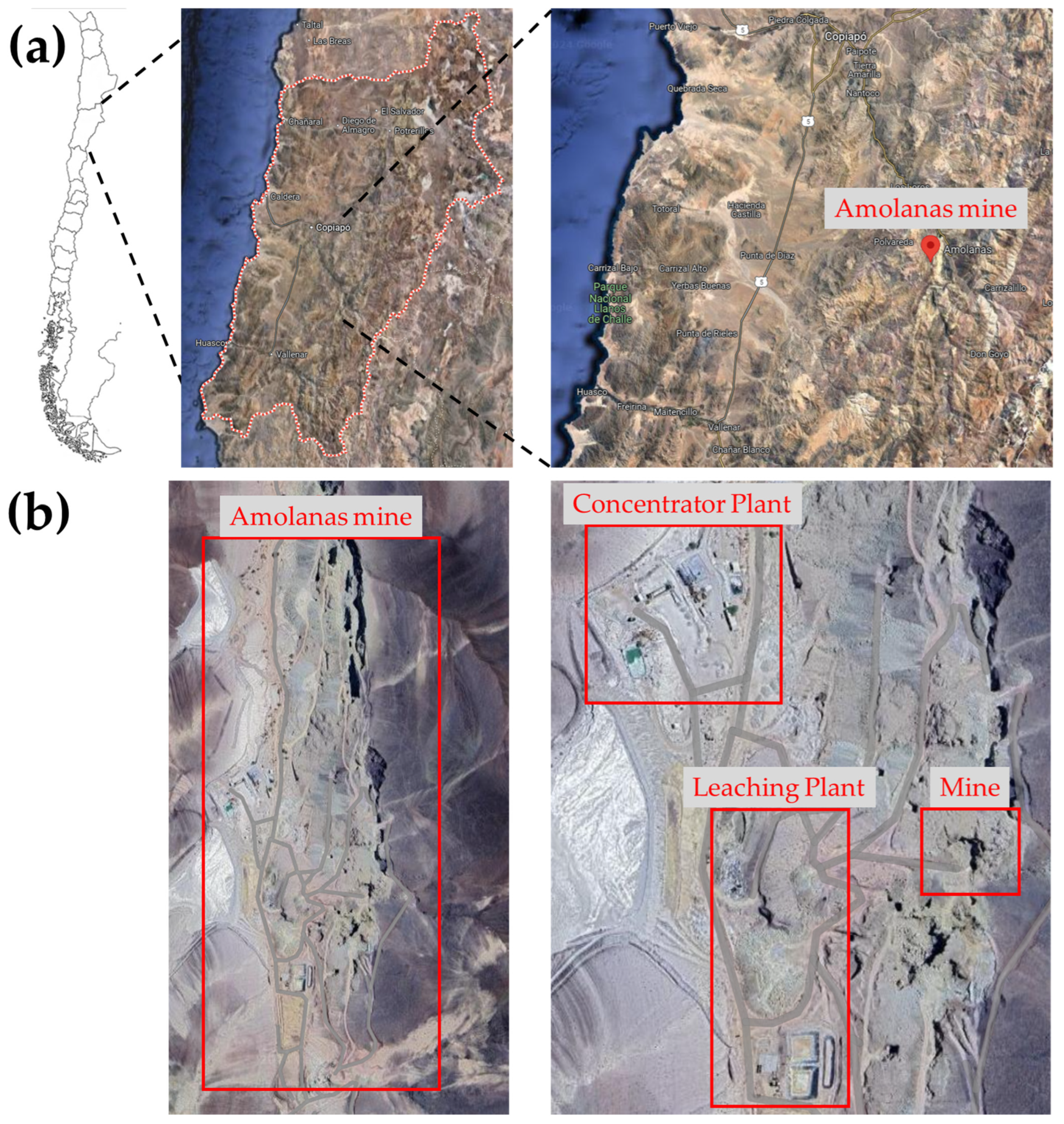
2.1. Mine Sample Collection

Samples were collected from three different places in the Amolanas Mine, Copiapo, Atacama Region, Chile (−28.07333, −70.03366): a pregnant leach solution (PLS) pond, an underground well, and an oxide leaching heap (Figure 1). At the PLS pond, sediment samples were collected. Samples of rock and water were collected at the underground well. At the oxide bioleaching heap, samples were collected from the surface, at 30 cm deep, and from the leaching solution outflow. The mineral characterization of the primary ore consisted of chalcocite (Cu2S) (0.1%), chalcopyrite (CuFeS2) (0.2%), bornite (Cu5FeS4) (0.4%), pyrite (FeS2) (0.01%), chrysocolla (Cu2_xAlx(2H2Si2O5)(OH)4·nH2O) (2.4%), malachite (Cu2CO3.(OH)2 (0.5%), atacamite (Cu2Cl·(OH)3) (0.01%), Cu native (0.02%), cuprite (Cu2O) (0.1%), Cu-biotite (0.2%), kaolinite (Al2.Si2O5(OH)4) (1.7%), clays (2.7%), quartz (42.6%), phlogopite (0.6%), K feldspar (23.6%), plagioclase (6.3%), sericite (11.5%), chlorite (1.4%), calcite (CaCO3) (2.1%), siderite (FeCO3) (0.2%), Fe oxides/hydroxides (0.3%), and others (2.9%). The samples were aseptically collected in 50 mL sterile polypropylene tubes, transported, and stored at 4 °C until analysis.

2.2. Enrichment and Growth Conditions

The samples were inoculated with 10% w/v or v/v and enriched in MAC medium, sterilized by autoclaving at 121 °C for 20 min, for iron- and sulfur-oxidizing acidophilic microorganisms. The MAC medium contained (NH4)2SO4 (0.13 g/L), H2SO4 (0.9 g/L), KH2PO4 (0.03 g/L), MgCl2·6H2O (0.03 g/L), CaCl2·2H2O (0.2 g/L), MnCl2·4H2O (0.1 mg/L), ZnCl2 (0.07 mg/L), CoCl2·6H2O (0.1 mg/L), H3BO3 (0.03 mg/L), Na2MoO4·2H2O (0.01 mg/L), and CuCl2·2H2O (0.09 mg/L) [20]. The pH of the medium was adjusted to 2.5 with sulfuric acid 98% (v/v). The medium was supplemented with Fe(II) as FeSO4·7H2O (4 g/L) or potassium tetrathionate (0.3 g/L), with or without yeast extract (YE; 0.02%), and filter-sterilized (0.02 µm pore size) [21,22]. Microbial enrichments were cultivated at 30 °C, 37 °C, and 50 °C at 150 rpm for 15 days. The enrichments were then subcultured twice at 10% v/v in fresh MAC medium, as described above.

2.3. Isolation and Characterization of Iron-Oxidizing Acidophile Bacteria

The microorganisms were isolated using two methodologies. First, double-layer solid medium plates were prepared with sterile MAC medium (pH 1.7) and Phytagel as a gelling agent, supplemented with Fe(II) (4 g/L) [23,24,25]. The plates were incubated at 30 °C and 37 °C for 30 days, and the grown colonies on the solid MAC medium were purified through a dilution series up to 10−8 and subcultured in MAC medium plus Fe(II) (4 g/L) twice. Second, dilution-to-extinction series up to 10−8 were prepared from the enrichments and then cultivated in MAC medium (pH 1.7) supplemented with Fe(II) (4 g/L) or potassium tetrathionate (0.3 g/L), with 0.02% YE for thermophilic enrichments [26,27]. The subcultures were incubated at 30 °C, 37 °C, or 50 °C at 150 rpm for five days. This dilution series process was repeated at least four times to purify the microorganisms. The isolates were cultivated with filtered sterilized Cu(II) (16 g/L), provided as CuSO4 to inhibit heterotrophic bacteria. The original enrichments were also kept for additional analysis. The enrichments and isolates were cultured, concentrated at 1010 cells/mL, and cryopreserved in dimethyl sulfoxide (DMSO) 8% at −80 °C. The cultures were tested for their iron- or sulfur-oxidizing capacity by adding Fe(II) (4 g/L) or 1% (w/v) S0 (sterilized at 100 °C for 30 min), respectively, with growth at temperatures at 30 °C, 37 °C, 40 °C, and 45 °C using Fe or S as electron donors. One isolate from each sampling place was tested for NaCl tolerance at 11.7, 17.5, and 23.4 g/L (200 mM, 300 mM, and 400 mM, respectively).

2.4. Molecular Identification and Characterization

For molecular identification, cells from each isolate were collected from 100 mL of the culture by centrifugation at 9600 rpm for 10 min. The cell pellets were washed twice with sodium citrate (2.6 g/L) [28]. The total DNA from each isolate pellet was extracted following the protocol of the ExgeneTM Cell SV kit (GeneAll Biotechnology Co., Seoul, Korea) and stored at −20 °C. The DNA quality and concentration were checked with a Nanodrop Take3TM plate of EpochTM spectrophotometer (BioTek Instruments, Winooski, VA, USA).
The 16S rRNA gene from each isolate was amplified by PCR using the universal primers 8F (5′-AGAGTTTGATCCTGGCTCAG-3′) and 1492R (5′-GGTTACCTTGTTACGACTT-3′). The amplicons were sequenced by Sanger at the Omics Technologies and Sequencing Unit of the Pontificia Universidad Católica, Santiago, Chile. The obtained sequences were edited manually with BioEdit 7.2.5 and MEGA 11: Molecular Evolutionary Genetics Analysis version 11 [29], and analyzed using the Ribosomal Database Project (RDP) and the classifier tool to identify the isolates.

2.5. Microbial Composition Identification

DNA from the enrichments was also extracted as described above, along with DNA from the isolates, and sent to the AUSTRAL-OMICS Core-Facility (Facultad de Ciencias, Universidad Austral de Chile) for 16S amplicon sequencing of the V3 and V4 variable regions of the 16S rRNA gene by using the Illumina MiSeq sequencing platform with primers 341 F (5′-CCTACGGGNGGCWGCAG-3′) and 785 R (5′-GACTACHVGGGTATCTAATCC-3′). The obtained data were processed using the DADA2 pipeline (https://benjjneb.github.io/dada2/, accessed on 25 November 2022) in R language (http://www.r-project.org/, accessed on 25 November 2022) in the Rstudio environment (http://www.rstudio.com/ accessed on 25 November 2022). The SILVA v138 database (http://www.arb-silva.de/, accessed on 25 November 2022) was used for sequence alignment and taxonomic assignment analysis of the 16S rRNA gene amplicon sequence variants (ASVs).
The DNA sequences of the strains isolated from the Amolanas Mine have been submitted to GenBank under accession numbers OR889289 to OR889299.

2.6. Mineral Preparation

The pyrite (Peru) used in this study was sterilized by cooking it with HCl 6 N for 30 min, washing it several times with deionized water, rinsing it with acetone, and drying it at 120 °C for 6 h. The chalcopyrite originated from Boliden AB mine (Stockholm, Sweden) and was described previously [11]. The chalcopyrite characterization (copper content of 29.5% w/w) and sterilization procedures were carried out following the protocol reported in [11]. Both minerals were fractioned in particle sizes between 50 and 100 µm. The pyrite was characterized by digesting it with four different types of acids (HCl, H2SO4, HNO3, and HF) and incubating it for one hour. The solution was analyzed by atomic absorption spectroscopy (AAS) in a PinAacle 900F spectrometer (PerkinElmer, Shelton, CT, USA), and it contained 46% iron.

2.7. Bioleaching Experiments

Before the bioleaching experiments, five isolated strains and two mesophilic and two thermotolerant enrichments were maintained in basal salt medium (pH 1.7) at 37 °C or 50 °C, supplemented with pyrite 1% (w/v) or Fe(II) (2 g/L) as an electron donor. The sterile basal salt medium contained (NH4)2SO4 (1 g/L), CaCl2·2H2O (0.02 g/L), MgSO4·7H2O (0.1 g/L), KH2PO4·2H2O (0.05 g/L), and NaH2PO4·2H2O (0.2 g/L), plus MAC trace elements. Pyrite bioleaching assays were performed in duplicate in 250 mL flasks with basal salt medium (pH 1.7) at a final volume of 150 mL plus 2% (w/v) pyrite (particle size between 50 and 100 µm) and Fe(II) (2 g/L). Abiotic controls were included. The cultures were incubated at 37 °C or 50 °C and 150 rpm for 30 days. Chalcopyrite bioleaching assays were conducted in triplicate in basal salt medium (pH 1.7) at a final volume of 150 mL, supplemented with 1% (w/v) sterile chalcopyrite concentrate. The chalcopyrite cultures were incubated at 37 °C or 50 °C at 150 rpm for 9 or 14 days. Abiotic controls were included. Each culture inoculum was washed twice with basal salt medium, centrifuged (9600 rpm, 10 min), and 107 cells/mL were added. The experiments were measured for cell density using a Thoma cell counting chamber under a light microscope (Zeiss, Oberkochen, Germany). The pH and redox potential (vs. Ag/AgCl with 3.5M KCl) were measured with Hanna® Instruments, HI 5222 (Woonsocket, RI, USA).
The iron and copper concentrations were measured by a UV-vis spectrophotometer (HACH DR 3900, Hach, Loveland, CO, USA) using the 1–10 phenanthroline (HACH FerroVer iron reagent) and bicinchoninate methods (HACH CuVer 1 copper reagent), respectively.

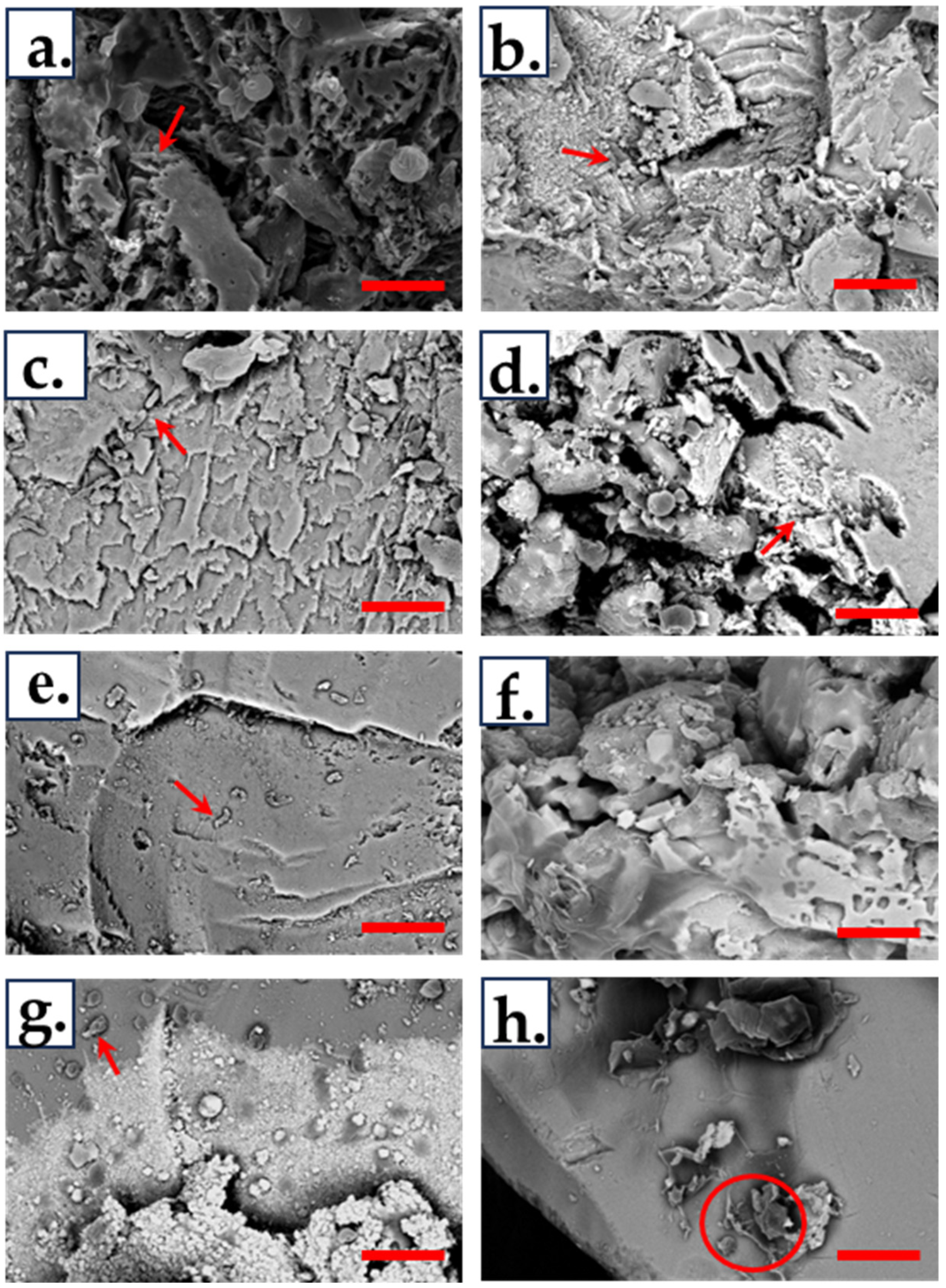
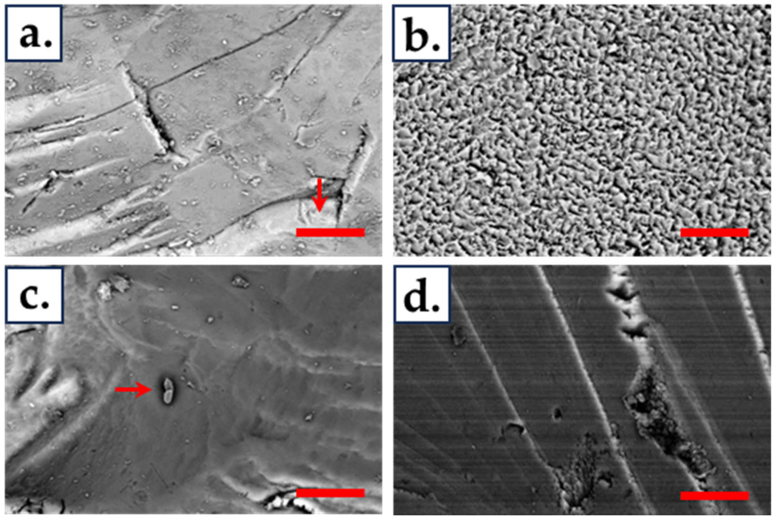
2.8. Scanning Electron Microscopy (SEM)

Chalcopyrite grains were collected after 2, 4, or 14 days of incubation and fixed in glutaraldehyde 2%. The samples were dehydrated, dried at a critical point, and sprayed with gold. Afterward, the samples were visualized in a scanning electron microscope (FEI QUANTA FEG 250, ELECMI, Zaragoza, Spain), and elemental analysis of the mineral was performed using energy dispersive spectrometry (EDS), operated at 15 kV.

2.9. Statistical Methods

Statistical significance of the differences among the enrichments, isolates, and controls in the cell density, pH, ORP, and metal concentrations were analyzed by two-way analysis of variance (ANOVA) and Tukey’s post hoc test. Significant differences were considered at a level of p < 0.05. Statistical analysis was conducted using IBM SPSS statistics (Version 22).

3. Results

3.1. Strains Molecular Identification and Characterization

Ten iron-oxidizing strains were isolated at 30 °C and one at 37 °C (IRPS17 strain) from the samples from the PLS pond, underground well, and oxide heap of the Amolanas Mine (Table 1). The I2CS27, I2CS28, I2CS29, and I2CS30 strains were isolated from double-layer plates, and the seven remaining ones were isolated by an extinction dilution technique. Sanger sequencing analysis indicated that two isolates were identified as Acidithiobacillus, and nine as Leptospirillum. A sequence comparison analysis of the 16S rRNA gene by multiple alignment showed that the Leptospirillum strains shared more than 99.9% similarity.
All strains could grow at 30 °C and 37 °C and oxidize ferrous iron. Not surprisingly, both Acidithiobacillus strains oxidized sulfur, and only four strains, IRPS15, IRPS17, I2CS21, and I2CS27, were also able to grow at 40 °C, but growth was not possible at 45 °C. The strains were also tested for growth at NaCl concentrations of 11.7 and 17.5 g/L (200 mM and 300 mM, respectively). The IPLS5, IRP17, and I2CS27 strains could grow at 11.7 g/L and 17.5 g/L NaCl (Table 2), but their growth was inhibited at 23.4 g/L.

3.2. Microbial Diversity

3.2.1. Analysis of Isolates by Next-Generation Sequencing

To support their identification, the isolates were examined by 16S rRNA gene next-generation sequencing (NGS) analysis. L. ferriphilum and At. ferrooxidans were the most representative species in all 11 isolates (Figure 2). All strains presented over 98% relative abundance, except for the I2C3031 culture, which displayed 80% of At. ferrooxidans and nearly 20% of L. ferriphilum. The taxonomic assignment made by DADA2 showed that a few percentages of the reads (the IPL5, I2CS21, I2CS27, ICS28, ICS29, and ICS30 strains) were uniquely assigned to genera (Leptospirillum sp. or Acidithiobacillus sp.)

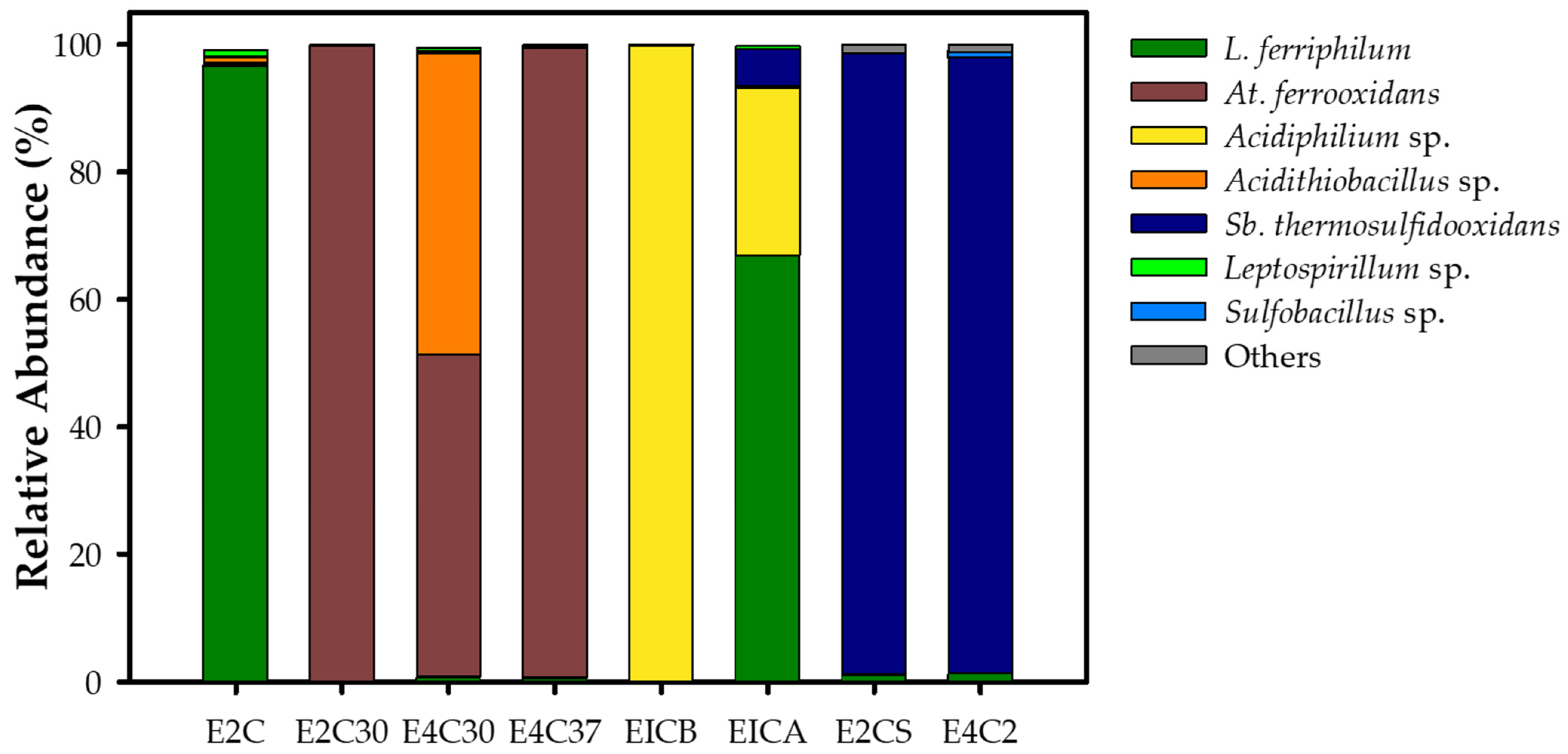
3.2.2. Enrichment Community Composition

Four enrichments were obtained at 30 °C, two at 37 °C, and two at 50 °C (Table 1). Microbial diversity was analyzed using the Illumina MiSeq platform for 16S rRNA gene sequencing. The enrichment abundance analysis showed Leptospirillum, Acidithiobacillus, and Acidiphilium as the most abundant genera in the mesophilic enrichments (Figure 3). The E2C, E2C30, E4C37, and EICB enrichments presented over 98% of a unique species/genus (L. ferriphilum, At. ferrooxidans, or Acidiphilium sp.). The E4C30 enrichment showed particular abundance, where nearly 50% of the reads were identified as species (At. ferrooxidans), and almost the remaining 50% of reads were identified as members of Acidithiobacillus. EICA was the only enrichment that showed higher species diversity, containing 60% L. ferriphilum, 30% Acidiphilium, and around 10% Sulfobacillus thermosulfidooxidans. The abundance of the enrichments E2CS and E4C2 at 50 °C primarily presented Sb. thermosulfidooxidans (Figure 3). Some small amounts of reads of L. ferriphilum were found in both enrichments.

3.3. Bioleaching of Pyrite

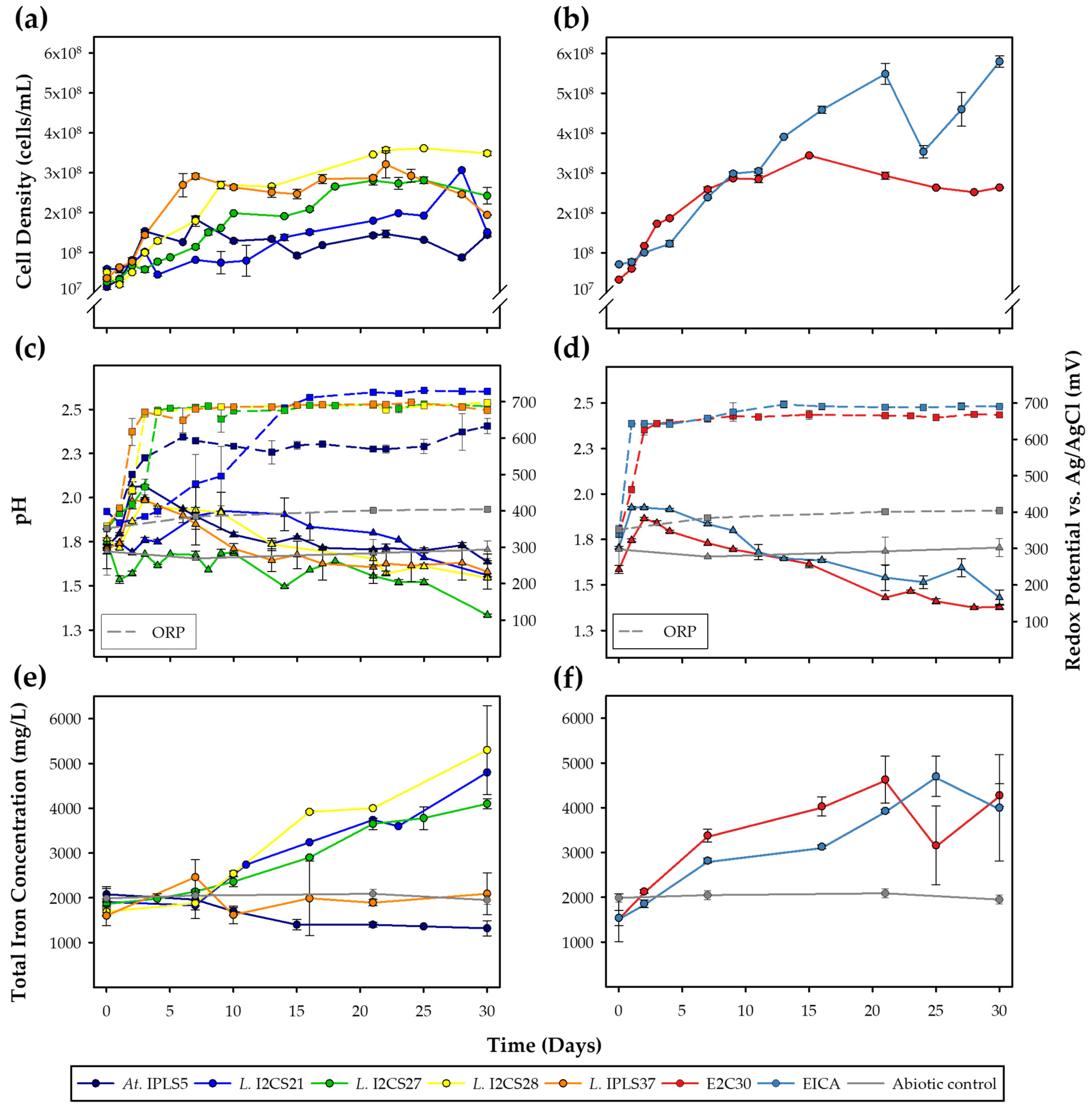
3.3.1. Mesophile Cultures

Pyrite bioleaching was evaluated for five isolates and two enrichments obtained from the Amolanas Mine at 37 °C for 30 days (Figure 4). The uninoculated controls showed barely any variation in the pH, ORP, or total iron concentration. The ORP changed from around 352 to 404 mV (vs. Ag/AgCl), and the total iron concentration did not change significantly (Figure 4c–f). The Leptospirillum I2CS28 strain and EICA enrichment significantly presented the highest cell density after 30 days (<0.05) (between 3 × 10⁸ and 5.5 × 10⁸ cells/mL) (Figure 4a,b), whereas Acidithiobacillus IPLS5 and Leptospirillum I2CS21 significantly showed the lowest cell density (<0.05) (about 1.1 × 10⁸ cells/mL) (Figure 4a). The pH presented a similar behavior amongst the seven mesophilic cultures; it increased to above 2 on the first day and then decreased to around 1.5 after 30 days (Figure 4c,d). The I2CS27 strain was the only one showing significantly low pH values (<0.05), of approximately 1.3 (Figure 4c). The redox potential of I2CS27, I2CS28, IPLS37, and E2CS increased sharply within the first days and remained stable at nearly 700 mV (Figure 4c,d). Acidithiobacillus IPLS5 maintained a lower ORP, reaching a maximum of 630 mV, and Leptospirillum I2CS21 and EICA enrichment reached the highest at around 730 mV (Figure 4c,d). The highest iron concentration was released by the I2CS27, I2CS21, I2CS28, and EICA enrichments (approximately 2000 mg/L), while IPLS5 and IPLS37 were not significantly (>0.05) different from the uninoculated control after 30 days of incubation (Figure 4e,f).

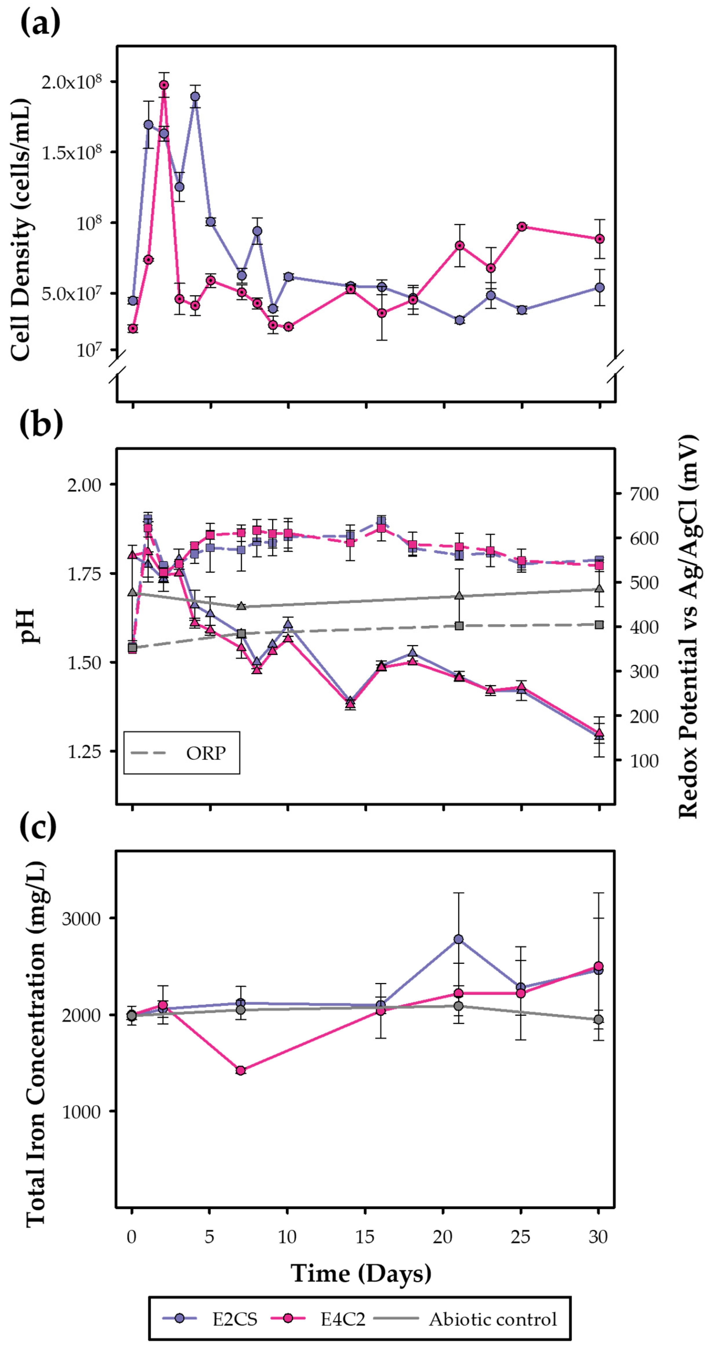
3.3.2. Moderate Thermophile Cultures

Pyrite bioleaching of the two thermotolerant enrichment cultures obtained from the Amolanas Mine at 50 °C for 30 days was evaluated (Figure 5). Both the E2CS and E4C2 enrichments showed identical behavior. They grew sharply after the first days of incubation (over 1.8 × 10⁸ cells/mL) and followed an abrupt reduction (about 5 × 10⁷ cells/mL) (Figure 5a). The pH values constantly decreased from 1.8 to 1.2, and the ORP increased sharply to 600 mV on the second day to remain steady at around 550 mV (Figure 5b). The total iron concentration did not differ significantly from the abiotic control (Figure 5c).

3.4. Bioleaching of Chalcopyrite

3.4.1. Mesophile Cultures

Chalcopyrite bioleaching was determined for four isolates and two enrichment cultures at 37 °C (Figure 6). The isolate growth displayed a period of mineral attachment within the first four days of incubation and then showed a constant increase (Figure 6a). The same performance occurred with EICA enrichment, but the E2C30 enrichment showed a continuous decline until the end of the experiment (Figure 6b). I2CS27 and I2CS28 showed the highest cell densities (over 1.8 × 10⁸ cells/mL) (Figure 6a). The pH behavior of the IPLS5 and I2CS21 strains and E2C30 enrichment did not show any differences in comparison to the control (Figure 6c). The I2CS28 strain and EICA enrichment increased slightly (to pH 2) (Figure 6d), and the pH of the I2CS27 strain reached up to 2.5 at the end of the incubation (Figure 6c). During the experiment, the ORP of the I2CS27 and I2CS28 strains remained high, increasing from 530 to 650 mV (Figure 6c). In the case of the IPLS5 and I2CS21 strains and EICA enrichment, the ORP underwent a reduction from over 550 to 400 mV within the first days, but then they reached the same potential of over 650 mV as the IPLS5, I2CS27, and I2C28 strains (Figure 6c,d). The E2C30 enrichment was the only one that did not show a significant change in the ORP, reaching around 500 mV (Figure 6c). The evaluation of the iron release from the chalcopyrite displayed a constant release. The IPLS5, I2SC21, and I2CS27 strains and EICA enrichment presented significantly similar iron concentrations as the uninoculated control (Figure 6e,f). Only the I2CS28 strain and E2C30 enrichment showed a higher iron release (230–280 mg/L) (Figure 6e). Concerning the copper release, on day 14, the IPLS5, I2CS21, and I2CS28 strains released over 200 mg/L of copper (about 10% of copper recovery) (Figure 6g and Figure S1), whereas the EICA enrichment reached a similar concentration on day seven. The E2C30 strain did not show significant differences compared to the uninoculated control (Figure 6h). The I2SC27 strain released the highest copper concentration on day 14 (750 mg/L) (Figure 6g), recovering about 25% of copper contained in the chalcopyrite concentrate (Figure S1).

3.4.2. Moderate Thermophile Cultures

The chalcopyrite bioleaching potential of the thermophile enrichments was assessed for 14 days (Figure 7). The E2CS and E4C2 enrichments showed comparable growth, reaching the highest cell densities between days 2 and 4 (1.7 × 10⁸ cells/mL) (Figure 7a). Both enrichments maintained a pH near 1.7 and did not change significantly compared to the uninoculated control (Figure 7b). The ORP increased from 430 to over 630 mV after two days of incubation; then, it remained steady until day 11, when it decreased to 400 mV and 500 mV for E2CS and E4C2, respectively (Figure 7b). The total iron release did not show significant differences (>0.05) between the enrichments and the uninoculated control (125 mg/L) (Figure 7c). The copper release of E2CS increased up to 200 mg/L, with nearly 7% of the copper recovered (Figure S2), while E4C2 showed the same concentration as the control (>0.05) (Figure 7d).

3.5. Biofilm Formation and Attachment on Chalcopyrite

Attachment and biofilm formation on the chalcopyrite surfaces were evaluated by SEM-EDS. Figure 7 and Figure S3 show the cell attachment of isolates to the mineral surfaces at days 4 and 14. No differences in cell attachment were observed. Individual cells could be observed at day 4, mainly in the I2CS27 and I2CS28 strains (Figure 8e,g). At day 14, it was noticed that more visible deterioration occurred on the mineral surface. Cells colonizing the mineral were recognized in the IPLS5 and I2CS21 strains, while biofilm formation was observed in the I2CS28 strain (Figure 8h and Figure S3h). All cultures presented a low atomic percentage of carbon by EDS (Figures S5–S8). The most noticeable attack on the mineral was observed by the I2CS27 strain (Figure 8f and Figure S3f), and the copper atomic percentage declined from 21.7% on day 4 to 1.1% on day 14 (Figure S6b,d), but no evident cell attachment was observed. A similar copper atomic percentage reduction was measured for the I2CS28 strain, which decreased from 20.9% to 7.5% from day 4 to day 14, respectively (Figure S7a–d).
The colonization of chalcopyrite by mesophilic enrichments after four days is shown in Figure 9 and Figure S8. Differences in the chalcopyrite surfaces were observed. The mineral of the E2C30 enrichment showed a smooth surface with no cells attached (Figure 9a). Instead, the chalcopyrite grains of the EICA enrichment seemed to have more attached cells on the surface (Figure 9b). EDS did not show differences in the atomic percentages of copper and carbon between the enrichments at day 4 (Figure S9).
The thermotolerant enrichments showed different patterns of colonization (Figure 10 and Figure S10). While the E4C2 enrichment grains displayed a smooth surface and little evidence of colonization at days 2 and 14 (Figure 10c,d and Figure S10b,d), it was observed in the E2CS enrichment that many cells were on the smooth surface of the mineral on day 2 (Figure 10a and Figure S10a). Then, the surface became rugged, with few single cells, but with evidence of precipitates at day 14 (Figure 10b and Figure S10c). EDS detected a null carbon atomic percentage of the E4C2 enrichment and low percentage of the E2CS enrichment at day 2 (Figures S11a,c and S12a,c), while the carbon percentages were 31.2% and 30.8% at day 14 for enrichments E4C2 and E2CS, respectively (Figures S11b,d and S12b,d).

4. Discussion

4.1. Microbial Biodiversity of the Amolanas Mine

The extreme environments where acidophilic bacteria and archaea thrive possess low complexity in their biodiversity, mainly due to low pH, high concentrations of heavy metals, and a low carbon source, which limit them to a few groups of microorganisms [30]. Here, we isolated predominantly Leptospirillum and Acidithiobacillus strains at mesophilic temperatures. Leptospirillum and Acidithiobacillus, the dominant bacteria found in our results, coincide with previous phylogenetic analysis, which showed that both genera are the most frequent microorganisms isolated worldwide in copper mine environments [3,31,32].
Acidophiles, in general, have been isolated by extinction dilution or double-layer solid medium techniques. Extinction dilution points to isolating the predominant microorganism in the culture. Here, we identified four strains using the double-layer technique and seven using extinction dilution (Table 1). NGS analysis confirmed their identities, and it was observed that the I2C3031 culture presented nearly 80% At. ferrooxidans, indicating extinction dilution worked in six of the seven cases (Figure 2). Likewise, the I2CS27, IC2S28, ICS29, and I2CS30 strains isolated by double-layer solid media displayed a few traces of other microorganisms (Figure 2), demonstrating that this methodology was equally effective as extinction dilution.
Acidithiobacillus IPLS5, Leptospirillum IRPS17, and I2CS27 showed relatively high chloride tolerance (17.5 g/L; 300 mM) (Table 2), though their growth was slower. Also, the strains were cultivated at 23.4 g/L (400 mM), but growth was not observed. Similarly, L. ferriphilum DSM 14647T displayed a delay in the maximum cell density, which went from 52 h to 84 h when NaCl (11.7 g/L) (200 mM) was added [33]. Previous studies showed that strains of L. ferriphilum and At. ferrooxidans tolerated NaCl maximum concentrations near 17.5 g/L [34,35]. Studies have focused on microorganisms able to tolerate high chloride concentrations, which could inhibit acidophile growth and affect the bioleaching rate [36]. Chloride-tolerant acidophiles, such as the strains isolated in this study (Table 2), could have enormous potential to be applied to ores containing Chilean minerals like atacamite, where the chloride content is elevated. Also, adding chloride solutions to the process might enhance leaching rates and modify sulfur deposits, allowing leaching to continue [37,38].
The characterization showed that all strains were iron-oxidizing, and both Acidithiobacillus strains could also oxidize sulfur, as expected (Table 2). Although phylogenetic analysis showed Leptospirillum strains had a similarity of 99.9%, the growth test at different temperatures showed that the IRPS15, IRPS17, I2CS21, I2CS27, and I2CS30 Leptospirillum strains could grow up to 40 °C (Table 2). This suggests that these five isolated strains might be part of Leptospirillum group II, whose representative species is L. ferriphilum. Their growth was reported to occur at temperatures up to 45 °C [30]. In contrast, the I2CS22, I2CS28, I2CS29, and ISPL37 Leptospirillum strains might belong to group I (L. ferrooxidans), which grow at temperatures lower than 40 °C [30,39]. This preliminary characterization gives some insights to elucidate the classification. Leptospirillum isolates could be separated into at least two different types. More analysis must be carried out, such as multi-locus sequence analysis (MLSA), C+G, and polar lipid composition, to determine their exact taxonomic classification and complement the 16S rDNA sequence results [25,40].
L. ferriphilum and At. ferrooxidans were the most abundant species identified in the mesophilic enrichments (Figure 3). The EICB culture was the only enrichment containing predominantly Acidiphilium, a dominant heterotrophic bacterium found in acidophilic environments (Figure 3) [31]. Acidiphilium spp. interacts mutually with iron- and sulfur-oxidizing acidophiles, metabolizing small-molecular-weight organic compounds that could be toxic for autotrophic bacteria [30]. In the case of EICA enrichment and thermophile enrichments E2CS and E4C2, the occurrence of Sb. thermosulfidooxidans was observed (Figure 3). This Gram-positive bacterium is an iron- and sulfur-oxidizing facultative mixotroph acidophile widely found in extreme environments, such as copper mines [32]. Although Sulfobacillus is a moderate thermophile whose optimal temperature varies between 40 and 59 °C, including the species Sb. acidophilus, Sb. thermosulfidooxidans, and Sb. thermotolerans, among others [32], it could also be found at mesophilic temperatures, e.g., Sb. benefaciens, whose optimal temperature is between 15 and 39 °C [3,30]. At moderate temperatures, F. caldus is well-characterized, growing optimally at 45 °C [32]. This microorganism was found in a low relative abundance percentage within enrichments grown at 50 °C (<0.5% relative abundance), grouped in the “others” category. Interestingly, nearly 2% of the relative abundance of L. ferriphilum was identified in both thermotolerant enrichments, E2CS and E4C2 (Figure 3). One study reported a Leptospirillum strain named L. thermoferrooxidans growing up to about 45 °C or higher, but the strain was lost [41,42]. In stirred tank reactors operated using a copper concentrate from black shale ore deposits at temperatures 42–46 °C, L. ferriphilum was identified with 3% abundance [43]. Archaea ASVs (Acidianus spp. and Sulfolobus spp.) were also found in the thermotolerant cultures, even though their relative abundance was lower than 0.2%.

4.2. Bioleaching of Metal Sulfides

We evaluated the pyrite bioleaching potential of strains and enrichments obtained from the Amolanas Mine for 30 days. The mesophilic Leptospirillum and Acidithiobacillus isolated strains and the E2C30 and EICA enrichments reached redox potential values near 700 and 630 mV, respectively (Figure 4c,d). These potentials coincide with those of previous studies that reported values up to 745 mV and 645 mV [11,42,44]. Although the EICA enrichment contained other predominant species of microorganisms (Sb. thermosulfidooxidans and Acidiphilium sp.), apart from L. ferriphilum, they could not counteract the Leptospirillum influence on potential. Also, the potential was affected by the initial Fe(II) (2 g/L) concentration. The IPLS37 and IPLS5 strains and thermotolerant enrichments were the only cultures with an equal or lower total iron concentration than the abiotic control (Figure 4e and Figure 5c). Prior research has demonstrated that iron might precipitate as hydroxides or sulfates at pH < 2, declining the solubilized iron [45,46,47,48]. Fe(III) concentrations above 3 g/L could lead to jarosite precipitation [49,50]. Also, Fe(III) presented in the culture could have formed heavier complexes with sulfates and hydroxides and decanted before the measuring sample was collected, which could explain the low total iron concentration. Furthermore, precipitate formation and sulfur oxidation could acidify the cultures and lower the pH [3,48,51]. As seen in the experiments, when almost all of the Fe(II) was oxidized into Fe(III), causing the pH to rise to nearly 1.9, the precipitates formed, and finally, the pH declined gradually to 1.5 (Figure 4d and Figure 5b). Thermotolerant enrichments presented a lower redox potential compared to mesophilic cultures near 500 mV (Figure 4d and Figure 5b). It was observed that the redox potential decreased at high temperatures, which was related to the absence of L. ferriphilum and the presence of Sb. thermosulfidooxidans [11,43]. This weak iron-oxidizing bacterium, the most abundant in the enrichments, was characterized for maintaining a low ORP close to 550 mV. Low redox potential values are convenient for the design of acidophilic consortia, which may improve copper extraction in bioleaching processes [11].
Microbial communities are implicated in several challenges for copper recovery from chalcopyrite, such as low bioleaching rates and passivation, which must be overcome [6,11]. This study was focused on indigenous acidophiles from the Amolanas Mine and their potential to recover copper from chalcopyrite to improve copper bioleaching processes. The mesophilic strains and EICA enrichment growth on the chalcopyrite had different behavior than on the pyrite. The cell densities and redox potentials did not reach the high values compared to the pyrite (Figure 4a–d and Figure 6a–d). The cell growth on the pyrite was almost double that of the growth on the chalcopyrite, and the potential redox took longer to reach a maximum of 650 mV. The redox potential of the I2CS28 strain and E2C30 enrichment on chalcopyrite was significantly lower (near 500 mV) in comparison to the growth on pyrite (Figure 4c,d and Figure 6c,d). This could be explained by the addition of Fe(II) in the pyrite leaching assays, which stimulated higher growth and increased the redox potential when Fe(II) was oxidized to Fe(III) [3,42]. The effect of the elevated Fe(III)/Fe(II) ratio, and mainly the acid consumption of chalcopyrite, was also reflected; it was observed that the pH remained stable in the chalcopyrite leaching assays (no Fe(II) supplemented), in contrast to the pyrite ones (Figure 4c,d and Figure 6c,d). This coincides with previous studies, where excess Fe(III) caused precipitate formation and, consequently, decreasing pH values [48,51]. Interestingly, both the I2CS28 and E2C30 cultures released more total iron (>200 mg/L) than the other mesophilic cultures (Figure 6e,f). The I2CS27 strain was the most efficient copper bioleaching culture, with an almost fourfold increase in copper (700 mg/L) in comparison to IPLS5, I2CS21, and I2CS28, and a total copper recovery of 25% after 14 days of bioleaching (Figure 6g and Figure S1).
The I2CS27 strain and EICA enrichment released the highest copper concentrations after seven days of incubation (255 mg/L and 320 mg/L, respectively) (9% and 11%, respectively) (Figure 6g,h and Figure S1). The differences between the iron and copper concentrations for each culture could be due to the covellite and chalcocite formation by Cu(II)—Fe(II) substitution, which might precipitate [52]. Recent studies showed that the At. ferrooxidans DSM 14882T strain and an F. caldus DSM 8584TSb. thermosulfidooxidans DSM 9293T co-culture recovered 34% and 15% of copper after 14 days, respectively, using the same chalcopyrite concentrate [11,53]. Here, we report the indigenous Leptospirillum I2CS27 strain and EICA enrichment, which present promising copper bioleaching rates. Subsequent subculture conditioning experiments may help to improve copper release. Surprisingly, the release of iron and copper by thermotolerant cultures was minimal compared to the mesophilic cultures.
SEM-EDS analyses of the chalcopyrite surface during leaching showed, in general, low degrees of cell attachment. Degradation of the mineral grains was enhanced over time in all cultures. The strains and enrichments that showed the most iron and/or copper release presented more mineral damage and debris on their surface, namely I2CS27, which released the highest total copper concentration after 14 days and the highest sulfur atomic percentage on the surface on day 14 (Figure 8f and Figure S6). In contrast, the E2C30 and E4C2 enrichments did not have significant iron and copper releases at days 2 and 4, respectively, and displayed smooth surfaces (Figure 9a and Figure 10c). The E2CS and E4C2 thermotolerant enrichments shared similar biodiversity, characterized almost totally by the abundance of Sb. thermosulfidooxidans. However, the E2CS enrichment released nearly 200 mg/L of copper (a total copper recovery of 8%) (Figure 7d and Figure S2), presented more cell colonization at day 2, and rugged chalcopyrite grains after 14 days (Figure 10a,b). This suggests that both enrichments could differ in biodiversity, though deeper identification studies are needed. Our results show that copper release occurs with low cell attachment, which might indicate cell–surface direct contact was not required for metal release. In the case of the E2CS and E4C2 enrichments, Sb. thermosulfidooxidans presented low adhesion and fast detachment rates, and they were weak iron-oxidizers, as reported previously [54]. The enrichments might have produced a low Fe(III) concentration, an essential agent for mineral attack and initial adhesion [42,54]. In addition to SEM-EDS analyses, epifluorescence microscopy (EFM) assays with diamino-2-phenylindole (DAPI) [55,56] were carried out to evaluate isolate and enrichment colonization on chalcopyrite, but minimal epifluorescence signals were obtained, confirming the information shown by SEM-EDS analyses.
The colonization of the chalcopyrite grains by the strains and enrichments was typical of single-cell monolayers, decreasing their presence in later days. It has been seen that during bioleaching, the cells remain mostly planktonic [3], and biofilms are characterized as monolayers with selective attachment sites [3,9]. The E2CS28 strain was the only one that evidenced biofilm formation with EPS at day 14 (Figure 8h). Also, SEM-EDS analysis indicated the accumulation of lighter elements, such as carbon. The redox potentials in the chalcopyrite experiments were higher in comparison to the optimal redox potential recommended for copper recovery (415–435 mV) [7], and exceeded the value when passivation occurred (>475 mV) [8]. Also, only the I2CS27 strain presented a rise in the sulfur atomic percentage on chalcopyrite grains, which could be associated with high chalcopyrite bioleaching and the beginning of a passivation process.

5. Conclusions

The results highlight the promising metal sulfide bioleaching potential of a novel Leptospirillum I2CS27 strain and EICA community obtained from the Amolanas Mine. The redox potential was more favorable when the iron concentration remained low in the chalcopyrite experiments, although enrichments with high Sb. thermosulfidooxidans abundance could require small amounts of Fe(III) to improve copper bioleaching. In general, the strains and enrichments did not show a high biofilm formation capacity, yet the Leptospirillum I2CS27 strain and EICA enrichment displayed high copper bioleaching rates with no observable attachment. Therefore, discovering new indigenous acidophiles could be a promising tool to apply in situ during bioleaching, although following studies on reactors must be carried out to evaluate and complement their potential.

Supplementary Materials

The following supporting information can be downloaded at https://www.mdpi.com/article/10.3390/min14090867/s1, Figure S1: Copper recovery of mesophilic cultures from Amolanas mine at 37 °C of incubation; Figure S2: Copper recovery of thermotolerant cultures from Amolanas mine at 50 °C of incubation; Figure S3: Representative SEM images of chalcopyrite grains from mesophilic isolates after 4 and 14 days of incubation at 37 °C; Figure S4; EDS spectrum and absolute quantification results of Acidithiobacillus IPLS5 growth on day 4 and 14; Figure S5: EDS spectrum (a, b) and absolute quantification results of Leptospirillum I2CS21 growth on day 4 and 14; Figure S6: EDS spectrum and absolute quantification results of Leptospirillum I2CS27 growth on day 4 and 14; Figure S7: EDS spectrum and absolute quantification results of Leptospirillum I2CS28 growth on day 4 and 14; Figure S8: Representative SEM images of chalcopyrite grains from mesophilic enrichments after four days of incubation at 37 °C; Figure S9: EDS spectrum and absolute quantification results of E2C30 and EICA enrichments on day 4; Figure S10: Representative SEM images of chalcopyrite grains from thermotolerant enrichments after 2 and 14 days of incubation at 50 °C; Figure S11: EDS spectrum and absolute quantification results of E2CS enrichment growth on day 2 and 14; Figure S12: EDS spectrum and absolute quantification results of E4C2 enrichment growth on day 2 and 14; Figure S13: EDS spectrum and absolute quantification results of Abiotic control.

Author Contributions

Conceptualization, J.C.C.-V. and M.V.; sampling, C.M.-B., Á.V. and M.V.; laboratory experiments, J.C.C.-V.; formal analysis, J.C.C.-V.; investigation, J.C.C.-V. and M.V.; curation data, J.C.C.-V.; writing—original draft preparation, J.C.C.-V.; writing—review and editing, C.M.-B., Á.V. and M.V.; supervision, M.V.; project administration, M.V.; funding acquisition, Á.V. and M.V. All authors have read and agreed to the published version of the manuscript.

Funding

This research was funded by The European Union ERA-NET Confound on Raw Materials (ERA-MIN), given in Chile through Agencia Nacional de Investigación y Desarrollo (ANID) grant number PCI ERAMIN 71. A.V. acknowledges support from ANID Fondecyt Regular Grant ID1211768. J.C.-V. received ANID Doctoral Fellowship grant number 2020-23230360.

Data Availability Statement

Data is contained within the article and Supplementary Material.

Acknowledgments

The support from Cristian Molina, of Coyancura SA, is greatly acknowledged for providing access to the Amolanas Mine site and facilities for sampling.

Conflicts of Interest

The authors declare no conflicts of interest.

References

  1. Brierley, C.L.; Brierley, J.A. Progress in Bioleaching: Part B: Applications of Microbial Processes by the Minerals Industries. Appl. Microbiol. Biotechnol. 2013, 97, 7543–7552. [Google Scholar] [CrossRef] [PubMed]
  2. Schippers, A.; Sand, W. Bacterial Leaching of Metal Sulfides Proceeds by Two Indirect Mechanisms via Thiosulfate or via Polysulfides and Sulfur. Appl. Environ. Microbiol. 1999, 65, 319–321. [Google Scholar] [CrossRef]
  3. Vera, M.; Schippers, A.; Hedrich, S.; Sand, W. Progress in Bioleaching: Fundamentals and Mechanisms of Microbial Metal Sulfide Oxidation—Part A. Appl. Microbiol. Biotechnol. 2022, 106, 6933–6952. [Google Scholar] [CrossRef] [PubMed]
  4. Buetti-Dinh, A.; Herold, M.; Christel, S.; Hajjami, M.E.; Bellenberg, S.; Ilie, O.; Wilmes, P.; Poetsch, A.; Sand, W.; Vera, M.; et al. Systems Biology of Acidophile Biofilms for Efficient Metal Extraction. Sci. Data 2020, 7, 215. [Google Scholar] [CrossRef]
  5. Panda, S.; Akcil, A.; Pradhan, N.; Deveci, H. Current Scenario of Chalcopyrite Bioleaching: A Review on the Recent Advances to Its Heap-Leach Technology. Bioresour. Technol. 2015, 196, 694–706. [Google Scholar] [CrossRef]
  6. Hiroyoshi, N.; Tsunekawa, M.; Okamoto, H.; Nakayama, R.; Kuroiwa, S. Improved Chalcopyrite Leaching through Optimization of Redox Potential. Can. Metall. Q. 2008, 47, 253–258. [Google Scholar] [CrossRef]
  7. Li, Y.; Kawashima, N.; Li, J.; Chandra, A.P.; Gerson, A.R. A Review of the Structure, and Fundamental Mechanisms and Kinetics of the Leaching of Chalcopyrite. Adv. Colloid Interface Sci. 2013, 197–198, 1–32. [Google Scholar] [CrossRef]
  8. Watling, H.R. Chalcopyrite Hydrometallurgy at Atmospheric Pressure: 1. Review of Acidic Sulfate, Sulfate–Chloride and Sulfate–Nitrate Process Options. Hydrometallurgy 2013, 140, 163–180. [Google Scholar] [CrossRef]
  9. Zhang, R.; Bellenberg, S.; Neu, T.R.; Sand, W.; Vera, M. The Biofilm Lifestyle of Acidophilic Metal/Sulfur-Oxidizing Microorganisms. In Grand Challenges in Biology and Biotechnology; Rampelotto, P., Ed.; Springer: Cham, Switzerland, 2016; Volume 1, pp. 177–213. ISBN 978-3-319-13521-2. [Google Scholar]
  10. Debernardi, G.; Carlesi, C. Chemical-Electrochemical Approaches to the Study Passivation of Chalcopyrite. Miner. Process. Extr. Metall. Rev. 2013, 34, 10–41. [Google Scholar] [CrossRef]
  11. Christel, S.; Herold, M.; Bellenberg, S.; Buetti-Dinh, A.; El Hajjami, M.; Pivkin, I.V.; Sand, W.; Wilmes, P.; Poetsch, A.; Vera, M.; et al. Weak Iron Oxidation by Sulfobacillus thermosulfidooxidans Maintains a Favorable Redox Potential for Chalcopyrite Bioleaching. Front. Microbiol. 2018, 9, 3059. [Google Scholar] [CrossRef]
  12. Sand, W.; Gehrke, T. Extracellular Polymeric Substances Mediate Bioleaching/Biocorrosion via Interfacial Processes Involving Iron(III) Ions and Acidophilic Bacteria. Res. Microbiol. 2006, 157, 49–56. [Google Scholar] [CrossRef] [PubMed]
  13. Castro, C.; Donati, E.R.; Vera, M. Characterization of Extracellular Polymeric Substances Produced by an Acidianus Species and Their Relevance to Bioleaching. Minerals 2023, 13, 310. [Google Scholar] [CrossRef]
  14. Riekkola-Vanhanen, M. Talvivaara Mining Company—From a Project to a Mine. Miner. Eng. 2013, 48, 2–9. [Google Scholar] [CrossRef]
  15. Johnson, D.B.; Quatrini, R. Acidophile Microbiology in Space and Time. Curr. Issues Mol. Biol. 2020, 39, 63–76. [Google Scholar] [CrossRef]
  16. Rossoni, S.; Beard, S.; Segura-Bidermann, M.I.; Duarte-Ramírez, J.; Osorio, F.K.; Varas-Godoy, M.; Martínez-Bellange, P.; Vera, M.; Quatrini, R.; Castro, M. Membrane Vesicles in Acidithiobacillia Class Extreme Acidophiles: Influence on Collective Behaviors of ‘Fervidacidithiobacillus caldus’. Front. Microbiol. 2024, 14, 1331363. [Google Scholar] [CrossRef]
  17. Okibe, N.; Johnson, D.B. Biooxidation of Pyrite by Defined Mixed Cultures of Moderately Thermophilic Acidophiles in PH-controlled Bioreactors: Significance of Microbial Interactions. Biotechnol. Bioeng. 2004, 87, 574–583. [Google Scholar] [CrossRef] [PubMed]
  18. Romo, E.; Weinacker, D.F.; Zepeda, A.B.; Figueroa, C.A.; Chavez-Crooker, P.; Farias, J.G. Bacterial Consortium for Copper Extraction from Sulphide Ore Consisting Mainly of Chalcopyrite. Braz. J. Microbiol. 2013, 44, 523–528. [Google Scholar] [CrossRef]
  19. Watling, H.R. The Bioleaching of Sulphide Minerals with Emphasis on Copper Sulphides—A Review. Hydrometallurgy 2006, 84, 81–108. [Google Scholar] [CrossRef]
  20. Mackintosh, M.E. Nitrogen Fixation by Thiobacillus ferrooxidans. J. Gen. Microbiol. 1978, 105, 215–218. [Google Scholar] [CrossRef]
  21. de Bruyn, J.C.; Boogerd, F.C.; Bos, P.; Kuenen, J.G. Floating Filters, a Novel Technique for Isolation and Enumeration of Fastidious, Acidophilic, Iron-Oxidizing, Autotrophic Bacteria. Appl. Environ. Microbiol. 1990, 56, 2891–2894. [Google Scholar] [CrossRef]
  22. Natarajan, K.A. Biotechnology of Metals: Principles, Recovery Methods and Environmental Concerns; Elsevier: Amsterdam, The Netherlands, 2018; ISBN-10: 012804022X; ISBN-13: 978-0128040225. [Google Scholar]
  23. Lin, C.C.; Casida, L.E. GELRITE as a Gelling Agent in Media for the Growth of Thermophilic Microorganisms. Appl. Environ. Microbiol. 1984, 47, 427–429. [Google Scholar] [CrossRef] [PubMed]
  24. Lindström, E.B.; Sehlin, H.M. High Efficiency of Plating of the Thermophilic Sulfur-Dependent Archaebacterium Sulfolobus acidocaldarius. Appl. Environ. Microbiol. 1989, 55, 3020–3021. [Google Scholar] [CrossRef] [PubMed]
  25. Giaveno, M.A.; Urbieta, M.S.; Ulloa, J.R.; González Toril, E.; Donati, E.R. Physiologic Versatility and Growth Flexibility as the Main Characteristics of a Novel Thermoacidophilic Acidianus Strain Isolated from Copahue Geothermal Area in Argentina. Microb. Ecol. 2013, 65, 336–346. [Google Scholar] [CrossRef]
  26. Schut, F.; de Vries, E.J.; Gottschal, J.C.; Robertson, B.R.; Harder, W.; Prins, R.A.; Button, D.K. Isolation of Typical Marine Bacteria by Dilution Culture: Growth, Maintenance, and Characteristics of Isolates under Laboratory Conditions. Appl. Environ. Microbiol. 1993, 59, 2150–2160. [Google Scholar] [CrossRef]
  27. Sun, Y.; Liu, Y.; Pan, J.; Wang, F.; Li, M. Perspectives on Cultivation Strategies of Archaea. Microb. Ecol. 2020, 79, 770–784. [Google Scholar] [CrossRef]
  28. Muñoz-Villagrán, C.; Grossolli-Gálvez, J.; Acevedo-Arbunic, J.; Valenzuela, X.; Ferrer, A.; Díez, B.; Levicán, G. Characterization and Genomic Analysis of Two Novel Psychrotolerant Acidithiobacillus ferrooxidans Strains from Polar and Subpolar Environments. Front. Microbiol. 2022, 13, 960324. [Google Scholar] [CrossRef]
  29. Tamura, K.; Stecher, G.; Kumar, S. MEGA11: Molecular Evolutionary Genetics Analysis Version 11. Mol. Biol. Evol. 2021, 38, 3022–3027. [Google Scholar] [CrossRef] [PubMed]
  30. Johnson, D.B.; Aguilera, A. Extremophiles and Acidic Environments. In Reference Module in Life Sciences; Elsevier: Amsterdam, The Netherlands, 2019; pp. 206–227. [Google Scholar] [CrossRef]
  31. Latorre, M.; Cortés, M.P.; Travisany, D.; Di Genova, A.; Budinich, M.; Reyes-Jara, A.; Hödar, C.; González, M.; Parada, P.; Bobadilla-Fazzini, R.A.; et al. The Bioleaching Potential of a Bacterial Consortium. Bioresour. Technol. 2016, 218, 659–666. [Google Scholar] [CrossRef] [PubMed]
  32. Dopson, M. Physiological and Phylogenetic Diversity of Acidophilic Bacteria. In Acidophiles: Life in Extremely Acidic Environments; Quatrini, R., Johnson, D.B., Eds.; Caister Academic Press: Wymondham, UK, 2016; pp. 79–92. [Google Scholar]
  33. Huynh, D.; Giebner, F.; Kaschabek, S.R.; Rivera-Araya, J.; Levican, G.; Sand, W.; Schlömann, M. Effect of Sodium Chloride on Leptospirillum ferriphilum DSM 14647T and Sulfobacillus thermosulfidooxidans DSM 9293T: Growth, Iron Oxidation Activity and Bioleaching of Sulfidic Metal Ores. Miner. Eng. 2019, 138, 52–59. [Google Scholar] [CrossRef]
  34. Romero, J.; Yañez, C.; Vásquez, M.; Moore, E.R.B.; Espejo, R.T. Characterization and Identification of an Iron-Oxidizing, Leptospirillum-like Bacterium, Present in the High Sulfate Leaching Solution of a Commercial Bioleaching Plant. Res. Microbiol. 2003, 154, 353–359. [Google Scholar] [CrossRef]
  35. Watkin, E.; Zammit, C. Adaptation to Extreme Acidity and Osmotic Stress. In Acidophiles: Life in Extremely Acidic Environments; Quatrini, R., Johnson, D.B., Eds.; Caister Academic Press: Wymondham, UK, 2016; pp. 49–62. [Google Scholar]
  36. Rivera-Araya, J.; Huynh, N.D.; Kaszuba, M.; Chávez, R.; Schlömann, M.; Levicán, G. Mechanisms of NaCl-Tolerance in Acidophilic Iron-Oxidizing Bacteria and Archaea: Comparative Genomic Predictions and Insights. Hydrometallurgy 2020, 194, 105334. [Google Scholar] [CrossRef]
  37. Neira, A.; Pizarro, D.; Quezada, V.; Velásquez-Yévenes, L. Pretreatment of Copper Sulphide Ores Prior to Heap Leaching: A Review. Metals 2021, 11, 1067. [Google Scholar] [CrossRef]
  38. Velásquez-Yévenes, L.; Torres, D.; Toro, N. Leaching of Chalcopyrite Ore Agglomerated with High Chloride Concentration and High Curing Periods. Hydrometallurgy 2018, 181, 215–220. [Google Scholar] [CrossRef]
  39. Battaglia, F.; Morin, D.; Garcia, J.L.; Ollivier, P. Isolation and Study of Two Strains of Leptospirillum-like Bacteria from a Natural Mixed Population Cultured on a Cobaltiferous Pyrite Substrate. Antonie Van Leeuwenhoek 1994, 66, 295–302. [Google Scholar] [CrossRef] [PubMed]
  40. Nuñez, H.; Moya-Beltrán, A.; Covarrubias, P.C.; Issotta, F.; Cárdenas, J.P.; González, M.; Atavales, J.; Acuña, L.G.; Johnson, D.B.; Quatrini, R. Molecular Systematics of the Genus Acidithiobacillus: Insights into the Phylogenetic Structure and Diversification of the Taxon. Front. Microbiol. 2017, 8, 30. [Google Scholar] [CrossRef]
  41. Rawlings, D.E. Heavy Metal Mining Using Microbes. Annu. Rev. Microbiol. 2002, 56, 65–91. [Google Scholar] [CrossRef] [PubMed]
  42. Rawlings, D.E.; Johnson, D.B. Biomining, 1st ed.; Springer: Berlin/Heidelberg, Germany, 2007; ISBN 978-3-540-34909-9. [Google Scholar]
  43. Hedrich, S.; Joulian, C.; Graupner, T.; Schippers, A.; Guézennec, A.-G. Enhanced Chalcopyrite Dissolution in Stirred Tank Reactors by Temperature Increase during Bioleaching. Hydrometallurgy 2018, 179, 125–131. [Google Scholar] [CrossRef]
  44. Boon, M.; Brasser, H.J.; Hansford, G.S.; Heijnen, J.J. Comparison of the Oxidation Kinetics of Different Pyrites in the Presence of Thiobacillus ferrooxidans or Leptospirillum ferrooxidans. Hydrometallurgy 1999, 53, 57–72. [Google Scholar] [CrossRef]
  45. Lazaroff, N. Mineral Leaching, Iron Precipitation, and the Sulfate Requirement for Chemolithotrophic Iron Oxidation. In Studies in Environmental Science; Elsevier: Amsterdam, The Netherlands, 1997; Volume 66, pp. 61–75. [Google Scholar] [CrossRef]
  46. Larsson, L.; Olsson, G.; Holst, O.; Karlsson, H.T. Pyrite Oxidation by Thermophilic Archaebacteria. Appl. Environ. Microbiol. 1990, 56, 697–701. [Google Scholar] [CrossRef]
  47. Bigham, J.M.; Nordstrom, D.K. Iron and Aluminum Hydroxysulfates from Acid Sulfate Waters. Rev. Mineral. Geochem. 2000, 40, 351–403. [Google Scholar] [CrossRef]
  48. Kaksonen, A.H.; Morris, C.; Rea, S.; Li, J.; Wylie, J.; Usher, K.M.; Ginige, M.P.; Cheng, K.Y.; Hilario, F.; du Plessis, C.A. Biohydrometallurgical Iron Oxidation and Precipitation: Part I—Effect of PH on Process Performance. Hydrometallurgy 2014, 147–148, 255–263. [Google Scholar] [CrossRef]
  49. Behrad Vakylabad, A.; Nazari, S.; Darezereshki, E. Bioleaching of Copper from Chalcopyrite Ore at Higher NaCl Concentrations. Miner. Eng. 2022, 175, 107281. [Google Scholar] [CrossRef]
  50. Tao, J.; Liu, X.; Luo, X.; Teng, T.; Jiang, C.; Drewniak, L.; Yang, Z.; Yin, H. An Integrated Insight into Bioleaching Performance of Chalcopyrite Mediated by Microbial Factors: Functional Types and Biodiversity. Bioresour. Technol. 2021, 319, 124219. [Google Scholar] [CrossRef]
  51. Borilova, S.; Mandl, M.; Zeman, J.; Kucera, J.; Pakostova, E.; Janiczek, O.; Tuovinen, O.H. Can Sulfate Be the First Dominant Aqueous Sulfur Species Formed in the Oxidation of Pyrite by Acidithiobacillus ferrooxidans? Front. Microbiol. 2018, 9, 3134. [Google Scholar] [CrossRef] [PubMed]
  52. Yang, C.; Jiao, F.; Qin, W. Leaching of Chalcopyrite: An Emphasis on Effect of Copper and Iron Ions. J. Cent. South Univ. 2018, 25, 2380–2386. [Google Scholar] [CrossRef]
  53. Ríos, D.; Bellenberg, S.; Christel, S.; Lindblom, P.; Giroux, T.; Dopson, M. Potential of Single and Designed Mixed Cultures to Enhance the Bioleaching of Chalcopyrite by Oxidation-Reduction Potential Control. Hydrometallurgy 2024, 224, 106245. [Google Scholar] [CrossRef]
  54. Li, Q.; Sand, W.; Zhang, R. Enhancement of Biofilm Formation on Pyrite by Sulfobacillus thermosulfidooxidans. Minerals 2016, 6, 71. [Google Scholar] [CrossRef]
  55. Zhang, R.; Blanchard, V.; Vera, M.; Sand, W. EPS Characterization of a Cell Wall-Lacking Archaeon Ferroplasma acidiphilum. Solid State Phenom. 2017, 262 SSP, 434–438. [Google Scholar] [CrossRef]
  56. Zhang, R.Y.; Vera, M.; Bellenberg, S.; Sand, W. Attachment to Minerals and Biofilm Development of Extremely Acidophilic Archaea. Adv. Mater. Res. 2013, 825, 103–106. [Google Scholar] [CrossRef]
Figure 1. (a) Map of Chile highlighting the location of the Amolanas Mine in the Atacama Region in northern Chile. (b) Highlighted zones of Amolanas Mine.
Minerals 14 00867 g001
Figure 2. Relative abundance of mesophilic isolates obtained from the Amolanas Mine samples, obtained by Illumina NGS analysis. Genera/species that represented ≤1% were classified as “others”.
Minerals 14 00867 g002
Figure 3. Relative abundance of the enrichments at 37 °C and 50 °C obtained from the Amolanas Mine samples. Genera/species that represented ≤1% were classified as “others”.
Minerals 14 00867 g003
Figure 4. Pyrite bioleaching of mesophilic isolated strains and enrichments from the Amolanas Mine. The panels show the cell density (a,b), pH and ORP (c,d), and total iron concentration (e,f) of isolates (a,c,e) and enrichments (b,d,f). The triangles indicate the pH values, and the box-dotted lines represent the ORP values.
Minerals 14 00867 g004
Figure 5. Pyrite bioleaching of thermotolerant enrichments from the Amolanas Mine. The panels show the cell density (a), pH and ORP (b), and total iron concentration (c). The triangles indicate the pH values, and the box-dotted lines represent the ORP values.
Minerals 14 00867 g005
Figure 6. Chalcopyrite bioleaching of mesophilic isolated strains and enrichments from the Amolanas Mine. The panels show the cell density (a,b), pH and ORP (c,d), total iron concentration (e,f), and total copper concentration (g,h) of the isolates (a,c,e,g) and enrichments (b,d,f,h). The triangles indicate the pH values, and the box-dotted lines represent the ORP values.
Minerals 14 00867 g006
Figure 7. Chalcopyrite bioleaching of thermotolerant enrichments from the Amolanas Mine. The panels show the cell density (a), pH and ORP (b), total iron concentration (c), and total copper concentration (d). The triangles indicate the pH values, and the box-dotted lines represent the ORP values.
Minerals 14 00867 g007
Figure 8. Representative SEM images of chalcopyrite grains from mesophilic isolates after 4 (a,c,e,g) and 14 days (b,d,f,h) of incubation at 37 °C. (a,b) IPLS5 strain (c,d), I2CS21 strain, (e,f) I2CS27 strain, and (g,h) I2CS28 strain. The red bars indicate 5 µm of 15,000 ×, and the red arrows indicate cell presence. The red circle shows the biofilm formation.
Minerals 14 00867 g008
Figure 9. Representative SEM images of chalcopyrite grains from mesophilic enrichments after four days of incubation at 37 °C. (a) E2C30 enrichment, and (b) EICA enrichment. The red bars indicate 5 µm of 15,000×, and the red arrows indicate cell presence.
Minerals 14 00867 g009
Figure 10. Representative SEM images of chalcopyrite grains from thermotolerant enrichments after 2 (a,c) and 14 days (b,d) of incubation at 50 °C. (a,b) E2CS enrichment, and (c,d) E4C2 enrichment. The red bars indicate 5 µm of 15,000×, and the red arrows indicate cell presence.
Minerals 14 00867 g010
Table 1. List of enrichment and isolates obtained from Amolanas Mine.
Sample CodeMicroorganismsCollection SiteIsolation MethodIsolation Temperature (°C)
IPLS5AcidithiobacillusPLS pondExtinction dilution30
IRPS15LeptospirillumUnderground well—rocksExtinction dilution30
IRPS17LeptospirillumUnderground well—rocksExtinction dilution37
I2CS21LeptospirillumOxide leaching heap—surfaceExtinction dilution30
I2CS22LeptospirillumOxide leaching heap—surfaceExtinction dilution30
I2CS27LeptospirillumOxide leaching heap—surfaceDouble-layer solid30
I2CS28LeptospirillumOxide leaching heap—surfaceDouble-layer solid30
I2CS29LeptospirillumOxide leaching heap—surfaceDouble-layer solid30
I2CS30LeptospirillumOxide leaching heap—surfaceDouble-layer solid30
I2C3031AcidithiobacillusOxide leaching heap—30 cm deepExtinction dilution30
ISPL37LeptospirillumOxide leaching heap—outflowExtinction dilution30
E2CCommunityUnderground well—waterEnrichment30
E2C30CommunityOxide leaching heap—30 cm deepEnrichment37
E4C30CommunityPLS pondEnrichment30
E4C37CommunityPLS pondEnrichment37
EICBCommunityUnderground well—rocksEnrichment30
EICACommunityUnderground well—rocksEnrichment30
E2CSCommunityOxide leaching heap—surfaceEnrichment50
E4C2CommunityPLS pondEnrichment50
Table 2. Strain growth under different conditions.
IsolateTemperature (°C)NaCl
11.7 g/L
NaCl
17.5 g/L
Fe(II) OxidationS0 Oxidation
303740
Acidithiobacillus IPLS5++-++++
Leptospirillum IRPS15+++xx+-
Leptospirillum IRPS17++++++-
Leptospirillum I2CS21+++xx+-
Leptospirillum I2CS22++-xx+-
Leptospirillum I2CS27++++++-
Leptospirillum I2CS28++-xx+-
Leptospirillum I2CS29++-xx+-
Leptospirillum I2CS30+++xx+-
Acidithiobacillus I2C3031++-xx++
Leptospirillum ISPL37++-xx+-
+: growth; -: no growth; x: not tested.
Disclaimer/Publisher’s Note: The statements, opinions and data contained in all publications are solely those of the individual author(s) and contributor(s) and not of MDPI and/or the editor(s). MDPI and/or the editor(s) disclaim responsibility for any injury to people or property resulting from any ideas, methods, instructions or products referred to in the content.

