
Long-Term Macular Vascular Changes after Primary Rhegmatogenous Retinal Detachment Surgery Resolved with Different Tamponade or Different Surgical Techniques

 ,
,  ,
,  ,
,  ,
,  and
and 
Abstract
Simple Summary
Abstract
1. Introduction
2. Materials and Methods
2.1. Design and Setting of the Study
2.2. Participants
2.3. Surgical Procedure
2.4. Functional Parameter: Best-Corrected Visual Acuity
2.5. Perfusion Parameters: OCTA Image Acquisition and Analysis
2.6. Statistical Analysis
3. Results
3.1. Macula-ON Group
3.2. Macula-OFF Group
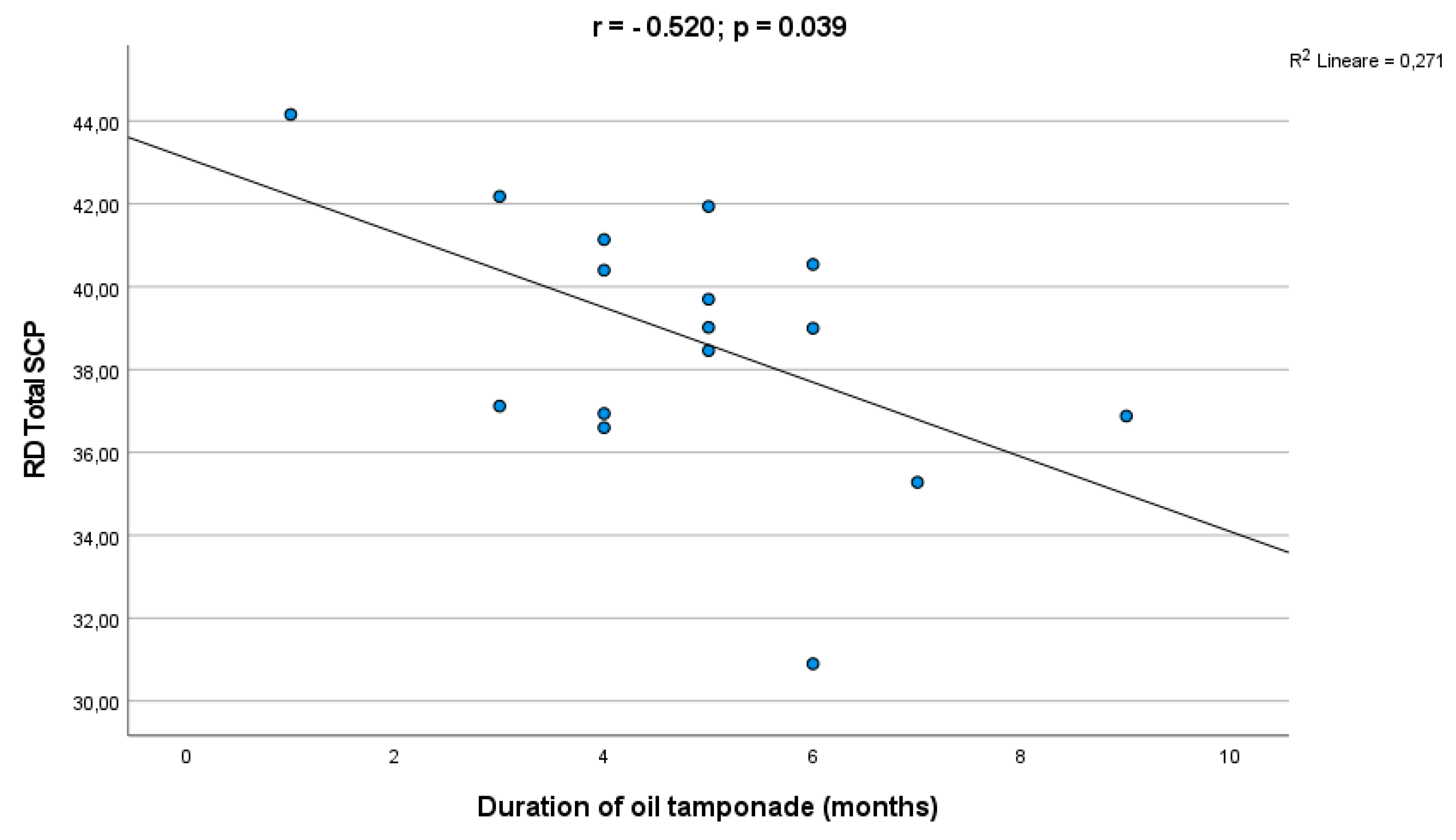
3.3. Surgery Subgroup Analysis
3.4. OCT Findings
4. Discussion
5. Conclusions
Author Contributions
Funding
Institutional Review Board Statement
Informed Consent Statement
Data Availability Statement
Conflicts of Interest
References
- Cheng, K.C.; Cheng, K.Y.; Cheng, K.H.; Chen, K.J.; Chen, C.H.; Wu, W.C. Using optical coherence tomography to evaluate macular changes after surgical management for rhegmatogenous retinal detachment. Kaohsiung J. Med. Sci. 2016, 32, 248–254. [Google Scholar] [CrossRef]
- Coppola, M.; Marchese, A.; Cicinelli, M.V.; Rabiolo, A.; Giuffrè, C.; Gomarasca, S.; Querques, G.; Bandello, F. Macular optical coherence tomography findings after vitreoretinal surgery for rhegmatogenous retinal detachment. Eur. J. Ophthalmol. 2020, 30, 805–816. [Google Scholar] [CrossRef]
- Gupta, R.R.; Iaboni, D.S.M.; Seamone, M.E.; Sarraf, D. Inner, outer, and full-thickness retinal folds after rhegmatogenous retinal detachment repair: A review. Surv. Ophthalmol. 2019, 64, 135–161. [Google Scholar] [CrossRef]
- Mastropasqua, R.; Toto, L.; Mattei, P.A.; di Nicola, M.; Zecca, I.A.L.; Carpineto, P.; di Antonio, L. Reproducibility and Repeatability of Foveal Avascular Zone Area Measurements Using Swept-Source Optical Coherence Tomography Angiography in Healthy Subjects. Eur. J. Ophthalmol. 2017, 27, 336–341. [Google Scholar] [CrossRef]
- de Carlo, T.E.; Romano, A.; Waheed, N.K.; Duker, J.S. A review of optical coherence tomography angiography (OCTA). Int. J. Retin. Vitr. 2015, 1, 5. [Google Scholar] [CrossRef]
- Spaide, R.F.; Klancnik, J.M., Jr.; Cooney, M.J. Retinal vascular layers imaged by fluorescein angiography and optical coherence tomography angiography. JAMA Ophthalmol. 2015, 133, 45–50. [Google Scholar] [CrossRef]
- Christou, E.E.; Stavrakas, P.; Batsos, G.; Christodoulou, E.; Stefaniotou, M. Association of OCT-A characteristics with postoperative visual acuity after rhegmatogenous retinal detachment surgery: A review of the literature. Int. Ophthalmol. 2021, 41, 2283–2292. [Google Scholar] [CrossRef]
- Cardillo Piccolino, F. Vascular changes in rhegmatogenous retinal detachment. Ophthalmologica 1983, 186, 17–24. [Google Scholar] [CrossRef]
- Satoh, Y. Retinal circulation in rhegmatogenous retinal detachment demonstrated by videofluorescence angiography and image analysis. I. The condition of retinal circulation before retinal detachment surgery. Nippon Ganka Gakkai Zasshi 1989, 93, 1002–1008. [Google Scholar]
- Sato, T.; Kanai, M.; Busch, C.; Wakabayashi, T. Foveal avascular zone area after macula-off rhegmatogenous retinal detachment repair: An optical coherence tomography angiography study. Graefes Arch. Clin. Exp. Ophthalmol. 2017, 255, 2071–2072. [Google Scholar] [CrossRef]
- Wang, H.; Xu, X.; Sun, X.; Ma, Y.; Sun, T. Macular perfusion changes assessed with optical coherence tomography angiography after vitrectomy for rhegmatogenous retinal detachment. Graefes Arch. Clin. Exp. Ophthalmol. 2019, 257, 733–740. [Google Scholar] [CrossRef]
- van Bussel, E.M.; van der Valk, R.; Bijlsma, W.R.; La Heij, E.C. Impact of duration of macula-off retinal detachment on visual outcome: A systematic review and meta-analysis of literature. Retina 2014, 34, 1917–1925. [Google Scholar] [CrossRef] [PubMed]
- Funatsu, R.; Terasaki, H.; Koriyama, C.; Yamashita, T.; Shiihara, H.; Sakamoto, T. Silicone oil versus gas tamponade for primary rhegmatogenous retinal detachment treated successfully with a propensity score analysis: Japan Retinal Detachment Registry. Br. J. Ophthalmol. 2022, 106, 1044–1050. [Google Scholar] [CrossRef] [PubMed]
- Liu, Y.; Lei, B.; Jiang, R.; Huang, X.; Zhou, M.; Xu, G. Changes of macular vessel density and thickness in gas and silicone oil tamponades after vitrectomy for macula-on rhegmatogenous retinal detachment. BMC Ophthalmol. 2021, 21, 392. [Google Scholar] [CrossRef] [PubMed]
- Kang, J.W.; Yoo, R.; Jo, Y.H.; Kim, H.C. Correlation of microvascular structures on optical coherence tomography angiography with visual acuity in retinal vein occlusion. Retina 2017, 37, 1700–1709. [Google Scholar] [CrossRef] [PubMed]
- Williams, G.A.; Reeser, F.; O’Brien, W.J.; Fleischman, J.A. Prostacyclin and thromboxane A2 derivatives in rhegmatogenous subretinal fluid. Arch. Ophthalmol. 1983, 101, 463–464. [Google Scholar] [CrossRef] [PubMed]
- Quintyn, J.C.; Brasseur, G. Subretinal fluid in primary rhegmatogenous retinal detachment: Physiopathology and composition. Surv. Ophthalmol. 2004, 49, 96–108. [Google Scholar] [CrossRef]
- Ricker, L.J.; Kijlstra, A.; de Jager, W.; Liem, A.T.; Hendrikse, F.; La Heij, E.C. Chemokine levels in subretinal fluid obtained during scleral buckling surgery after rhegmatogenous retinal detachment. Investig. Ophthalmol. Vis. Sci. 2010, 51, 4143–4150. [Google Scholar] [CrossRef]
- Roldán-Pallarés, M.; Musa, A.S.; Hernández-Montero, J.; Bravo-Llatas, C. Preoperative duration of retinal detachment and preoperative central retinal artery hemodynamics: Repercussion on visual acuity. Graefes Arch. Clin. Exp. Ophthalmol. 2009, 247, 625–631. [Google Scholar] [CrossRef]
- Iandiev, I.; Uhlmann, S.; Pietsch, U.C.; Biedermann, B.; Reichenbach, A.; Wiedemann, P.; Bringmann, A. Endothelin receptors in the detached retina of the pig. Neurosci. Lett. 2005, 384, 72–75. [Google Scholar] [CrossRef]
- Çetinkaya Yaprak, A.; Küçük, M.F.; Yaprak, L.; Erol, M.K. Change in retinal and choroidal microvascular structures after rhegmatogenous retinal detachment surgery and effects on visual recovery. J. Fr. Ophtalmol. 2021, 44, 804–812. [Google Scholar] [CrossRef] [PubMed]
- McKay, K.M.; Vingopoulos, F.; Wang, J.C.; Papakostas, T.D.; Silverman, R.F.; Marmalidou, A.; Lains, I.; Eliott, D.; Vavvas, D.G.; Kim, L.A.; et al. Retinal Microvasculature Changes After Repair of Macula-off Retinal Detachment Assessed with Optical Coherence Tomography Angiography. Clin. Ophthalmol. 2020, 14, 1759–1767. [Google Scholar] [CrossRef]
- Chatziralli, I.; Theodossiadis, G.; Parikakis, E.; Chatzirallis, A.; Dimitriou, E.; Theodossiadis, P. Inner retinal layers’ alterations and microvasculature changes after vitrectomy for rhegmatogenous retinal detachment. Int. Ophthalmol. 2020, 40, 3349–3356. [Google Scholar] [CrossRef] [PubMed]
- Tsen, C.; Sheu, S.; Chen, S.; Wu, T. Imaging analysis with optical coherence tomography angiography after primary repair of macula-off rhegmatogenous retinal detachment. Graefes Arch. Clin. Exp. Ophthalmol. 2019, 257, 1847–1855. [Google Scholar] [CrossRef] [PubMed]
- D’Aloisio, R.; Gironi, M.; Verdina, T.; Vivarelli, C.; Leonelli, R.; Mariotti, C.; Kaleci, S.; Toto, L.; Mastropasqua, R. Early Structural and Vascular Changes after Within-24 Hours Vitrectomy for Recent Onset Rhegmatogenous Retinal Detachment Treatment: A Pilot Study Comparing Bisected Macula and Not Bisected Macula. J. Clin. Med. 2022, 11, 3498. [Google Scholar] [CrossRef]
- Hong, E.H.; Cho, H.; Kim, D.R.; Kang, M.H.; Shin, Y.U.; Seong, M. Changes in Retinal Vessel and Retinal Layer Thickness After Vitrectomy in Retinal Detachment via Swept-Source OCT Angiography. Investig. Ophthalmol. Vis. Sci. 2020, 61, 35. [Google Scholar] [CrossRef]
- Barca, F.; Bacherini, D.; Dragotto, F.; Tartaro, R.; Lenzetti, C.; Finocchio, L.; Virgili, G.; Caporossi, T.; Giansanti, F.; Savastano, A.; et al. OCT Angiography Findings in Macula-ON and Macula-OFF Rhegmatogenous Retinal Detachment: A Prospective Study. J. Clin. Med. 2020, 9, 3982. [Google Scholar] [CrossRef]
- Jiang, J.; Chen, S.; Jia, Y.D.; Li, R.; Zhou, J.X.; Li, R.M. Evaluation of macular vessel density changes after vitrectomy with silicone oil tamponade in patients with rhegmatogenous retinal detachment. Int. J. Ophthalmol. 2021, 14, 881–886. [Google Scholar] [CrossRef]
- Bonfiglio, V.; Ortisi, E.; Scollo, D.; Reibaldi, M.; Russo, A.; Pizzo, A.; Faro, G.; Macchi, I.; Fallico, M.; Toro, M.D.; et al. Vascular changes after vitrectomy for rhegmatogenous retinal detachment: Optical coherence tomography angiography study. Acta Ophthalmol. 2019, 98, e563–e569. [Google Scholar] [CrossRef]
- Hassanpoor, N.; Milani, A.E.; Kordestani, A.; Niyousha, M.R. Analysis of Retinal Layers’ Thickness and Vascular Density after Successful Scleral Buckle Surgery. J. Curr. Ophthalmol. 2021, 33, 304–309. [Google Scholar] [CrossRef]
- Nam, S.H.; Kim, K.; Kim, E.S.; Kim, D.G.; Yu, S.Y. Longitudinal Microvascular Changes on Optical Coherence Tomographic Angiography after Macula-Off Rhegmatogenous Retinal Detachment Repair Surgery. Ophthalmologica 2021, 244, 34–41. [Google Scholar] [CrossRef] [PubMed]
- Resch, M.D.; Balogh, A.; Lászik, G.; Nagy, Z.Z.; Papp, A. Association between retinal vessel density and postoperative time after primary repair of rhegmatogenous retinal detachment. PLoS ONE 2021, 16, e0258126. [Google Scholar] [CrossRef]
- Borrelli, E.; Balasubramanian, S.; Triolo, G.; Barboni, P.; Sadda, S.R.; Sadun, A.A. Topographic Macular Microvascular Changes and Correlation With Visual Loss in Chronic Leber Hereditary Optic Neuropathy. Am. J. Ophthalmol. 2018, 192, 217–228. [Google Scholar] [CrossRef] [PubMed]
- Borrelli, E.; Toto, L.; Viggiano, P.; Evangelista, F.; Palmieri, M.; Mastropasqua, R. Widefield topographical analysis of the retinal perfusion and neuroretinal thickness in healthy eyes: A pilot study. Eye 2020, 34, 2264–2270. [Google Scholar] [CrossRef] [PubMed]
- Agarwal, A.; Aggarwal, K.; Akella, M.; Agrawal, R.; Khandelwal, N.; Bansal, R.; Singh, R.; Gupta, V.; OCTA Study Group. Fractal dimension and optical coherence tomography angiography features of the central macula after repair of rhegmatogenous retinal detachments. Retina 2019, 39, 2167–2177. [Google Scholar] [CrossRef] [PubMed]
- Kim, K.; Kim, E.S.; Yu, S.Y. Optical coherence tomography angiography analysis of foveal microvascular changes and inner retinal layer thinning in patients with diabetes. Br. J. Ophthalmol. 2018, 102, 1226–1231. [Google Scholar] [CrossRef] [PubMed]
- Menke, M.N.; Kowal, J.H.; Dufour, P.; Wolf-Schnurrbusch, U.E.; Ceklic, L.; Framme, C.; Wolf, S. Retinal layer measurements after successful macula-off retinal detachment repair using optical coherence tomography. Investig. Ophthalmol. Vis. Sci. 2014, 55, 6575–6579. [Google Scholar] [CrossRef]
- Lee, J.Y.; Kim, J.Y.; Lee, S.Y.; Jeong, J.H.; Lee, E.K. Foveal Microvascular Structures in Eyes with Silicone Oil Tamponade for Rhegmatogenous Retinal Detachment: A Swept-source Optical Coherence Tomography Angiography Study. Sci. Rep. 2020, 10, 2555. [Google Scholar] [CrossRef]
- Williams, P.D.; Fuller, C.G.; Scott, I.U.; Fuller, D.G.; Flynn, H.W. Vision loss associated with the use and removal of intraocular silicone oil. Clin. Ophthalmol. 2008, 2, 955–959. [Google Scholar]
- Dogramaci, M.; Williams, K.; Lee, E.; Williamson, T.H. Foveal light exposure is increased at the time of removal of silicone oil with the potential for phototoxicity. Graefes Arch. Clin. Exp. Ophthalmol. 2013, 251, 35–39. [Google Scholar] [CrossRef]
- García-Ayuso, D.; Salinas-Navarro, M.; Agudo-Barriuso, M.; Alarcón-Martínez, L.; Vidal-Sanz, M.; Villegas-Pérez, M.P. Retinal ganglion cell axonal compression by retinal vessels in light-induced retinal degeneration. Mol. Vis. 2011, 17, 1716–1733. [Google Scholar]
- Grzybowski, A.; Pieczynski, J.; Ascaso, F.J. Neuronal complications of intravitreal silicone oil: An updated review. Acta Ophthalmol. 2014, 92, 201–204. [Google Scholar] [CrossRef] [PubMed]
- Winter, M.; Eberhardt, W.; Scholz, C.; Reichenbach, A. Failure of potassium siphoning by Müller cells: A new hypothesis of perfluorocarbon liquid-induced retinopathy. Investig. Ophthalmol. Vis. Sci. 2000, 41, 256–261. [Google Scholar]
- Inoue, M.; Iriyama, A.; Kadonosono, K.; Tamaki, Y.; Yanagi, Y. Effects of perfluorocarbon liquids and silicone oil on human retinal pigment epithelial cells and retinal ganglion cells. Retina 2009, 29, 677–681. [Google Scholar] [CrossRef] [PubMed]
- Bambas, B.; Eckardt, C.; Vowinkel, E.; Kruse, H. Toxische Substanzen im Silikonöl nach intraokularer Injektion [Toxic substances with silicone oil after intraocular injections]. Ophthalmologe 1995, 92, 663–667. (In German) [Google Scholar] [PubMed]
- Wickham, L.; Asaria, R.H.; Alexander, R.; Luthert, P.; Charteris, D.G. Immunopathology of intraocular silicone oil: Enucleated eyes. Br. J. Ophthalmol. 2007, 91, 253–257. [Google Scholar] [CrossRef]
- Asaria, R.H.; Kon, C.H.; Bunce, C.; Sethi, C.S.; Limb, G.A.; Khaw, P.T.; Aylward, G.W.; Charteris, D.G. Silicone oil concentrates fibrogenic growth factors in the retro-oil fluid. Br. J. Ophthalmol. 2004, 88, 1439–1442. [Google Scholar] [CrossRef]


| Variables | |
|---|---|
| Patients, n | 82 |
| Age, mean yrs ± SD | 63 ± 12 |
| Male, n (%) | 50 (61) |
| Diabetes | 6 (7.3) |
| Hypertension | 38 (46.3) |
| Pre-operative factors | |
| Right eye: Left eye, % | 55: 45 |
| Macula OFF, n (%) | 49 (59.8) |
| Pseudophakic | 20 (24.4) |
| Intraoperative factors | |
| Type of surgery, n (%) | |
| PPV | 70 (85.4) |
| SB | 12 (14.6) |
| Type of surgery | |
| Gas | 65 (79.3) |
| SO | 17 (20.7) |
| Combined cataract surgery | 42 (51.2) |
| 360° laser | 12 (14.6) |
| Operation time, min (mean ± SD) | 91.27 ± 34.9 |
| Post-operative factors | |
| Duration of SO tamponade, months (mean ± SD) | 4.81 ± 1.8 |
| Hypotony (IOP < 5 mmHg) | 0 |
| Secondary glaucoma (IOP > 21 mmHg), n (%) | 8 (9.8) |
| Intraretinal cysts, % | 12 (14.6) |
| Follow-up period (months; m ± SD) | 13.1 ± 2 |
| BCVA-Post | Macula-ON Group | Macula-OFF Group | ||
|---|---|---|---|---|
| β ± SE | p-Value | β ± SE | p-Value | |
| Type of surgery | −0.009 ± 0.043 | 0.841 | −0.059 ± 0.084 | 0.487 |
| Tamponade | 0.273 ± 0.088 | 0.004 * | −0.007 ± 0.119 | 0.955 |
| Follow-up period | 0.002 ± 0.005 | 0.640 | −0.001 ± 0.007 | 0.878 |
| Macula-ON Group | Macula-OFF Group | |||||
|---|---|---|---|---|---|---|
| Study Eyes | Control Eyes | p-Value | Study Eyes | Control Eyes | p-Value | |
| CENTRAL SCP VD | 30.20 ± 5.70 | 32.22 ± 5.66 | 0.002 * | 28.31 ± 5.79 | 30.32 ± 5.78 | 0.002 * |
| INF SCP VD | 40.78 ± 4.51 | 41.80 ± 3.52 | 0.275 | 40.33 ± 3.64 | 42.24 ± 2.38 | 0.002 * |
| NAS SCP VD | 39.45 ± 4.48 | 40.90 ± 3.77 | 0.038 * | 38.51 ± 3.73 | 40.19 ± 3.18 | 0.010 * |
| SUP SCP VD | 40.32 ± 4.34 | 42.14 ± 2.78 | 0.030 * | 39.65 ± 3.31 | 41.42 ± 2.88 | 0.002 * |
| TEMP SCP VD | 38.42 ± 3.78 | 39.87 ± 3.35 | 0.056 | 38.06 ± 2.85 | 39.76 ± 2.56 | 0.000 * |
| CENTRAL DCP VD | 30.30 ± 8.90 | 30.67 ± 8.15 | 0.752 | 28.76 ± 7.44 | 29.05 ± 8.15 | 0.784 |
| INF DCP VD | 42.76 ± 3.41 | 42.61 ± 2.65 | 0.829 | 42.08 ± 3.08 | 42.82 ± 2.99 | 0.185 |
| NAS DCP VD | 42.74 ± 3.94 | 43.00 ± 2.36 | 0.698 | 41.54 ± 2.79 | 42.37 ± 2.93 | 0.117 |
| SUP DCP VD | 42.28 ± 4.06 | 43.03 ± 2.47 | 0.307 | 41.85 ± 2.83 | 42.40 ± 2.63 | 0.176 |
| TEMP DCP VD | 41.83 ± 3.69 | 41.96 ± 2.61 | 0.842 | 41.43 ± 2.97 | 41.58 ± 2.70 | 0.731 |
| CENTRAL SCP VLD | 16.17 ± 2.83 | 17.65 ± 2.86 | 0.001 * | 15.23 ± 2.87 | 16.53 ±2.96 | 0.001 * |
| INF SCP VLD | 21.01 ± 3.52 | 21.70 ± 3.69 | 0.221 | 21.11 ± 3.66 | 22.24 ± 3.35 | 0.004 * |
| NAS SCP VLD | 20.88 ± 3.33 | 21.83 ± 3.25 | 0.086 | 21.04 ± 3.29 | 21.92 ± 2.99 | 0.032 * |
| SUP SCP VLD | 20.55 ± 3.48 | 21.59 ± 4.04 | 0.090 | 20.73 ± 3.27 | 22.08 ± 3.65 | 0.001 * |
| TEMP SCP VLD | 20.18 ± 2.98 | 20.78 ± 3.55 | 0.259 | 20.46 ± 2.77 | 21.50 ± 2.85 | 0.002 * |
| CENTRAL DCP VLD | 16.54 ± 4.40 | 16.80 ± 4.58 | 0.739 | 16.10 ± 4.25 | 16.30 ± 4.72 | 0.766 |
| INF DCP VLD | 25.29 ± 3.89 | 24.66 ± 4.04 | 0.288 | 25.06 ± 3.95 | 25.28 ± 3.90 | 0.598 |
| NAS DCP VLD | 24.66 ± 3.64 | 25.06 ± 3.72 | 0.537 | 24.82 ± 3.60 | 25.01 ± 3.46 | 0.655 |
| SUP DCP VLD | 24.78 ± 3.74 | 25.10 ± 4.46 | 0.600 | 24.90 ± 3.65 | 25.41 ± 3.79 | 0.089 |
| TEMP DCP VLD | 24.30 ± 3.17 | 24.12 ± 3.62 | 0.747 | 24.34 ± 3.34 | 24.26 ± 3.40 | 0.796 |
Publisher’s Note: MDPI stays neutral with regard to jurisdictional claims in published maps and institutional affiliations. |
© 2022 by the authors. Licensee MDPI, Basel, Switzerland. This article is an open access article distributed under the terms and conditions of the Creative Commons Attribution (CC BY) license (https://creativecommons.org/licenses/by/4.0/).
Share and Cite
Gironi, M.; D’Aloisio, R.; Verdina, T.; Vivarelli, C.; Leonelli, R.; Kaleci, S.; Toto, L.; Mastropasqua, R. Long-Term Macular Vascular Changes after Primary Rhegmatogenous Retinal Detachment Surgery Resolved with Different Tamponade or Different Surgical Techniques. Life 2022, 12, 1525. https://doi.org/10.3390/life12101525
Gironi M, D’Aloisio R, Verdina T, Vivarelli C, Leonelli R, Kaleci S, Toto L, Mastropasqua R. Long-Term Macular Vascular Changes after Primary Rhegmatogenous Retinal Detachment Surgery Resolved with Different Tamponade or Different Surgical Techniques. Life. 2022; 12(10):1525. https://doi.org/10.3390/life12101525
Chicago/Turabian StyleGironi, Matteo, Rossella D’Aloisio, Tommaso Verdina, Chiara Vivarelli, Riccardo Leonelli, Shaniko Kaleci, Lisa Toto, and Rodolfo Mastropasqua. 2022. "Long-Term Macular Vascular Changes after Primary Rhegmatogenous Retinal Detachment Surgery Resolved with Different Tamponade or Different Surgical Techniques" Life 12, no. 10: 1525. https://doi.org/10.3390/life12101525
APA StyleGironi, M., D’Aloisio, R., Verdina, T., Vivarelli, C., Leonelli, R., Kaleci, S., Toto, L., & Mastropasqua, R. (2022). Long-Term Macular Vascular Changes after Primary Rhegmatogenous Retinal Detachment Surgery Resolved with Different Tamponade or Different Surgical Techniques. Life, 12(10), 1525. https://doi.org/10.3390/life12101525