
Near-Infrared Fluorescence with Indocyanine Green to Assess Bone Perfusion: A Systematic Review


 and
and
Abstract
:1. Introduction
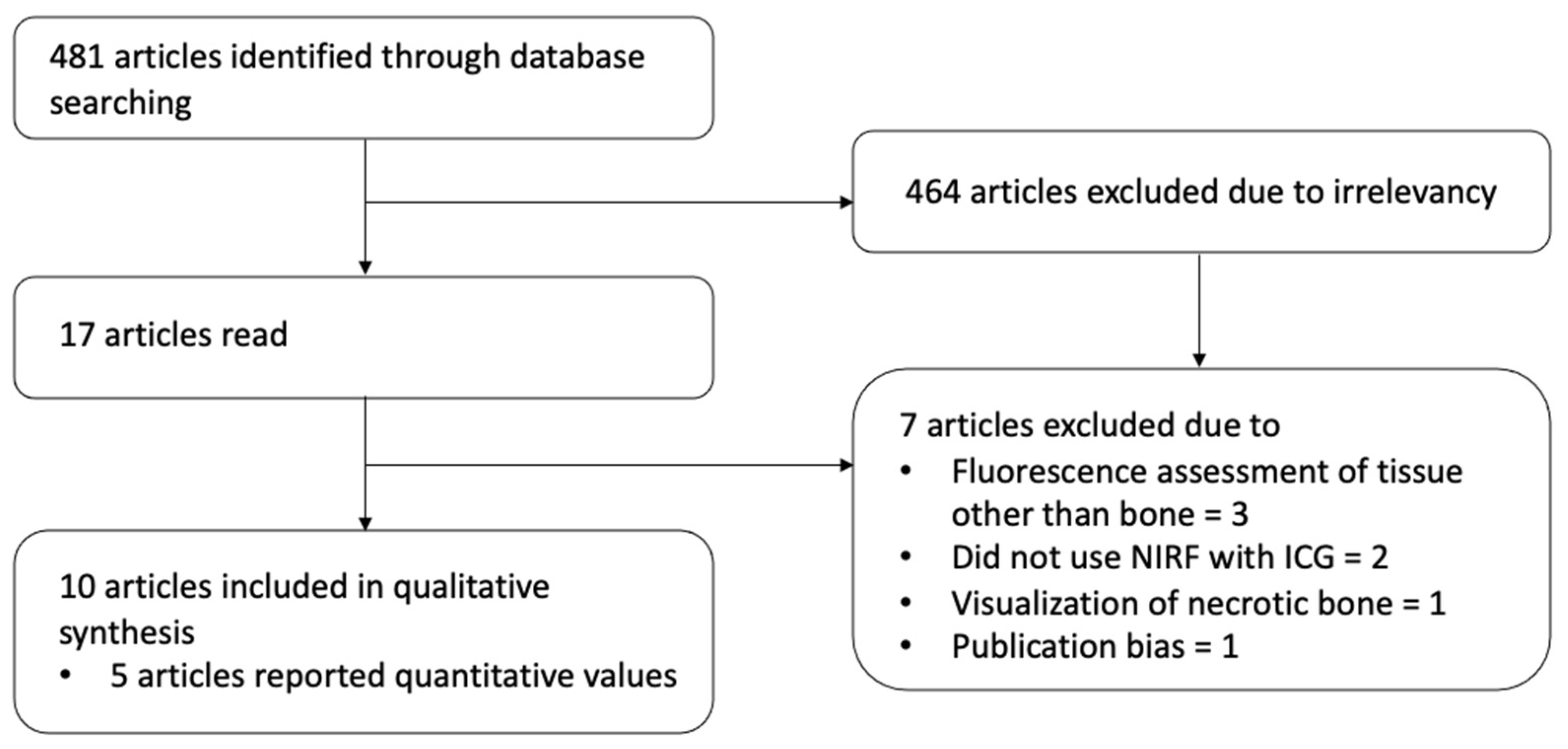
2. Methods
3. Data Collection
4. Results
4.1. NIRF Imaging Methodology
4.2. Objective Perfusion Parameters
4.3. Maximum Bone Fluorescence Intensity
4.4. Relative Bone Fluorescence Intensity
4.5. Other Parameters
5. Discussion
6. Conclusions
Author Contributions
Funding
Institutional Review Board Statement
Informed Consent Statement
Acknowledgments
Conflicts of Interest
References
- Muntean, M.V.; Muntean, V.; Ardelean, F.; Georgescu, A. Dynamic perfusion assessment during perforator flap surgery: An up-to-date. Clujul Med. 2015, 88, 293–297. [Google Scholar] [CrossRef] [PubMed] [Green Version]
- Kohlert, S.; Quimby, A.E.; Saman, M.; Ducic, Y. Postoperative Free-Flap Monitoring Techniques. Semin. Plast. Surg. 2019, 33, 13–16. [Google Scholar] [CrossRef] [PubMed]
- Cierny, G., 3rd; Mader, J.T.; Penninck, J.J. A clinical staging system for adult osteomyelitis. Clin. Orthop. Relat. Res. 2003, 7–24. [Google Scholar] [CrossRef] [PubMed] [Green Version]
- Parsons, B.; Strauss, E. Surgical management of chronic osteomyelitis. Am. J. Surg. 2004, 188 (Suppl. 1A), 57–66. [Google Scholar] [CrossRef]
- Harris, L.; Goldstein, D.; Hofer, S.; Gilbert, R. Impact of vasopressors on outcomes in head and neck free tissue transfer. Microsurgery 2012, 32, 15–19. [Google Scholar] [CrossRef]
- Brown, J.S.; Lowe, D.; Kanatas, A.; Schache, A. Mandibular reconstruction with vascularised bone flaps: A systematic review over 25 years. Br. J. Oral. Maxillofac. Surg. 2017, 55, 113–126. [Google Scholar] [CrossRef]
- Knitschke, M.; Sonnabend, S.; Bäcker, C.; Schmermund, D.; Böttger, S.; Howaldt, H.P.; Attia, S. Partial and Total Flap Failure after Fibula Free Flap in Head and Neck Reconstructive Surgery: Retrospective Analysis of 180 Flaps over 19 Years. Cancers 2021, 13, 865. [Google Scholar] [CrossRef] [PubMed]
- Smit, J.M.; Negenborn, V.L.; Jansen, S.M.; Jaspers, M.E.H.; de Vries, R.; Heymans, M.W.; Winters, H.A.H.; van Leeuwen, T.G.; Mullender, M.G.; Krekel, N.M.A. Intraoperative evaluation of perfusion in free flap surgery: A systematic review and meta-analysis. Microsurgery 2018, 38, 804–818. [Google Scholar] [CrossRef]
- Reinhart, M.B.; Huntington, C.R.; Blair, L.J.; Heniford, B.T.; Augenstein, V.A. Indocyanine Green: Historical Context, Current Applications, and Future Considerations. Surg. Innov. 2016, 23, 166–175. [Google Scholar] [CrossRef]
- Momeni, A.; Sheckter, C. Intraoperative Laser-Assisted Indocyanine Green Imaging Can Reduce the Rate of Fat Necrosis in Microsurgical Breast Reconstruction. Plast. Reconstr. Surg. 2020, 145, 507e–513e. [Google Scholar] [CrossRef]
- Varela, R.; Casado-Sanchez, C.; Zarbakhsh, S.; Diez, J.; Hernandez-Godoy, J.; Landin, L. Outcomes of DIEP Flap and Fluorescent Angiography: A Randomized Controlled Clinical Trial. Plast. Reconstr. Surg. 2020, 145, 1–10. [Google Scholar] [CrossRef]
- Malagón-López, P.; Vilà, J.; Carrasco-López, C.; García-Senosiain, O.; Priego, D.; Julian Ibañez, J.F.; Higueras-Suñe, C. Intraoperative Indocyanine Green Angiography for Fat Necrosis Reduction in the Deep Inferior Epigastric Perforator (DIEP) Flap. Aesthet. Surg. J. 2019, 39, Np45–Np54. [Google Scholar] [CrossRef]
- Driessen, C.; Arnardóttir, T.H.; Lorenzo, A.R.; Mani, M.R. How should indocyanine green dye angiography be assessed to best predict mastectomy skin flap necrosis? A systematic review. J. Plast. Reconstr. Aesthet. Surg. 2020, 73, 1031–1042. [Google Scholar] [CrossRef] [PubMed]
- Slooter, M.D.; Jansen, S.; Bloemen, P.R.; van den Elzen, R.M.; Wilk, L.S.; van Leeuwen, T.G.; van Berge Henegouwen, M.I.; de Bruin, D.M.; Gisbertz, S.S. Comparison of Optical Imaging Techniques to Quantitatively Assess the Perfusion of the Gastric Conduit during Oesophagectomy. J. Appl. Sci. 2020, 10, 5522. [Google Scholar] [CrossRef]
- Nguyen, J.T.; Ashitate, Y.; Buchanan, I.A.; Ibrahim, A.M.; Gioux, S.; Patel, P.P.; Frangioni, J.V.; Lee, B.T. Bone flap perfusion assessment using near-infrared fluorescence imaging. J. Surg. Res. 2012, 178, e43–e50. [Google Scholar] [CrossRef] [PubMed] [Green Version]
- Fichter, A.M.; Ritschl, L.M.; Georg, R.; Kolk, A.; Kesting, M.R.; Wolff, K.D.; Mücke, T. Effect of Segment Length and Number of Osteotomy Sites on Cancellous Bone Perfusion in Free Fibula Flaps. J. Reconstr. Microsurg. 2019, 35, 108–116. [Google Scholar]
- Valerio, I.; Green, J.M., 3rd; Sacks, J.M.; Thomas, S.; Sabino, J.; Acarturk, T.O. Vascularized osseous flaps and assessing their bipartate perfusion pattern via intraoperative fluorescence angiography. J. Reconstr. Microsurg. 2015, 31, 45–53. [Google Scholar] [PubMed]
- Elliott, J.T.; Jiang, S.; Pogue, B.W.; Gitajn, I.L. Bone-specific kinetic model to quantify periosteal and endosteal blood flow using indocyanine green in fluorescence guided orthopedic surgery. J. Biophotonics 2019, 12, e201800427. [Google Scholar] [CrossRef] [PubMed]
- Gitajn, I.L.; Elliott, J.T.; Gunn, J.R.; Ruiz, A.J.; Henderson, E.R.; Pogue, B.W.; Jiang, S. Evaluation of bone perfusion during open orthopedic surgery using quantitative dynamic contrast-enhanced fluorescence imaging. Biomed. Opt. Express 2020, 11, 6458–6469. [Google Scholar] [CrossRef]
- Phillips, B.T.; Lanier, S.T.; Conkling, N.; Wang, E.D.; Dagum, A.B.; Ganz, J.C.; Khan, S.U.; Bui, D.T. Intraoperative perfusion techniques can accurately predict mastectomy skin flap necrosis in breast reconstruction: Results of a prospective trial. Plast. Reconstr. Surg. 2012, 129, 778e–788e. [Google Scholar] [CrossRef]
- Yoshimatsu, H.; Steinbacher, J.; Meng, S.; Hamscha, U.M.; Weninger, W.J.; Tinhofer, I.E.; Harima, M.; Fuse, Y.; Yamamoto, T.; Tzou, C.H.J. Superficial Circumflex Iliac Artery Perforator Flap: An Anatomical Study of the Correlation of the Superficial and the Deep Branches of the Artery and Evaluation of Perfusion from the Deep Branch to the Sartorius Muscle and the Iliac Bone. Plast. Reconstr. Surg. 2019, 143, 589–602. [Google Scholar] [CrossRef]
- Yoshimatsu, H.; Steinbacher, J.; Meng, S.; Hamscha, U.M.; Weninger, W.J.; Tinhofer, I.E.; Yamamoto, T.; Iida, T.; Tzou, C.H. Feasibility of Bone Perfusion Evaluation in Cadavers Using Indocyanine Green Fluorescence Angiography. Plast. Reconstr. Surg. Glob. Open 2017, 5, e1570. [Google Scholar] [CrossRef] [PubMed]
- Muangsiri, P.; Tanjapatkul, R.; Sriswadpong, P.; Jomkoh, P.; Jirawatnotai, S. Indocyanine Green Fluorescence Angiography of the Transverse Cervical Arterial Supply to Clavicle Flaps: An Anatomical Study. Otolaryngol. Head. Neck. Surg. 2021, 166. [Google Scholar] [CrossRef] [PubMed]
- Tyrell, R.; Kilmartin, C.; Acevedo, E.; Keshavamurthy, S.; Gassman, A. Is non-invasive indocyanine-green angiography a useful adjunct for the debridement of infected sternal wounds? JPRAS Open 2018, 16, 117–120. [Google Scholar] [CrossRef] [PubMed]
- Reece, E.M.; Davis, M.J.; Wagner, R.D.; Abu-Ghname, A.; Cruz, A.; Kaung, G.; Verla, T.; Winocour, S.; Ropper, A.E. Vascularized Bone Grafts for Spinal Fusion-Part 1: The Iliac Crest. Oper. Neurosurg. 2021, 20, 493–496. [Google Scholar] [CrossRef] [PubMed]
- Oni, O.O.; Stafford, H.; Gregg, P.J. An experimental study of the patterns of periosteal and endosteal damage in tibial shaft fractures using a rabbit trauma model. J. Orthop. Trauma 1989, 3, 142–147. [Google Scholar] [CrossRef]
- Wei, F.C.; Chen, H.C.; Chuang, C.C.; Noordhoff, M.S. Fibular osteoseptocutaneous flap: Anatomic study and clinical application. Plast. Reconstr. Surg. 1986, 78, 191–200. [Google Scholar] [CrossRef]
- Menck, J.; Sander, A. Periosteal and endosteal blood supply of the human fibula and its clinical importance. Acta Anat. 1992, 145, 400–405. [Google Scholar] [CrossRef]
- Nguyen, C.L.; Barry, N.; Lindsay, A.; Seah, J.L.; Easwaralingam, N.; Pulitano, C.; Warrier, S. Indocyanine green angiography in breast reconstruction surgery: A systematic review of cost-analysis studies. J. Plast. Reconstr. Aesthet. Surg. 2021, 74, 3196–3211. [Google Scholar] [CrossRef]
- Van Den Hoven, P.; Goncalves, L.N.; Quax, P.H.A.; Van Rijswijk, C.S.P.; Van Schaik, J.; Schepers, A.; Vahrmeijer, A.L.; Hamming, J.F.; Van Der Vorst, J.R. Perfusion Patterns in Patients with Chronic Limb-Threatening Ischemia versus Control Patients Using Near-Infrared Fluorescence Imaging with Indocyanine Green. Biomedicines 2021, 9, 1417. [Google Scholar] [CrossRef]
- Alander, J.T.; Kaartinen, I.; Laakso, A.; Pätilä, T.; Spillmann, T.; Tuchin, V.V.; Venermo, M.; Välisuo, P. A review of indocyanine green fluorescent imaging in surgery. Int. J. Biomed. Imaging 2012, 2012, 940585. [Google Scholar] [CrossRef] [PubMed]
- Cornelissen, A.J.M.; van Mulken, T.J.M.; Graupner, C.; Qiu, S.S.; Keuter, X.H.A.; van der Hulst, R.; Schols, R.M. Near-infrared fluorescence image-guidance in plastic surgery: A systematic review. Eur. J. Plast. Surg. 2018, 41, 269–278. [Google Scholar] [CrossRef] [PubMed] [Green Version]
- Matsui, A.; Lee, B.T.; Winer, J.H.; Laurence, R.G.; Frangioni, J.V. Quantitative assessment of perfusion and vascular compromise in perforator flaps using a near-infrared fluorescence-guided imaging system. Plast. Reconstr. Surg. 2009, 124, 451–460. [Google Scholar] [CrossRef] [PubMed] [Green Version]


| Study | Level of Evidence | Study Design | Total Number of Cases | Reported Objective NIRF Values | Non-Viable Tissue Excised | |
|---|---|---|---|---|---|---|
| 1 | Nguyen, 2012 | 5 | Experimental research (animal) | 8 animals/16 limbs | Yes | No |
| 2 | Valerio, 2015 | 5 | Retrospective case series | 16 | Yes | No |
| 3 | Yoshimatsu, 2017 | 5 | Experimental research (cadavers) | 4 cadavers/8 limbs | No | No |
| 4 | Tyrell, 2018 | 5 | Retrospective case series | 2 | No | Yes |
| 5 | Fichter, 2019 | 2 | Prospective case series | 39 | Yes | No |
| 6 | Yoshimatsu, 2019 | 5 | Experimental research (cadavers) | 8 cadavers | No | No |
| 7 | Elliot, 2019 | 5 | Experimental research (animal) | 2 animals | Yes | No |
| 8 | Gitajn, 2020 | 5 | Experimental research (animal) | 12 animals | Yes | No |
| 9 | Muangsiri, 2021 | 5 | Experimental research (cadavers) | 11 cadavers/22 flaps | No | No |
| 10 | Reece, 2021 | 5 | Retrospective case series | 14 | No | No |
| Study | ICG Dose | ICG Fluorescence System | Software | Image Timeframe | Camera Position | Area of Interest | |
|---|---|---|---|---|---|---|---|
| 1 | Nguyen, 2012 | 36 ug/kg IV | FLARE imaging system | Custom | NR | 18 inches from surgical field | 1. Radius of forelimb 2. Distal osteotomy site of fibula |
| 2 | Valerio, 2015 | 7.5 mg IV | SPY and SPY Elite | SPY-Q | NR | Laser assisted | Periosteum and cancellous bone of several flaps |
| 3 | Yoshimatsu, 2017 | NR (5 mL Pulsion ICG solution) | Sony HD, Handycam CM 05 | NR | NR | NR | Periosteum and cancellous bone of the iliac crest bone flap |
| 4 | Tyrell, 2018 | NR | SPY | NR | NR | NR | Sternum |
| 5 | Fichter, 2019 | 0.3 mg/kg IV | Pulsion Photodynamic Eye | ImageJ and Prism | 3 min | 30 cm | Distal osteotomy line of distal fibular segment |
| 6 | Yoshimatsu, 2019 | Sony HD, Handycam CM 05 | NR | NR | NR | Periosteum and cancellous bone of medial condyle of femur bone flap | |
| 7 | Elliot, 2019 | NR | Zeiss Pentero OPMI 800 in FLOW 800 mode | MatLab | NR | 30 cm | Tibia, at baseline, after osteotomy with intact and disrupted periosteum, proximal or distal |
| 8 | Gitajn, 2020 | 0.1 mg/kg | Zeiss Pentero OPMI 800 in FLOW 800 mode | MatLab | 4 min | 30 cm | Tibia, at baseline, after osteotomy with intact and disrupted periosteum, proximal or distal |
| 9 | Muangsiri, 2021 | 25 mg (Daiichi Sankyo, Diagnogreen) | Fluoptics | NR | NR | NR | Periosteum, cortex and cancellous bone of clavicula flaps |
| 10 | Reece, 2021 | NR | SPY elite | NR | NR | NR | Visualization of bone surface of ilac crest bone flap |
| Study | ICG Fluorescence System | Perfusion Parameters | |
|---|---|---|---|
| 1 | Nguyen, 2012 | Flare | Absolute perfusion: NR |
| Relative perfusion: Fluorescence intensity of the region of interest/Fluorescence Intensity of background
| |||
| Other parameters: NR | |||
| 2 | Valerio, 2015 | SPY and SPY Elite | Absolute perfusion: SPY-Q score greater than 6.0 |
| Relative perfusion: NR | |||
| Other parameters: NR | |||
| 5 | Fichter, 2019 | Pulsion Photodynamic Eye | Absolute perfusion: Peak Enhancement (PE)
|
| Relative perfusion: NR | |||
| Other parameters: Wash in rate (WiR):
Wash in perfusion index (WiPI)
Other reported parameters Ti—Time local TTP—Time to peak WiAUC—Wash in area under the curve RT—Raise time | |||
| 7 | Elliot, 2019 | Zeiss Pentero | Absolute perfusion: NR |
| Relative perfusion: NR | |||
| Other parameters: PBF (periosteal blood flow): Baseline 6.8 ± 1.1 mL/min/100 gr After periosteal damage
Total Blood Flow (TBF) = PBF + EBF EBF fractional flow (EFF) = EBF/TBF | |||
| 8 | Gitajn, 2020 | Zeiss Pentero en Spy Elite | Absolute perfusion: Imax (a.u) Normal bone 201 ± 50 Injured bone 163 ± 58 No significant difference |
| Relative perfusion: NR | |||
| Other parameters: Total bone perfusion (TBP) normal vs. injured bone: 3.2 ± 0.8 vs. 0.8 ± 0.4 mL/min/100 gr Early bone perfusion (EBP) normal vs. injured bone: 3.0 ± 0.8 vs. 0.7 ± 0.4 mL/min/100 gr Late bone perfusion (LBP) normal vs. injured bone: 0.1 ± 0.0 vs. 0.2 ± 0.1 mL/min/100 gr Late perfusion fraction (LPF) normal vs. injured bone: 2.5 ± 1.1 vs. 21.4 ± 10 |
Publisher’s Note: MDPI stays neutral with regard to jurisdictional claims in published maps and institutional affiliations. |
© 2022 by the authors. Licensee MDPI, Basel, Switzerland. This article is an open access article distributed under the terms and conditions of the Creative Commons Attribution (CC BY) license (https://creativecommons.org/licenses/by/4.0/).
Share and Cite
Michi, M.; Madu, M.; Winters, H.A.H.; de Bruin, D.M.; van der Vorst, J.R.; Driessen, C. Near-Infrared Fluorescence with Indocyanine Green to Assess Bone Perfusion: A Systematic Review. Life 2022, 12, 154. https://doi.org/10.3390/life12020154
Michi M, Madu M, Winters HAH, de Bruin DM, van der Vorst JR, Driessen C. Near-Infrared Fluorescence with Indocyanine Green to Assess Bone Perfusion: A Systematic Review. Life. 2022; 12(2):154. https://doi.org/10.3390/life12020154
Chicago/Turabian StyleMichi, Marlies, Max Madu, Henri A. H. Winters, Daniel M. de Bruin, Joost R. van der Vorst, and Caroline Driessen. 2022. "Near-Infrared Fluorescence with Indocyanine Green to Assess Bone Perfusion: A Systematic Review" Life 12, no. 2: 154. https://doi.org/10.3390/life12020154
APA StyleMichi, M., Madu, M., Winters, H. A. H., de Bruin, D. M., van der Vorst, J. R., & Driessen, C. (2022). Near-Infrared Fluorescence with Indocyanine Green to Assess Bone Perfusion: A Systematic Review. Life, 12(2), 154. https://doi.org/10.3390/life12020154