Perspective

Lymphatic and Blood Endothelial Extracellular Vesicles: A Story Yet to Be Written

by
Johanna Trisko
1,2,†,
Johanna Fleck
1,2,†,
Silvio Kau
1,2,3,
Johannes Oesterreicher
1,2 and
Wolfgang Holnthoner
1,2,*
1
Ludwig-Boltzmann-Institute for Traumatology, The Research Center in Cooperation with AUVA, A-1200 Vienna, Austria
2
Austrian Cluster for Tissue Regeneration, A-1200 Vienna, Austria
3
Institute of Morphology, University of Veterinary Medicine Vienna, A-1210 Vienna, Austria
*
Author to whom correspondence should be addressed.
These authors contributed equally to this work.
Life 2022, 12(5), 654; https://doi.org/10.3390/life12050654
Submission received: 6 April 2022 / Revised: 22 April 2022 / Accepted: 27 April 2022 / Published: 28 April 2022
(This article belongs to the Special Issue Early Career Stars in Cell Biology and Tissue Engineering)

Abstract

:
Extracellular vesicles (EVs), such as exosomes, microvesicles, and apoptotic bodies, are cell-derived, lipid bilayer-enclosed particles mediating intercellular communication and are therefore vital for transmitting a plethora of biological signals. The vascular endothelium substantially contributes to the circulating particulate secretome, targeting important signaling pathways that affect blood cells and regulate adaptation and plasticity of endothelial cells in a paracrine manner. Different molecular signatures and functional properties of endothelial cells reflect their heterogeneity among different vascular beds and drive current research to understand varying physiological and pathological effects of blood and lymphatic endothelial EVs. Endothelial EVs have been linked to the development and progression of various vascular diseases, thus having the potential to serve as biomarkers and clinical treatment targets. This review aims to provide a brief overview of the human vasculature, the biology of extracellular vesicles, and the current knowledge of endothelium-derived EVs, including their potential role as biomarkers in disease development.

1. The Human Vasculature and Endothelial Cell Heterogeneity

The human vascular system can be broadly separated into the blood and the lymphatic system. The blood vasculature is responsible for the active supply and distribution of blood and its components, whereas the lymphatic system removes accumulating interstitial fluid to ensure tissue homeostasis. Thus, the formation, maintenance, and remodeling of a functioning vascular network is essential for oxygen and nutrient supply as well as lymphatic fluid drainage in the healthy human body [1]. Vasculogenesis describes the formation of new vessels from endothelial progenitor cells [2]. In contrast, angiogenesis refers to the formation of new vessels splitting and sprouting from preexisting ones [3]. Endothelial cells (ECs) arise from the mesodermal stem cell lineage. Mesodermal progenitor cells develop to angioblasts under the influence of a group of transcription factors including E26 transformation-specific (ETS) [4]. Specification into endothelial and hematopoietic lineage is primarily driven through the expression of transcription factors Etv2 and Npas4l [5]. During this process, activation of distinct signaling networks leads to further differentiation into arterial, venous, and lymphatic subtypes [1]. The specification into arterial or venous fate is further driven by regulators such as SoxF transcription factors, Notch receptor proteins, bone morphogenic proteins (BMPs), and transforming growth factor-beta (TGF-ß) [5]. The upregulation of Notch has been shown to lead simultaneously to the expression of arterial markers and the downregulation of venous ones [6]. By suppressing of Notch signaling, the nuclear receptor Coup-TFII acts as a key regulator of venous identity [4]. Regarding the “second” vascular system, lymphatic cell commitment is influenced by vascular endothelial growth factor-C (VEGF-C)/vascular endothelial growth factor receptor-3 (VEGFR3, FLT4)-signaling and activation of transcriptional programs by prospero homeobox-1 (Prox1) and the SRY-related HMG-box family member Sox18 [5]. Finally, circulating endothelial progenitor cells (EPCs), which can be experimentally isolated and differentiated into highly proliferating endothelial colony-forming cells (ECFCs), are thought to aid postnatal vasculogenesis and to contribute to the regeneration of damaged vasculature [7].
The heterogeneity of ECs reflects tissue-dependent differences in their functionality. Meeting these different demands requires a certain morphological variability, which is partially manifested by regional differences in intercellular junctions [3]. The endothelium of arteries and veins is continuous, while the endothelium of capillaries may also be fenestrated or discontinuous depending on the different underlying needs of the tissue [8]. In most capillaries of the brain, lungs, and skeletal muscle, a continuous sheet of ECs is held together by tight junctions and is anchored to the basal membrane [3]. Other tissues build on a fenestrated endothelium with pores of around 60–80 nm in diameter that are spanned by a diaphragm, which increase permeability and allow small molecules to diffuse [7]. This type of endothelium is found in organs involved in filtration or secretion, including exocrine and endocrine glands, gastric and intestinal mucosa, choroid plexus, kidney glomeruli, and a subpopulation of renal tubules [3,8]. In contrast to the intact basal membrane in fenestrated endothelium, the underlying basement membrane in discontinuous endothelium is poorly formed and exhibits larger fenestration (100–200 nm) among ECs, which are devoid of diaphragm [9]. Discontinuous endothelium is found in the sinusoids of the liver, spleen, and bone marrow [3]. Other morphologic differences are seen in the size and thickness of ECs. Aortic ECs are 1 µm thick compared to the cells of capillaries, which are 0.1 µm thick, and veinous ECs are 0.1–0.2 µm [9]. ECs can be remodeled in response to hemodynamic shear stress [8]. Changed flow patterns do not only result in region-specific phenotypic differences but also lead to changes on a molecular level by modifying gene expression [5]. The special role of the endothelium as an interface between underlying tissue and blood or lymphatic fluid makes it an important contributor to the circulating secretome. Therefore, it can have an impact on the plasticity and adaptation of ECs in different vascular beds as well as on circulating immune cells [4]. Taken together, ECs in different vascular beds reflect their respective physiological roles, which meet tissue-specific demands, by highly adopted and therefore heterogeneous phenotypes. The question that remains is if those heterogeneities of endothelial cells are transferred to their respective extracellular vesicles.

2. Biology of Extracellular Vesicles

Extracellular vesicles (EVs) have been found to influence a variety of pathological and physiological processes, such as inflammation, coagulation, or atherosclerosis, specifically in the context of vascular biology. However, their small subfractions (<200 nm) especially, together with possible differences between different vascular origins, remain poorly understood to this day. In general, EVs are defined as lipid membrane-enclosed vesicles with cellular origin that transport bioactive cargo including lipids, proteins, and nucleic acids. Not considering their cellular origin or cargo, they are commonly differentiated based on their size and release pathway into exosomes (30–150 nm), microvesicles (100 nm–1 µm), and apoptotic bodies (50 nm–5 µm) as the three main subcategories [10]. The small subset of EVs, most often termed exosomes, are vesicles that are actively secreted via an endosomal pathway. After the inward budding of the plasma membrane during endocytosis, membrane-bound proteins as well as extracellular components are internalized [11]. Subsequently, after scission from the membrane, the internally formed vesicles, now termed early endosomes, experience further regulated inward budding and cargo sorting during the maturation to late endosomes. The regulated inward budding and transport of cytoplasmic cargo leads to the formation of intraluminal vesicles (ILVs), which hallmarks the transition of late endosomes to multivesicular bodies (MVBs) [12]. These sorting and vesicle-formation processes are a topic of ongoing research that has revealed the high complexity and involvement of a variety of different proteins such as the endosomal sorting complex required for transport (ESCRT) or RAS-related protein RAB31. Mature MVBs are either fused with lysosomes for content degradation by hydrolytic enzymes through a ubiquitin- and clathrin-dependent manner or are trafficked to the cell membrane, where the subsequent fusion causes the release of the ILVs into the extracellular space in which they are then termed exosomes [12,13,14,15,16,17,18]. The commonly larger species of EVs, termed microvesicles (MV), originate, in contrast to exosomes, from the budding of the plasma membrane into the extracellular space and subsequent dissociation from the membrane. Specific adaptation of physical properties by changes of lipid and protein components thereby allows the remodeling of the cellular membrane, leading to separation and release of the MVs. Various molecules such as the ARF6 GTPase or vesicle-associated membrane protein 3 (VAMP3) have been shown to facilitate the active transport of cargo, such as proteins, enzymes, as well as nucleic acids, to the membrane prior to the budding process [19,20,21]. Although similar in size, apoptotic bodies show distinct differences to their exosomal and microvesicular counterparts. Apoptotic bodies are generally released through internal changes of the cytoskeleton and increasing hydrostatic pressure during cell death. These vesicles mainly function as a means of disposal of the dying cell, resulting in a highly similar proteomic profile to the cell lysate itself. The release of apoptotic bodies has been shown to influence various biological processes, such as the activation and modulation of the immune system by transferring antigens, increasing cell proliferation in stem cells as well as tumor niche formation [10,22,23,24,25]

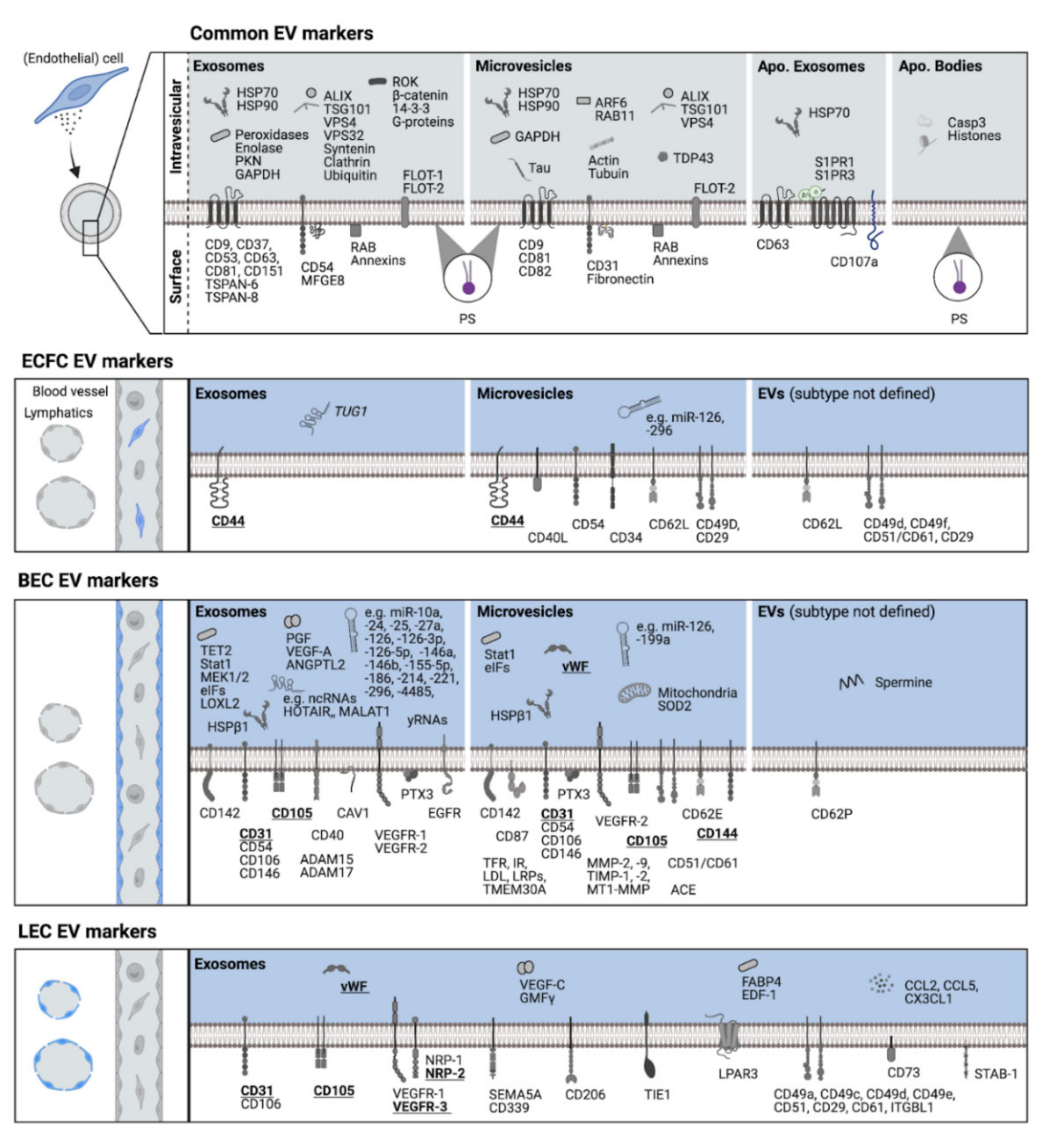
3. Molecular Signatures of Endothelial Cells and Endo-EVs

Identifying molecular profiles that could serve as endothelial EV (endoEV)-specific biomarkers relies on solid evidence of tissue-specific signatures of parental ECs. In addition to the pan EC markers vascular endothelial (VE)-cadherin (CD144), platelet endothelial cell adhesion molecule-1 (PECAM-1, CD31), and van Willebrand factor [26], recent studies revealed further constitutively expressed markers in circulating precursors and mature ECs of different vascular beds. ECFCs express homing-associated cell adhesion molecule (HCAM, CD44), mast/stem cell growth factor receptor (SCFR, c-kit, CD117), endoglin (ENG, CD105), 5′-nucleotidase (NT5, CD73), and stemness-associated transcription factors Klf4, Oct4 and c-Myc [27,28,29,30,31]. Blood vascular endothelial cells (BECs) express neural (N)-cadherin (CD325), pathologische Anatomie Leiden-endothelium (PAL-E) reactive antigen, urokinase, versican, collagen 8A1, ENG, and endothelial cell specific molecule-1 (ESM-1, endocan) [26,31,32,33,34]. More specifically, PAL-E is characteristic for ECs of blood capillaries and small veins [35,36]. Lymphatic endothelial cells (LECs) also express lymphatic vessel endothelial hyaluronan receptor-1 (LYVE-1), VEGFR-3, neuropilin-2 (NRP-2), podoplanin (PDPN), Prox-1, forkhead box protein C2 (FOXC2), and integrin α9 [32,33,37,38,39]. Generally, ECs can differ in their marker expression in a tissue-specific manner or share certain markers across vascular beds. Highlighting just a few uniquely expressed BEC markers, glucose transporter type 1 (GLUT-1) and integral membrane protein 2A (ITM2A) are seen to be specific to brain vascular ECs in mice, just as fatty acid binding protein 4 (FABP4) is to coronary vessel ECs and natriuretic peptide receptor C (NPR3) and cytokine-like protein 1 (C17) are to endocardial ECs [40]. VEGFR-3 is widely accepted to be LEC-specific although also expressed in BECs, constituting fenestrated capillaries in bone marrow, liver, spleen, and kidney glomeruli [37]. LECs that line different lymph node regions differentially express LYVE-1 [41]. The intricacy of EC marker expression profiles reflects the need for combinatorial detection strategies. This is equally important for characterizing endoEVs from different vascular beds in different host tissues and under varying stimuli.
EndoEVs as liquid biopsy-derived biomarkers are increasingly coming into focus as their abundance and cargo provide valuable information on EC response to certain stimuli and can be pathognomonic for EC damage. EndoEVs can be characterized by markers inherent to their biogenesis or parental cells or those accumulating under certain conditions (Figure 1). Contrary to common assumptions, phosphatidylserine (PS) is not only found in the outer membrane leaflet of MVs but also exosomes [42]. PS on endoEVs facilitates tethering to distant ECs and conveys pro-coagulant effects, as was shown by tissue factor (CD142)-bearing BEC-EVs [43,44]. Recent evidence suggests an apoptosis-related subtype of exosomes (apoExos) that in BECs are shed in a caspase 3-dependent manner [45]. ApoExos, alongside exosome-specific tetraspanin CD63, express the lysosomal marker LAMP1 (CD107a), heat-shock protein 70 (HSP70), and sphingosine 1-phosphate receptors 1 and 3 (S1PR1, S1PR3) [46,47]. BEC apoExos also enrich pro-inflammatory non-ribosomal non-coding viral-like RNAs [45].
ECFC-EVs constitutively express HCAM (CD44), which is intrinsic to their parental cells [29,48], and further reveal an expression of certain adhesion molecules, e.g., hematopoietic progenitor cell antigen (CD34), intercellular adhesion molecule-1 (ICAM-1, CD54), L-selectin (CD62L), integrins α4 (CD49d), α6 (CD49f), ß1 (CD29), and αvß3 (CD51/61), but also CD40 ligand, which is involved in activation of antigen presenting cells [48,49,50]. ECFC-exosomes exert anti-inflammatory effects through lncRNA TUG1-mediated alteration of macrophage histone deacetylation [51]. ECFC-MVs convey pro-angiogenic signals via transfer of miR-126 and miR-296, while specific sorting of further miRNAs to MVs was also described [49].
BEC-EVs were found to express EC markers VE-cadherin, PECAM-1, vWF, and ENG [52,53,54,55,56,57,58,59,60,61]. In addition, angiogenesis-implicated growth factor receptors VEGFR-1 and VEGFR-2 and their ligand VEGF-A were detected, with the latter’s expression being increased under hypoxic conditions [52,62,63]. VE-cadherin expression on BEC-EVs is also primed under hypoxia [55,57]. PECAM-1 and ENG were upregulated on BEC-EVs upon growth factor depletion-mediated EC apoptosis [54]. While ENG is downregulated in blood—brain barrier ECs under TNF stimulation, it is highly upregulated in their MVs but consistently expressed in exosomes [56]. TNF pro-inflammatory stimuli further exceeded baseline expression of the cell adhesion molecules intercellular adhesion molecule-1 (ICAM-1, CD54), vascular cell adhesion molecule-1 (VCAM-1, CD106), and E-selectin (CD62E), the classical complement pathway activator pentraxin-related protein (PTX3), and the transcription factor signal transducer and activator of transcription 1 (STAT1) in both BEC-MVs and -exosomes [54,55,56]. MVs from blood—brain barrier ECs also increased expression of eukaryotic translation initiating factors (eIFs) and cell-death-preventing superoxide dismutase 2 (SOD2) [56]. As a transcription regulator, Tet methylcytosine dioxygenase 2 (TET2) was found in BEC-exosomes and is downregulated by CD137 TNF receptor signaling [64]. EV-mediated transfer of CD142 by TNF-stimulated BECs was linked to marked coagulation factor activation [44] although BECs line up an otherwise hemocompatible interface between blood and extravascular tissue. As widely described for CD142 in MSC-derived therapeutics [65], BEC-EVs may pose a similar procoagulant risk when administered intravascularly, necessitating proper endoEV characterization. MVs from brain-BECs express receptors that transfer molecules across the blood—brain barrier, i.e., transferrin receptor, insulin receptor (CD220), low-density lipoproteins (LDL), LDL receptor-related proteins (LRPs), and transmembrane protein 30A (TMEM30A) [66]. Further adhesion molecules found on BEC-EVs are melanoma cell adhesion molecule (MCAM, CD146), P-selectin (CD62P), and integrin αvß3 [53,54,55,58]. BEC-EVs were also shown carrying matrix degrading metalloproteinases, i.e., ADAM15 and ADAM17 in exosomes [56] and MMP-2, MMP-9, and MT1-MMP in MVs [67]. Furthermore, metalloproteinase inhibitors TIMP-1 and TIMP-2 could be found [67]. EVs from hypoxic BECs were also shown to promote extracellular matrix crosslinking via lysyl oxidase-like 2 (LOXL2) transfer [68]. BEC-exosomes further express pro-angiogenic molecules, e.g., angiopoietin-like protein 2 (ANGPTL2) [69]. Pro-angiogenic effects of BEC-EVs could also be observed due to transfer of miR-24, miR-214, and miR-126 [61,70,71,72]. Beside cardioprotective miR-126 and miR-199a [61] and pro-proliferative miR-25, miR-186, and miR-221 [71], BEC-EVs transfer a plethora of further miRNAs, yRNA fragments [71], and lncRNAs [73]. Cardioprotective EV properties could be hampered by caveolin-1 exosomes from interleukin-3 (IL-3)-stimulated BECs [74]. In vitro, EC morphology also regulates inflammatory response, as EVs from elongated BECs carry markedly higher amounts of anti-inflammatory miR-10a [75]. Over and above this excerpt of molecules, RNA sequencing and proteomic studies on BEC-EVs revealed a variety of further RNAs and proteins, respectively [52,56,71].
In LEC-EVs, there is sparse information on molecular patterns. In one proteomic study using exosomes from primary human LECs, more than 1700 proteins were detected [76]. Beside vWF, PECAM-1, and ENG, in this study, LEC markers VEGFR-3 and NRP-2 (also NRP-1) and the lymphangiogenic factor VEGF-C were also found. Further adhesion molecules, e.g., VCAM-1 and integrins α1, α3, α4, α5, αV, ß1, ß3, and subunit beta-like 1, could also be detected. Treatment of LECs with TNF resulted in an overall motility-promoting protein signature [76]. In conclusion, further studies are needed to elucidate additional and specific molecular signatures for different vascular bed-derived endoEVs.

4. The Role of EndoEVs in Pathology

The secretion of EVs represents one form of cell—cell communication and is thus an important component in physiological and pathological processes [5,83]. Although little is known about the exact identity of endoEVs, recent studies suggest a high degree of heterogeneity and plasticity influencing cells, tissues, and organs both locally and systemically [5,84]. EndoEVs exhibit molecular patterns, which suggests that they contribute to the maintenance of tissue homeostasis, including cell survival and protection, angiogenesis, and an anti-inflammatory state. Moreover, through the production of nitric oxide (NO) ECs are able to control vasodilation, vasoconstriction, and thrombogenesis. In this context, it has been shown that the endothelial NO release triggered by high shear stress can impair the release of endoEVs [83]. A recent study investigated the effect of impaired blood flow on endothelial MV release in healthy subjects using an occlusion cuff model. In comparison to the control arm, significantly higher levels of CD62E+ and CD31+/CD42b- endoMVs were observed [85]. Accordingly, the levels of circulating endoEVs in the physiological state are considered to be rather low [83,86]. Nonetheless, EndoEVs exhibit molecular patterns that suggest that they contribute to the maintenance of tissue homeostasis, including cell survival and protection, angiogenesis, and an anti-inflammatory state. Other studies provide evidence that exosomes derived from ECs exposed to hypoxia and inflammatory cues contain proteins and mRNA that indicate the state of the cell of origin [87]. In addition, ECs have been shown to actively protect themselves from apoptosis and cellular stress by releasing endoMVs. This is achieved by the encapsulation of intercellular caspase-3 into endoMVs and their subsequent release from ECs, which results in a reduction of respective molecule levels within the cell [88]. Moreover, by presenting the endothelial protein C receptor, endoMVs can accelerate the activation of protein C, which may have anti-inflammatory and anti-apoptotic effects and thereby promote cell survival [89]. By transferring miRNA-rich microvesicles, protein and mRNA expression in recipient cells has been shown to be influenced by endoEVs [90]. For example, the horizontal transfer of miR-126 can inhibit the expression of SPRED-1 (an intracellular inhibitor of angiogenic signaling), leading to the promotion of angiogenesis [91]. Additionally, endoMVs were shown to express the urokinase-type plasminogen activator (uPA) and its respective receptor (uPAR). Therefore, endoMVs may contribute to the generation of plasmin, which in turn can have beneficial effects on tissue remodeling and in vitro tube formation [78]. Matrix metalloproteinase-2 and -9 in secreted endothelial vesicles can be associated with degradation of the surrounding extracellular matrix, release of growth factors, and thereby promotion of angiogenesis [67]. Some studies indicate that these effects are dose-dependent, with low (physiological) concentrations of endoMVs appearing to favor the formation of capillary-like structures and high concentrations inhibiting tube formation [78]. EndoEV-dependent transfer of functional miRNA-222 into ECs can decrease the expression of ICAM-1, which appears to have anti-inflammatory effects [92]. Moreover, endoEVs containing anti-inflammatory miRNAs are able to limit monocyte activation [93]. In contrast to the observation that rather low levels of endoEVs can be detected in physiological conditions, elevated levels of endoEVs may be indicative of diseases associated with endothelial dysfunctions [83]. Increased amounts of endoEVs are produced and released upon activation or apoptosis of endothelial cells. These EVs may play a role in the onset and progression of various vascular diseases [83]. Activating stimuli for ECs include proinflammatory cytokines such as TNF-a, bacterial lipopolysaccharide, reactive oxygen species (ROS), plasminogen activator inhibitor, thrombin, C-reactive protein, low shear stress [94], hypoxia, cell injury, and senescence [95]. EndoEVs released in response to proinflammatory signals may further drive inflammation through paracrine signal transduction, thereby promoting endothelial dysfunction [5]. These findings were further demonstrated by showing that endoMVs were able to activate human pulmonary microvascular endothelial cells and to induce the production of proinflammatory cytokines [96]. EndoEVs released upon TNF-α stimulation showed a significant change in the amount of proinflammatory mRNA, such as IL-8, MCP-1, IL-32, and VCAM-1, the latter being involved in the mobilization of leukocytes, which in turn also favors a proinflammatory state [83,97]. Endothelial dysfunction is a critical feature of type 2 diabetes and is considered a major cause of diabetic cardiovascular complications [98]. Koga et al. observed that significantly elevated levels of CD144+endoMVs were found in patients suffering from diabetes mellitus compared to nondiabetic controls. In addition, they described that the CD144+endoMVs levels in diabetes mellitus patients with coronary artery disease (CAD) were significantly higher than in diabetic patients without CAD [98].
In addition, endothelial dysfunction can be associated with the development of atherosclerosis [90]. EndoEVs carrying miR-155 were able to enhance the activation of monocytes and shift the balance from an anti-inflammatory to a pro-inflammatory phenotype, causing these EVs to contribute to atherosclerosis [99]. Partially, endoEVs released by inflammatory stimuli can be characterized by co-expression of tissue factor (TF) and phosphatidylserine (PS) on their outer surface, which facilitates the binding of EVs and provides them with procoagulant properties. Thus, the release of endoEVs seems to be associated with the activation of coagulation cascades and the formation of thrombi, suggesting that they might be also be linked to the development of stroke [86,90]. Simak et al. showed higher PS+endoMVs levels in patients with acute ischemic stroke compared to controls. A correlation between endo-EV level and lesion volume and clinical outcome was observed [100]. Additionally, it was found that elevated plasma levels of endoMVs can be linked to acute coronary syndrome, which includes myocardial infarction, angina pectoris, and myocardial ischemia. [55].

5. Conclusions

Taken together, increasing evidence supports the potential roles of EVs derived from blood and lymphatic endothelial cells in order to maintain physiological homeostasis as well as their roles in pathological settings. Further studies specifically addressing the vascular bed-specific molecular cargos in endoEVs will be needed to clarify their various roles and functions throughout biological processes. To achieve this goal, special attention must be given to the isolation and characterization of EVs, which demands general standards in EV research. As importantly corroborated by the International Society of Extracellular Vesicles (ISEV), publishing guidelines to help researchers in this fast-growing field studies on crucial issues, such as the comparability of EV, are of importance beyond any specificity of EVs in the vascular system [101].

Funding

This research received no external funding.

Institutional Review Board Statement

Not applicable.

Informed Consent Statement

Not applicable.

Data Availability Statement

Not applicable.

Acknowledgments

We thank James Fergueson for his help in language editing. We are grateful to Marion Gröger (Medical University of Vienna, Austria), who sadly passed away, for her continous support.

Conflicts of Interest

The authors declare no conflict of interest.

References

  1. Qiu, J.; Hirschi, K.K. Endothelial Cell Development and Its Application to Regenerative Medicine. Circ. Res. 2019, 125, 489–501. [Google Scholar] [CrossRef]
  2. Yoder, M.C. Human Endothelial Progenitor Cells. Cold Spring Harb. Perspect. Med. 2012, 2, a006692. [Google Scholar] [CrossRef] [PubMed]
  3. Krüger-Genge; Blocki; Franke. Jung Vascular Endothelial Cell Biology: An Update. Int. J. Mol. Sci. 2019, 20, 4411. [Google Scholar] [CrossRef] [PubMed] [Green Version]
  4. De Val, S.; Black, B.L. Transcriptional Control of Endothelial Cell Development. Dev. Cell 2009, 16, 180–195. [Google Scholar] [CrossRef] [PubMed] [Green Version]
  5. Mathiesen, A.; Hamilton, T.; Carter, N.; Brown, M.; McPheat, W.; Dobrian, A. Endothelial Extracellular Vesicles: From Keepers of Health to Messengers of Disease. Int. J. Mol. Sci. 2021, 22, 4640. [Google Scholar] [CrossRef] [PubMed]
  6. Corada, M.; Orsenigo, F.; Morini, M.F.; Pitulescu, M.E.; Bhat, G.; Nyqvist, D.; Breviario, F.; Conti, V.; Briot, A.; Iruela-Arispe, M.L.; et al. Sox17 Is Indispensable for Acquisition and Maintenance of Arterial Identity. Nat. Commun. 2013, 4, 2609. [Google Scholar] [CrossRef] [PubMed] [Green Version]
  7. Paschalaki, K.E.; Randi, A.M. Recent Advances in Endothelial Colony Forming Cells Toward Their Use in Clinical Translation. Front. Med. 2018, 5, 295. [Google Scholar] [CrossRef] [Green Version]
  8. Aird, W.C. Endothelial Cell Heterogeneity. Cold Spring Harb. Perspect. Med. 2012, 2, a006429. [Google Scholar] [CrossRef]
  9. Aird, W.C. Phenotypic Heterogeneity of the Endothelium: I. Structure, Function, and Mechanisms. Circ. Res. 2007, 100, 158–173. [Google Scholar] [CrossRef]
  10. Doyle, L.; Wang, M. Overview of Extracellular Vesicles, Their Origin, Composition, Purpose, and Methods for Exosome Isolation and Analysis. Cells 2019, 8, 727. [Google Scholar] [CrossRef] [Green Version]
  11. Kaksonen, M.; Roux, A. Mechanisms of Clathrin-Mediated Endocytosis. Nat. Rev. Mol. Cell Biol. 2018, 19, 313–326. [Google Scholar] [CrossRef] [PubMed]
  12. Piper, R.C.; Katzmann, D.J. Biogenesis and Function of Multivesicular Bodies. Annu. Rev. Cell Dev. Biol. 2007, 23, 519–547. [Google Scholar] [CrossRef] [Green Version]
  13. Mellman, I. Endocytosis and Molecular Sorting. Annu. Rev. Cell Dev. Biol. 1996, 12, 575–625. [Google Scholar] [CrossRef] [PubMed] [Green Version]
  14. Grant, B.D.; Donaldson, J.G. Pathways and Mechanisms of Endocytic Recycling. Nat. Rev. Mol. Cell Biol. 2009, 10, 597–608. [Google Scholar] [CrossRef] [PubMed] [Green Version]
  15. Luzio, J.P.; Parkinson, M.D.J.; Gray, S.R.; Bright, N.A. The Delivery of Endocytosed Cargo to Lysosomes. Biochem. Soc. Trans. 2009, 37, 1019–1021. [Google Scholar] [CrossRef] [PubMed]
  16. Henne, W.M.; Buchkovich, N.J.; Emr, S.D. The ESCRT Pathway. Dev. Cell 2011, 21, 77–91. [Google Scholar] [CrossRef] [Green Version]
  17. Nagano, M.; Toshima, J.Y.; Siekhaus, D.E.; Toshima, J. Rab5-Mediated Endosome Formation Is Regulated at the Trans-Golgi Network. Commun. Biol. 2019, 2, 419. [Google Scholar] [CrossRef] [Green Version]
  18. Wei, D.; Zhan, W.; Gao, Y.; Huang, L.; Gong, R.; Wang, W.; Zhang, R.; Wu, Y.; Gao, S.; Kang, T. RAB31 Marks and Controls an ESCRT-Independent Exosome Pathway. Cell Res. 2021, 31, 157–177. [Google Scholar] [CrossRef]
  19. Tricarico, C.; Clancy, J.; D’Souza-Schorey, C. Biology and Biogenesis of Shed Microvesicles. Small GTPases 2017, 8, 220–232. [Google Scholar] [CrossRef] [Green Version]
  20. D’Souza-Schorey, C.; Clancy, J.W. Tumor-Derived Microvesicles: Shedding Light on Novel Microenvironment Modulators and Prospective Cancer Biomarkers. Genes Dev. 2012, 26, 1287–1299. [Google Scholar] [CrossRef] [Green Version]
  21. Clancy, J.W.; Schmidtmann, M.; D’Souza-Schorey, C. The Ins and Outs of Microvesicles. FASEB BioAdv. 2021, 3, 399–406. [Google Scholar] [CrossRef] [PubMed]
  22. Wickman, G.; Julian, L.; Olson, M.F. How Apoptotic Cells Aid in the Removal of Their Own Cold Dead Bodies. Cell Death Differ. 2012, 19, 735–742. [Google Scholar] [CrossRef] [PubMed] [Green Version]
  23. Xu, X.; Lai, Y.; Hua, Z.-C. Apoptosis and Apoptotic Body: Disease Message and Therapeutic Target Potentials. Biosci. Rep. 2019, 39, BSR20180992. [Google Scholar] [CrossRef] [Green Version]
  24. Brock, C.K.; Wallin, S.T.; Ruiz, O.E.; Samms, K.M.; Mandal, A.; Sumner, E.A.; Eisenhoffer, G.T. Stem Cell Proliferation Is Induced by Apoptotic Bodies from Dying Cells during Epithelial Tissue Maintenance. Nat. Commun. 2019, 10, 1044. [Google Scholar] [CrossRef] [PubMed] [Green Version]
  25. Gregory, C.D.; Dransfield, I. Apoptotic Tumor Cell-Derived Extracellular Vesicles as Important Regulators of the Onco-Regenerative Niche. Front. Immunol. 2018, 9, 1111. [Google Scholar] [CrossRef] [PubMed] [Green Version]
  26. Kriehuber, E.; Breiteneder-Geleff, S.; Groeger, M.; Soleiman, A.; Schoppmann, S.F.; Stingl, G.; Kerjaschki, D.; Maurer, D. Isolation and Characterization of Dermal Lymphatic and Blood Endothelial Cells Reveal Stable and Functionally Specialized Cell Lineages. J. Exp. Med. 2001, 194, 797–808. [Google Scholar] [CrossRef] [Green Version]
  27. Rose, J.A.; Erzurum, S.; Asosingh, K. Biology and Flow Cytometry of Proangiogenic Hematopoietic Progenitors Cells: Proangiogenic Hematopoietic Progenitor Cells. Cytom. A 2015, 87, 5–19. [Google Scholar] [CrossRef] [PubMed]
  28. Zhang, H.; Tao, Y.; Ren, S.; Liu, H.; Zhou, H.; Hu, J.; Tang, Y.; Zhang, B.; Chen, H. Isolation and Characterization of Human Umbilical Cord-Derived Endothelial Colony-Forming Cells. Exp. Ther. Med. 2017, 14, 4160–4166. [Google Scholar] [CrossRef]
  29. Marra, K.V.; Aguilar, E.; Ouchi, A.; Sakimoto, S.; Friedlander, M. Extracellular Vesicles Shed from Endothelial Colony Forming Cells (ECFCs) with High Expression of CD44 Are Paracrine Mediators of Neurovasculotrophic Retinal Repair. Investig. Ophthalmol. Vis. Sci. 2019, 60, 6389. [Google Scholar]
  30. Guillevic, O.; Ferratge, S.; Pascaud, J.; Driancourt, C.; Boyer-Di-ponio, J.; Uzan, G. A Novel Molecular and Functional Stemness Signature Assessing Human Cord Blood-Derived Endothelial Progenitor Cell Immaturity. PLoS ONE 2016, 11, e0152993. [Google Scholar] [CrossRef] [Green Version]
  31. Rossi, E.; Bernabeu, C.; Smadja, D.M. Endoglin as an Adhesion Molecule in Mature and Progenitor Endothelial Cells: A Function beyond TGF-β. Front. Med. 2019, 6, 10. [Google Scholar] [CrossRef] [PubMed]
  32. Hirakawa, S.; Hong, Y.K.; Harvey, N.; Schacht, V.; Matsuda, K.; Libermann, T.; Detmar, M. Identification of Vascular Lineage-Specific Genes by Transcriptional Profiling of Isolated Blood Vascular and Lymphatic Endothelial Cells. Am. J. Pathol. 2003, 162, 575–586. [Google Scholar] [CrossRef] [Green Version]
  33. Petrova, T.V.; Mäkinen, T.; Mäkelä, T.P.; Saarela, J.; Virtanen, I.; Ferrell, R.E.; Finegold, D.N.; Kerjaschki, D.; Ylä-Herttuala, S.; Alitalo, K. Lymphatic Endothelial Reprogramming of Vascular Endothelial Cells by the Prox-1 Homeobox Transcription Factor. EMBO J. 2002, 21, 4593–4599. [Google Scholar] [CrossRef] [PubMed] [Green Version]
  34. Sarrazin, S.; Adam, E.; Lyon, M.; Depontieu, F.; Motte, V.; Landolfi, C.; Lortat-Jacob, H.; Bechard, D.; Lassalle, P.; Delehedde, M. Endocan or Endothelial Cell Specific Molecule-1 (ESM-1): A Potential Novel Endothelial Cell Marker and a New Target for Cancer Therapy. Biochim. Biophys. Acta BBA Rev. Cancer 2006, 1765, 25–37. [Google Scholar] [CrossRef]
  35. Schlingemann, R.O.; Dingjan, G.M.; Emeis, J.J.; Blok, J.; Warnaar, S.O.; Ruiter, D.J. Monoclonal Antibody PAL-E Specific for Endothelium. Lab. Investig. J. Technol. Methods Pathol. 1985, 52, 71–76. [Google Scholar]
  36. Ruiter, D.J.; Schlingemann, R.O.; Rietveld, F.J.R.; De Waal, R.M.W. Monoclonal Antibody-Defined Human Endothelial Antigens as Vascular Markers. J. Investig. Dermatol. 1989, 93, 25S–32S. [Google Scholar] [CrossRef]
  37. Rakocevic, J.; Orlic, D.; Mitrovic-Ajtic, O.; Tomasevic, M.; Dobric, M.; Zlatic, N.; Milasinovic, D.; Stankovic, G.; Ostojić, M.; Labudovic-Borovic, M. Endothelial Cell Markers from Clinician’s Perspective. Exp. Mol. Pathol. 2017, 102, 303–313. [Google Scholar] [CrossRef]
  38. Kong, L.-L.; Yang, N.-Z.; Shi, L.-H.; Zhao, G.-H.; Zhou, W.; Ding, Q.; Wang, M.-H.; Zhang, Y.-S. The Optimum Marker for the Detection of Lymphatic Vessels. Mol. Clin. Oncol. 2017, 7, 515–520. [Google Scholar] [CrossRef] [Green Version]
  39. Marziano, C.; Genet, G.; Hirschi, K.K. Vascular Endothelial Cell Specification in Health and Disease. Angiogenesis 2021, 24, 213–236. [Google Scholar] [CrossRef]
  40. Fleischer, J.R.; Jodszuweit, C.A.; Ghadimi, M.; De Oliveira, T.; Conradi, L.-C. Vascular Heterogeneity With a Special Focus on the Hepatic Microenvironment. Front. Physiol. 2020, 11, 591901. [Google Scholar] [CrossRef]
  41. Ulvmar, M.H.; Werth, K.; Braun, A.; Kelay, P.; Hub, E.; Eller, K.; Chan, L.; Lucas, B.; Novitzky-Basso, I.; Nakamura, K.; et al. The Atypical Chemokine Receptor CCRL1 Shapes Functional CCL21 Gradients in Lymph Nodes. Nat. Immunol. 2014, 15, 623–630. [Google Scholar] [CrossRef] [PubMed]
  42. Van Niel, G.; D’Angelo, G.; Raposo, G. Shedding Light on the Cell Biology of Extracellular Vesicles. Nat. Rev. Mol. Cell Biol. 2018, 19, 213–228. [Google Scholar] [CrossRef] [PubMed]
  43. Reddy, A.T.; Lakshmi, S.P.; Maruthi Prasad, E.; Varadacharyulu, N.C.; Kodidhela, L.D. Epigallocatechin Gallate Suppresses Inflammation in Human Coronary Artery Endothelial Cells by Inhibiting NF-ΚB. Life Sci. 2020, 258, 118136. [Google Scholar] [CrossRef] [PubMed]
  44. Holnthoner, W.; Bonstingl, C.; Hromada, C.; Muehleder, S.; Zipperle, J.; Stojkovic, S.; Redl, H.; Wojta, J.; Schöchl, H.; Grillari, J.; et al. Endothelial Cell-Derived Extracellular Vesicles Size-Dependently Exert Procoagulant Activity Detected by Thromboelastometry. Sci. Rep. 2017, 7, 3707. [Google Scholar] [CrossRef] [PubMed]
  45. Hardy, M.P.; Audemard, É.; Migneault, F.; Feghaly, A.; Brochu, S.; Gendron, P.; Boilard, É.; Major, F.; Dieudé, M.; Hébert, M.J.; et al. Apoptotic Endothelial Cells Release Small Extracellular Vesicles Loaded with Immunostimulatory Viral-like RNAs. Sci. Rep. 2019, 9, 7203. [Google Scholar] [CrossRef] [PubMed] [Green Version]
  46. Kakarla, R.; Hur, J.; Kim, Y.J.; Kim, J.; Chwae, Y.J. Apoptotic Cell-Derived Exosomes: Messages from Dying Cells. Exp. Mol. Med. 2020, 52, 1–6. [Google Scholar] [CrossRef] [PubMed] [Green Version]
  47. Park, S.J.; Kim, J.M.; Kim, J.; Hur, J.; Park, S.; Kim, K.; Shin, H.J.; Chwae, Y.J. Molecular Mechanisms of Biogenesis of Apoptotic Exosome-like Vesicles and Their Roles as Damage-Associated Molecular Patterns. Proc. Natl. Acad. Sci. USA 2018, 115, E11721–E11730. [Google Scholar] [CrossRef] [Green Version]
  48. Deregibus, M.C.; Cantaluppi, V.; Calogero, R.; Lo Iacono, M.; Tetta, C.; Biancone, L.; Bruno, S.; Bussolati, B.; Camussi, G. Endothelial Progenitor Cell–Derived Microvesicles Activate an Angiogenic Program in Endothelial Cells by a Horizontal Transfer of MRNA. Blood 2007, 110, 2440–2448. [Google Scholar] [CrossRef] [Green Version]
  49. Cantaluppi, V.; Gatti, S.; Medica, D.; Figliolini, F.; Bruno, S.; Deregibus, M.C.; Sordi, A.; Biancone, L.; Tetta, C.; Camussi, G. Microvesicles Derived from Endothelial Progenitor Cells Protect the Kidney from Ischemia–Reperfusion Injury by MicroRNA-Dependent Reprogramming of Resident Renal Cells. Kidney Int. 2012, 82, 412–427. [Google Scholar] [CrossRef] [Green Version]
  50. Medica, D.; Franzin, R.; Stasi, A.; Castellano, G.; Migliori, M.; Panichi, V.; Figliolini, F.; Gesualdo, L.; Camussi, G.; Cantaluppi, V. Extracellular Vesicles Derived from Endothelial Progenitor Cells Protect Human Glomerular Endothelial Cells and Podocytes from Complement- and Cytokine-Mediated Injury. Cells 2021, 10, 1675. [Google Scholar] [CrossRef]
  51. Ma, W.; Zhang, W.; Cui, B.; Gao, J.; Liu, Q.; Yao, M.; Ning, H.; Xing, L. Functional Delivery of LncRNA TUG1 by Endothelial Progenitor Cells Derived Extracellular Vesicles Confers Anti-Inflammatory Macrophage Polarization in Sepsis via Impairing MiR-9-5p-Targeted SIRT1 Inhibition. Cell Death Dis. 2021, 12, 1056. [Google Scholar] [CrossRef] [PubMed]
  52. Yadid, M.; Lind, J.U.; Ardoña, H.A.M.; Sheehy, S.P.; Dickinson, L.E.; Eweje, F.; Bastings, M.M.C.; Pope, B.; O’Connor, B.B.; Straubhaar, J.R.; et al. Endothelial Extracellular Vesicles Contain Protective Proteins and Rescue Ischemia-Reperfusion Injury in a Human Heart-on-Chip. Sci. Transl. Med. 2020, 12, eaax8005. [Google Scholar] [CrossRef] [PubMed]
  53. Penna, C.; Femminò, S.; Tapparo, M.; Lopatina, T.; Fladmark, K.E.; Ravera, F.; Comità, S.; Alloatti, G.; Giusti, I.; Dolo, V.; et al. The Inflammatory Cytokine IL-3 Hampers Cardioprotection Mediated by Endothelial Cell-Derived Extracellular Vesicles Possibly via Their Protein Cargo. Cells 2020, 10, 13. [Google Scholar] [CrossRef]
  54. Jimenez, J.J.; Jy, W.; Mauro, L.M.; Soderland, C.; Horstman, L.L.; Ahn, Y.S. Endothelial Cells Release Phenotypically and Quantitatively Distinct Microparticles in Activation and Apoptosis. Thromb. Res. 2003, 109, 175–180. [Google Scholar] [CrossRef]
  55. Hromada, C.; Mühleder, S.; Grillari, J.; Redl, H.; Holnthoner, W. Endothelial Extracellular Vesicles—Promises and Challenges. Front. Physiol. 2017, 8, 275. [Google Scholar] [CrossRef] [PubMed]
  56. Dozio, V.; Sanchez, J.C. Characterisation of Extracellular Vesicle-Subsets Derived from Brain Endothelial Cells and Analysis of Their Protein Cargo Modulation after TNF Exposure. J. Extracell. Vesicles 2017, 6, 1302705. [Google Scholar] [CrossRef] [PubMed] [Green Version]
  57. Berezin, A.E.; Kremzer, A.A.; Martovitskaya, Y.V.; Samura, T.A.; Berezina, T.A.; Zulli, A.; Klimas, J.; Kruzliak, P. The Utility of Biomarker Risk Prediction Score in Patients with Chronic Heart Failure. Int. J. Clin. Exp. Med. 2015, 8, 18255–18264. [Google Scholar] [CrossRef] [Green Version]
  58. Fujimoto, S.; Fujita, Y.; Kadota, T.; Araya, J.; Kuwano, K. Intercellular Communication by Vascular Endothelial Cell-Derived Extracellular Vesicles and Their MicroRNAs in Respiratory Diseases. Front. Mol. Biosci. 2021, 7, 515. [Google Scholar] [CrossRef]
  59. Chatterjee, V.; Yang, X.; Ma, Y.; Wu, M.H.; Yuan, S.Y. Extracellular Vesicles: New Players in Regulating Vascular Barrier Function. Am. J. Physiol. Heart Circ. Physiol. 2020, 319, H1181–H1196. [Google Scholar] [CrossRef]
  60. Hujacova, A.; Sirc, J.; Pekarkova, K.; Brozova, T.; Kostelanska, M.; Soukup, J.; Mosko, T.; Holada, K.; Stranak, Z. Large Platelet and Endothelial Extracellular Vesicles in Cord Blood of Preterm Newborns: Correlation with the Presence of Hemolysis. Diagn 2021, 11, 1316. [Google Scholar] [CrossRef]
  61. Jansen, F.; Yang, X.; Proebsting, S.; Hoelscher, M.; Przybilla, D.; Baumann, K.; Schmitz, T.; Dolf, A.; Endl, E.; Franklin, B.S.; et al. MicroRNA Expression in Circulating Microvesicles Predicts Cardiovascular Events in Patients with Coronary Artery Disease. J. Am. Heart Assoc. 2014, 3, e001249. [Google Scholar] [CrossRef] [PubMed] [Green Version]
  62. Jarad, M.; Kuczynski, E.A.; Morrison, J.; Viloria-Petit, A.M.; Coomber, B.L. Release of Endothelial Cell Associated VEGFR2 during TGF-β Modulated Angiogenesis in Vitro. BMC Cell Biol. 2017, 18, 10. [Google Scholar] [CrossRef] [PubMed] [Green Version]
  63. Maiullari, F.; Chirivì, M.; Costantini, M.; Ferretti, A.M.; Recchia, S.; Maiullari, S.; Milan, M.; Presutti, D.; Pace, V.; Raspa, M.; et al. In Vivo Organized Neovascularization Induced by 3D Bioprinted Endothelial-Derived Extracellular Vesicles. Biofabrication 2021, 13, 035014. [Google Scholar] [CrossRef] [PubMed]
  64. Li, B.; Zang, G.; Zhong, W.; Chen, R.; Zhang, Y.; Yang, P.; Yan, J. Activation of CD137 Signaling Promotes Neointimal Formation by Attenuating TET2 and Transferrring from Endothelial Cell-Derived Exosomes to Vascular Smooth Muscle Cells. Biomed. Pharmacother. 2020, 121, 109593. [Google Scholar] [CrossRef] [PubMed]
  65. Moll, G.; Ankrum, J.A.; Kamhieh-Milz, J.; Bieback, K.; Ringdén, O.; Volk, H.-D.; Geissler, S.; Reinke, P. Intravascular Mesenchymal Stromal/Stem Cell Therapy Product Diversification: Time for New Clinical Guidelines. Trends Mol. Med. 2019, 25, 149–163. [Google Scholar] [CrossRef] [PubMed] [Green Version]
  66. Haqqani, A.S.; Delaney, C.E.; Tremblay, T.L.; Sodja, C.; Sandhu, J.K.; Stanimirovic, D.B. Method for Isolation and Molecular Characterization of Extracellular Microvesicles Released from Brain Endothelial Cells. Fluids Barriers CNS 2013, 10, 4. [Google Scholar] [CrossRef] [PubMed]
  67. Taraboletti, G.; D’Ascenzo, S.; Borsotti, P.; Giavazzi, R.; Pavan, A.; Dolo, V. Shedding of the Matrix Metalloproteinases MMP-2, MMP-9, and MT1-MMP as Membrane Vesicle-Associated Components by Endothelial Cells. Am. J. Pathol. 2002, 160, 673–680. [Google Scholar] [CrossRef] [Green Version]
  68. De Jong, O.G.; van Balkom, B.W.M.; Gremmels, H.; Verhaar, M.C. Exosomes from Hypoxic Endothelial Cells Have Increased Collagen Crosslinking Activity through Up-Regulation of Lysyl Oxidase-like 2. J. Cell. Mol. Med. 2016, 20, 342–350. [Google Scholar] [CrossRef] [Green Version]
  69. Huang, D.; Sun, G.; Hao, X.; He, X.; Zheng, Z.; Chen, C.; Yu, Z.; Xie, L.; Ma, S.; Liu, L.; et al. ANGPTL2-Containing Small Extracellular Vesicles from Vascular Endothelial Cells Accelerate Leukemia Progression. J. Clin. Investig. 2021, 131, e138986. [Google Scholar] [CrossRef]
  70. Balkom, B.W.M.v.; Jong, O.G.d.; Smits, M.; Brummelman, J.; den Ouden, K.; de Bree, P.M.; van Eijndhoven, M.A.J.; Pegtel, D.M.; Stoorvogel, W.; Würdinger, T.; et al. Endothelial Cells Require MiR-214 to Secrete Exosomes That Suppress Senescence and Induce Angiogenesis in Human and Mouse Endothelial Cells. Blood 2013, 121, 3997–4006. [Google Scholar] [CrossRef] [Green Version]
  71. Van Balkom, B.W.M.; Eisele, A.S.; Michiel Pegtel, D.; Bervoets, S.; Verhaar, M.C. Quantitative and Qualitative Analysis of Small RNAs in Human Endothelial Cells and Exosomes Provides Insights into Localized RNA Processing, Degradation and Sorting. J. Extracell. Vesicles 2015, 4, 26760. [Google Scholar] [CrossRef] [PubMed]
  72. Roig-Carles, D.; Willms, E.; Fontijn, R.D.; Martinez-Pacheco, S.; Mäger, I.; de Vries, H.E.; Hirst, M.; Sharrack, B.; Male, D.K.; Hawkes, C.A.; et al. Endothelial-Derived Extracellular Vesicles Induce Cerebrovascular Dysfunction in Inflammation. Pharmaceutics 2021, 13, 1525. [Google Scholar] [CrossRef]
  73. Lamichhane, T.N.; Leung, C.A.; Douti, L.Y.; Jay, S.M. Ethanol Induces Enhanced Vascularization Bioactivity of Endothelial Cell-Derived Extracellular Vesicles via Regulation of MicroRNAs and Long Non-Coding RNAs. Sci. Rep. 2017, 7, 13794. [Google Scholar] [CrossRef] [PubMed]
  74. Alfì, E.; Thairi, C.; Femminò, S.; Alloatti, G.; Moccia, F.; Brizzi, M.F.; Pagliaro, P.; Penna, C. Extracellular Vesicles (EVs) in Ischemic Conditioning and Angiogenesis: Focus on Endothelial Derived EVs. Vascul. Pharmacol. 2021, 140, 106873. [Google Scholar] [CrossRef] [PubMed]
  75. Liang, J.; Gu, S.; Mao, X.; Tan, Y.; Wang, H.; Li, S.; Zhou, Y. Endothelial Cell Morphology Regulates Inflammatory Cells Through MicroRNA Transferred by Extracellular Vesicles. Front. Bioeng. Biotechnol. 2020, 8, 369. [Google Scholar] [CrossRef]
  76. Brown, M.; Johnson, L.A.; Leone, D.A.; Majek, P.; Vaahtomeri, K.; Senfter, D.; Bukosza, N.; Schachner, H.; Asfour, G.; Langer, B.; et al. Lymphatic Exosomes Promote Dendritic Cell Migration along Guidance Cues. J. Cell Biol. 2018, 217, 2205–2221. [Google Scholar] [CrossRef]
  77. Todorova, D.; Simoncini, S.; Lacroix, R.; Sabatier, F.; Dignat-George, F. Extracellular Vesicles in Angiogenesis. Circ. Res. 2017, 120, 1658–1673. [Google Scholar] [CrossRef]
  78. Lacroix, R.; Sabatier, F.; Mialhe, A.; Basire, A.; Pannell, R.; Borghi, H.; Robert, S.; Lamy, E.; Plawinski, L.; Camoin-Jau, L.; et al. Activation of Plasminogen into Plasmin at the Surface of Endothelial Microparticles: A Mechanism That Modulates Angiogenic Properties of Endothelial Progenitor Cells in Vitro. Blood 2007, 110, 2432–2439. [Google Scholar] [CrossRef]
  79. Zarà, M.; Guidetti, G.F.; Camera, M.; Canobbio, I.; Amadio, P.; Torti, M.; Tremoli, E.; Barbieri, S.S. Biology and Role of Extracellular Vesicles (EVs) in the Pathogenesis of Thrombosis. Int. J. Mol. Sci. 2019, 20, 2840. [Google Scholar] [CrossRef] [Green Version]
  80. Takei, Y.; Yamada, M.; Saito, K.; Kameyama, Y.; Sugiura, H.; Makiguchi, T.; Fujino, N.; Koarai, A.; Toyama, H.; Saito, K.; et al. Increase in Circulating ACE-Positive Endothelial Microparticles during Acute Lung Injury. Eur. Respir. J. 2019, 54, 1801188. [Google Scholar] [CrossRef]
  81. Zhu, L.; Xiao, R.; Zhang, X.; Lang, Y.; Liu, F.; Yu, Z.; Zhang, J.; Su, Y.; Lu, Y.; Wang, T.; et al. Spermine on Endothelial Extracellular Vesicles Mediates Smoking-Induced Pulmonary Hypertension Partially through Calcium-Sensing Receptor. Arterioscler. Thromb. Vasc. Biol. 2019, 39, 482–495. [Google Scholar] [CrossRef] [PubMed] [Green Version]
  82. D’Souza, A.; Burch, A.; Dave, K.M.; Sreeram, A.; Reynolds, M.J.; Dobbins, D.X.; Kamte, Y.S.; Zhao, W.; Sabatelle, C.; Joy, G.M.; et al. Microvesicles Transfer Mitochondria and Increase Mitochondrial Function in Brain Endothelial Cells. J. Control. Release 2021, 338, 505–526. [Google Scholar] [CrossRef] [PubMed]
  83. Desideri, E.; Ciccarone, F.; Ciriolo, M.R.; Fratantonio, D. Extracellular Vesicles in Endothelial Cells: From Mediators of Cell-to-Cell Communication to Cargo Delivery Tools. Free Radic. Biol. Med. 2021, 172, 508–520. [Google Scholar] [CrossRef] [PubMed]
  84. Milasan, A.; Farhat, M.; Martel, C. Extracellular Vesicles as Potential Prognostic Markers of Lymphatic Dysfunction. Front. Physiol. 2020, 11, 476. [Google Scholar] [CrossRef]
  85. Jenkins, N.T.; Padilla, J.; Boyle, L.J.; Credeur, D.P.; Laughlin, M.H.; Fadel, P.J. Disturbed Blood Flow Acutely Induces Activation and Apoptosis of the Human Vascular Endothelium. Hypertension 2013, 61, 615–621. [Google Scholar] [CrossRef] [Green Version]
  86. Chironi, G.N.; Boulanger, C.M.; Simon, A.; Dignat-George, F.; Freyssinet, J.-M.; Tedgui, A. Endothelial Microparticles in Diseases. Cell Tissue Res. 2009, 335, 143–151. [Google Scholar] [CrossRef]
  87. de Jong, O.G.; Verhaar, M.C.; Chen, Y.; Vader, P.; Gremmels, H.; Posthuma, G.; Schiffelers, R.M.; Gucek, M.; van Balkom, B.W.M. Cellular Stress Conditions Are Reflected in the Protein and RNA Content of Endothelial Cell-Derived Exosomes. J. Extracell. Vesicles 2012, 1, 18396. [Google Scholar] [CrossRef]
  88. Abid Hussein, M.; Böing, A.; Sturk, A.; Hau, C.; Nieuwland, R. Inhibition of Microparticle Release Triggers Endothelial Cell Apoptosis and Detachment. Thromb. Haemost. 2007, 98, 1096–1107. [Google Scholar] [CrossRef] [Green Version]
  89. Perez-Casal, M.; Downey, C.; Cutillas-Moreno, B.; Zuzel, M.; Fukudome, K.; Toh, C.H. Microparticle-Associated Endothelial Protein C Receptor and the Induction of Cytoprotective and Anti-Inflammatory Effects. Haematologica 2009, 94, 387–394. [Google Scholar] [CrossRef] [Green Version]
  90. Jansen, F.; Li, Q.; Pfeifer, A.; Werner, N. Endothelial- and Immune Cell-Derived Extracellular Vesicles in the Regulation of Cardiovascular Health and Disease. JACC Basic Transl. Sci. 2017, 2, 790–807. [Google Scholar] [CrossRef]
  91. Wang, S.; Aurora, A.B.; Johnson, B.A.; Qi, X.; McAnally, J.; Hill, J.A.; Richardson, J.A.; Bassel-Duby, R.; Olson, E.N. The Endothelial-Specific MicroRNA MiR-126 Governs Vascular Integrity and Angiogenesis. Dev. Cell 2008, 15, 261–271. [Google Scholar] [CrossRef] [PubMed] [Green Version]
  92. Jansen, F.; Yang, X.; Baumann, K.; Przybilla, D.; Schmitz, T.; Flender, A.; Paul, K.; Alhusseiny, A.; Nickenig, G.; Werner, N. Endothelial Microparticles Reduce ICAM-1 Expression in a MicroRNA-222-Dependent Mechanism. J. Cell. Mol. Med. 2015, 19, 2202–2214. [Google Scholar] [CrossRef] [PubMed]
  93. Njock, M.-S.; Cheng, H.S.; Dang, L.T.; Nazari-Jahantigh, M.; Lau, A.C.; Boudreau, E.; Roufaiel, M.; Cybulsky, M.I.; Schober, A.; Fish, J.E. Endothelial Cells Suppress Monocyte Activation through Secretion of Extracellular Vesicles Containing Antiinflammatory MicroRNAs. Blood 2015, 125, 3202–3212. [Google Scholar] [CrossRef] [PubMed] [Green Version]
  94. Leroyer, A.; Anfosso, F.; Lacroix, R.; Sabatier, F.; Simoncini, S.; Njock, S.; Jourde, N.; Brunet, P.; Camoin-Jau, L.; Sampol, J.; et al. Endothelial-Derived Microparticles: Biological Conveyors at the Crossroad of Inflammation, Thrombosis and Angiogenesis. Thromb. Haemost. 2010, 104, 456–463. [Google Scholar] [CrossRef] [PubMed]
  95. Karpman, D.; Ståhl, A.; Arvidsson, I. Extracellular Vesicles in Renal Disease. Nat. Rev. Nephrol. 2017, 13, 545–562. [Google Scholar] [CrossRef]
  96. Liu, Y.; Zhang, R.; Qu, H.; Wu, J.; Li, L.; Tang, Y. Endothelial Microparticles Activate Endothelial Cells to Facilitate the Inflammatory Response. Mol. Med. Rep. 2017, 15, 1291–1296. [Google Scholar] [CrossRef] [Green Version]
  97. Chistiakov, D.A.; Orekhov, A.N.; Bobryshev, Y.V. Extracellular Vesicles and Atherosclerotic Disease. Cell. Mol. Life Sci. 2015, 72, 2697–2708. [Google Scholar] [CrossRef] [PubMed]
  98. Koga, H.; Sugiyama, S.; Kugiyama, K.; Watanabe, K.; Fukushima, H.; Tanaka, T.; Sakamoto, T.; Yoshimura, M.; Jinnouchi, H.; Ogawa, H. Elevated Levels of VE-Cadherin-Positive Endothelial Microparticles in Patients With Type 2 Diabetes Mellitus and Coronary Artery Disease. J. Am. Coll. Cardiol. 2005, 45, 1622–1630. [Google Scholar] [CrossRef] [Green Version]
  99. He, S.; Wu, C.; Xiao, J.; Li, D.; Sun, Z.; Li, M. Endothelial Extracellular Vesicles Modulate the Macrophage Phenotype: Potential Implications in Atherosclerosis. Scand. J. Immunol. 2018, 87, e12648. [Google Scholar] [CrossRef] [Green Version]
  100. Simak, J.; Gelderman, M.P.; Yu, H.; Wright, V.; Baird, A.E. Circulating Endothelial Microparticles in Acute Ischemic Stroke: A Link to Severity, Lesion Volume and Outcome. J. Thromb. Haemost. 2006, 4, 1296–1302. [Google Scholar] [CrossRef]
  101. Théry, C.; Witwer, K.W.; Aikawa, E.; Alcaraz, M.J.; Anderson, J.D.; Andriantsitohaina, R.; Antoniou, A.; Arab, T.; Archer, F.; Atkin-Smith, G.K.; et al. Minimal Information for Studies of Extracellular Vesicles 2018 (MISEV2018): A Position Statement of the International Society for Extracellular Vesicles and Update of the MISEV2014 Guidelines. J. Extracell. Vesicles 2018, 7, 1535750. [Google Scholar] [CrossRef] [PubMed] [Green Version]
Figure 1. Overview on molecules commonly expressed with EVs of different biogenesis pathways and molecules found in association with EVs from endothelial colony-forming (progenitor) cells (ECFCs) and ECs of blood (BEC) and lymphatic vascular beds (LEC). Underlined bold molecules indicate parental cell markers. Common EV markers [5,42,46,77]; ECFC EV markers [29,48,49,50,51]; BEC EV markers [44,52,53,54,55,56,57,58,61,62,63,64,66,67,68,69,70,71,72,73,75,78,79,80,81,82]; and LEC EV markers [76]. ACE, angiotensin converting enzyme; ADAM15/17, disintegrin and metalloproteinase domain-containing protein 15/17; ALIX, apoptosis-linked gene (ALG)-2 interacting protein X; ANGPTL2, angiopoieitin-like protein 2; ARF6, ADP-ribosylation factor 6; Casp3; caspase 3; CAV1, caveolin 1; CCL2/5, chemokine ligand 2/5; CD, cluster of differentiation; CX3CL1, fractalkine; EDF-1, endothelial differentiation-related factor-1; EGFR, epidermal growth factor receptor; eIFs, eukaryotic initiation factors; FABP4, fatty acid binding protein 4; FLOT-1/2, flotillin 1/2; GAPDH, glyceraldehyde-3-phosphate dehydrogenase; GMFγ, glia maturation factor gamma; HSP70/90, heat-shock protein 70/90; HSPß1, heat-shock protein ß1; IR, insulin receptor; ITGBL1, integrin subunit beta-like 1; LDL, low-density lipoprotein; (l)ncRNA, (long) non-coding RNA; LOXL2, lysyl oxidase-like 2; LPAR3, lysophosphatidic acid receptor 3; LRPs, low-density lipoprotein receptor-related proteins; MEK1/2, mitogen-activated protein kinase 1/2; MFGE8, milk fat globule-epidermal growth factor 8 protein (lactadherin); miR, micro RNA; MMP-2/-9, matrix metalloproteinase-2/-9; MT1-MMP, membrane-type 1 matrix metalloproteinase; NRP-1/-2, neuropilin-1/-2; PGF, placental growth factor; PKN, protein kinases; PS, phosphatidylserine; PTX3, pentraxin-related protein; RAB/RAB11, Ras superfamily of small G proteins (GTPases); ROK, Roh-associated kinases; S1PR1/3, sphingosine-1-phosphate receptor 1/3; SEMA5A, semaphoring 5A; SOD2, superoxide dismutase 2; STAB-1, stabilin-1; Stat1, signal transducer and activator of transcription-1; TDP43, transactive response DNA binding protein 43; TET2, Tet methylcytosine dioxygenase 2; TFR, transferrin receptor; TIE1, tyrosine kinase with immunoglobulin-like and EGF-like domains 1; TIMP-1/-2, tissue inhibitor of matrix metalloproteinase-1/-2; TMEM30A, transmembrane protein 30A; TSG101, tumor susceptibility gene 101 protein; TSPAN-6/8, tetraspanin 6/8; TUG1, taurine upregulated gene 1 long non-coding RNA (lncRNA); VEGF-A/-C, vascular endothelial growth factor-A/-C; VEGFR-1/2/3, vascular endothelial growth factor receptor-1/-2/-3; VPS4/32, vacuolar protein sorting-associated protein 4/32; vWF, van Willebrand factor; yRNA, small non-coding RNA; 14-3-3, signaling protein superfamily.
Life 12 00654 g001
Publisher’s Note: MDPI stays neutral with regard to jurisdictional claims in published maps and institutional affiliations.

