Article

Validation of AmpliSeq NGS Panel for BRCA1 and BRCA2 Variant Detection in Canine Formalin-Fixed Paraffin-Embedded Mammary Tumors

by
Daniela Di Giacomo
1,2,*,†,
Marco Di Domenico
1,2,†,
Sabrina Vanessa Patrizia Defourny
1,
Daniela Malatesta
1,
Giovanni Di Teodoro
1,
Michele Martino
1,
Antonello Viola
3,
Nicola D’Alterio
1,
Cesare Cammà
1,2,
Paola Modesto
4 and
Antonio Petrini
1
1
Istituto Zooprofilattico Sperimentale dell’Abruzzo e del Molise “G. Caporale”, Campo Boario, 64100 Teramo, Italy
2
National Reference Center for Whole Genome Sequencing of Microbial Pathogens: Database and Bioinformatic Analysis, Istituto Zooprofilattico Sperimentale dell’Abruzzo e del Molise, Campo Boario, 64100 Teramo, Italy
3
Veterinary Practitioner, Centro Veterinario Nova Julia, Via Galileo Galilei 177, 64021 Giulianova, Italy
4
Istituto Zooprofilattico Sperimentale del Piemonte, Liguria e Valle d’Aosta, Via Bologna 148, 10154 Torino, Italy
*
Author to whom correspondence should be addressed.
These authors contributed equally to this work.
Life 2022, 12(6), 851; https://doi.org/10.3390/life12060851
Submission received: 3 May 2022 / Revised: 25 May 2022 / Accepted: 30 May 2022 / Published: 7 June 2022
(This article belongs to the Section Cell Biology and Tissue Engineering)

Abstract

:
Mammary carcinomas are the most common neoplasms observed in women and in female dogs. Canine mammary tumors show epidemiological, clinical, genetic, and prognostic characteristics comparable to human breast cancers. The recent introduction of next generation sequencing (NGS) technologies has greatly improved research and diagnostics for humans, while these new tools still need to be implemented in animal models. In this study we developed and validated an AmpliSeq Panel assay for the identification of BRCA variants in twenty-two different dogs. The amplicon mean coverage was 5499× and uniformity was higher than 98% in all samples. The results of germline single nucleotide variants (SNVs) and insertions/deletions (INDELs) were fully concordant regardless of the types of samples considered (blood, fresh and FFPE tissues). Moreover, despite the high DNA degradation observed in older FFPE blocks (>5 years), the assay allowed full coverage of all amplicons for downstream analyses. We consider the NGS panel developed in this study as a useful tool for expanding information on BRCA genes in the veterinary field and for human health from a comparative oncology perspective.

1. Introduction

BRCA1 and BRCA2 are tumor suppressor genes that have been associated with increased risk, mainly for breast and ovarian human cancers, when carrying specific mutations. These genes are also involved in canine mammary tumors (CMTs), which are the most common neoplasms in female animals, representing a major health threat also for dogs [1].
Similarities in the biological, epidemiological, and genetic characteristics of canine and human mammary tumors suggested that dogs may provide a useful model for human breast cancer research. Humans and dogs share over 80% of the aminoacidic residues of BRCA genes [2]. Moreover, the clinically common traits of human and dog breast cancer include spontaneous tumor incidence, onset age, hormonal etiology, and the identical course of the disease. In addition, canine tumors also exhibit the principal pathologic features of human cancers, including a long-term oncogenic setting, intratumoral heterogeneity, acquired resistance to treatments, tumor size, clinical stage, lymph node invasion and distant metastases [3,4].
The recent introduction of next-generation sequencing (NGS) is changing the genetic analysis and research of human BRCA due to its high throughput and cost-effectiveness [5,6,7]. These new tools allow researchers to detect already known SNVs, but also make possible the discovery of new variants within these two large genes.
The most common technique for downstream diagnosis, classification, and storage of cancer tissues in many veterinary pathology laboratories is formalin-fixed, paraffin-embedded (FFPE). The use of this technique has also intensified to enlarge the biobanks. All this has led to a significant increase in FFPE samples that could be available for further analysis, such as retrospective investigations. FFPE blocks also provide useful information on histotypes, in addition to the presence of precancerous lesions. Such data, combined with other techniques such as immunohistochemistry and NGS, can complete the scenario of specific lesions.
In human medicine, many studies have shown the effectiveness of using the NGS technique applied to FFPE tissues [8,9]; however, despite this, in veterinary medicine there are few reports that use this technique, many of which are focused on etiological agent detection [10]. An AmpliSeq panel for BRCA1 and BRCA2 has recently been published for cats [11], while the great potential of NGS is not yet explored for BRCA research and diagnostics in dogs. To date, studies on dog BRCA mutations and the possible association with mammary cancer are very limited. Unfortunately, most investigations have focused on highly conserved regions only, which often represent sites of interaction with the RAD51 protein [12,13,14].
Aiming to fill in this gap, we present here the development and validation of a new AmpliSeq BRCA Panel assay on an Illumina platform for the identification of variants across BRCA1 and BRCA2 genes in dogs. Rather than the differentiation between somatic or germline variations introduced in neoplastic tissues and those inherited, this study focuses instead on the analysis of FFPE blocks. It is also worth considering that this method may open a new window for the retrospective analysis of historical samples.
Finally, we compared variants found in dogs with those already described in humans to evaluate the possible clinical effects and a new strategy for classification in canine samples.

2. Materials and Methods

2.1. Sample Selection

Twenty-two FFPE tissue samples from pathological (n. 20) and normal (n. 2) canine mammary tissues were retrieved from the archives of the Diagnostic Department of Istituto Zooprofilattico Sperimentale dell’Abruzzo e del Molise (Table 1). Seven out of twenty mammary tumors were submitted to the laboratory immediately after surgery, together with a 1 mL blood sample collected in EDTA. These neoplastic specimens were sectioned in half: one part was fixed in 10% buffered formalin for 48 h, processed and embedded in paraffin wax, and the other part was frozen at −80 °C. From these seven animals (ID: 1, 2, 3, 4, 7, 8, 9), we sampled blood, FFPE blocks and fresh tissues. Normal mammary gland tissues were collected during routine necropsies from female dogs who died from causes unrelated to oncologic diseases. Written consent for specimen collection and analysis was provided by each owner. Medical history and anamnesis, including breed, age and sex, were taken when available. FFPE samples were also grouped as old (2014–2016), medium (2017–2019) and recent (2020–2021), depending on the time of storage.

2.2. DNA Extraction

Genomic DNA was extracted from blood, fresh tissues, and FFPE specimens. More precisely, DNA from blood samples (200 µL) and fresh tissues (25 mg) were extracted by means of the Maxwell 16 Blood DNA Purification Kit (Promega, Madison, CA, USA) and the Maxwell 16 Tissue DNA Purification Kit (Promega), respectively. FFPE samples were extracted using the Maxwell® RSC DNA FFPE Kit (Promega) with a slight modification to the protocol at point 7 where the samples were incubated on a heat block at 65 °C overnight instead of 80 °C for 4 h. Cartridges provided by the three different kits were loaded on the Maxwell 16 Instrument (Promega). Quality control was performed on DNA by the Agilent 4200 TapeStation System (Agilent Technologies, Santa Clara, CA, USA) using the Genomic DNA ScreenTape and Genomic DNA reagents (Agilent Technologies). DNA was also quantified by the Qubit 2.0 Fluorometer (Thermo Fisher Scientific, Waltham, MA, USA) and then diluted to 4 ng/µL using Low TE buffer (10 mM Tris-HCl (pH 8.0) + 0.1 mM EDTA) for library preparation.

2.3. Sequencing and Data Analysis

A total of 197 custom amplicons (Supplementary Materials) were used for specific amplification of 19,804 bp BRCA1 and BRCA2 exonic regions of Canis familiaris (UCSC canFam3). The amplicon average length was 162 bp, with a maximum length of 175 bp. Libraries were prepared by means of the AmpliSeq Library PLUS (Illumina, San Diego, CA, USA) and AmpliSeq UD Indexes for Illumina (IDT, Coralville, IA, USA), following the protocol described in the AmpliSeq for Illumina On-demand, Custom, and Community Panels Reference Guide (Document # 1000000036408 v09). Quality control of the individual libraries (size and concentration) was fulfilled by the Agilent 4200 TapeStation System (Agilent Technologies), using the High Sensitivity D5000 ScreenTape and reagents (Agilent Technologies), and Qubit 2.0 Fluorometer (Thermo Fisher Scientific). Manually normalized libraries were then pooled, diluted to 1.2 pM and loaded in the MiniSeq Mid Output reagent cartridge (Illumina). Sequencing was performed by the Illumina MiniSeq Sequencer setting 300 cycles (150 bp paired-end reads), followed by primary analysis performed by the on-instrument software SCS/RTA (Illumina). Secondary analysis was accomplished by the DNA Amplicon workflow available on Local Run Manager Version 2.4.1 (Illumina, San Diego, CA, USA), using the manifest file (IAA19398_179_manifest.txt) (Supplementary Materials). Analyses were performed by using the software aligner BWA-MEM Whole-Genome (v0.7.9a-isis-1.0.2), Pisces Variant Caller (v5.2.9.23), Bam Metrics (v0.0.22) and SAMtools (v0.1.19-isis-1.0.3), all provided by Illumina (San Diego, CA, USA). Only variants with quality values 100 and filter “pass” were considered for the analysis. Finally, tertiary analysis was established by using both the Genome Browser, available online at https://genome.ucsc.edu/ (accessed on 27 May 2022), on CanFam3/CanFam3.1 genome, and Alamut Plus software v.1.0 (SOPHiA GENETICS SA, Lausanne, VD, CH), in order to gain biological significance for BRCA1/BRCA2 variants from dog versus human comparisons. Statistical analysis was completed by the Kruskal–Wallis non-parametric test.

3. Results

3.1. DNA Quality

Results from Qubit quantification showed a DNA concentration higher than the input required by the protocol (4 ng/µL) in all the samples considered in this study. DNA integrity (DIN) evidenced different grades of fragmentation according to the time of storage of FFPE samples and to the different matrices considered (FFPE, blood, fresh tissue). As expected, the highest level of degradation was observed for FFPE samples and, within them, for those stored for a larger period (Table 2, Figure 1).

3.2. Amplicon Coverage and Variants Found

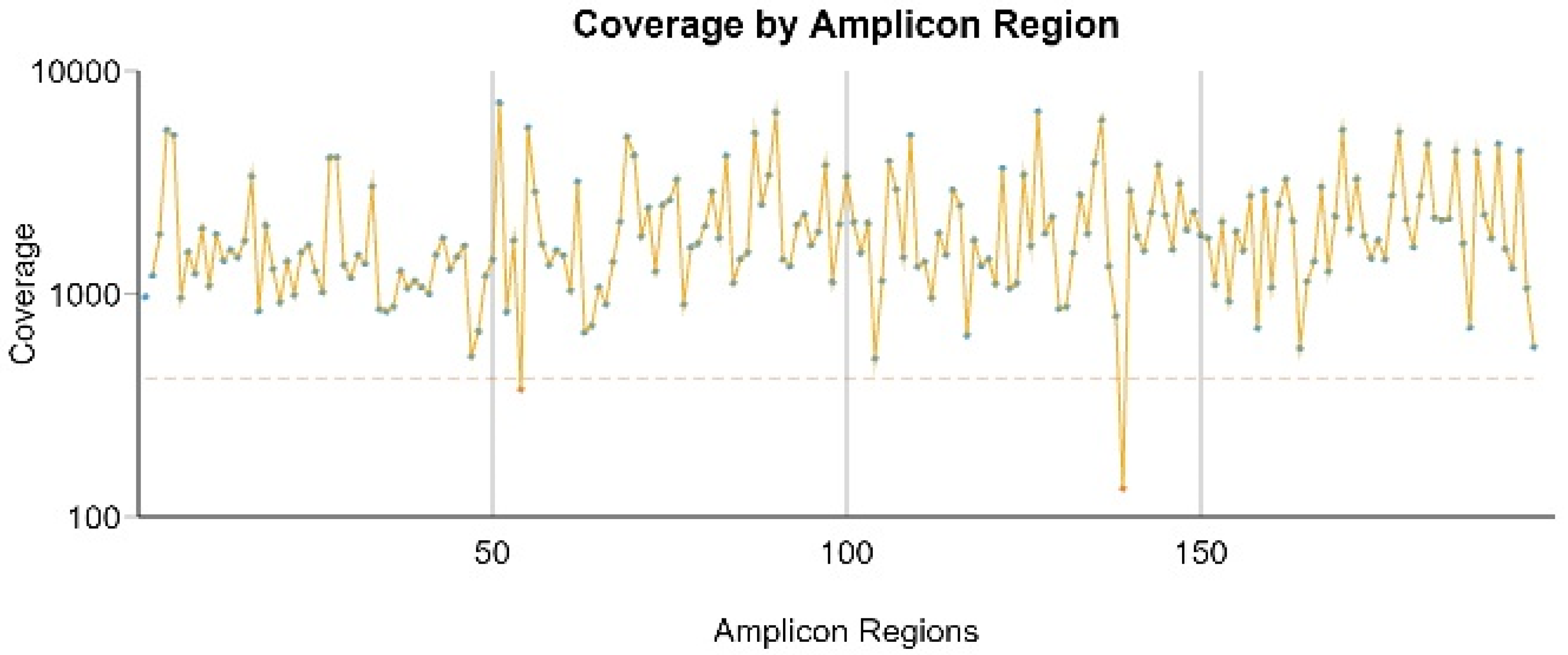
The amplicon mean (deep) coverage was 5499×, and a mean of 1.2 M passing filter reads (Q score > 30) were obtained from the samples. Uniformity of coverage was higher than 98% in all samples (Figure 2). Reproducibility of single nucleotide variants (SNVs) and insertions/deletions (INDELs) found in the three different matrices (blood, fresh and FFPE tissues) of 7 animals was always 100%. Moreover, no statistically significant differences were observed for both total reads number and amplicon mean coverage in the three matrices (p > 0.05).
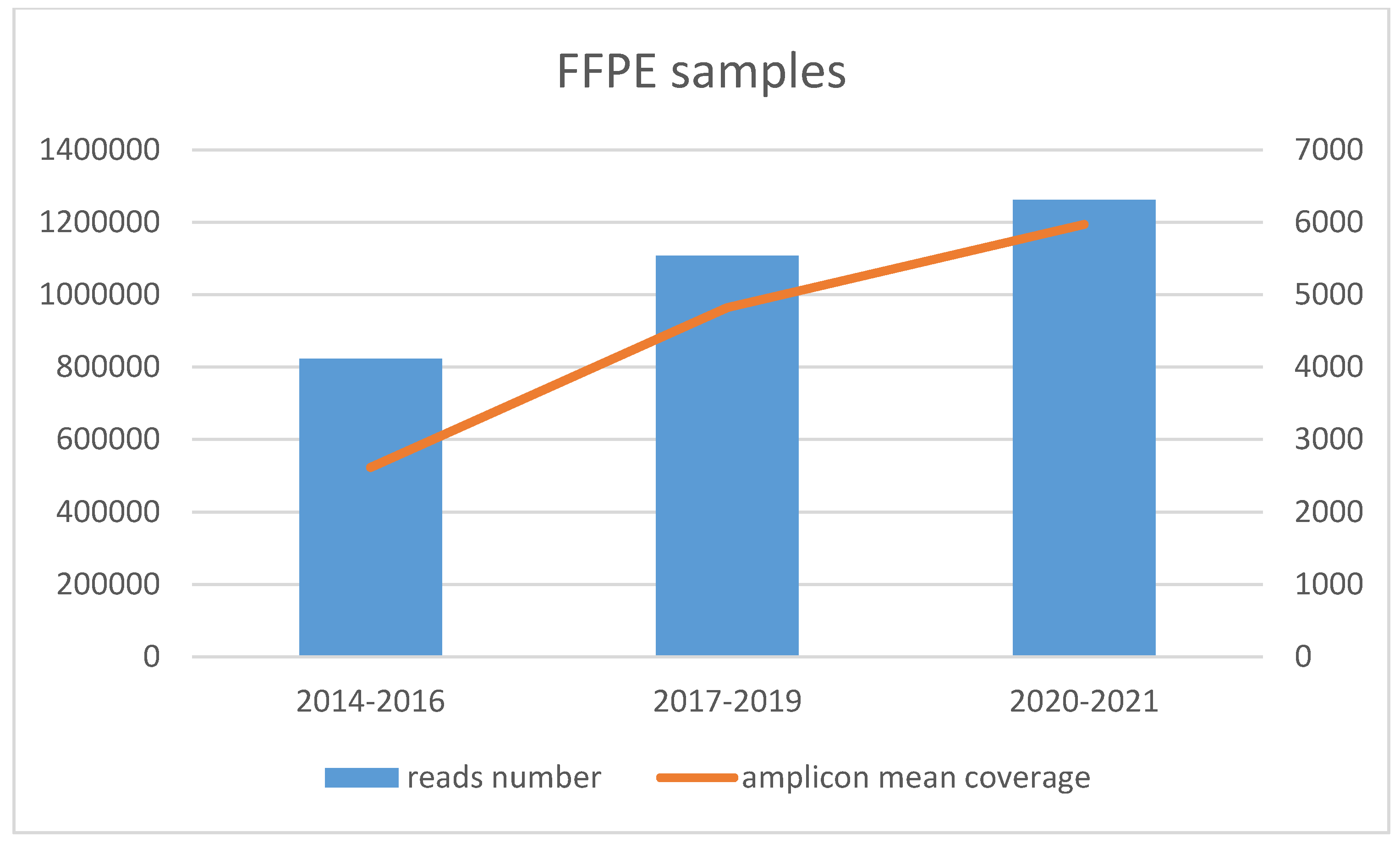
Conversely, FFPE samples stored for different numbers of years showed progressively better results from the oldest to the recent ones in terms of total reads number and amplicon mean coverage (Figure 3). These results are statistically significant with p-values 0.00819 and 0.00174 for total reads and coverage, respectively.
A total of 42 different genetic variants were identified in 22 dogs. In particular, 12 exonic and 4 intronic variants were found in BRCA1, while 23 exonic and 3 intronic variants were identified in BRCA2 (Table 3). According to human classification, variations in BRCA1 were catalogued as likely benign (n. 2), and not referenced (n. 14), while mutations in BRCA2 were classified as likely benign (n. 3), variants of unknown significance (n. 5), not referenced (n. 15), and not found in humans (n. 3).
Regarding the exonic mutations, we observed 10 coding single nucleotide polymorphisms (SNPs) and 2 synonymous SNPs in BRCA1, whereas in BRCA2, 17 variants were coding SNPs, 4 synonymous SNPs, 1 deletion and 1 insertion both of 3 bp (Table 4).

4. Discussion

Amplicon sequencing (AmpliSeq) is a targeted approach which enables researchers to analyze genetic variation in specific genomic regions. The ultra-deep sequencing of PCR products allows efficient variant identification and characterization in a wide range of fields [17,18,19]. AmpliSeq is particularly useful for the discovery of rare somatic mutations in complex samples such as tumors mixed with germline DNA. Moreover, the information generated by targeted gene panels can be helpful for diagnostic classification, therapeutic decisions, and/or prognostic insights [20]. In this study, we described the development and validation of an AmpliSeq approach for the identification BRCA1 and BRCA2 variants in dogs (Canis familiaris). The Amplicon panel has been designed to cover the whole BRCA1/BRCA2 exome and the proximal intronic regions (~30–50 bp), in both directions. The short-length PCR panel has been properly designed for fragmented DNA (e.g., FFPE samples). Indeed, despite the high degradation of DNA observed in FFPE samples, particularly true for DNA obtained from older FFPE blocks (>5 years), the assay was able to produce good quality results for all the samples. In the first sequencing attempt, we pooled together all the samples without previous individual quantification and normalization. The pool was then quantified and diluted as recommended but the resulting coverage was unbalanced between samples and sometimes unsuitable for downstream analysis. The poorer the DNA quality the lower the total reads number and amplicon mean coverage occurred. The same libraries were thus quantified and normalized at the equal molar concentration before pooling and sequencing. Following this procedure, we obtained more standardized reads number and amplicon mean coverage among the samples. Despite the variation observed in FFPE samples, depending on the time of storage, all the samples were successfully analyzed. Individual normalization is thus a critical point to obtain reliable quality results for strong fragmented DNA samples.
Variant analysis of the seven dogs for which all three matrices were available in this study, confirmed the high reproducibility of the whole workflow, from DNA extraction to variant calling. In such samples, we could establish with certainty the germinal nature of the variants found, although this was not among our goals.
In addition to the method validation, this study also attempted the interpretation of the variants found in dogs. In humans, family information is generally integrated to evaluate the probability that sequence variants are associated with genetic predisposition [21]. In contrast, much of this information is not available for dogs. The results of dog samples were thus interpreted on the basis of software and classifications developed for humans.
In this study, the dog versus human BRCA1/BRCA2 comparison was established by using Alamut Visual Plus, while the interpretation of the dog variants found in both humans and dogs were carried out using ClinVar, a software that archives and aggregates information about relationships among variation and health in humans. Only two BRCA1 variants were referenced in humans and classified as likely benign, while eight variants of BRCA2 were also reported in humans; five were classified as of uncertain significance (UV) and three as likely benign. Among UVs, the human BRCA2 variants p.(Lys1440Arg), p.(Arg1997Lys) and p.(Trp395Cys), showed the same amino acid substitution described here in dogs, while at the position of human BRCA2 p.(His150Arg), we found p.(His143Pro).
Four out of 22 different dogs (ID: 2, 4, 14, 15) showed c.4314A>G—p.(Lys1435Arg) together with c.4283A>C—p.(Thr1425Pro), both in exon 11 of BRCA2 and previously described in the literature as implicated in carcinogenesis of dogs [13,14,22]. Both mutations were also confirmed by Sanger sequencing (data not shown). Of them, two showed an intraductal papillary carcinoma, one a solid carcinoma and one a complex carcinoma in a broad range of grade of malignancy (I–III); correlation to a specific histotype is thus excluded.
In healthy dogs (n = 2), we did not find c.4314A>G nor c.4283A>C in FFPE samples, nor in fresh tissues. These variations are located in the BRC3 repeat domain, which plays a fundamental role in the interaction with the protein RAD51, and affect well conserved positions [23,24] in available vertebrate species. Only one of these two dog BRCA2 variants, c.4314A>G—p.(Lys1435Arg), was also reported by ClinVar and classified in humans as of uncertain significance. We found this mutation alone in 18 out of 22 dogs (81%). Another dog BRCA2 variant, found in exon 11, c.4175G>A—p.(Arg2022Lys), was reported in humans and classified by ClinVar as Uncertain Significance. This variant was found on the mammary gland of a male dog, described as intraductal carcinoma, located in one of the BRCA repeats critical for binding to RAD51 [25,26]. Further analyses are needed to evaluate possible correlations.
Finally, it is worth considering the c.10204_10206insAAA variation that is positioned in the nuclear localization signal 2 (NLS2), which plays a key role in the mislocation of BRCA2 in humans [27]. Recent studies suggest that c.10204_10206insAAA may be associated with significantly higher mammary tumor morbidity rates in dogs [13,14]. We found this insertion in 18 out of 22 dogs (81%) in exon 27 of BRCA2.

5. Conclusions

Canine and human tumors share numerous traits: genetic and histopathological characteristics, biological behavior, molecular targets and response to standard antineoplastic treatments. For this reason, dogs offer an extraordinary opportunity to improve our knowledge of tumor biology in both species. The AmpliSeq NGS assay developed in this study for dog BRCA1 and BRCA2 will help to generate reproducible and robust results. The data obtained here are part of a database of variants that will be further increased by new findings from pathologic and healthy tissues of both historical and new samples. All these data will certainly lead to a better understanding of the disease and a more careful management of health care in both humans and dogs.

Supplementary Materials

The following supporting information can be downloaded at: https://www.mdpi.com/article/10.3390/life12060851/s1. Custom amplicons and manifest file.

Author Contributions

Writing—original draft: D.D.G. and M.D.D., Writing—review and editing: D.D.G., M.D.D., S.V.P.D., A.P., P.M. and C.C., Conceptualization: M.D.D., D.D.G. and A.P., Resources: D.M., S.V.P.D., G.D.T., M.M. and A.V., Funding acquisition: A.P. and N.D. All authors have read and agreed to the published version of the manuscript.

Funding

This work was supported by a grant from Italian Ministry of Health (IZS AM 06/19 RC, Ricerca Corrente 2019 “Le neoplasie mammarie: approccio multidisciplinare in oncologia comparata mediante l’utilizzo di tecnologie innovative”, recipient Antonio Petrini).

Institutional Review Board Statement

All procedures on animals were performed for therapeutic purposes and for routine testing and each owner provided written consent for specimen collection and analysis. No animal ethics committee is available to review applications. In this study we did not consider experimental animals, but only post mortem examination of spontaneous tumors.

Informed Consent Statement

Not applicable.

Data Availability Statement

Data are available upon request to the corresponding author.

Acknowledgments

The authors would like to thank Mario Tosi (Inserm U1245, University of Rouen, Rouen, France) for fruitful discussions.

Conflicts of Interest

None of the authors have conflict of interest to declare. Mention of trade names or commercial products in this article is solely for the purpose of providing specific information and does not imply recommendation or endorsement by the IZSAM.

References

  1. Scully, R.; Livingston, D.M. In search of the tumor-suppressor functions of BRCA1 and BRCA2. Nature 2000, 408, 429–432. [Google Scholar] [CrossRef] [PubMed]
  2. Szabo, C.I.; Wagner, L.A.; Francisco, L.V.; Roach, J.C.; Argonza, R.; King, M.C.; Ostrander, E.A. Human, canine and murine BRCA1 genes: Sequence comparison among species. Hum. Mol. Genet. 1996, 5, 1289–1298. [Google Scholar] [CrossRef] [PubMed]
  3. Nguyen, F.; Peña, L.; Ibisch, C.; Loussouarn, D.; Gama, A.; Rieder, N.; Belousov, A.; Campone, M.; Abadie, J. Canine invasive mammary carcinomas as models of human breast cancer. Part 1: Natural history and prognostic factors. Breast Cancer Res. Treat. 2018, 167, 635–648. [Google Scholar] [CrossRef] [PubMed] [Green Version]
  4. Shafiee, R.; Javanbakht, J.; Atyabi, N.; Kheradmand, P.; Kheradmand, D.; Bahrami, A.; Daraei, H.; Khadivar, F. Diagnosis, classification and grading of canine mammary tumours as a model to study human breast cancer: A Clinico-Cytohistopathological study with environmental factors influencing public health and medicine. Cancer Cell Int. 2013, 13, 79. [Google Scholar] [CrossRef] [Green Version]
  5. Feliubadaló, L.; Lopez-Doriga, A.; Castellsagué, E.; del Valle, J.; Menéndez, M.; Tornero, E.; Montes, E.; Cuesta, R.; Gómez, C.; Campos, O.; et al. Next-generation sequencing meets genetic diagnostics: Development of a comprehensive workflow for the analysis of BRCA1 and BRCA2 genes. Eur. J. Hum. Genet. 2013, 21, 864–870. [Google Scholar] [CrossRef]
  6. Strom, C.M.; Rivera, S.; Elzinga, C.; Angeloni, T.; Rosenthal, S.H.; Goos-Root, D.; Siaw, M.; Platt, J.; Braastadt, C.; Cheng, L.; et al. Development and Validation of a Next Generation Sequencing Assay for BRCA1 and BRCA2 Variants for the clinical Laboratory. PLoS ONE 2015, 10, e0136419. [Google Scholar] [CrossRef] [Green Version]
  7. Nicolussi, A.; Belardinilli, F.; Mahdavian, Y.; Colicchia, V.; D’Inzeo, S.; Petroni, M.; Zani, M.; Ferraro, S.; Valentini, V.; Ottini, L.; et al. Next-generation sequencing of BRCA1 and BRCA2 genes for rapid detection of germline mutations in hereditary breast/ovarian cancer. PeerJ 2019, 7, e6661. [Google Scholar] [CrossRef] [Green Version]
  8. Mercatante Carrick, D.; Mehaffey, M.G.; Sachs, M.C.; Altekruse, S.; Camalier, C.; Chuaqui, R.; Cozen, W.; Das, B.; Hernandez, B.Y.; Lih, C.J.; et al. Robustness of Next Generation Sequencing on Older Formalin-Fixed Paraffin-Embedded Tissue. PLoS ONE 2015, 10, e0127353. [Google Scholar]
  9. Einaga, N.; Yoshida, A.; Noda, H.; Suemitsu, M.; Nakayama, Y.; Sakurada, A.; Kawaji, Y.; Yamaguchi, H.; Sasaki, Y.; Tokino, T.; et al. Assessment of the quality of DNA from various formalin-fixed paraffin-embedded (FFPE) tissues and the use of this DNA for next-generation sequencing (NGS) with no artifactual mutation. PLoS ONE 2017, 12, e0176280. [Google Scholar] [CrossRef]
  10. He, Y.; Taylor, L.T.; Dimitrov, K.D.; Butt, S.L.; Stanton, J.B.; Goraichuk, I.V.; Fenton, H.; Poulson, R.; Zhang, J.; Brown, C.C.; et al. Whole-genome sequencing of genotype VI Newcastle disease viruses from formalin-fixed paraffin-embedded tissues from wild pigeons reveals continuous evolution and previously unrecognized genetic diversity in the US. Virol. J. 2018, 15, 9. [Google Scholar] [CrossRef] [Green Version]
  11. Govoni, V.M.; Da Silva, T.C.; Guerra, J.M.; Pereira, I.V.A.; Queiroga, F.L.; Cogliati, B. Genetic variants of BRCA1 and BRCA2 genes in cats with mammary gland carcinoma. Vet. Comp. Oncol. 2021, 19, 404–408. [Google Scholar] [PubMed]
  12. Engingler, S.O.; Akış, I.; Toydemir, T.S.F.; Oztabak, K.; Haktanir, D.; Gündüz, M.C.; Kırşan, I.; Fırat, I. Genetic variations of BRCA1 and BRCA2 genes in dogs with mammary tumours. Vet. Res. Commun. 2014, 38, 21–27. [Google Scholar]
  13. Ozmen, O.; Kul, S.; Risvanli, A.; Ozalp, G.; Sabuncu, A.; Kul, O. Somatic SNPs of the BRCA2 gene at the fragments encoding RAD51 binding sites of canine mammary tumors. Vet. Comp. Oncol. 2017, 15, 1479–1486. [Google Scholar] [CrossRef] [PubMed]
  14. Thumser-Henner, P.; Nytko, K.J.; Rohrer Bley, C. Mutations of BRCA2 in canine mammary tumors and their targeting potential in clinical therapy. BMC Vet. Res. 2020, 16, 30. [Google Scholar] [CrossRef] [PubMed] [Green Version]
  15. Goldschmidt, M.; Peña, L.; Rasotto, R.; Zappulli, V. Classification and grading of canine mammary tumors. Vet. Pathol. 2021, 48, 117–131. [Google Scholar] [CrossRef]
  16. Peña, L.; De Andrés, P.J.; Clemente, M.; Cuesta, P.; Pérez-Alenza, M.D. Prognostic value of histological grading in non-inflammatory canine mammary carcinomas in a prospective study with two-year follow-up: Relationship with clinical and histological characteristics. Vet. Pathol. 2013, 50, 94–105. [Google Scholar] [CrossRef]
  17. Bhoyar, R.C.; Jain, A.; Sehgal, P.; Divakar, M.K.; Sharma, D.; Imran, M. High throughput detection and genetic epidemiology of SARS-CoV-2 using COVIDSeq next-generation sequencing. PLoS ONE 2021, 16, e0247115. [Google Scholar] [CrossRef]
  18. Ogiso-Tanaka, E.; Shimizu, T.; Hajika, M.; Kaga, A.; Ishimoto, M. Highly multiplexed AmpliSeq technology identifies novel variation of flowering time-related genes in soybean (Glycine Max). DNA Res. 2019, 26, 243–260. [Google Scholar] [CrossRef] [Green Version]
  19. Tariq, H.; Gul, A.; Zubair, M.; Jaffer, S.R.; Zafar, N.; Sadaf, G. Targetedet Next-generation Sequencing for Reliable Detection of Genetic Status in Breast Cancer. J. Coll. Physicians Surg. Pak. 2021, 30, 837–840. [Google Scholar]
  20. Jennings, L.J.; Arcila, M.E.; Corless, C.; Kamel-Reid, S.; Lubin, I.M.; Pfeifer, J.; Temple-Smolkin, R.L.; Voelkerding, K.V.; Nikiforova, M.N. Guidelines for Validation of Next-Generation Sequencing-Based Oncology Panels: A Joint Consensus Recommendation of the Association for Molecular Pathology and College of American Pathologists. J. Mol. Diagn. 2017, 19, 341–365. [Google Scholar] [CrossRef] [Green Version]
  21. Lu, K.H.; Wood, M.E.; Daniels, M.; Burke, C.; Ford, J.; Kauff, N.D.; Kohlmann, W.; Lindor, N.M.; Mulvey, T.M.; Robinson, L.; et al. American Society of Clinical Oncology Expert Statement: Collection and Use of a Cancer Family History for Oncology Providers. J. Clin. Oncol. 2014, 32, 833–840. [Google Scholar] [CrossRef] [PubMed]
  22. Maués, T.; El-Jaick, K.B.; Costa, F.B.; Araujo, G.E.F.; Soares, M.V.G.; Moreira, A.S.; Ferreira, M.L.G.; Ferreira, A.M.R. Common germline haplotypes and genotypes identified in BRCA2 exon 11 of dogs with mammary tumours and histopathological analyses. Vet. Comp. Oncol. 2018, 16, 379–384. [Google Scholar] [CrossRef] [PubMed]
  23. Davies, A.A.; Masson, J.Y.; McIlwraith, M.J.; Stasiak, A.Z.; Stasiak, A.; Venkitaraman, A.R.; West, S.C. Role of BRCA2 in control of the RAD51 recombination and DNA repair protein. Mol. Cell 2001, 7, 273–282. [Google Scholar] [CrossRef]
  24. Mizuta, R.; La Salle, J.M.; Cheng, H.L.; Shinohara, A.; Ogawa, H.; Copeland, N.; Jenkins, N.A.; Lalande, M.; Alt, F.W. RAB22 and RAB163/mouse BRCA2: Proteins that specifically interact with the RAD51 protein. Proc. Natl. Acad. Sci. USA 1997, 94, 6927–6932. [Google Scholar] [CrossRef] [PubMed] [Green Version]
  25. Bork, P.; Blomberg, N.; Nilges, M. Internal repeats in the BRCA2 protein sequence. Nat. Genet. 1996, 13, 22–23. [Google Scholar] [CrossRef]
  26. Wong, A.K.; Pero, R.; Ormonde, P.A.; Tavitigian, S.V.; Bartel, P.L. RAD51 interacts with the evolutionarily conserved BRC motifs in the human breast cancer susceptibility gene BRCA2. J. Biol. Chem. 1997, 272, 31941–33194. [Google Scholar] [CrossRef] [Green Version]
  27. Yoshikawa, Y.; Morimatsu, M.; Ochiai, K.; Nagano, M.; Yamane, Y.; Tomizawa, N.; Sasaki, N.; Hashizume, K.J. Analysis of genetic variations in the exon 27 region of the canine BRCA2 locus. Vet. Med. Sci. 2005, 67, 1013–1017. [Google Scholar] [CrossRef] [Green Version]
Figure 1. Three samples per group were randomly used. A1: Ladder, B1-C1-D1: FFPE old samples (2014 – 2016), E1-F1-G1: FFPE medium samples (2017–2019), H1-A2-B2: FFPE recent samples (2020 – 2021), C2-D2-E2: Blood samples, F2-G2-H2: Fresh tissue samples.
Life 12 00851 g001
Figure 2. Details of the amplicon coverage. The 197 regions are uniformly covered (green dot), except for 2 amplicons (red dot). The threshold (dotted red line) marks coverage values above 20% of the mean value (Pct > 0.2 mean).
Life 12 00851 g002
Figure 3. FFPE blocks grouped as old (2014–2016), medium (2017–2019), and recent (2020–2021). The output significantly improved from the ancient to the new samples, in terms of total reads number and amplicon mean coverage.
Life 12 00851 g003
Table 1. Anamnestic and histological data of the dogs involved in the study.
Dog IDYear of CollectionSex and Hormonal StatusBreedAge (Years)TissueHistologic Type Grade of Malignancy
1 §2019FiYorkshire terrier8MammaryTubular carcinomaI
2 §2019FiMaremma sheepdog11MammaryIntraductal papillary carcinoma (cystic type)I
3 §2020FiMixed breed10MammaryTubular carcinomaIII
4 §2020FiMixed breed10MammarySolid carcinomaI
52020FiGolden retriever9MammaryIntraductal papillary carcinoma (cystic type)I
62021FiPinscher15MammaryTubulopapillary carcinomaI
7 §2021FiMixed breed12MammaryAnaplastic carcinomaII
8 §2021F-Pinscher17MammaryTubular carcinoma, with foci of necrosisII
9 §2021FiGerman shepherd12MammaryDuctal carcinomaI
102016MiMixed breed13MammaryIntraductal carcinomaI
112020MiMixed breed13MammaryTubulopapillary carcinoma, with foci of necrosisII
122018FiGerman shepherd6MammaryAnaplastic carcinoma with multifocal aspects of intraductal papillary carcinoma and foci of necrosisIII
132016FnGerman shepherd5MammaryTubular carcinomaI
142016FnMixed breed4Mammary Complex carcinoma I
152016FnMixed breed5MammaryIntraductal papillary carcinoma (cystic type), with focal areas of squamous metaplasiaIII
162015FnAmerican pit bull terrier11MammarySolid carcinoma with foci of necrosisII
172015FnPoodle4MammaryTubular carcinomaII
182015FnSiberian husky-MammarySolid carcinomaII
192015FnGolden retriever3MammaryTubulopapillary carcinomaI
202014FiGerman shepherd9MammaryIntraductal papillary carcinomaII
212021FiItalian Cane Corso4MammaryNormal tissuen/a
222021F-German shepherd4MammaryNormal tissuen/a
Fi = Intact female; Fn = Neutered female; F- = no information regarding the hormonal status; Mi = Intact male. n/a = not applicable. Histological classification for mammary [15]. Grading System for tumor mammary tissue [16] § Dogs for which all three matrices blood, FFPE blocks and fresh tissues, were used in this study.
Table 2. DNA quality control.
ParametersFFPEBloodFresh Tissue
OldMediumRecent
DIN1.5–2.22.3–4.23.4–6.28.3–9.17.7–8.2
Concentration (ng/µL)8.2–19.626.2–34.512.8–24.812.0–28.616.3–31.0
FFPE old samples: 2014–2016, FFPE medium samples: 2017–2019, FFPE recent samples: 2020–2021.
Table 3. Variants found in BRCA genes.
N° TotN° Exonic VariantsN° Intronic Variants
BRCA1 variants16124
BRCA2 variants26233
Table 4. Details on germline and somatic variants found in the dogs enrolled in the study.
GeneChromosome
(Dog)
Exon/IntronNucleotide Change §Protein ChangeHuman Variant
at Corresponding Position §
Human Clinical Classification
BRCA1chr 9INT 2c.81-26delC-c.81-26delCNot referenced
BRCA1chr 9INT 2c.81-18delC-c.81-12delCBenign/Likely benign
BRCA1chr 9INT 3c.135-43delA-c.135-43delANot referenced
BRCA1chr 9EX 5c.169G>Ap.(Gly57Arg)c.169G>A; p.(Gly57Arg)Not referenced
BRCA1chr 9EX 10c.606A>Gp.(Asp200Gly)c.602A>G; p.(Asp201Gly)Not referenced
BRCA1chr 9EX 10c.611A>Gp.(Lys202Glu)c.607G>A; p.(Glu203Lys)Not referenced
BRCA1chr 9EX 11c.723G>Ap.(Gly239Ser)c.720A>G; p.(Gln240=)Not referenced
BRCA1chr 9EX 11c.1315C>Tp.(Ala436Val)c.1316C>T; p.(Ala439Val)Not referenced
BRCA1chr 9EX 11c.1337A>Cp.(Arg443Ser)c.1338A>C; p.(Arg446Ser)Not referenced
BRCA1chr 9EX 11c.3628A>Gp.(Thr1207Ala)c.3616G>A; p.(Ala1206Thr)Not referenced
BRCA1chr 9EX 11c.3963G>Ap.(Ser1318=)c.3951G>A; p.(Leu1317=)Likely benign
BRCA1chr 9EX 13c.4219C>Tp.(Thr1403=)c.4197C>T; p.(Thr1399=)Not referenced
BRCA1chr 9INT 13c.4381-19C>T-c.4358-16C>T Not referenced
BRCA1chr 9EX 16c.4777C>Tp.(Ser1588Pro)c.4754C>T; p.(Pro1585Leu)Not referenced
BRCA1chr 9EX 19c.5203G>Ap.(Arg1729Lys)c.5177G>A; p.(Arg1726Lys) Not referenced
BRCA1chr 9EX 24c.5525 G>Ap.(Ala1869Thr)c.5581A>G; p.(Ser1861Gly)Not referenced
BRCA2chr 25EX 4c.308T>Cp.(Ile103Thr)c.326T>C; p.(Val109Ala)Not referenced
BRCA2chr 25INT 4c.408-9_408-8del--Not found
BRCA2chr 25EX 5c.428A>Gp.(His143Pro)c.449A>G; p.(His150Arg) Uncertain significance
BRCA2chr 25INT 5c.455-40del--Not found
BRCA2chr 25EX 10c.1122C>Tp.(Thr371Ile)c.1139G>T; p.(Ser380Ile)Not referenced
BRCA2chr 25EX 10c.1168T>Gp.(Cys386Trp)c.1185G>T; p.(Trp395Cys)Uncertain significance
BRCA2chr 25EX 11c.1937G>Ap.(Glu643Lys)c.1957G>A; p.(Glu653Lys)Not referenced
BRCA2chr 25EX 11c.2131T>Cp.(His707=)c.2148G>C; p.(Gln716His)Uncertain significance
BRCA2chr 25EX 11c.2154A>Cp.( Pro715 Gln)c.2170A>C; p.(Lys724Gln) Not referenced
BRCA2chr 25EX 11c.2164C>Ap.(Ser718=)c.2181A>C; p.(Ser727=) Likely benign
BRCA2chr 25EX 11c.2193C>Tp.(Ala728Val)c.2210C>T; p.(Ala737Val) Not referenced
BRCA2chr 25EX 11c.2232A>Gp.(Asp741Ser)c.2246G>A; p.(Ser749Asn)Not referenced
BRCA2chr 25EX 11c.2269A>Cp.(Lys801Gln)c.2431A>C; p.(Lys811Gln)Not referenced
BRCA2chr 25EX 11c.4283A>Cp.(Thr1425Pro)c.4288A>C; p.(Thr1430Pro)Not referenced
BRCA2chr 25EX 11c.4314A>Gp.(Lys1435Arg)c.4319A>G; p.(Lys1440Arg)Uncertain significance
BRCA2chr 25EX 11c.4147G>Ap.(Val2013Ile)c.5962G>A; p.(Val1988Ile)Likely benign
BRCA2chr 25EX 11c.4175G>Ap.(Arg2022Lys)c.5990G>A; p.(Arg1997Lys)Uncertain significance
BRCA2chr 25EX 11c.4198G>Ap.(Asp2030Asn)c.6013G>A; p.(Asp2005Asn) Not referenced
BRCA2chr 25EX 11c.4204G>Ap.(Ala2032Thr)c.6019A>G; p.(Thr2007Ala)Not referenced
BRCA2chr 25EX 12c.6952_6954delp.(Leu2306 o 2307del)c.6868_6870del; p.(Leu2290del)Not referenced
BRCA2chr 25EX 12c.6966C>Tp.(Phe2310=)c.6879T>C; p.(Phe2293=)Not referenced
BRCA2chr 25EX 16G>Ap.(Asp2611Asn)c.7774G>A; p.(Asp2592Asn)Not referenced
BRCA2chr 25EX 18T>Cp.(Ile2683=)c.7992T>C; p.(Ile2664=)Likely benign
BRCA2chr 25INT 25c.9692+57A>T--Not found
BRCA2chr 25EX 27c.10204_1026insAAAp.(Met3332_Ile3333insLys)c.9936_9938dup; p.(Lys3315dup)Not referenced
BRCA2chr 25EX 27c.10450A>Cp.(Thr3405Pro)c.101111A>C; p.(Thr3371Pro) Not referenced
§ prefix “c.” for cDNA sequence; the nucleotide number; a wild-type nucleotide; the symbol “>” (indicating a change), the prefix “del.” (indicating deletion) or “ins.” (indicating insertion); and the mutant nucleotide http://www.hgvs.org/mutnomen/recs-DNA.html (accessed on 27 May 2022). The DNA sequence numbering is based on the cDNA sequence of BRCA1/2 (NCBI RefSeq NM_001013416.1 and NM_001006653.4, respectively) following the recommendations of the Human Genome Variation Society (first position of the translation initiation codon ATG c.1). prefix “p.” for protein sequence; reference amino acid (three-letter code); position; amino acid change (three-letter code) or “fs” prefix (indicating frameshift) http://www.hgvs.org/mutnomen/recs-prot.html (accessed on 27 May 2022).
Publisher’s Note: MDPI stays neutral with regard to jurisdictional claims in published maps and institutional affiliations.