
ANCA-Associated Vasculitis May Result as a Complication to Both SARS-CoV-2 Infection and Vaccination

 , ,
, ,
Abstract
:1. Introduction
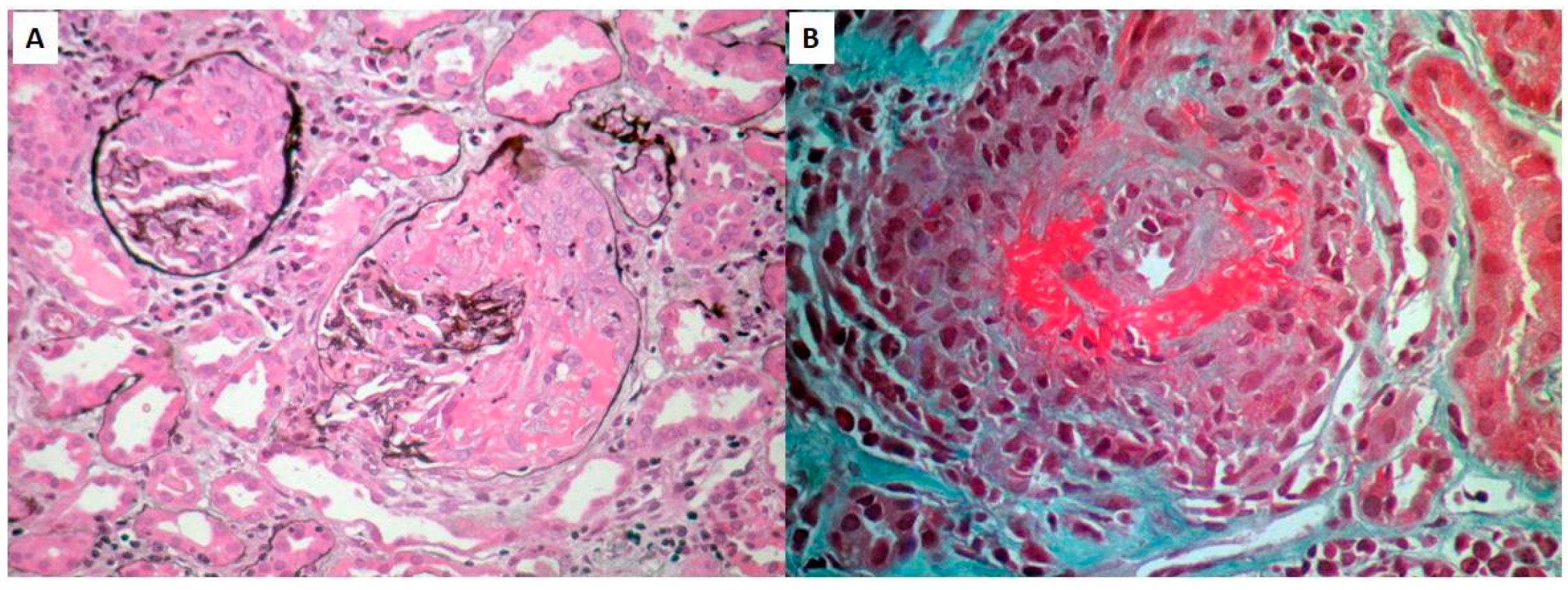
Case Presentation
2. Discussion
Author Contributions
Funding
Informed Consent Statement
Data Availability Statement
Conflicts of Interest
References
- Li, Y.D.; Chi, W.Y.; Su, J.H.; Ferrall, L.; Hung, C.F.; Wu, T.C. Coronavirus vaccine development: From SARS and MERS to COVID-19. J. Biomed. Sci. 2020, 27, 104. [Google Scholar] [CrossRef] [PubMed]
- Yadav, T.; Srivastava, N.; Mishra, G.; Dhama, K.; Kumar, S.; Puri, B.; Saxena, S.K. Recombinant vaccines for COVID-19. Hum. Vaccines Immunother. 2020, 16, 2905–2912. [Google Scholar] [CrossRef] [PubMed]
- Park, J.W.; Lagniton, P.N.P.; Liu, Y.; Xu, R.H. mRNA vaccines for COVID-19: What, why and how. Int. J. Biol. Sci. 2021, 17, 1446–1460. [Google Scholar] [CrossRef] [PubMed]
- Paces, J.; Strizova, Z.; Smrz, D.; Cerny, J. COVID-19 and the immune system. Physiol. Res. 2020, 69, 379–388. [Google Scholar] [CrossRef] [PubMed]
- Villa, M.; Díaz-Crespo, F.; Pérez de José, A.; Verdalles, Ú.; Verde, E.; Almeida Ruiz, F.; Acosta, A.; Mijaylova, A.; Goicoechea, M. A case of ANCA-associated vasculitis after AZD1222 (Oxford-AstraZeneca) SARS-CoV-2 vaccination: Casualty or causality? Kidney Int. 2021, 100, 937–938. [Google Scholar] [CrossRef] [PubMed]
- Caso, F.; Costa, L.; Ruscitti, P.; Navarini, L.; Del Puente, A.; Giacomelli, R.; Scarpa, R. Could Sars-coronavirus-2 trigger autoimmune and/or autoinflammatory mechanisms in genetically predisposed subjects? Autoimmun. Rev. 2020, 19, 102524. [Google Scholar] [CrossRef]
- Izci Duran, T.; Turkmen, E.; Dilek, M.; Sayarlioglu, H.; Arik, N. ANCA-associated vasculitis after COVID-19. Rheumatol. Int. 2021, 41, 1523–1529. [Google Scholar] [CrossRef]
- Gapud, E.J.; Seo, P.; Antiochos, B. ANCA-Associated Vasculitis Pathogenesis: A Commentary. Curr. Rheumatol. Rep. 2017, 19, 15. [Google Scholar] [CrossRef]
- Watad, A.; De Marco, G.; Mahajna, H.; Druyan, A.; Eltity, M.; Hijazi, N.; Haddad, A.; Elias, M.; Zisman, D.; Naffaa, M.E.; et al. Immunemediated disease flares or new-onset disease in 27 subjects following mRNA/DNA SARS-CoV-2 vaccination. Vaccines 2021, 9, 435. [Google Scholar] [CrossRef]
- Kitching, A.R.; Anders, H.J.; Basu, N.; Brouwer, E.; Gordon, J.; Jayne, D.R.; Kullman, J.; Lyons, P.A.; Merkel, P.A.; Savage, C.O.S.; et al. ANCA-associated vasculitis. Nat. Rev. Dis. Primers Nat. Res. 2020, 6, 71. [Google Scholar] [CrossRef]
- Stangou, M.; Papagianni, A.; Bantis, C.; Liakou, H.; Pliakos, K.; Giamalis, P.; Gionanlis, L.; Pantzaki, A.; Efstratiadis, G.; Memmos, D. Detection of multiple cytokines in the urine of patients with focal necrotising glomerulonephritis may predict short and long term outcome of renal function. Cytokine 2012, 57, 120–126. [Google Scholar] [CrossRef] [PubMed]
- Bantis, K.; Stangou, M.; Kalpakidis, S.; Hatziadamou, M.; Daikidou, D.V.; Lioulios, G.; Mitsoglou, Z.; Chatzidrosou, H.; Nikolaidou, C.; Fylaktou, A.; et al. Systemic complement activation in anti-neutrophil cytoplasmic antibody-associated vasculitis and necrotizing glomerulonephritis. Nephrology 2021, 26, 30–37. [Google Scholar] [CrossRef] [PubMed]
- Toor, S.M.; Saleh, R.; Sasidharan Nair, V.; Taha, R.Z.; Elkord, E. T-cell responses and therapies against SARS-CoV-2 infection. Immunology 2021, 162, 30–43. [Google Scholar] [CrossRef] [PubMed]
- Shimagami, H.; Yamaguchi, Y.; Kato, Y.; Kumanogoh, A. Marked increase of interferon-beta after BNT162b2 mRNA vaccination: A case of polyarthritis with pleurisy. BMJ Case Rep. 2022, 15, e246533. [Google Scholar] [CrossRef]
- Chen, Y.; Xu, Z.; Wang, P.; Li, X.M.; Shuai, Z.W.; Ye, D.Q.; Pan, H.F. New-onset autoimmune phenomena post-COVID-19 vaccination. Immunology 2022, 165, 386–401. [Google Scholar] [CrossRef]
- Vojdani, A.; Kharrazian, D. Potential antigenic cross-reactivity between SARS-CoV-2 and human tissue with a possible link to an increase in autoimmune diseases. Clin. Immun. 2020, 217, 108480. [Google Scholar] [CrossRef]
- Ishay, Y.; Kenig, A.; Tsemach-Toren, T.; Amer, R.; Rubin, L.; Hershkovitz, Y.; Kharouf, F. Autoimmune phenomena following SARS-CoV-2 vaccination. Int. Immunopharmacol. 2021, 99, 107970. [Google Scholar] [CrossRef]
- Stephenson, K.E.; Le Gars, M.; Sadoff, J.; de Groot, A.M.; Heerwegh, D.; Truyers, C.; Atyeo, C.; Loos, C.; Chandrashekar, A.; McMahan, K.; et al. Immunogenicity of the Ad26.COV2.S Vaccine for COVID-19. JAMA 2021, 325, 1535–1544. [Google Scholar] [CrossRef]
- Revannasiddaiah, S.; Kumar Devadas, S.; Palassery, R.; Kumar Pant, N.; Maka, V.V. A potential role for cyclophosphamide in the mitigation of acute respiratory distress syndrome among patients with SARS-CoV-2. Med. Hypotheses 2020, 144, 109850. [Google Scholar] [CrossRef]
- Azkur, A.K.; Akdis, M.; Azkur, D.; Sokolowska, M.; van de Veen, W.; Brüggen, M.-C.; O’Mahony, L.; Gao, Y.; Nadeau, K.; Akdis, C.A. Immune response to SARS-CoV-2 and mechanisms of immunopathological changes in COVID-19. Allergy 2020, 75, 1564–1581. [Google Scholar] [CrossRef]
- Mahroum, N.; Zoubi, M.; Lavine, N.; Ohayon, A.; Amital, H.; Shoenfeld, Y. The mosaic of autoimmunity—A taste for more. The 12th international congress of autoimmunity 2021 (AUTO12) virtual. Autoimmun. Rev. 2021, 20, 102945. [Google Scholar] [CrossRef] [PubMed]
- Weiskopf, D.; Schmitz, K.; Raadsen, M.; Grifoni, A.; Okba, N.M.A.; Endeman, H.; van den Akker, J.P.C.; Molenkamp, R.; Koopmans, M.P.G.; van Gorp, E.C.M.; et al. Phenotype and kinetics of SARS-CoV-2-specific T cells in COVID-19 patients with acute respiratory distress syndrome. Sci. Immunol. 2020, 5, eabd2071. [Google Scholar] [CrossRef] [PubMed]

| Prior to 2nd Dose | Initial Diagnosis | Follow-Up | Relapse | Normal Range | |
|---|---|---|---|---|---|
| Date | 5 July 2021 | 10 August 2021 | - | 17 February 2022 | |
| Clinical manifestations | - | Hemoptysis, dyspnea, fever | - | fever | |
| Serum creatinine (mg/dL) | 1.01 | 3.45 | 1.2 | 1.7 | 0.5–1.2 |
| BUN (mg/dL) | 36 | 170 | 65 | 83 | 10–40 |
| MPO ANCA (U/mL) | - | 20.3 | 7.1 | 27.6 | 0–5.5 |
| Urine sediment | |||||
| RBC (/hpf) | 1–2 | >100 | 2–4 | 26–35 | 1–2 |
| WBC (/hpf) | 1–2 | 16–25 | 2–4 | 26–35 | 1–2 |
| Proteinuria (mg/24 h) | - | 2052 | 576 | 1106 | <150 |
| Pulmonary hemorrhage | - | YES | - | NO | |
| Renal-replacement therapy | - | NO | - | NO | |
| AAV treatment | - | IV steroids+ IV CYC | po steroids+ IV CYC | IV steroids+ IV CYC |
| After COVID-19 Infection | After COVID-19 Vaccination | ||
|---|---|---|---|
| Large-vessel vasculitis | Takayasu’s arteritis | 0 | 0 |
| Temporal arteritis | 7 | 5 | |
| Medium-vessel vasculitis | Berger’s disease | 0 | 0 |
| Kawasaki disease * | 40 * | 4 * | |
| Polyarteritis nodosa | 1 | 5 | |
| Small-vessel vasculitis | Behcet’s syndrome | 0 | 4 |
| Eosinophilic granulomatosis with polyangiitis (EGPA) | 1 ** | 3 | |
| Cutaneous vasculitis | 15 *** | 31 *** | |
| Granulomatosis with polyangiitis (GPA) | 2 | 1 | |
| Microscopic polyangiitis (MPA) | 13 | 10 | |
| IgA vasculitis | 10 | 11 | |
Publisher’s Note: MDPI stays neutral with regard to jurisdictional claims in published maps and institutional affiliations. |
© 2022 by the authors. Licensee MDPI, Basel, Switzerland. This article is an open access article distributed under the terms and conditions of the Creative Commons Attribution (CC BY) license (https://creativecommons.org/licenses/by/4.0/).
Share and Cite
Christodoulou, M.; Iatridi, F.; Chalkidis, G.; Lioulios, G.; Nikolaidou, C.; Badis, K.; Fylaktou, A.; Papagianni, A.; Stangou, M. ANCA-Associated Vasculitis May Result as a Complication to Both SARS-CoV-2 Infection and Vaccination. Life 2022, 12, 1072. https://doi.org/10.3390/life12071072
Christodoulou M, Iatridi F, Chalkidis G, Lioulios G, Nikolaidou C, Badis K, Fylaktou A, Papagianni A, Stangou M. ANCA-Associated Vasculitis May Result as a Complication to Both SARS-CoV-2 Infection and Vaccination. Life. 2022; 12(7):1072. https://doi.org/10.3390/life12071072
Chicago/Turabian StyleChristodoulou, Michalis, Fotini Iatridi, George Chalkidis, Georgios Lioulios, Christina Nikolaidou, Kostas Badis, Asimina Fylaktou, Aikaterini Papagianni, and Maria Stangou. 2022. "ANCA-Associated Vasculitis May Result as a Complication to Both SARS-CoV-2 Infection and Vaccination" Life 12, no. 7: 1072. https://doi.org/10.3390/life12071072
APA StyleChristodoulou, M., Iatridi, F., Chalkidis, G., Lioulios, G., Nikolaidou, C., Badis, K., Fylaktou, A., Papagianni, A., & Stangou, M. (2022). ANCA-Associated Vasculitis May Result as a Complication to Both SARS-CoV-2 Infection and Vaccination. Life, 12(7), 1072. https://doi.org/10.3390/life12071072