Article

Identification of Allergenic Proteins in Velvet Mesquite (Prosopis velutina) Pollen: An Immunoproteomics Approach

by
José Ángel Huerta-Ocampo
1,2,*,
Lino Gerardo Batista-Roche
2,
Martha Beatriz Morales-Amparano
2,
María del Refugio Robles-Burgueño
2,
Gabriela Ramos-Clamont Montfort
2,
Luz Vázquez-Moreno
2,
Fernando Ramírez-Jiménez
3 and
Luis M. Terán
3,*
1
Consejo Nacional de Ciencia y Tecnología, Mexico City 03940, Mexico
2
Centro de Investigación en Alimentación y Desarrollo, A.C. Hermosillo, Hermosillo 83304, Mexico
3
Instituto Nacional de Enfermedades Respiratorias “Ismael Cosío Villegas”, Mexico City 14080, Mexico
*
Authors to whom correspondence should be addressed.
Life 2022, 12(9), 1421; https://doi.org/10.3390/life12091421
Submission received: 12 August 2022 / Revised: 25 August 2022 / Accepted: 26 August 2022 / Published: 13 September 2022
(This article belongs to the Section Proteins and Proteomics)

Abstract

:
Velvet mesquite (Prosopis velutina) is a native legume of the southwestern United States and northwestern Mexico, contributing significantly to the desert ecosystem and playing key ecological roles. It is also an important cause of allergic respiratory disease widely distributed in the Sonoran, Chihuahuan, and Mojave Deserts. However, no allergens from velvet mesquite pollen have been identified to date. Pollen proteins were extracted and analyzed by one- and two-dimensional electrophoresis and immunoblotting using a pool of 11 sera from mesquite-sensitive patients as the primary antibody. IgE-recognized protein spots were identified by mass spectrometry and bioinformatics analysis. Twenty-four unique proteins, including proteins well known as pollen, food, airway, or contact allergens and four proteins not previously reported as pollen allergens, were identified. This is the first report on allergenic proteins in velvet mesquite pollen. These findings will contribute to the development of specific diagnosis and treatment of mesquite pollen allergy.

1. Introduction

Prosopis spp. are members of a genus with more than 40 species that belong to the Fabaceae family and are widely distributed in the desert and semi-desert regions of Africa, Asia, and America. Some species within the genus are invasive and potentially harmful in the ecosystems where they are introduced [1]. Whereas P. juliflora is found in almost all of Mexico, mainly in arid places, from Baja California and Chihuahua to Oaxaca, and from Tamaulipas to Veracruz, P. velutina (velvet mesquite) is a legume native to the Sonoran, Chihuahuan, and Mojave Deserts (northwestern Mexico and the southwestern United States) where it plays key ecological roles, contributing significantly to the desert ecosystem to which this plant species is restricted [2,3]. Tree pollens are the most abundant in Mexico, outnumbering weed and grass pollens [4]. Positivity to mesquite pollen in allergic patients is high (21%) [5]. In addition, mesquite pollen represents an important source of pollinosis in cities of the Sonoran Desert such as Hermosillo [6,7].
Some studies reported the immunodetection of IgE-binding proteins with molecular weights between 10 and 99 kDa in P. juliflora pollen [1]. Additionally, only two mesquite allergens (Pro j 1, Pro j 2) have been identified, characterized, and officially included in the allergen.org database by the IUIS committee [2,5,8]. However, no velvet mesquite pollen allergens have been identified so far. Interestingly, the Mexican Immunotherapy Guide is considering including Prosopis pollen in the set of skin tests [9]. The scarce number of identified and characterized mesquite pollen proteins constitutes a limitation for the development of diagnostic and immunotherapy strategies [10]. Thus, the aim of this work was to immunodetect, for the first time, the allergenic proteins of velvet mesquite pollen using two-dimensional Western blot analysis and to identify them by tandem mass spectrometry.

2. Materials and Methods

2.1. Selection of Patients and Healthy Volunteers

Eleven polysensitized patients reactive to mesquite pollen and 4 non-allergic subjects were recruited by the allergy clinic of the National Institute of Respiratory Diseases (INER), Mexico. Skin prick test was performed on the forearm by applying a drop of a commercial allergenic extract (Hollister Stier, Elkhart, IN, USA) with the most common aeroallergens (including mesquite). Subjects with wheal diameter ≥ 3 mm after 20 min of exposure to the extract were considered positive for the test. Histamine (10 mg/mL) was used as positive control and saline solution as negative control. Serum and anticoagulated whole blood collection (for positive and negative patients) was approved by the Ethics and Research Committees of INER, and the study was conducted under the ethical principles of the 1975 Declaration of Helsinki (as revised in 1983), and it was consistent with Good Clinical Practice Guidelines. None of the patients at the time of sampling was receiving immunotherapy, corticosteroids, or antihistamine treatment. Peripheral blood samples were obtained by venipuncture. Anticoagulated whole blood samples were used to estimate the number of leukocytes by using a cell counter (Sysmex XP-300, Sysmex Corporation, Kobe, Japan), and the percentage of eosinophils was estimated by direct observation through Wright’s stain blood preparations. Serum samples were stored at −80 °C until use.

2.2. Pollen Collection and Protein Extraction

Inflorescences were collected from velvet mesquite trees (May 2020) in the city of Hermosillo, Sonora, Mexico. Trees were identified as Prosopis velutina, according to the dichotomous key reported by Palacios [3]. Anthers were removed from the dry inflorescences using a 2 mm mesh. Pollen grains were separated from the anthers with a 63 μM mesh (VWR International, Radnor, Pennsylvania, USA). Pollen was weighed, and 100 mg of pollen was transferred to 2 mL polypropylene tubes. The tubes were immersed in liquid nitrogen, and the frozen pollen was stored at −80 °C until use. Soluble proteins were extracted according to the method reported previously for Ligustrum lucidum pollen [11] with slight modifications. Briefly, 100 mg of pollen was mixed with 3 mL of extraction solution (sucrose, 30%, sodium dodecyl sulphate (SDS), 2%, Tris-HCl, 0.1 M, β-mercaptoethanol, 2%, phenylmethylsulfonyl fluoride, 1 mM), sonicated three times for 1 min at an amplitude of 30% (GE-505 Ultrasonic Processor, Sonics & Materials, Inc., Newtown, CO, USA), then shaken on ice for 10 min at 120 rpm. Phenol solution (equilibrated with 10 mM Tris-HCl, 1 mM EDTA, pH 8.0) was added to the above, mixed, and centrifuged at 5000 rpm/15 min/4 °C. Phenolic phase was recovered and mixed with 100 mM ammonium acetate and incubated overnight at −20 °C. Samples were then centrifuged at 13,000 rpm/15 min/4 °C, the pellet was washed once with cold absolute acetone, and twice with 80% acetone. Pellet was vacuum dried at room temperature and suspended in 600 μL of rehydration solution (8 M urea, 2% 3-((3-cholamidopropyl)dimethylammonio)-1-propanesulfonate, 20 mM dithiothreitol). The resulting protein solution was desalted using a PD-10 column (Cytiva, Uppsala, Sweden), precipitated again with ammonium acetate, centrifuged, and suspended in 600 μL of rehydration solution. Pollen protein concentration was estimated with the RC DC Protein Assay (Bio-Rad, Hercules, CA, USA).

2.3. SDS-PAGE and Two-Dimensional Gel Electrophoresis (2-DE)

Pollen soluble proteins (20 µg) were separated by SDS-PAGE and stained with Coomassie blue. For 2-DE, pollen proteins (1.3 mg) were loaded onto 13 cm IPG strips, pH 4–7 (Cytiva, Uppsala, Sweden). Passive rehydration, isoelectric focusing, and protein separation were conducted as reported previously [12]. Preparative gels were stained with Coomassie blue, digitized (Typhoon FLA-9500, GE-Healthcare Bio-Sciences, Uppsala, Sweden), and images were analyzed with PDQuest v8.0.1 (Bio-Rad). Proteins from analytical gels were transferred to a polyvinylidene difluoride (PVDF) membrane (Immun-Blot, Bio-Rad) using a semi-dry transfer system (Hoefer TE77XP, Thermo-Fisher Scientific, Waltham, MA, USA). Membranes were washed with PBS pH 7.5 and blocked with sodium-azide-free casein (0.5%) at 50 rpm and 4 °C overnight.

2.4. Immunodetection Analysis

Western blot tests were performed to immunodetect velvet mesquite pollen proteins using serum IgE from allergic patients. PVDF membranes were incubated overnight at 4 °C with individual sera from patients and controls diluted 1:10 in PBS with 0.5% sodium-azide-free casein for one-dimensional Western blot, whereas pooled sera were diluted 1:20 in PBS with 0.5% sodium-azide-free casein for two-dimensional Western blot. After washing (5 times for 5 min with PBS-Tween 20), membranes were incubated with a monoclonal anti-human IgE antibody conjugated to peroxidase diluted to 1 mg/mL in PBS (mouse monoclonal (B3102E8) anti-human IgE Fc (HRP), ABCAM Laboratories, Cambridge, MA, USA) for 2 h at room temperature. Membranes were washed again (4 times for 5 min with PBS-Tween and 1 time with PBS for 5 min). Chemiluminescent development of the membranes was carried out with the Clarity Western ECL substrate (Bio-Rad) and imaged (Chemidoc MP, Bio-Rad). Exposition parameters were automatically optimized by Image-Lab software (Bio-Rad).

2.5. In-Gel Protein Digestion and Mass Spectrometry Analysis

Immunodetected spots were cut from preparative 2-DE gels, reduced with 10 mM dithiothreitol, and alkylated with 55 mM iodoacetamide. Proteins were digested with mass-spectrometry-grade trypsin (Pierce Biotechnology, Rockford, IL, USA) overnight at 37 °C and tryptic peptides desalted using C18 Zip-Tips (Waters, Milford, MA, USA). Chromatographic separation of tryptic peptides was performed using the 1290 Infinity LC System (Agilent Technologies, Santa Clara, CA, USA) equipped with a ZORBAX 300SB-C8 column (5 µm × 2.1 mm × 150 mm, Agilent Technologies), and analysis was performed using the Agilent 6530 Q-TOF mass spectrometer, as reported previously [13].

2.6. Protein Identification

Mass spectrometry data were searched against the Prosopis alba subset of the NCBInr protein database (57,575 sequences, October 2021) using the Spectrum Mill MS Proteomics Workbench server (Agilent Technologies, Santa Clara, CA, USA). Trypsin was used as the specific protease, and one missed cleavage was allowed. Mass error tolerance for precursor and fragment ions was set at 20 ppm and 0.1 Da, respectively. Methionine oxidation and asparagine deamidation were specified as variable modifications, while cysteine carbamidomethylation was specified as fixed modification. Two good peptides (individual peptide ion score > 9 and scored peak intensity ≥ 60) and a protein score > 24 were needed for confident protein identifications.

3. Results

3.1. Patients

Clinical features of the allergic patients (four with allergic rhinitis, two with asthma, five with asthma + allergic rhinitis) and the control patients (four non-atopic subjects) that participated in the study are shown in Table 1.

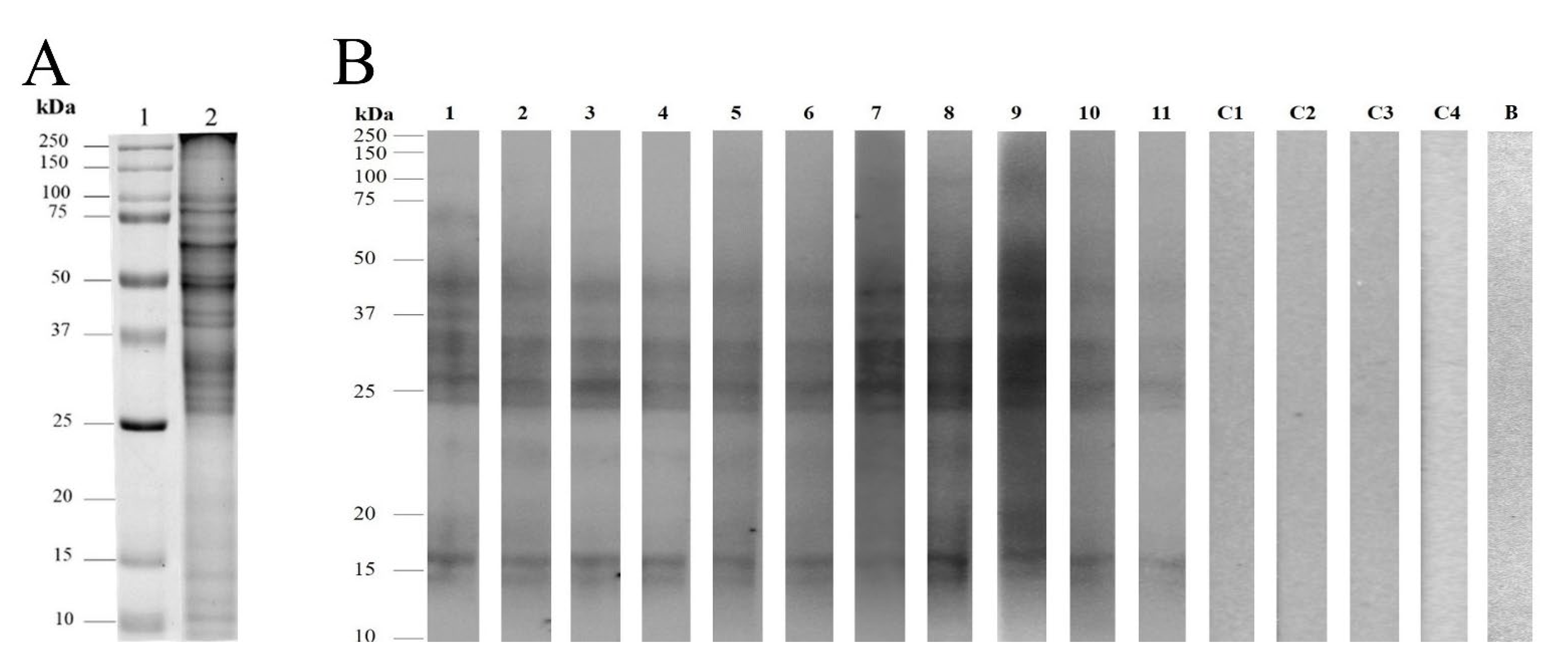
3.2. Pollen Protein Profiles and Immunodetection of Velvet Mesquite Allergens

Pollen protein extraction (100 mg starting material) yielded 87.5 mg/g of pollen. The SDS-PAGE profile showed that the most intense bands migrated between 25 and 100 kDa (Figure 1A); one-dimensional immunodetection using individual sera from allergic patients revealed nine major IgE-binding protein bands (14.5; 16; 24.5; 26; 33; 36; 41; 44; 81 kDa), since they were immunodetected in more than 80% of the patients. In contrast, sera from healthy volunteers did not exhibit any IgE-binding reactivity (Figure 1B).
The 2-DE profile was resolved into 652 spots (Figure 2A). Protein spots were in the 10–100 kDa range of molecular masses and pH range of 4–7. The 2D Western blot revealed 41 spots recognized by serum IgE antibodies from allergic patients. Immunodetected spots were found between 14.5 and 75 kDa and isoelectric points from 4.5 to 6.9 (Figure 2B). Only five spots (spots 1, 2, 40, 39, and 41) with molecular masses below 25 kDa were immunodetected.

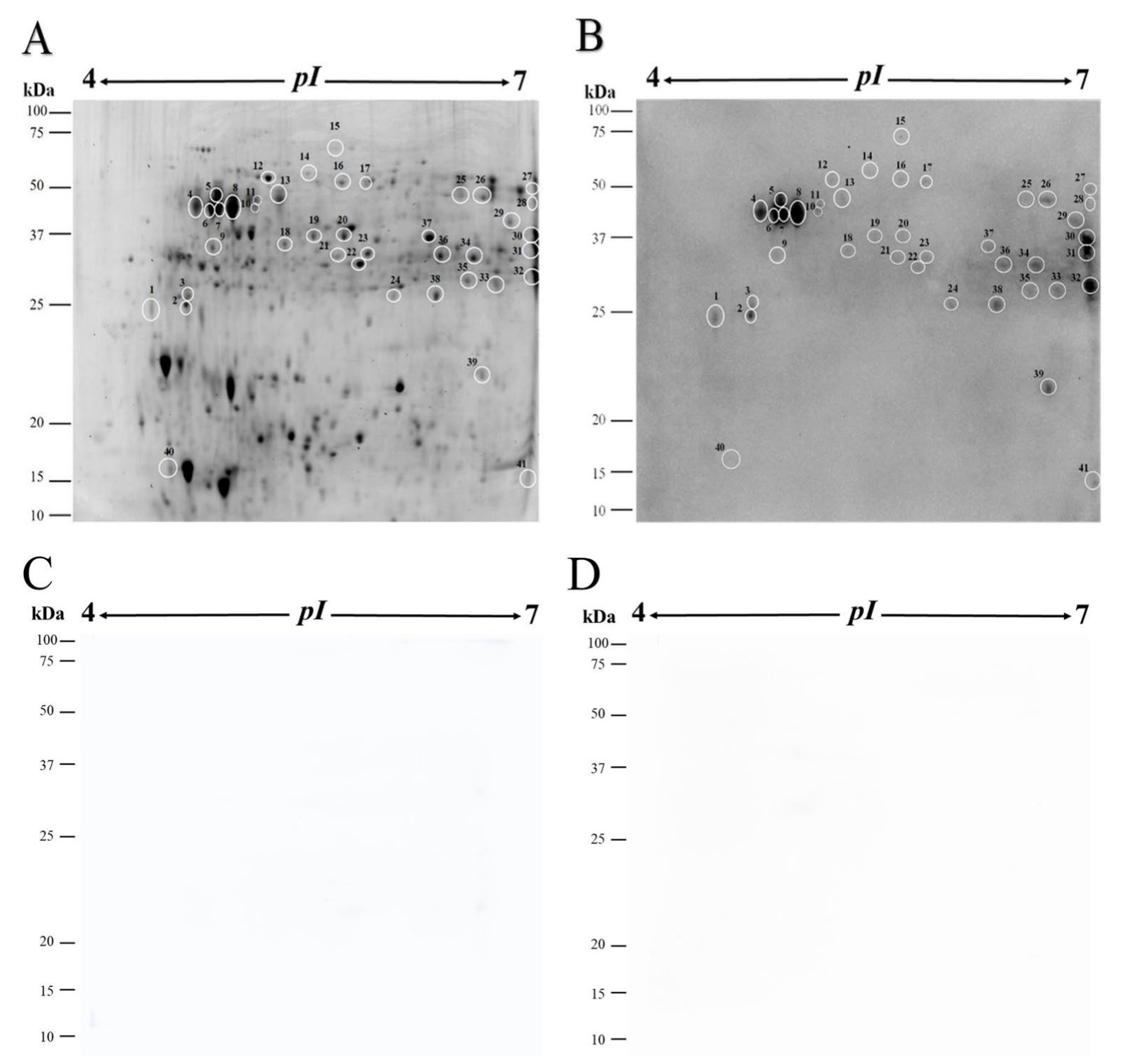
3.3. Protein Identification

A database search using Spectrum Mill MS Proteomics Workbench server Version B.06.00 (Agilent Technologies, Santa Clara, CA, USA), and mass spectrometry data allowed the identification of proteins in 36 of 41 immunodetected spots (Table 2). Whereas three spots (3, 10, and 15) were not identified, spots 8 and 11 presented excellent peptides (one-hit wonders) with scores of 17.25 and 20.89, respectively, corresponding to a polygalacturonase-like protein. Interestingly, 24 unique proteins were identified among the protein spots. Immunodetected proteoforms included triosephosphate isomerase (n = 2); polygalacturonase (n = 6); enolase (n = 3); glutelin type-D 1 (n = 2); UDP-arabinopyranose mutase 1 (n = 2); S-adenosylmethionine synthase 1 (n = 2); and glyceraldehyde-3-phosphate dehydrogenase (n = 2) (Table 2, Supplementary Materials Table S1).

4. Discussion

Mesquite pollen allergy is clinically relevant worldwide, particularly in desert and semi-desert regions. In addition, the influence of climate change and environmental pollution is expected to increase the allergenicity of this pollen [2]. To date, only two mesquite allergens have been identified and characterized. The allergens Pro j 2 [8] and Pro j 1 from P. juliflora [14] have been recombinantly expressed and characterized. In this study, we identified for the first time velvet mesquite pollen allergenic proteins using an immunoproteomics discovery approach (Table 2).
Polygalacturonases are major pollen allergens in Cupressaceae trees [15]. Additionally, a polygalacturonase was purified and identified as a major allergen (Pla a 2) in Platanus acerifolia pollen [16]. Glucan endo-1, 3-beta-glucosidase (spot 32), has been reported as allergenic in peach tree pollen [17]. Fructokinases (spot 22) have been identified as allergenic in coconut and pecan pollen [18,19]. We also identified two proteoforms of UDP-arabinopyranose mutase 1 (spots 19 and 20) in velvet mesquite pollen. This enzyme was recently reported as an allergen in Delonix regia pollen [20].
Two proteoforms of triosephosphate isomerase were immunodetected (spots 24 and 38) in this study, similar to those reported in pecan pollen and latex [18,21]. Pyruvate dehydrogenase E1 component subunit beta, mitochondrial (spot 18) was, for the first time, immunodetected in pollen protein using the serum from allergic patients. We also identified three enolase proteoforms (spots 17, 21, and 23). Enolases are highly conserved among organisms from different taxonomical groups. Enolases derived from diverse sources (fish, fungi, pollens, and latex) have been extensively investigated and associated with allergic diseases [22].
Glyceraldehyde-3-phosphate dehydrogenase (spots 9, 31, and 35) has been previously described as allergenic in pecan and sunflower pollen [18,23]. Uridine triphosphate-glucose-1-phosphate uridylyltransferase (spot 16) was also immunodetected in peach and red oak pollen [17,24]. Fructose-bisphosphate aldolase (spot 30) is allergenic in pecan and cashew tree pollen [18,25]. Mitochondrial malate dehydrogenase (spot 37) has been reported as allergenic in Senecio and olive pollen [26,27]. The major allergen Mal f 4 of yeast Malassezia furfur, which causes atopic dermatitis in allergic patients, is also a mitochondrial malate dehydrogenase [28].
Thaumatin-like protein (spot 1) belongs to the pathogenesis-related-5 family. Thaumatin-like proteins are important pollen (e.g., cypress, cedar, olea) and food (e.g., kiwi, chili, apple, banana, peach, and cherry) allergens [29,30]. Two proteins were identified in spot 41: nucleoside diphosphate kinase, which has been reported as an allergen in largemouth bass [31] and was reported for the first time as a pollen protein recognized by IgE in pecan nut [18], and pathogenesis-related protein 1, which has been previously identified as a pollen (Bermuda grass, wormwood and mugwort) and food (Muskmelon) allergen [32,33].
Proteasome subunit alpha (spot 33) was immunodetected as allergenic in corn flour and latex [34,35]. Mitochondrial-processing peptidase subunit alpha (spot 14) was identified for the first time as IgE-recognized in the present work. GDSL esterase/lipase (spot 29) was reported as allergenic in cashew tree pollen [25]. Manganese superoxide dismutase (spot 39) is known as a contact allergen in Hevea brasiliensis latex, as a food allergen (pistachio nut), and as an airway allergen from fungi (Alternaria, Aspergillus) [36,37,38,39]. It was also reported as allergenic in grass pollen [40]. The ATP synthase subunit beta (spot 12) has been reported to be allergenic in pollen from date palm, red oak, and gulmohar tree (among others) [20,24,41].
Rho GDP dissociation inhibitor 1 (spot 2) and glutelin type-D 1 (spots 34 and 36) have not been reported as pollen allergens before. However, the allergenic capacity of glutelin-type proteins was verified by immunodetection of recombinant proteins obtained from buckwheat seeds [42]. Carbonic anhydrase (spot 13) has been recognized by IgE from patients allergic to palm and pecan pollen [18,41].
S-adenosylmethionine synthase (spots 25 and 26) was identified as an IgE-binding protein in coconut pollen and gulmohar tree [19,20]. Profilin (spot 40) has previously been identified as an allergen in Prosopis juliflora and was named Pro j 2 [8]. Allergenic profilins are reported in various pollens, including olive, London planetree, amaranth, and sugar beet, among many others and are considered clinically relevant panallergens, showing extensive cross-reactivity between pollens and other foods of plant origin. Furthermore, the cross-reaction of pollen and fruit profilins can trigger oral allergy syndrome in allergic patients [43,44].

5. Conclusions

We used an immunoproteomics approach to uncover velvet mesquite pollen allergens. This represents the first report for allergenic proteins in velvet mesquite pollen. We identified 24 unique proteins, comprising proteins extensively recognized as pollen allergens and panallergens. Interestingly, four unique identified proteins have not been reported before as pollen allergens. However, more studies (Western blot/ELISA) using purified natural or recombinant proteins using patient’s serum are necessary to irrefutably confirm IgE reactivity in these four new, suspected allergens. These findings may contribute to the development of immunotherapeutic strategies for respiratory allergies, comprising the production of recombinant versions of these allergenic proteins with applications also in the clinic as diagnostic agents and to replace the use of whole pollen extracts, thus, eliminating unwanted side effects and making allergen immunotherapy safer and diagnosis more specific.

Supplementary Materials

The following supporting information can be downloaded at: https://www.mdpi.com/article/10.3390/life12091421/s1, Table S1: Allergenic proteins identified in velvet mesquite (Prosopis velutina) pollen by tandem mass spectrometry and bioinformatic analysis.

Author Contributions

Conceptualization, J.Á.H.-O. and L.M.T.; methodology, M.B.M.-A., F.R.-J., M.d.R.R.-B., G.R.-C.M., L.V.-M. and L.G.B.-R.; software, J.Á.H.-O., M.d.R.R.-B. and L.G.B.-R.; validation, J.Á.H.-O. and L.G.B.-R.; formal analysis J.Á.H.-O. and L.G.B.-R.; data curation, J.Á.H.-O.; original draft preparation, J.Á.H.-O. and L.G.B.-R.; writing—review and editing, all authors; supervision, project administration, and funding acquisition, J.Á.H.-O. and L.M.T. All authors have read and agreed to the published version of the manuscript.

Funding

This research was partially funded by CONACYT-Mexico, grant number 251744 (infrastructure) and by the Institutional Analytical Platform of CIAD, project number PAI-10363.

Institutional Review Board Statement

The study was conducted in accordance with the Declaration of Helsinki and approved by the Institutional Ethics Committee of Instituto Nacional de Enfermedades Respiratorias “Isamel Cosío Villegas” CDMX, Mexico, and it was consistent with Good Clinical Practice Guidelines.

Informed Consent Statement

Informed consent was obtained from all subjects involved in the study.

Data Availability Statement

Data availability upon request.

Acknowledgments

Batista-Roche and Morales-Amparano thank the Mexican National Council for Science and Technology (CONACYT) for the Ms.C. and Ph.D. scholarships, respectively.

Conflicts of Interest

The authors declare no conflict of interest.

References

  1. Hussain, M.I.; Shackleton, R.T.; El-Keblawy, A.; Del Mar Trigo Pérez, M.; González, L. Invasive Mesquite (Prosopis juliflora), an Allergy and Health Challenge. Plants 2020, 9, 141. [Google Scholar] [CrossRef] [PubMed]
  2. Batista-Roche, L.G.; Huerta-Ocampo, J.Á. Mezquite: Una planta alergénica de relevancia clínica en México. TIP Rev. Espec. En Cienc. Químico-Biológicas 2021, 24, 1–15. [Google Scholar] [CrossRef]
  3. Palacios, R.A. Los mezquites mexicanos: Biodiversidad y distribución geográfica. Boletín Soc. Argent. Botánica 2006, 41, 99–121. [Google Scholar]
  4. Calderon-Ezquerro, M.C.; Guerrero-Guerra, C.; Galán, C.; Serrano-Silva, N.; Guidos-Fogelbach, G.; Jiménez-Martínez, M.C.; Larenas-Linnemann, D.; López Espinosa, E.D.; Ayala-Balboa, J. Pollen in the atmosphere of Mexico City and its impact on the health of the pediatric population. Atmos. Environ. 2018, 186, 198–208. [Google Scholar] [CrossRef]
  5. Velázquez-Sámano, G.; Mendoza-Gertrudis, M.D.L.; Velasco-Medina, A.A.; Rosas-Alvarado, A. Prosopis spp. en enfermedades alérgicas. Rev. Alerg. México 2021, 68, 242–250. [Google Scholar] [CrossRef]
  6. Ortega-Rosas, C.I.; Meza-Figueroa, D.; Vidal-Solano, J.R.; González-Grijalva, B.; Schiavo, B. Association of airborne particulate matter with pollen, fungal spores, and allergic symptoms in an arid urbanized area. Environ. Geochem. Health 2021, 43, 1761–1782. [Google Scholar] [CrossRef]
  7. Romero, C.L.; Romero, J.H.; Frías-Mendívil, M. Sensibilización a alérgenos en pacientes pediátricos mayores de 2 años en el Hospital Infantil del Estado de Sonora. Boletín Clínico Hosp. Infant. Estado Sonora 2018, 34, 90–96. [Google Scholar]
  8. Ali-Sadeghi, H.; Khodadadi, A.; Amini, A.; Assarehzadegan, M.-A.; Sepahi, N.; Zarinhadideh, F. Pro j 2 is mesquite profilin: Molecular characteristics and specific IgE binding activity. Asian Pac. J. Allergy Immunol. 2015, 33, 90–98. [Google Scholar] [CrossRef]
  9. Larenas-innemann, D.; Luna-Pech, J.A.; Rodríguez-Pérez, N.; Rodríguez -González, M.; Arias-Cruz, A.; Blandón-Vijil, M.V.; Costa-Domínguez, M.D.C.; Del Río-Navarro, B.E.; Estrada-Cardona, A.; Navarrete-Rodríguez, E.M. GUIMIT 2019, Guía Mexicana de Inmunoterapia. Guía de diagnóstico de alergia mediada por IgE e inmunoterapia aplicando el método ADAPTE. Rev. Alerg. México 2019, 66, 10. [Google Scholar] [CrossRef]
  10. Larsen, J.N.; Broge, L.; Jacobi, H. Allergy immunotherapy: The future of allergy treatment. Drug Discov. Today 2016, 21, 26–37. [Google Scholar] [CrossRef]
  11. Mani, B.M.; Huerta-Ocampo, J.A.; Garcia-Sanchez, J.R.; Barrera-Pacheco, A.; de la Rosa, A.P.B.; Teran, L.M. Identification of Ligustrum lucidum pollen allergens using a proteomics approach. Biochem. Biophys. Res. Commun. 2015, 468, 788–792. [Google Scholar] [CrossRef] [PubMed]
  12. Maldonado-Cervantes, E.; Huerta-Ocampo, J.A.; Montero-Morán, G.M.; Barrera-Pacheco, A.; Espitia-Rangel, E.; Barba de la Rosa, A.P. Characterization of Amaranthus cruentus L. seed proteins by 2-DE and LC/MS–MS: Identification and cloning of a novel late embryogenesis-abundant protein. J. Cereal Sci. 2014, 60, 172–178. [Google Scholar] [CrossRef]
  13. Morales-Amparano, M.B.; Ramos-Clamont Montfort, G.; Baqueiro-Peña, I.; Robles-Burgueño, M.D.R.; Vázquez-Moreno, L.; Huerta-Ocampo, J.Á. Proteomic response of Saccharomyces boulardii to simulated gastrointestinal conditions and encapsulation. Food Sci. Biotechnol. 2019, 28, 831–840. [Google Scholar] [CrossRef] [PubMed]
  14. Fatemeh, D.; Mohammad-Ali, A.; Payam, M.; Gholam Reza, K.; Bahareh, A. Molecular cloning and expression of Pro J 1: A new allergen of Prosopis juliflora pollen. Iran. J. Allergy Asthma Immunol. 2016, 15, 122–131. [Google Scholar]
  15. Di Felice, G.; Barletta, B.; Tinghino, R.; Pini, C. Cupressaceae pollinosis: Identification, purification and cloning of relevant allergens. Int. Arch. Allergy Immunol. 2001, 125, 280–289. [Google Scholar] [CrossRef]
  16. Ibarrola, I.; Arilla, M.C.; Martínez, A.; Asturias, J.A. Identification of a polygalacturonase as a major allergen (Pla a 2) from Platanus acerifolia pollen. J. Allergy Clin. Immunol. 2004, 113, 1185–1191. [Google Scholar] [CrossRef]
  17. Blanca, M.; Victorio Puche, L.; Garrido-Arandia, M.; Martin-Pedraza, L.; Romero Sahagún, A.; López-Sánchez, J.D.; Galán, C.; Marin, A.; Villaba, M.; Díaz-Perales, A.; et al. Pru p 9, a new allergen eliciting respiratory symptoms in subjects sensitized to peach tree pollen. PLoS ONE 2020, 15, e0230010. [Google Scholar] [CrossRef]
  18. Morales-Amparano, M.B.; Valenzuela-Corral, A.; Ramos-Clamont Montfort, G.; Vázquez-Moreno, L.; Escobedo-Moratilla, A.; Pastor-Palacios, G.; Ovando-Vázquez, C.; Teran, L.M.; Huerta-Ocampo, J.Á. Immunoproteomic identification of allergenic proteins in pecan (Carya illinoinensis) pollen. J. Proteom. 2021, 248, 104348. [Google Scholar] [CrossRef]
  19. Saha, B.; Sircar, G.; Pandey, N.; Gupta Bhattacharya, S. Mining novel allergens from coconut pollen employing manual de novo sequencing and homology-driven proteomics. J. Proteome Res. 2015, 14, 4823–4833. [Google Scholar] [CrossRef]
  20. Bhowmik, M.; Ghosh, N.; Gupta Bhattacharya, S. Allergenicity assessment of Delonix regia pollen grain and identification of allergens by immunoproteomic approach. Heliyon 2021, 7, e06014. [Google Scholar] [CrossRef]
  21. Posch, A.; Chen, Z.; Dunn, M.J.; Wheeler, C.H.; Petersen, A.; Leubner-Metzger, G.; Baur, X. Latex allergen database. Electrophoresis 1997, 18, 2803–2810. [Google Scholar] [CrossRef] [PubMed]
  22. Morales-Amparano, M.B.; Huerta-Ocampo, J.Á.; Pastor-Palacios, G.; Teran, L.M. The Role of Enolases in Allergic Disease. J. Allergy Clin. Immunol. Pract. 2021, 9, 3026–3032. [Google Scholar] [CrossRef] [PubMed]
  23. Ghosh, N.; Sircar, G.; Saha, B.; Pandey, N.; Bhattacharya, S.G. Search for allergens from the pollen proteome of sunflower (Helianthus annuus L.): A major sensitizer for respiratory allergy patients. PLoS ONE 2015, 10, e0138992. [Google Scholar] [CrossRef] [Green Version]
  24. Huerta-Ocampo, J.Á.; Valenzuela-Corral, A.; Robles-Burgueño, M.D.R.; Guzmán-Partida, A.M.; Hernández-Oñate, M.Á.; Vázquez-Moreno, L.; Pavón-Romero, G.F.; Terán, L.M. Proteomic identification of allergenic proteins in red oak (Quercus rubra) pollen. World Allergy Organ. J. 2020, 13, 100111. [Google Scholar] [CrossRef] [PubMed]
  25. Danella Figo, D.; De Amicis, K.; Neiva Santos de Aquino, D.; Pomiecinski, F.; Gadermaier, G.; Briza, P.; Eduardo Santos Galvão, C.; Bussador do Amaral, J.; de Oliveira Martins, C.; Fernandes Morato Castro, F.; et al. Cashew Tree Pollen: An Unknown Source of IgE-Reactive Molecules. Int. J. Mol. Sci. 2019, 20, 2397. [Google Scholar] [CrossRef] [PubMed]
  26. Luengo, O.; Mollá, R.; Gámez, C.; Cardona, V.; Lopez, E.; Sastre, B.; Waisel, Y.; Belmonte, J.; Cadahia, A.; Lahoz, C. Allergenicity and cross-reactivity of Senecio pollen: Identification of novel allergens using the immunoproteomics approach. Clin. Exp. Allergy 2008, 38, 1048–1060. [Google Scholar] [CrossRef]
  27. San Segundo-Acosta, P.; Oeo-Santos, C.; Benedé, S.; de los Ríos, V.; Navas, A.; Ruiz-Leon, B.; Moreno, C.; Pastor-Vargas, C.; Jurado, A.; Villalba, M.; et al. Delineation of the olive pollen proteome and its allergenome unmasks cyclophilin as a relevant cross-reactive allergen. J. Proteome Res. 2019, 18, 3052–3066. [Google Scholar] [CrossRef]
  28. Onishi, Y.; Kuroda, M.; Yasueda, H.; Saito, A.; Sono-Koyama, E.; Tunasawa, S.; Hashida-Okado, T.; Yagihara, T.; Uchida, K.; Yamaguchi, H. Two-dimensional electrophoresis of Malassezia allergens for atopic dermatitis and isolation of Mal f 4 homologs with mitochondrial malate dehydrogenase. Eur. J. Biochem. 1999, 261, 148–154. [Google Scholar] [CrossRef]
  29. De Jesús-Pires, C.; Ferreira-Neto, R.C.J.; Pacifico Bezerra-Neto, J.; Kido, A.E.; de Oliveira Silva, L.R.; Pandolfi, V.; Wanderley-Nogueira, C.A.; Binneck, E.; da Costa, F.A.; Pio-Ribeiro, G.; et al. Plant Thaumatin-like Proteins: Function, Evolution and Biotechnological Applications. Curr. Protein Pept. Sci. 2020, 21, 36–51. [Google Scholar] [CrossRef]
  30. Midoro-Horiuti, T.; Goldblum, R.M.; Kurosky, A.; Wood, T.G.; Brooks, E.G. Variable expression of pathogenesis-related protein allergen in mountain cedar (Juniperus ashei) pollen. J. Immunol. 2000, 164, 2188–2192. [Google Scholar] [CrossRef]
  31. Liu, C.-Y.; Tao, S.; Xue, J.-Y.; Zhang, H.; Xue, W.-T.; Chen, F.-S. Identification and purification of a novel fish allergen from largemouth bass (Micropterus salmoides). Food Agric. Immunol. 2014, 25, 70–81. [Google Scholar] [CrossRef]
  32. Chow, L.-P.; Chiu, L.-L.; Khoo, K.-H.; Peng, H.-J.; Yang, S.-Y.; Huang, S.-W.; Su, S.-N. Purification and structural analysis of the novel glycoprotein allergen Cynd24, a pathogenesis-related protein PR-1, from Bermuda grass pollen. FEBS J. 2005, 272, 6218–6227. [Google Scholar] [CrossRef] [PubMed]
  33. Sankian, M.; Hajavi, J.; Moghadam, M.; Varasteh, A.-R. Identification and molecular characterization of the cDNA encoding Cucumis melo allergen, Cuc m 3, a plant pathogenesis-related protein. Rep. Biochem. Mol. Biol. 2014, 2, 82–87. [Google Scholar]
  34. D’Amato, A.; Bachi, A.; Fasoli, E.; Boschetti, E.; Peltre, G.; Sénéchal, H.; Sutra, J.P.; Citterio, A.; Righetti, P.G. In-depth exploration of Hevea brasiliensis latex proteome and “hidden allergens” via combinatorial peptide ligand libraries. J. Proteom. 2010, 73, 1368–1380. [Google Scholar] [CrossRef] [PubMed]
  35. Fonseca, C.; Planchon, S.; Pinheiro, C.; Renaut, J.; Ricardo, C.P.; Oliveira, M.M.; Batista, R. Maize IgE binding proteins: Each plant a different profile? Proteome Sci. 2014, 12, 17. [Google Scholar] [CrossRef]
  36. Ayuso, R.; Grishina, G.; Ahn, K.; Bardina, L.; Beyer, K.; Sampson, H. Identification of a MnSOD-like protein as a new major pistachio allergen. J. Allergy Clin. Immunol. 2007, 119, S115. [Google Scholar] [CrossRef]
  37. Boluda, L.; Alonso, C.; Fernández-Caldas, E. Purification, characterization, and partial sequencing of two new allergens of Olea europaea. J. Allergy Clin. Immunol. 1998, 101, 210–216. [Google Scholar] [CrossRef]
  38. Noorbakhsh, R.; Mortazavi, S.A.; Sankian, M.; Shahidi, F.; Assarehzadegan, M.A.; Varasteh, A. Cloning, Expression, Characterization, and Computational Approach for Cross-Reactivity Prediction of Manganese Superoxide Dismutase Allergen from Pistachio Nut. Allergol. Int. 2010, 59, 295–304. [Google Scholar] [CrossRef]
  39. Wagner, S.; Sowka, S.; Mayer, C.; Crameri, R.; Focke, M.; Kurup, V.P.; Scheiner, O.; Breiteneder, H. Identification of a Hevea brasiliensis Latex Manganese Superoxide Dismutase (Hev b 10) as a Cross-Reactive Allergen. Int. Arch. Allergy Immunol. 2001, 125, 120–127. [Google Scholar] [CrossRef] [PubMed]
  40. Conti, A.; Alessio, M.; Pesca, M.; Soldarini, A.; Breda, D.; Pigatto, P.D.; Burastero, S.E. Phleum pratense manganese superoxide dismutase identified by proteomic: A new candidate grass allergen. Ann. Allergy Asthma Immunol. 2014, 112, 261–262.e263. [Google Scholar] [CrossRef] [PubMed]
  41. Saha, B.; Bhattacharya, S.G. Charting novel allergens from date palm pollen (Phoenix sylvestris) using homology driven proteomics. J. Proteom. 2017, 165, 1–10. [Google Scholar] [CrossRef] [PubMed]
  42. Fujino, K.; Funatsuki, H.; Inada, M.; Shimono, Y.; Kikuta, Y. Expression, Cloning, and Immunological Analysis of Buckwheat (Fagopyrum esculentum Moench) Seed Storage Proteins. J. Agric. Food Chem. 2001, 49, 1825–1829. [Google Scholar] [CrossRef] [PubMed]
  43. Högerle, C.; San Nicolo, M.; Gellrich, D.; Eder, K.; Gröger, M. Clinical relevance of profilin sensitization concerning oral allergy syndrome in birch pollen sensitized patients. J. Asthma Allergy 2022, 15, 249–255. [Google Scholar] [CrossRef] [PubMed]
  44. Santos, A.; Van Ree, R. Profilins: Mimickers of allergy or relevant allergens? Int. Arch. Allergy Immunol. 2011, 155, 191–204. [Google Scholar] [CrossRef] [PubMed]
Figure 1. Immunoscreening of the allergens of velvet mesquite (Prosopis velutina) pollen grains. (A) One-dimensional electrophoresis of Prosopis velutina pollen proteins. Lane 1, molecular weight marker. Lane 2, total soluble pollen proteins (20 μg). (B) Western blot showing IgE-based immunodetection of protein bands in 11 atopic patient sera. Lanes C1–C4, negative control blots with individual serum from four healthy volunteers. Lane B, reagent blank (without primary antibody).
Life 12 01421 g001
Figure 2. 2-DE and immunoblotting using pollen proteins from velvet mesquite. (A) 2-DE profile of total soluble proteins stained with Coomassie blue. (B) Protein spots detected with pooled sera from 11 allergic patients using 2-DE immunoblotting. (C) Reagent control (without primary antibody). (D) Negative control (pooled serum samples from four healthy volunteers).
Life 12 01421 g002
Table 1. Clinical characteristics of allergic and non-atopic subjects.
Allergic PatientsControl Subjects
No. subjects114
Age (years)42 ± 3.736 ± 3.0
Females73
Males41
FEV1%85% (81–93)104% (98–113) *
Total IgE (U/dL)330 (90–1480)60 (25–84) *
Eosinophils (cells/mm3)335 (190–680)105 (59–140) *
Atopyyesno
Asthma20
Asthma + Allergic rhinitis50
Allergic rhinitis40
FEV1 = forced expiratory volume at the end of the 1st second. * p < 0.05. Statistical analysis by Mann–Whitney U test.
Table 2. Allergenic proteins identified in velvet mesquite (Prosopis velutina) pollen.
ProteinAccession NumberMr/pI Theor. PM/SC §Score Spot
ATP synthase subunit beta, mitochondrial162408968360.1/5.9019/43.6%378.4912
Carbonic anhydrase162411405237.3/8.792/7.2%34.4713
Enolase162404002248.0/5.837/18.4%121.0117
Enolase162404002248.0/5.837/17.7%120.9323
Enolase162404002248.0/5.835/12.3%87.6521
Fructose-bisphosphate aldolase162410886438.8/6.6913/38.5%242.4730
GDSL esterase/lipase162402901437.9/6.077/16.7%101.429
Glucan endo-1,3-beta-glucosidase162404097038.9/8.815/18.6%92.6732
Glutelin type-D 1162406523338.4/6.337/20.5%121.2636
Glutelin type-D 1162406523338.4/6.335/14.3%85.2334
Glyceraldehyde-3-phosphate dehydrogenase162412779636.5/7.8112/43.9%183.4231
Glyceraldehyde-3-phosphate dehydrogenase162412779636.5/7.8111/37.3%182.9535
Glyceraldehyde-3-phosphate dehydrogenase162410260136.9/8.922/4.7%24.39
Malate dehydrogenase, mitochondrial162405115636.3/8.8110/30.7%188.2637
Mitochondrial-processing peptidase subunit α162411215655.2/5.876/13.2%79.4514
Nucleoside diphosphate kinase162407655216.4/6.856/39.8%109.0841
Pathogenesis-related protein 1162412663918.1/9.052/17.2%30.4441
Polygalacturonase-like162406584138.4/6.325/15.3%83.1427
Polygalacturonase-like162406585143.6/6.725/14.5%73.1728
Polygalacturonase-like162406572042.0/5.183/10.7%63.096
Polygalacturonase-like162406572042.0/5.183/10.7%56.244
Polygalacturonase-like162406572042.0/5.182/7.3%41.015
Polygalacturonase-like162406572042.0/5.182/7.3%29.117
Polygalacturonase-like *162402342742.2/5.201/3.3%20.8911
Polygalacturonase-like *162402342742.5/5.201/3.3%17.258
Probable fructokinase-5162405297235.3/5.7913/44.3%234.5522
Profilin162411223514.4/4.783/25.5%51.2840
Proteasome subunit alpha162412765927.5/6.756/26.4%109.3433
Pyruvate dehydrogenase E1 component subunit beta162402111740.2/5.774/11.1%65.5518
Rho GDP dissociation inhibitor 1162402309225.2/4.702/16.5%27.992
S-adenosylmethionine synthase162412154043.0/6.084/12.5%75.7926
S-adenosylmethionine synthase162412154043.0/6.083/11.7%47.0925
Superoxide dismutase, mitochondrial162412832226.5/7.204/18.8%62.5839
Thaumatin-like protein 1b162403976025.8/5.052/9.9%32.131
Triosephosphate isomerase, cytosolic162402185127.2/5.889/51.9%168.0538
Triosephosphate isomerase, cytosolic162402185127.2/5.884/16.2%80.1624
UDP-arabinopyranose mutase 1162408549641.1/5.6511/28.4%187.2120
UDP-arabinopyranose mutase 1162408549641.1/5.6510/27%157.5819
UTP-glucose-1-phosphate uridylyltransferase162402061251.9/5.615/34.1%273.5616
Theoretical molecular mass (kDa)/isoelectric point; § Number of peptides matched/protein sequence coverage. Spectrum Mill protein score (scores ≥ 24 and at least two peptides were necessary for confident protein identification). * Proteins putatively identified by one outstanding-scoring peptide.
Publisher’s Note: MDPI stays neutral with regard to jurisdictional claims in published maps and institutional affiliations.

