
PET Criteria by Cancer Type from Imaging Interpretation to Treatment Response Assessment: Beyond FDG PET Score

 , , , ,
  , , , ,
  , ,
, ,  and
on behalf of Young Italian Association of Nuclear Medicine (AIMN) Working Group
and
on behalf of Young Italian Association of Nuclear Medicine (AIMN) Working Group
Abstract
:1. Introduction
2. Research Strategy
3. Research Strategy Results
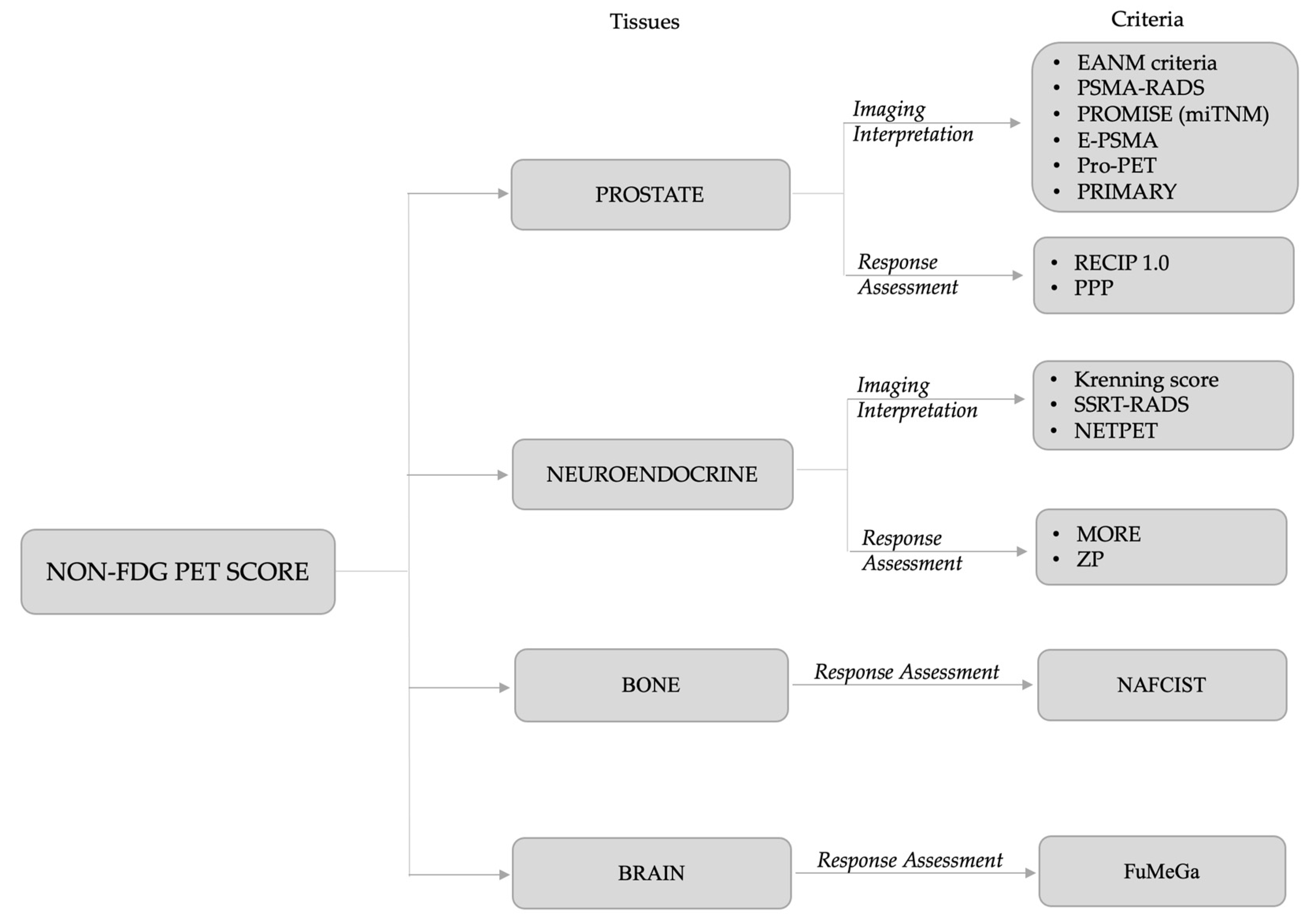
3.1. Prostate Cancer
3.1.1. Interpretation Criteria: EANM Criteria, PSMA-RADS, PROMISE (miTNM), E-PSMA, Pro-PET and PRIMARY Score
3.1.2. Response Assessment: PPP and RECIP 1.0 Criteria
3.2. Neuroendocrine Tumors
3.2.1. Interpretation Criteria: PET-Based Krenning Score, SSTR-RADS, and NETPET
3.2.2. Response Assessment: ZP and MORE

| MORE | ZP | ||
|---|---|---|---|
| Category | Description | Description | |
| Non PD | CR | Complete uptake disappearance in all lesions | No lesion (CT or PET) |
| PR | ≥25% reduction in the sum of SUVmax after more than one RLT cycle | ≥25% reduction in the product of SUVmean and HU | |
| SD | Does not meet other criteria | Does not meet other criteria | |
| PD | PD | ≥25% increase in the sum of SUVmax or at least one new lesion | ≥25% increase in the product of SUVmean and HU |
3.3. Bone Primary Cancer: [18F]NaF PET Response Criteria in Solid Tumors (NAFCIST)
3.4. Glioma: Functional and Metabolic Glioma Analysis (FuMeGA)
4. Discussion
5. Conclusions
Author Contributions
Funding
Institutional Review Board Statement
Informed Consent Statement
Data Availability Statement
Conflicts of Interest
References
- Basu, S.; Hess, S.; Nielsen Braad, P.E.; Olsen, B.B.; Inglev, S.; Høilund-Carlsen, P.F. The Basic Principles of FDG-PET/CT Imaging. PET Clin. 2014, 9, 355–370. [Google Scholar] [CrossRef] [PubMed]
- Dondi, F.; Pasinetti, N.; Gatta, R.; Albano, D.; Giubbini, R.; Bertagna, F. Comparison between Two Different Scanners for the Evaluation of the Role of 18F-FDG PET/CT Semiquantitative Parameters and Radiomics Features in the Prediction of Final Diagnosis of Thyroid Incidentalomas. J. Clin. Med. 2022, 11, 615. [Google Scholar] [CrossRef] [PubMed]
- Kim, J.H. Comparison of the EORTC criteria and PERCIST in solid tumors: A pooled analysis and review. Oncotarget 2016, 7, 58105–58110. [Google Scholar] [CrossRef] [Green Version]
- Wahl, R.L.; Jacene, H.; Kasamon, Y.; Lodge, M.A. From RECIST to PERCIST: Evolving Considerations for PET response criteria in solid tumors. J. Nucl. Med. 2009, 50, 122S–150S. [Google Scholar] [CrossRef] [PubMed] [Green Version]
- Annovazzi, A.; Vari, S.; Giannarelli, D.; Pasqualoni, R.; Sciuto, R.; Carpano, S.; Cognetti, F.; Ferraresi, V. Comparison of 18F-FDG PET/CT Criteria for the Prediction of Therapy Response and Clinical Outcome in Patients With Metastatic Melanoma Treated With Ipilimumab and PD-1 Inhibitors. Clin. Nucl. Med. 2020, 45, 187–194. [Google Scholar] [CrossRef] [PubMed]
- Cho, S.Y.; Lipson, E.J.; Im, H.J.; Rowe, S.P.; Gonzalez, E.M.; Blackford, A.; Chirindel, A.; Pardoll, D.M.; Topalian, S.L.; Wahl, R.L. Prediction of Response to Immune Checkpoint Inhibitor Therapy Using Early-Time-Point 18F-FDG PET/CT Imaging in Patients with Advanced Melanoma. J. Nucl. Med. 2017, 58, 1421–1428. [Google Scholar] [CrossRef] [Green Version]
- Anwar, H.; Sachpekidis, C.; Winkler, J.; Kopp-Schneider, A.; Haberkorn, U.; Hassel, J.C.; Dimitrakopoulou-Strauss, A. Absolute number of new lesions on 18F-FDG PET/CT is more predictive of clinical response than SUV changes in metastatic melanoma patients receiving ipilimumab. Eur. J. Nucl. Med. Mol. Imaging 2018, 45, 376–383. [Google Scholar] [CrossRef]
- Fanti, S.; Minozzi, S.; Morigi, J.J.; Giesel, F.; Ceci, F.; Uprimny, C.; Hofman, M.S.; Eiber, M.; Schwarzenbock, S.; Castellucci, P.; et al. Development of standardized image interpretation for 68Ga-PSMA PET/CT to detect prostate cancer recurrent lesions. Eur. J. Nucl. Med. Mol. Imaging 2017, 44, 1622–1635. [Google Scholar] [CrossRef]
- Rowe, S.P.; Pienta, K.J.; Pomper, M.G.; Gorin, M.A. Proposal for a Structured Reporting System for Prostate-Specific Membrane Antigen-Targeted PET Imaging: PSMA-RADS Version 1.0. J. Nucl. Med. 2018, 59, 479–485. [Google Scholar] [CrossRef] [Green Version]
- Eiber, M.; Herrmann, K.; Calais, J.; Hadaschik, B.; Giesel, F.L.; Hartenbach, M.; Hope, T.; Reiter, R.; Maurer, T.; Weber, W.A.; et al. Prostate Cancer Molecular Imaging Standardized Evaluation (PROMISE): Proposed miTNM Classification for the Interpretation of PSMA-Ligand PET/CT. J. Nucl. Med. 2018, 59, 469–478. [Google Scholar] [CrossRef] [Green Version]
- Ceci, F.; Oprea-Lager, D.E.; Emmett, L.; Adam, J.A.; Bomanji, J.; Czernin, J.; Eiber, M.; Haberkorn, U.; Hofman, M.S.; Hope, T.A.; et al. E-PSMA: The EANM standardized reporting guidelines v1.0 for PSMA-PET. Eur. J. Nucl. Med. Mol. Imaging 2021, 48, 1626–1638. [Google Scholar] [CrossRef]
- Adnan, A.; Basu, S. Concept proposal for a six-tier integrated dual tracer PET-CT (68Ga-PSMA and FDG) image scoring system (‘Pro-PET’ score) and examining its potential implications in metastatic castration-resistant prostate carcinoma theranostics and prognosis. Nucl. Med. Commun. 2021, 42, 566–574. [Google Scholar] [CrossRef]
- Emmett, L.; Papa, N.; Buteau, J.; Ho, B.; Liu, V.; Roberts, M.; Thompson, J.; Moon, D.; Sheehan-Dare, G.; Alghazo, O.; et al. The PRIMARY Score: Using Intraprostatic 68Ga-PSMA PET/CT Patterns to Optimize Prostate Cancer Diagnosis. J. Nucl. Med. 2022, 63, 1644–1650. [Google Scholar] [CrossRef]
- Fanti, S.; Hadaschik, B.; Herrmann, K. Proposal for Systemic-Therapy Response-Assessment Criteria at the Time of PSMA PET/CT Imaging: The PSMA PET Progression Criteria. J. Nucl. Med. 2020, 61, 678–682. [Google Scholar] [CrossRef]
- Gafita, A.; Rauscher, I.; Weber, M.; Hadaschik, B.; Wang, H.; Armstrong, W.R.; Tauber, R.; Grogan, T.R.; Czernin, J.; Rettig, M.B.; et al. Novel Framework for Treatment Response Evaluation Using PSMA PET/CT in Patients with Metastatic Castration-Resistant Prostate Cancer (RECIP 1.0): An International Multicenter Study. J. Nucl. Med. 2022, 63, 1651–1658. [Google Scholar] [CrossRef]
- Krenning, E.P.; Valkema, R.; Kooij, P.P.; Breeman, W.A.; Bakker, W.H.; de Herder, W.W.; van Eijck, C.H.; Kwekkeboom, D.J.; deJong, M.; Pauwels, S. Scintigraphy and radionuclide therapy with [indium-111-labelled-diethyl triamine penta-acetic acid-D-Phe1]-octreotide. Ital. J. Gastroenterol. Hepatol. 1999, 31, S219–S223. [Google Scholar]
- Werner, R.A.; Solnes, L.B.; Javadi, M.S.; Weich, A.; Gorin, M.A.; Pienta, K.J.; Higuchi, T.; Buck, A.K.; Pomper, M.G.; Rowe, S.P.; et al. SSTR-RADS Version 1.0 as a Reporting System for SSTR PET Imaging and Selection of Potential PRRT Candidates: A Proposed Standardization Framework. J. Nucl. Med. 2018, 59, 1085–1091. [Google Scholar] [CrossRef] [Green Version]
- Chan, D.L.; Pavlakis, N.; Schembri, G.P.; Bernard, E.J.; Hsiao, E.; Hayes, A.; Barnes, T.; Diakos, C.; Khasraw, M.; Samra, J.; et al. Dual Somatostatin Receptor/FDG PET/CT Imaging in Metastatic Neuroendocrine Tumours: Proposal for a Novel Grading Scheme with Prognostic Significance. Theranostics 2017, 7, 1149–1158. [Google Scholar] [CrossRef] [Green Version]
- Zwirtz, K.; Hardt, J.; Acker, G.; Baur, A.D.J.; Pavel, M.; Huang, K.; Brenner, W.; Prasad, V. Comparison of Choi, RECIST and Somatostatin Receptor PET/CT Based Criteria for the Evaluation of Response and Response Prediction to PRRT. Pharmaceutics 2022, 14, 1278. [Google Scholar] [CrossRef]
- Kairemo, K.; Rohren, E.M.; Anderson, P.M.; Ravizzini, G.; Rao, A.; Macapinlac, H.A.; Subbiah, V. Development of sodium fluoride PET response criteria for solid tumours (NAFCIST) in a clinical trial of radium-223 in osteosarcoma: From RECIST to PERCIST to NAFCIST. ESMO Open. 2019, 4, e000439. [Google Scholar] [CrossRef] [Green Version]
- García Vicente, A.M.; Pena Pardo, F.J.; Lozano Setien, E.; Sandoval Valencia, H.; Villena Martín, M. FuMeGA Criteria for Visual Assessment of Postoperative 18F-Fluorocholine PET in Patients With Glioma. Clin. Nucl. Med. 2020, 45, 448–450. [Google Scholar] [CrossRef]
- Werner, R.A.; Bundschuh, R.A.; Bundschuh, L.; Javadi, M.S.; Leal, J.P.; Higuchi, T.; Pienta, K.J.; Buck, A.K.; Pomper, M.G.; Gorin, M.A.; et al. Interobserver Agreement for the Standardized Reporting System PSMA-RADS 1.0 on 18F-DCFPyL PET/CT Imaging. J. Nucl. Med. 2018, 59, 1857–1864. [Google Scholar] [CrossRef] [Green Version]
- Chiu, L.W.; Lawhn-Heath, C.; Behr, S.C.; Juarez, R.; Perez, P.M.; Lobach, I.; Bucknor, M.D.; Hope, T.A.; Flavell, R.R. Factors Predicting Metastatic Disease in 68Ga-PSMA-11 PET-Positive Osseous Lesions in Prostate Cancer. J. Nucl. Med. 2020, 61, 1779–1785. [Google Scholar] [CrossRef]
- Letang, A.; Crombé, A.; Rousseau, C.; Sargos, P.; Merlin, C.; Cantarel, C.; Cazeau, A.L. Bone Uptake in Prostate Cancer Patients: Diagnostic Performances of PSMA-RADS v1.0, Clinical, Biological, and 68 Ga-PSMA-11 PET Features to Predict Metastasis After Biochemical Recurrence. Clin. Nucl. Med. 2022, 47, e529–e539. [Google Scholar] [CrossRef]
- Yin, Y.; Werner, R.A.; Higuchi, T.; Lapa, C.; Pienta, K.J.; Pomper, M.G.; Gorin, M.A.; Rowe, S.P. Follow-up of Lesions with Equivocal Radiotracer Uptake on PSMA-Targeted PET in Patients with Prostate Cancer: Predictive Values of the PSMA-RADS-3A and PSMA-RADS-3B Categories. J. Nucl. Med. 2019, 60, 511–516. [Google Scholar] [CrossRef]
- Kuten, J.; Dekalo, S.; Mintz, I.; Yossepowitch, O.; Mano, R.; Even-Sapir, E. The significance of equivocal bone findings in staging PSMA imaging in the preoperative setting: Validation of the PSMA-RADS version 1.0. EJNMMI Res. 2021, 11, 3. [Google Scholar] [CrossRef]
- Bhoil, A.; Seshadri, N.; Vinjamuri, S. Indeterminate skeletal and lymph node lesion on 18F PSMA 1007 PET/CT scanning: Lessons from a review at 12 months with PSMA-RADS. Nucl. Med. Commun. 2022, 43, 1034–1041. [Google Scholar] [CrossRef]
- Garg, T.; Werner, R.A.; Chung, H.W.; Khatri, W.; Pienta, K.J.; Pomper, M.G.; Gorin, M.A.; Saad, E.; Rowe, S.P. Association of True Positivity with Serum Prostate-Specific Antigen Levels and Other Clinical Factors in Indeterminate PSMA-RADS-3A Lesions Identified on 18F-DCFPyL PET/CT Scans. Tomography 2022, 8, 220. [Google Scholar] [CrossRef]
- Khatri, W.; Chung, H.W.; Werner, R.A.; Leal, J.P.; Pienta, K.J.; Lodge, M.A.; Gorin, M.A.; Pomper, M.G.; Rowe, S.P. Effect of Point-Spread Function Reconstruction for Indeterminate PSMA-RADS-3A Lesions on PSMA-Targeted PET Imaging of Men with Prostate Cancer. Diagnostics 2021, 11, 665. [Google Scholar] [CrossRef]
- Mihatsch, P.W.; Beissert, M.; Pomper, M.G.; Bley, T.A.; Seitz, A.K.; Kübler, H.; Buck, A.K.; Rowe, S.P.; Serfling, S.E.; Hartrampf, P.E.; et al. Changing Threshold-Based Segmentation Has No Relevant Impact on Semi-Quantification in the Context of Structured Reporting for PSMA-PET/CT. Cancers 2022, 14, 270. [Google Scholar] [CrossRef]
- Gültekin, A.; Yaylalı, O.; Şengöz, T.; Yüksel, D.; Şahin, B. Intraobserver and interobserver agreement for the interpretation of 68Ga-prostate-specific membrane antigen-I&T positron emission tomography/computed tomography imaging. Nucl. Med. Commun. 2019, 40, 1250–1255. [Google Scholar] [CrossRef]
- Wang, H.; Amiel, T.; Würnschimmel, C.; Langbein, T.; Steiger, K.; Rauscher, I.; Horn, T.; Maurer, T.; Weber, W.; Wester, H.J.; et al. PSMA-ligand uptake can serve as a novel biomarker in primary prostate cancer to predict outcome after radical prostatectomy. EJNMMI Res. 2021, 11, 76. [Google Scholar] [CrossRef]
- Hoberück, S.; Löck, S.; Borkowetz, A.; Sommer, U.; Winzer, R.; Zöphel, K.; Fedders, D.; Michler, E.; Kotzerke, J.; Kopka, K.; et al. Intraindividual comparison of [68Ga]-Ga-PSMA-11 and [18F]-F-PSMA-1007 in prostate cancer patients: A retrospective single-center analysis. EJNMMI Res. 2021, 11, 109. [Google Scholar] [CrossRef]
- Koehler, D.; Sauer, M.; Karimzadeh, A.; Apostolova, I.; Klutmann, S.; Adam, G.; Knipper, S.; Maurer, T.; Berliner, C. Evaluation of [68 Ga]Ga-PSMA-I&T PET/CT with additional late scans of the pelvis in prostate-specific antigen recurrence using the PROMISE criteria. EJNMMI Res. 2022, 12, 66. [Google Scholar] [CrossRef]
- Demirci, E.; Akyel, R.; Caner, B.; Alan-Selçuk, N.; Güven-Meşe, Ş.; Ocak, M.; Kabasakal, L. Interobserver and intraobserver agreement on prostate-specific membrane antigen PET/CT images according to the miTNM and PSMA-RADS criteria. Nucl. Med. Commun. 2020, 41, 759–767. [Google Scholar] [CrossRef]
- Toriihara, A.; Nobashi, T.; Baratto, L.; Duan, H.; Moradi, F.; Park, S.; Hatami, N.; Aparici, C.M.; Davidzon, G.; Iagaru, A. Comparison of 3 Interpretation Criteria for 68Ga-PSMA11 PET Based on Inter- and Intrareader Agreement. J. Nucl. Med. 2020, 61, 533–539. [Google Scholar] [CrossRef]
- Michalski, K.; Klein, C.; Brueggemann, T.; Meyer, P.T.; Jilg, C.A.; Ruf, J. Assessing Response to [177Lu]PSMA Radioligand Therapy using modified PSMA PET Progression Criteria. J. Nucl. Med. 2021, 62, 1741–1746. [Google Scholar] [CrossRef]
- Gafita, A.; Rauscher, I.; Fendler, W.P.; Murthy, V.; Hui, W.; Armstrong, W.R.; Herrmann, K.; Weber, W.A.; Calais, J.; Eiber, M.; et al. Measuring response in metastatic castration-resistant prostate cancer using PSMA PET/CT: Comparison of RECIST 1.1, aPCWG3, aPERCIST, PPP, and RECIP 1.0 criteria. Eur. J. Nucl. Med. Mol. Imaging 2022, 49, 4271–4281. [Google Scholar] [CrossRef]
- Hope, T.A.; Calais, J.; Zhang, L.; Dieckmann, W.; Millo, C. 111In-Pentetreotide Scintigraphy Versus 68Ga-DOTATATE PET: Impact on Krenning Scores and Effect of Tumor Burden. J. Nucl. Med. 2019, 60, 1266–1269. [Google Scholar] [CrossRef] [Green Version]
- Purandare, N.C.; Puranik, A.; Agrawal, A.; Shah, S.; Kumar, R.; Jiwnani, S.; Karimundackal, G.; Pramesh, C.S.; Rangarajan, V. Does 68Ga-DOTA-NOC-PET/CT impact staging and therapeutic decision making in pulmonary carcinoid tumors? Nucl. Med. Commun. 2020, 41, 1040–1046. [Google Scholar] [CrossRef]
- Menon, B.K.; Kalshetty, A.; Bhattacharjee, A.; Basu, S. Standardized uptake values and ratios on 68Ga-DOTATATE PET-computed tomography for normal organs and malignant lesions and their correlation with Krenning score in patients with metastatic neuroendocrine tumors. Nucl. Med. Commun. 2020, 41, 1095–1099. [Google Scholar] [CrossRef]
- Werner, R.A.; Derlin, T.; Rowe, S.P.; Bundschuh, L.; Sheikh, G.T.; Pomper, M.G.; Schulz, S.; Higuchi, T.; Buck, A.K.; Bengel, F.M.; et al. High Interobserver Agreement for the Standardized Reporting System SSTR-RADS 1.0 on Somatostatin Receptor PET/CT. J. Nucl. Med. 2021, 62, 514–520. [Google Scholar] [CrossRef]
- Chan, D.L.; Hayes, A.R.; Karfis, I.; Conner, A.; Furtado O’Mahony, L.; Mileva, M.; Bernard, E.; Roach, P.; Marin, G.; Pavlakis, N.; et al. Dual [68Ga]DOTATATE and [18F]FDG PET/CT in patients with metastatic gastroenteropancreatic neuroendocrine neoplasms: A multicentre validation of the NETPET score. Br. J. Cancer 2022. Epub ahead of print. [Google Scholar] [CrossRef]
- Hayes, A.R.; Furtado O’Mahony, L.; Quigley, A.M.; Gnanasegaran, G.; Caplin, M.E.; Navalkissoor, S.; Toumpanakis, C. The Combined Interpretation of 68Ga-DOTATATE PET/CT and 18F-FDG PET/CT in Metastatic Gastroenteropancreatic Neuroendocrine Tumors: A Classification System With Prognostic Impact. Clin. Nucl. Med. 2022, 47, 26–35. [Google Scholar] [CrossRef]
- Karfis, I.; Marin, G.; Levillain, H.; Drisis, S.; Muteganya, R.; Critchi, G.; Taraji-Schiltz, L.; Guix, C.A.; Shaza, L.; Elbachiri, M.; et al. Prognostic value of a three-scale grading system based on combining molecular imaging with 68Ga-DOTATATE and 18F-FDG PET/CT in patients with metastatic gastroenteropancreatic neuroendocrine neoplasias. Oncotarget 2020, 11, 589–599. [Google Scholar] [CrossRef] [Green Version]
- Chan, D.L.; Ulaner, G.A.; Pattison, D.; Wyld, D.; Ladwa, R.; Kirchner, J.; Li, B.T.; Lai, W.V.; Pavlakis, N.; Roach, P.J.; et al. Dual PET Imaging in Bronchial Neuroendocrine Neoplasms: The NETPET Score as a Prognostic Biomarker. J. Nucl. Med. 2021, 62, 1278–1284. [Google Scholar] [CrossRef]
- Kairemo, K.; Roszik, J.; Anderson, P.; Ravizzini, G.; Rao, A.; Macapinlac, H.A.; Subbiah, V. 18F-sodium fluoride positron emission tomography (NaF-18-PET/CT) radiomic signatures to evaluate responses to alpha-particle Radium-223 dichloride therapy in osteosarcoma metastases. Curr. Probl. Cancer 2021, 45, 100797. [Google Scholar] [CrossRef]
- García Vicente, A.M.; Pena Pardo, F.J.; Amo-Salas, M.; Villena Martín, M.; López Menéndez, C.; Soriano Castrejón, Á.M.; Pérez-Beteta, J. Prognostic Potential of Postoperative 18F-Fluorocholine PET/CT in Patients With High-Grade Glioma. Clinical Validation of FuMeGA Postoperative PET Criteria. Clin. Nucl. Med. 2022, 47, 480–487. [Google Scholar] [CrossRef]
- Deppen, S.A.; Blume, J.; Bobbey, A.J.; Shah, C.; Graham, M.M.; Lee, P.; Delbeke, D.; Walker, R.C. 68Ga-DOTATATE Compared with 111In-DTPA-Octreotide and Conventional Imaging for Pulmonary and Gastroenteropancreatic Neuroendocrine Tumors: A Systematic Review and Meta-Analysis. J. Nucl. Med. 2016, 57, 872–878. [Google Scholar] [CrossRef] [Green Version]
- Gabriel, M.; Decristoforo, C.; Kendler, D.; Dobrozemsky, G.; Heute, D.; Uprimny, C.; Kovacs, P.; Von Guggenberg, E.; Bale, R.; Virgolini, I.J. 68Ga-DOTA-Tyr3-octreotide PET in neuroendocrine tumors: Comparison with somatostatin receptor scintigraphy and CT. J. Nucl. Med. 2007, 48, 508–518. [Google Scholar] [CrossRef] [Green Version]
- Sadowski, S.M.; Neychev, V.; Millo, C.; Shih, J.; Nilubol, N.; Herscovitch, P.; Pacak, K.; Marx, S.J.; Kebebew, E. Prospective Study of 68Ga-DOTATATE Positron Emission Tomography/Computed Tomography for Detecting Gastro-Entero-Pancreatic Neuroendocrine Tumors and Unknown Primary Sites. J. Clin. Oncol. 2016, 34, 588–596. [Google Scholar] [CrossRef] [Green Version]
- Bodei, L.; Ambrosini, V.; Herrmann, K.; Modlin, I. Current Concepts in 68Ga-DOTATATE Imaging of Neuroendocrine Neoplasms: Interpretation, Biodistribution, Dosimetry, and Molecular Strategies. J. Nucl. Med. 2017, 58, 1718–1726. [Google Scholar] [CrossRef] [Green Version]
- Ma, G.; Du, J.; Zhang, X.; Liu, J.; Xu, X.; Xu, B.; Guan, Z. Quantitative analysis of 68Ga-DOTA(0)-Tyr(3)-octreotate positron emission tomography/computed tomography imaging for the differential diagnosis of primary pheochromocytoma and paraganglioma. Quant. Imaging. Med. Surg. 2022, 12, 2427–2440. [Google Scholar] [CrossRef]
- Bodei, L.; Mueller-Brand, J.; Baum, R.P.; Pavel, M.E.; Hörsch, D.; O’Dorisio, M.S.; O’Dorisio, T.M.; Howe, J.R.; Cremonesi, M.; Kwekkeboom, D.J.; et al. The joint IAEA, EANM, and SNMMI practical guidance on peptide receptor radionuclide therapy (PRRNT) in neuroendocrine tumours. Eur. J. Nucl. Med. Mol. Imaging 2013, 40, 800–816. [Google Scholar] [CrossRef]
- Bozkurt, M.F.; Virgolini, I.; Balogova, S.; Beheshti, M.; Rubello, D.; Decristoforo, C.; Ambrosini, V.; Kjaer, A.; Delgado-Bolton, R.; Kunikowska, J.; et al. Guideline for PET/CT imaging of neuroendocrine neoplasms with 68Ga-DOTA-conjugated somatostatin receptor targeting peptides and 18F-DOPA. Eur. J. Nucl. Med. Mol. Imaging 2017, 44, 1588–1601. [Google Scholar] [CrossRef] [Green Version]
- Fueger, B.J.; Hamilton, G.; Raderer, M.; Pangerl, T.; Traub, T.; Angelberger, P.; Baumgartner, G.; Dudczak, R.; Virgolini, I. Effects of chemotherapeutic agents on expression of somatostatin receptors in pancreatic tumor cells. J. Nucl. Med. 2001, 42, 1856–1862. [Google Scholar]
- Bartolomei, M.; Berruti, A.; Falconi, M.; Fazio, N.; Ferone, D.; Lastoria, S.; Pappagallo, G.; Seregni, E.; Versari, A. Clinical Management of Neuroendocrine Neoplasms in Clinical Practice: A Formal Consensus Exercise. Cancers 2022, 14, 2501. [Google Scholar] [CrossRef]
- Urso, L.; Panareo, S.; Castello, A.; Ambrosio, M.R.; Zatelli, M.C.; Caracciolo, M.; Tonini, E.; Valpiani, G.; Boschi, A.; Uccelli, L.; et al. Glucose Metabolism Modification Induced by Radioligand Therapy with [177Lu]Lu/[90Y]Y-DOTATOC in Advanced Neuroendocrine Neoplasms: A Prospective Pilot Study within FENET-2016 Trial. Pharmaceutics 2022, 14, 2009. [Google Scholar] [CrossRef]
- Luo, Y.; Chen, J.; Huang, K.; Lin, Y.; Chen, M.; Xu, L.; Li, Z.P.; Feng, S.T. Early evaluation of sunitinib for the treatment of advanced gastroenteropancreatic neuroendocrine neoplasms via CT imaging: RECIST 1.1 or Choi Criteria? BMC Cancer. 2017, 17, 154. [Google Scholar] [CrossRef] [Green Version]
- Duclos, V.; Iep, A.; Gomez, L.; Goldfarb, L.; Besson, F.L. PET Molecular Imaging: A Holistic Review of Current Practice and Emerging Perspectives for Diagnosis, Therapeutic Evaluation and Prognosis in Clinical Oncology. Int. J. Mol. Sci. 2021, 22, 4159. [Google Scholar] [CrossRef]
- Lindsay, M.J.; Siegel, B.A.; Tunis, S.R.; Hillner, B.E.; Shields, A.F.; Carey, B.P.; Coleman, R.E. The National Oncologic PET Registry: Expanded medicare coverage for PET under coverage with evidence development. Am. J. Roentgenol. 2007, 188, 1109–1113. [Google Scholar] [CrossRef]
- Maurer, T.; Gschwend, J.E.; Rauscher, I.; Souvatzoglou, M.; Haller, B.; Weirich, G.; Wester, H.J.; Heck, M.; Kübler, H.; Beer, A.J.; et al. Diagnostic Efficacy of (68)Gallium-PSMA Positron Emission Tomography Compared to Conventional Imaging for Lymph Node Staging of 130 Consecutive Patients with Intermediate to High Risk Prostate Cancer. J. Urol. 2016, 195, 1436–1443. [Google Scholar] [CrossRef]
- Eiber, M.; Maurer, T.; Souvatzoglou, M.; Beer, A.J.; Ruffani, A.; Haller, B.; Graner, F.P.; Kübler, H.; Haberkorn, U.; Eisenhut, M.; et al. Evaluation of Hybrid ⁶⁸Ga-PSMA Ligand PET/CT in 248 Patients with Biochemical Recurrence After Radical Prostatectomy. J. Nucl. Med. 2015, 56, 668–674. [Google Scholar] [CrossRef] [Green Version]
- Rowe, S.P.; Macura, K.J.; Mena, E.; Blackford, A.L.; Nadal, R.; Antonarakis, E.S.; Eisenberger, M.; Carducci, M.; Fan, H.; Dannals, R.F.; et al. PSMA-Based [(18)F]DCFPyL PET/CT Is Superior to Conventional Imaging for Lesion Detection in Patients with Metastatic Prostate Cancer. Mol. Imaging Biol. 2016, 18, 411–419. [Google Scholar] [CrossRef] [Green Version]
- Dondi, F.; Albano, D.; Cerudelli, E.; Gazzilli, M.; Giubbini, R.; Treglia, G.; Bertagna, F. Radiolabelled PSMA PET/CT or PET/MRI in Hepatocellular Carcinoma (HCC): A Systematic Review. Clin. Transl. Imaging. 2020, 8, 461–467. [Google Scholar] [CrossRef]
- Urso, L.; Castello, A.; Rocca, G.C.; Lancia, F.; Panareo, S.; Cittanti, C.; Uccelli, L.; Florimonte, L.; Castellani, M.; Ippolito, C.; et al. Role of PSMA-ligands imaging in Renal Cell Carcinoma management: Current status and future perspectives. J. Cancer Res. Clin. Oncol. 2022, 148, 1299–1311. [Google Scholar] [CrossRef]
- Rizzo, A.; Dall’Armellina, S.; Pizzuto, D.A.; Perotti, G.; Zagaria, L.; Lanni, V.; Treglia, G.; Racca, M.; Annunziata, S. PSMA Radioligand Uptake as a Biomarker of Neoangiogenesis in Solid Tumours: Diagnostic or Theragnostic Factor? Cancers 2022, 14, 4039. [Google Scholar] [CrossRef]
- Werner, R.A.; Bundschuh, R.A.; Bundschuh, L.; Fanti, S.; Javadi, M.S.; Higuchi, T.; Weich, A.; Pienta, K.J.; Buck, A.K.; Pomper, M.G.; et al. Novel Structured Reporting Systems for Theranostic Radiotracers. J. Nucl. Med. 2019, 60, 577–584. [Google Scholar] [CrossRef]
- Hennrich, U.; Eder, M. [177Lu]Lu-PSMA-617 (PluvictoTM): The First FDA-Approved Radiotherapeutical for Treatment of Prostate Cancer. Pharmaceuticals 2022, 15, 1292. [Google Scholar] [CrossRef]
- Scher, H.I.; Morris, M.J.; Stadler, W.M.; Higano, C.; Basch, E.; Fizazi, K.; Antonarakis, E.S.; Beer, T.M.; Carducci, M.A.; Chi, K.N.; et al. Prostate Cancer Clinical Trials Working Group 3. Trial Design and Objectives for Castration-Resistant Prostate Cancer: Updated Recommendations From the Prostate Cancer Clinical Trials Working Group 3. J. Clin. Oncol. 2016, 34, 1402–1418. [Google Scholar] [CrossRef] [Green Version]
- Eisenhauer, E.A.; Therasse, P.; Bogaerts, J.; Schwartz, L.H.; Sargent, D.; Ford, R.; Dancey, J.; Arbuck, S.; Gwyther, S.; Mooney, M.; et al. New response evaluation criteria in solid tumours: Revised RECIST guideline (version 1.1). Eur. J. Cancer. 2009, 45, 228–247. [Google Scholar] [CrossRef]
- Ceci, F.; Castellucci, P.; Graziani, T.; Schiavina, R.; Renzi, R.; Borghesi, M.; Di Tullio, P.; Brunocilla, E.; Ardizzoni, A.; Fanti, S. (11)C-Choline PET/CT in castration-resistant prostate cancer patients treated with docetaxel. Eur. J. Nucl. Med. Mol. Imaging 2016, 43, 84–91. [Google Scholar] [CrossRef]
- Urso, L.; Lancia, F.; Ortolan, N.; Frapoli, M.; Rauso, M.; Artioli, P.; Cittanti, C.; Uccelli, L.; Frassoldati, A.; Evangelista, L.; et al. 18F-Choline PET/CT or PET/MR and the evaluation of response to systemic therapy in prostate cancer: Are we ready? Clin. Transl. Imaging 2022, 10, 687–695. [Google Scholar] [CrossRef]
- Seitz, A.K.; Rauscher, I.; Haller, B.; Krönke, M.; Luther, S.; Heck, M.M.; Horn, T.; Gschwend, J.E.; Schwaiger, M.; Eiber, M.; et al. Preliminary results on response assessment using 68Ga-HBED-CC-PSMA PET/CT in patients with metastatic prostate cancer undergoing docetaxel chemotherapy. Eur. J. Nucl. Med. Mol. Imaging 2018, 45, 602–612. [Google Scholar] [CrossRef]
- Alongi, P.; Laudicella, R.; Lanzafame, H.; Farolfi, A.; Mapelli, P.; Picchio, M.; Burger, I.A.; Iagaru, A.; Minutoli, F.; Evangelista, L. PSMA and Choline PET for the Assessment of Response to Therapy and Survival Outcomes in Prostate Cancer Patients: A Systematic Review from the Literature. Cancers 2022, 14, 1770. [Google Scholar] [CrossRef]
- Shahrokhi, P.; Emami-Ardekani, A.; Karamzade-Ziarati, N. SSTR-based theranostics in neuroendocrine prostate cancer (NEPC). Clin. Transl. Imaging 2022, 1, 1–8. [Google Scholar] [CrossRef]
- Fanti, S.; Goffin, K.; Hadaschik, B.A.; Herrmann, K.; Maurer, T.; MacLennan, S.; Oprea-Lager, D.E.; Oyen, W.J.; Rouvière, O.; Mottet, N.; et al. Consensus statements on PSMA PET/CT response assessment criteria in prostate cancer. Eur. J. Nucl. Med. Mol. Imaging 2021, 48, 469–476. [Google Scholar] [CrossRef]
- Strosberg, J.; El-Haddad, G.; Wolin, E.; Hendifar, A.; Yao, J.; Chasen, B.; Mittra, E.; Kunz, P.L.; Kulke, M.H.; Jacene, H.; et al. NETTER-1 Trial Investigators. Phase 3 Trial of 177Lu-Dotatate for Midgut Neuroendocrine Tumors. N. Engl. J. Med. 2017, 376, 125–135. [Google Scholar] [CrossRef]
- Kwekkeboom, D.J.; de Herder, W.W.; Kam, B.L.; van Eijck, C.H.; van Essen, M.; Kooij, P.P.; Feelders, R.A.; van Aken, M.O.; Krenning, E.P. Treatment with the radiolabeled somatostatin analog [177 Lu-DOTA 0,Tyr3]octreotate: Toxicity, efficacy, and survival. J. Clin. Oncol. 2008, 26, 2124–2130. [Google Scholar] [CrossRef] [Green Version]
- Haug, A.R.; Auernhammer, C.J.; Wängler, B.; Schmidt, G.P.; Uebleis, C.; Göke, B.; Cumming, P.; Bartenstein, P.; Tiling, R.; Hacker, M. 68Ga-DOTATATE PET/CT for the early prediction of response to somatostatin receptor-mediated radionuclide therapy in patients with well-differentiated neuroendocrine tumors. J. Nucl. Med. 2010, 51, 1349–1356. [Google Scholar] [CrossRef] [Green Version]
- Gabriel., M.; Oberauer, A.; Dobrozemsky, G.; Decristoforo, C.; Putzer, D.; Kendler, D.; Uprimny, C.; Kovacs, P.; Bale, R.; Virgolini, I.J. 68Ga-DOTA-Tyr3-octreotide PET for assessing response to somatostatin-receptor-mediated radionuclide therapy. J. Nucl. Med. 2009, 50, 1427–1434. [Google Scholar] [CrossRef] [Green Version]
- Cui, M.; Zorrilla-Veloz, R.I.; Hu, J.; Guan, B.; Ma, X. Diagnostic Accuracy of PET for Differentiating True Glioma Progression From Post Treatment-Related Changes: A Systematic Review and Meta-Analysis. Front. Neurol. 2021, 12, 671867. [Google Scholar] [CrossRef]
- Santo, G.; Laudicella, R.; Linguanti, F.; Nappi, A.G.; Abenavoli, E.; Vergura, V.; Rubini, G.; Sciagrà, R.; Arnone, G.; Schillaci, O.; et al. The Utility of Conventional Amino Acid PET Radiotracers in the Evaluation of Glioma Recurrence also in Comparison with MRI. Diagnostics 2022, 12, 844. [Google Scholar] [CrossRef]

| Reference | Year | Radiotracer | Criteria | Description |
|---|---|---|---|---|
| Fanti et al. [8] | 2017 | PSMA-ligand | EANM criteria | All “anomalous” findings suggestive of recurrent PCa were noticed and categorized as “pathologic” unless another explanation could be hypothesized. |
| Rowe et al. [9] | 2018 | PSMA-ligand | PSMA-RADS | 5-point scale framework reflecting the level of confidence in interpreting PSMA-PET imaging in PCa. |
| Eiber et al. [10] | 2019 | PSMA-ligand | PROMISE (miTNM) | Standardized reporting framework based on miTNM system for PSMA-ligand PET/CT or PET/MRI imaging in PCa. |
| Ceci et al. [11] | 2021 | PSMA-ligand | E-PSMA | The E-PSMA standardized reporting guidelinesprovide consensus statements to develop a structuredreport for PSMA-PET in PCa. |
| Adnan et al. [12] | 2021 | PSMA/FDG | Pro-PET | An integrated dual tracer PET/CT (PSMA and FDG) image scoring system for mCRPCa patients referred to [177Lu]Lu-PSMA therapy. |
| Emmett et al. [13] | 2022 | PSMA-ligand | PRIMARY | A 5-level score for the diagnosis of clinically significant PCa. |
| Fanti et al. [14] | 2020 | PSMA-ligand | PSMA PET Progression (PPP) | PSMA-driven response evaluation criteria in patients with metastatic PCa. |
| Grafita et al. [15] | 2022 | PSMA-ligand | RECIP 1.0 | PSMA-driven response evaluation criteria in mCRPCa patients who underwent [177Lu]Lu-PSMA therapy. |
| Krenning et al. [16] * | 1999 | SSTR-ligand | Krenning | Visual interpretation criteria for evaluating NETs patients’ eligibility for [177Lu]Lu DOTA therapy. |
| Werner et al. [17] | 2018 | SSTR-ligand | SSTR-RADS | 5-point scale framework reflecting the level of confidence of interpreting SSTR-PET imaging in NET assessing the eligibility to [177Lu]Lu-DOTA therapy. |
| Chan et al. [18] | 2017 | SSTR/FDG | NETPET | An integrated dual tracer PET/CT (SSTR and FDG) image scoring system for NET patients. |
| Zwirtz et al. [19] | 2021 | SSTR-ligand | MORE; ZP | Response evaluation criteria in NETs patients who underwent [177Lu]Lu-DOTA therapy. |
| Kairemo et al. [20] | 2019 | [18F]NaF | NAFCIST | Response evaluation criteria for high-risk osteosarcoma patients who underwent [223Ra]Cl2 therapy. |
| García Vicente et al. [21] | 2020 | [18F]Fluorocholine | FuMeGa | Visual interpretation criteria for post-surgery evaluation of HGG patients |
| Reference | Radiotracer(s) | n pts | Key Point | |
|---|---|---|---|---|
| Prostate | Fanti et al. [8] | [68Ga]Ga-PSMA | 104 | EANM criteria showed a moderate interobserver agreement among multiple readers. |
| Werner et al. [22] | [18F]DCFPyL | 50 | PSMA-RADS showed excellent interobserver agreement for lymph nodes and for the overall scan impression (ICC 0.79 and 0.84, respectively). | |
| Chiu et al. [23] | [68Ga]Ga-PSMA-11 | 56 | PSMA-RADS ratings showed perfect interrater reliability to detect prostate bone metastasis. A SUVmax ratio (lesion-to-blood pool) > 2.2 presented a superior lesion detection rate and specificity when compared to PSMA-RADS. | |
| Letang et al. [24] | [68Ga]Ga-PSMA-11 | 53 | PSMA-RADS had a significantly higher AUROC than the initial reading for the assessment of bone metastasis. | |
| Yin et al. [25] | [18F]DCFPyL | 36 | PSMA-RADS-3A lesions are more likely than PSMA-RADS-3B lesions to represent sites of PCa. | |
| Kuten et al. [26] | [68Ga]Ga-PSMA-11[18F]PSMA-1007 | 15 | In intermediate and high-risk patients staged prior to radical prostatectomy, most PSMA-RADS-3B lesions are of no clinical relevance. | |
| Bhoil et al. [27] | [18F]PSMA-1007 | 203 | A structured reporting with PSMA-RADS grading helps in the proper classification of lesions and standardization of reports. | |
| Garg et al. [28] | [18F]DCFPyL | 28 | A PSA level ≥ 0.6–1.0 ng/mL can be used as a marker for a high index of suspicion for true positivity in PSMA-RADS-3A lesions. A Gleason score ≥ 7 showed a higher rate of PSMA-RADS-3A lesion positivity. | |
| Khatri et al. [29] | [18F]DCFPyL | 30 | The PSF reconstructions allowed the re-categorization of a small number of indeterminate PSMA-RADS-3A soft tissue lesions as more definitive PSMA-RADS-4 lesions. | |
| Mihatsch et al. [30] | [18F]PSMA-1007 | 18 | SUVmax was significantly higher in PSMA-RADS-5 lesions compared to all other categories. PSMA-RADS-3A lesions showed significantly lower SUVmax and SUVpeak compared to the entire PSMA-RADS-4 or -5 cohort. | |
| Gültekin et al. [31] | [68Ga]Ga-PSMA-I&T | 80 | The miN and miM categories showed almost perfect interobserver agreement (K = 0.93 and 0.94, respectively) as well as miM (K = 0.84). | |
| Wang et al. [32] | [68Ga]Ga-PSMA-11 | 187 | Preoperative miTNM correlated with postoperative Gleason score, surgical margin status and time to biochemical recurrence. | |
| Hoberück et al. [33] | [68Ga]Ga-PSMA-11; [18F]PSMA-1007 | 46 | In terms of miTNM staging, both [68Ga]Ga-PSMA-11 and [18F]PSMA-1007 appeared exchangeable, as no tracer outperformed the other. | |
| Koehler et al. [34] | [68Ga]Ga-PSMA-I&T | 297 | Additional late scans of the pelvis in [68Ga]Ga-PSMA-I&T PET/CT detected more lesions and an increasing contrast compared to early imaging, influencing the final miTNM-staging. | |
| Demirci et al. [35] | [68Ga]Ga-PSMA-11 | 133 | The miTNM provides a high level of concordance among readers, with the highest agreement level in miM and lowest agreement in miT. PSMA-RADS showed almost-perfect agreement between readers in terms of scoring (ICC analysis). However, if the results were grouped as benign (score of 1/2), indeterminate (3), and malignant (4/5), Fleiss’ κ analysis showed a moderate level of agreement. | |
| Toriihara et al. [36] | [68Ga]Ga-PSMA-11 | 104 | Comparing the 3 proposed interpretation criteria for PCa, intrareader agreement was moderate to almost perfect. The intercriteria agreement for each site was moderate to almost perfect. | |
| Adnan et al. [12] | [68Ga]Ga-PSMA-11 + [18F]FDG | 47 | The Pro-PET score significantly correlated with symptomatic, biochemical, metabolic, and anatomical responses, as well as with PFS (p = 0.03) and OS (p = 0.027). | |
| Emmett et al. [13] | [68Ga]Ga-PSMA-11 | 291 | The estimated AUC of the PRIMARY score was 0.85 (95%CI: 0.81–0.89) and exceeded that of PI-RADS 0.76 (95%CI: 0.71–0.81) (p = 0.003). Sensitivity, specificity, PPV and NPV for PRIMARY score 3 to 5 (high risk patterns) vs. PRIMARY score 1,2 (low risk patterns) was 88%, 64%, 76% and 81%, compared to 83%, 53%, 69% and 72% for PI-RADS 3–5 vs. 2. | |
| Michalski et al. [37] | [68Ga]Ga-PSMA-11; [18F]PSMA-1007 | 46 | Modified PPP criteria showed high reproducibility. A significant correlation was shown between progressive and non-progressive disease defined by PPP and OS. | |
| Gafita et al. [15] | PSMA-ligand | 124 | Patients with RECIP-PD had shorter OS compared to patients with RECIP-SD/PR | |
| Gafita et al. [38] | PSMA-ligand | 124 | PD patients according to RECIP 1.0 had a higher risk of death compared to non-PD, also in comparison with other response criteria (PPP, RECIST 1.1, aPERCIST) | |
| Neurendocrine | Hope et al. [39] | [68Ga]Ga-DOTA-TATE | 150 | The detection rate of SSTR-positive disease (KS 2–4) resulted of 23%, 38%, and 72% with [111In]Pentetreotide planar scintigraphy, SPECT and PET, respectively. Lesion size did not affect SSTR PET-based KS. |
| Purandare et al. [40] | [68Ga]Ga-DOTA-NOC | 119 | [68Ga]Ga-DOTA-NOC showed a high sensitivity for tumor detection (92.4%) and can help differentiate between typical and atypical carcinoid variants. | |
| Menon et al. [41] | [68Ga]Ga-DOTA-TATE | 32 | No significant change in KS was seen between the delayed scan compared to the standard one (1–1.5 h). | |
| Werner et al. [42] | [68Ga]Ga-DOTA-TOC | 51 | The interobserver agreement for overall scan impression based on SSTR-RADS was excellent (ICC, 0.88), as well as for lymph node and liver lesions (ICC, 0.91 and 0.77, respectively). | |
| Chan et al. [18] | [68Ga]Ga-DOTATATE + [18F]FDG | 62 | NETPET grade showed statistically significant correlation with OS at univariate analysis. NETPET grade was significantly associated with histological grade. | |
| Chan et al. [43] | [68Ga]Ga-DOTATATE + [18F]FDG | 319 | NETPET score significantly correlated with OS and TTP. NETPET showed both high inter-rater (kappa = 0.8) and intra-rater (kappa = 0.9) reliability. | |
| Hayes et al. [44] | [68Ga]Ga-DOTATATE + [18F]FDG | 87 | The dual-tracer PET classification was an independent predictor of OS (multivariate p = 0.016) and predicted PFS (univariate p = 0.030). | |
| Karfis et al. [45] | [68Ga]Ga-DOTATATE + [18F]FDG | 85 | Combined [68Ga]Ga-DOTATATE and [18F]FDG PET imaging classification reached high prognostic value in terms of PFS in GEPNET patients. | |
| Chan et al. [46] | [68Ga]Ga-DOTATATE + [18F]FDG | 38 | The NETPET score and histology were significantly correlated with OS (p = 0.003, p = 0.01) | |
| Zwirtz et al. [19] | [68Ga]Ga-DOTA-TATE/TOC | 34 | Only patients showing progressive disease after two PRRT cycles according to MORE criteria had a worse prognosis, while baseline ZP and ZPnormalized performed best in predicting lesion progression after three cycles of PRRT. | |
| Bone | Kairemo et al. [20] | [18F]NaF | 18 | NAFCIST correlates with changes in bone alkaline phosphatase levels, cumulative dose of [223Ra]Cl2 and OS. |
| Kairemo et al. [47] | [18F]NaF | 18 | Due to mixed response, neither PERCIST nor RECIST were able to predict response in osteosarcoma patients treated with [223Ra]Cl2. Radiomics can help in the assessment of intra-tumoral and inter-tumoral heterogeneity in the response to bone-forming osteosarcoma to [223Ra]Cl2 therapy. | |
| Brain | García Vicente et al. [48] | [18F]Fluorocholine | 47 | Significant differences were found for PFS and OS between incomplete versus complete metabolic resections assessed by the FuMeGa score |
| PSMA-RADS 1.0 | ||
|---|---|---|
| Category | Findings | Action |
| PSMA-RADS-1 (benign) | ||
| PSMA-RADS-1A | Benign lesion characterized by biopsy or pathognomonic finding on anatomic imaging and without abnormal uptake | |
| PSMA-RADS-1B | Benign lesion characterized by biopsy or pathognomonic finding on anatomic imaging and with focal radiotracer uptake | |
| PSMA-RADS-2 (likely benign) | Equivocal uptake (focal, but low level such as blood pool) in soft-tissue or in a bone site atypical of PCa involvement | |
| PSMA-RADS-3 (equivocal) | Consider further work-up | |
| PSMA-RADS-3A | Equivocal uptake in soft-tissue site typical of PCa involvement. | Lesion targetable: biopsy is suggested. Alternatively, follow-up imaging (either anatomic or PSMA-targeted PET/CT) after 3–6 months is recommended. |
| PSMA-RADS-3B | Equivocal uptake in bone lesion not definitive but also not atypical of PCa on anatomic imaging. | Comparison to other imaging modalities (bone scan, [18F]NaF PET, or tumor-protocol MRI images) or bone biopsy. Alternatively, follow-up imaging (either anatomic or PSMA-targeted PET/CT) after 3–6 months is recommended. |
| PSMA-RADS-3C | Lesions that would be atypical for PCa but have high levels of uptake. The likelihood of non-prostatic malignancy or another benign tumor is high. | Biopsy to histologically confirm the diagnosis is often preferred. Alternatively, organ-specific follow-up imaging may be done (e.g., liver-protocol MRI to evaluate possible primary hepatocellular carcinoma). |
| PSMA-RADS-3D | Lesion suggestive of malignancy on anatomic imaging but lacking uptake. | Consider non-prostatic malignancy, neuroendocrine PCa, and an uncommon case of prostate adenocarcinoma that fails to express PSMA. Biopsy to histologically confirm the diagnosis is often preferred; alternatively, organ-specific follow-up imaging may be done. |
| PSMA-RADS-4 (Pca highly likely) | Intense uptake in sites typical of PCa but lacking definitive findings on conventional imaging. | No biopsy confirmation will be needed, although tissue for genomic analysis (or other purposes) may be useful. |
| PSMA-RADS-5 (Pca almost certainly present) | Intense uptake in sites typical of PCand having corresponding findings on conventional imaging. | No biopsy confirmation will be needed, although tissue for genomic analysis (or other purposes) may be useful. |
| miTNM | |
|---|---|
| Class | Description |
| Local tumor (T) | |
| miT0 | No local tumor |
| miT2 | Organ confined tumor (unifocal or multifocal) |
| miT3 | Non-organ confined tumor |
| miT3a | Extracapsular extension |
| miT3b | Tumor invading seminal vesicles |
| miT4 | Tumor invading adjacent structures other than the seminal vesicles, such as external sphincter, rectum, bladder, elevator muscles, or pelvic wall |
| miTr | Presence of local recurrence after radical prostatectomy |
| Regional nodes (N) | |
| miN0 | No positive regional lymph nodes |
| miN1a | Single lymph node region with lymph node metastases |
| miN1b | Multiple lymph node regions with lymph node metastases |
| Distant metastases (M) | |
| miM0 | No distant metastasis |
| miM1 | Distant metastasis |
| miM1a | Extrapelvic lymph nodes |
| miM1b | Bones |
| miM1c | Other sites |
| miPSMA Score | |||
|---|---|---|---|
| Category | Score | PSMA Expression | Uptake |
| Negative | Score 0 | No | Below blood pool |
| Score 1 | Low | Equal to or above blood pool and lower than liver (or spleen) | |
| Positive | Score 2 | Intermediate | Equal to or above liver (or spleen) and lower than parotid gland |
| Score 3 | High | Equal to or above parotid gland | |
| Pro-PET Score | |||
|---|---|---|---|
| Score | PSMA-PET | FDG-PET | Description of Reference Lesion * (n° of Lesions) |
| Pro-PET 0 | − | − | NA |
| Pro-PET 1 | + | − | NA |
| Pro-PET 2a | + | + | FDG uptake less than PSMA (≤4 lesions) |
| Pro-PET 2b | + | + | FDG uptake less than PSMA (≤5 lesions) |
| Pro-PET 3a | + | + | FDG uptake equivalent to PSMA (≤4 lesions) |
| Pro-PET 3b | + | + | FDG uptake equivalent to PSMA (≥5 lesions) |
| Pro-PET 4a | + | + | FDG uptake more than PSMA (≤4 lesions) |
| Pro-PET 4b | + | + | FDG uptake more than PSMA (≥5 lesions) |
| Pro-PET 5 | − | + | NA |
| PRIMARY Score | |
|---|---|
| Score | Description |
| Score 1 | No pattern, low grade activity. |
| Score 2 | Diffuse TZ or symmetrical CZ activity without focal uptake (including diffuse TZ activity with irregular focal uptake not above background TZ activity). |
| Score 3 | Focal TZ activity (must be visually above twice background TZ activity). |
| Score 4 | Focal PZ activity |
| Score 5 | Any pattern with SUVmax ≥ 12 |
| PPP Criteria | ||
|---|---|---|
| Category | Progression Criterion | Description and Recommendations |
| A | ≥2 new PSMA-positive lesions | ≥2 new PSMA-positive distant lesions |
| B | 1 new PSMA-positive lesion | 1 new PSMA-positive lesion plus clinical or laboratory data. Recommended confirmation by biopsy or correlative imaging within 3 months from PSMA |
| C | No new lesions but size increase | Increase by ≥30% in size or uptake plus consistent clinical or laboratory data. Confirmation by biopsy or correlative imaging within 3 months of PSMA PET |
| RECIP 1.0 | |
|---|---|
| Progression Criterion | Description |
| RECIP-CR | Absence of any PSMA-ligand uptake on PET after 12 weeks from RLT |
| RECIP-PR | Decline ≥ 30% in PSMA-VOL and no appearance of new lesions |
| RECIP-PD | Increase ≥ 20% in PSMA-VOL and the appearance of new lesions |
| RECIP-SD | Any condition but RECIP-PR or RECIP-PD |
| PET-Based Krenning Score | ||
|---|---|---|
| Category | Score | Interpretation |
| Negative | Score 0 | no uptake |
| Score 1 | very low uptake | |
| Positive | Score 2 | uptake less than or equal to that of the liver |
| Score 3 | uptake greater than the liver | |
| Score 4 | uptake greater than that of the spleen | |
| SSTR-Expression | |
|---|---|
| Level 1 | ≤bloodpool |
| Level 2 | Uptake > bloodpool, but ≤physiological liver uptake |
| Level 3 | uptake > physiological liver uptake |
| SSTR-RADS | ||||
|---|---|---|---|---|
| Category | Findings | Uptake Level | Action | RLT |
| SSTR-RADS 1 (benign) | Benign lesion confirmed by biopsy or with a pathognomonic appearance on anatomic imaging | |||
| SSTR-RADS 1A | Benign lesion, characterized by biopsy or by anatomic imaging and without any abnormal uptake | 1 | Not to be considered | |
| SSTR-RADS 1B | Benign lesion, characterized by biopsy or by anatomic imaging but with increased (focal) uptake | 2–3 | Not to be considered | |
| SSTR-RADS 2 (likely benign) | Soft-tissue site atypical of metastatic NET or equivocal uptake in bone lesion atypical for NET | 1 | Not to be considered | |
| SSTR-RADS 3 | Further work-up might be required | |||
| SSTR-RADS 3A | Suggestive but not definitive for NET. Equivocal uptake in soft-tissue sites typical for NET metastases (e.g., regional lymph nodes). | 1–2 | Biopsy or initial fu imaging (SSTR-PET or whole-body MRI after 3 months), also depending on Ki-67/Grading. | Not to be considered |
| SSTR-RADS 3B | Suggestive but not definitive for NET. Uptake in bone lesions not atypical for NET. | 1–2 | Initial fu imaging (SSTR-PET or whole-body MRI after 3 months) might confirm diagnosis, also depending on Ki-67/Grading. | Single lesions: locoregional procedure. Increased number of lesions: RLT |
| SSTR-RADS 3C | Intense uptake in a site highly atypical for NET. Suggestive of an SSTR-expressing, non-NET benign tumor or malignant process. | 3 | Tissue confirmation of tumor histology should be considered. | Not to be considered |
| SSTR-RADS 3D | High likelihood of malignant NET lesion, but negative on an SSTR-PET scan (de-differentiated NET or another type of malignancy). | n/a | [18F]FDG PET should be considered. Tissue confirmation of tumor histology should be considered. | Not to be considered |
| SSTR-RADS 4 NET highly likely | Intense uptake in common site typical for NET lesion, but without confirmatory findings on anatomic imaging. | 3 | Further confirmation by biopsy might be not necessary. | To be considered |
| SSTR-RADS 5 NET almost certainly present | Intense uptake in site typical for NET with corresponding findings on conventional imaging. | 3 | Further confirmation by biopsy might be not necessary. | Definitely to be considered. |
| NETPET Grade | ||||
|---|---|---|---|---|
| Grade | SSTR-PET | FDG-PET | Description of Reference Lesion * (n° of Lesions) | Secondary Characteristics |
| P0 | − | − | NA | |
| P1 | + | − | NA | |
| P2a | + | + | FDG uptake less than SSRT (1–2 lesions) | |
| P2b | + | + | FDG uptake less than SSRT (≥3 lesions) | |
| P3a | + | + | FDG uptake equivalent to SSRT (1–2 lesions) | |
| P3b | + | + | FDG uptake equivalent to SSRT (≥3 lesions) | |
| P4a | + | + | FDG uptake more than SSRT (1–2 lesions) | |
| P4b | + | + | FDG uptake more than SSRT (≥3 lesions) | |
| + | + | FDG+/SSRT- (1 lesion) | One additional lesion FDG > SSRT | |
| P5 | + | + | FDG+/SSRT- (1 lesion) | ≥2 additional lesions FDG > >SSRT |
| + | + | FDG+/SSRT- (≥2 lesions) | ||
| − | + | NA | ||
| NAFCIST | |
|---|---|
| Category | Description |
| Complete metabolic response | Normalization of all lesions (target and non-target) to SUV less than the mean skeletal SUV and equal to the normal surrounding tissue SUV; verification with a follow-up study in 1 month if anatomical criteria indicate disease progression. |
| Partial metabolic response | >30% decrease in SUVpeak *; verification with follow-up study if anatomical criteria indicate disease progression. |
| Stable metabolic disease | Does not meet other criteria. |
| Progressive metabolic disease | >30% increase in SUVpeak *; >75% increase in total [18F]NaF burden of the five most active lesions; visible increase in the extent of [18F]NaF uptake; new lesions; verification with follow-up study if anatomical criteria indicate complete or partial response. |
| FuMeGa | ||
|---|---|---|
| Category | Score | Description |
| Metabolic complete resection | Score 1 | no uptake |
| Score 2 | faint uptake (lower than the contralateral skull) around the resection cavity | |
| Incomplete metabolic resection | Score 3 | moderate uptake around the resection cavity (like the contralateral skull) |
| Score 4 | moderate/high uptake around the resection cavity with focal deposits (unique) | |
| Score 5 | moderate/high uptake around the resection cavity with focal deposits (multiple) | |
Disclaimer/Publisher’s Note: The statements, opinions and data contained in all publications are solely those of the individual author(s) and contributor(s) and not of MDPI and/or the editor(s). MDPI and/or the editor(s) disclaim responsibility for any injury to people or property resulting from any ideas, methods, instructions or products referred to in the content. |
© 2023 by the authors. Licensee MDPI, Basel, Switzerland. This article is an open access article distributed under the terms and conditions of the Creative Commons Attribution (CC BY) license (https://creativecommons.org/licenses/by/4.0/).
Share and Cite
Dondi, F.; Lazzarato, A.; Gorica, J.; Guglielmo, P.; Borgia, F.; Filice, R.; Vento, A.; Pacella, S.; Camedda, R.; Caracciolo, M.; et al. PET Criteria by Cancer Type from Imaging Interpretation to Treatment Response Assessment: Beyond FDG PET Score. Life 2023, 13, 611. https://doi.org/10.3390/life13030611
Dondi F, Lazzarato A, Gorica J, Guglielmo P, Borgia F, Filice R, Vento A, Pacella S, Camedda R, Caracciolo M, et al. PET Criteria by Cancer Type from Imaging Interpretation to Treatment Response Assessment: Beyond FDG PET Score. Life. 2023; 13(3):611. https://doi.org/10.3390/life13030611
Chicago/Turabian StyleDondi, Francesco, Achille Lazzarato, Joana Gorica, Priscilla Guglielmo, Francesca Borgia, Rossella Filice, Antonio Vento, Sara Pacella, Riccardo Camedda, Matteo Caracciolo, and et al. 2023. "PET Criteria by Cancer Type from Imaging Interpretation to Treatment Response Assessment: Beyond FDG PET Score" Life 13, no. 3: 611. https://doi.org/10.3390/life13030611