
A Comparative Study of Two Imaging Techniques of Meibomian Glands


Abstract
1. Introduction
2. Materials and Methods
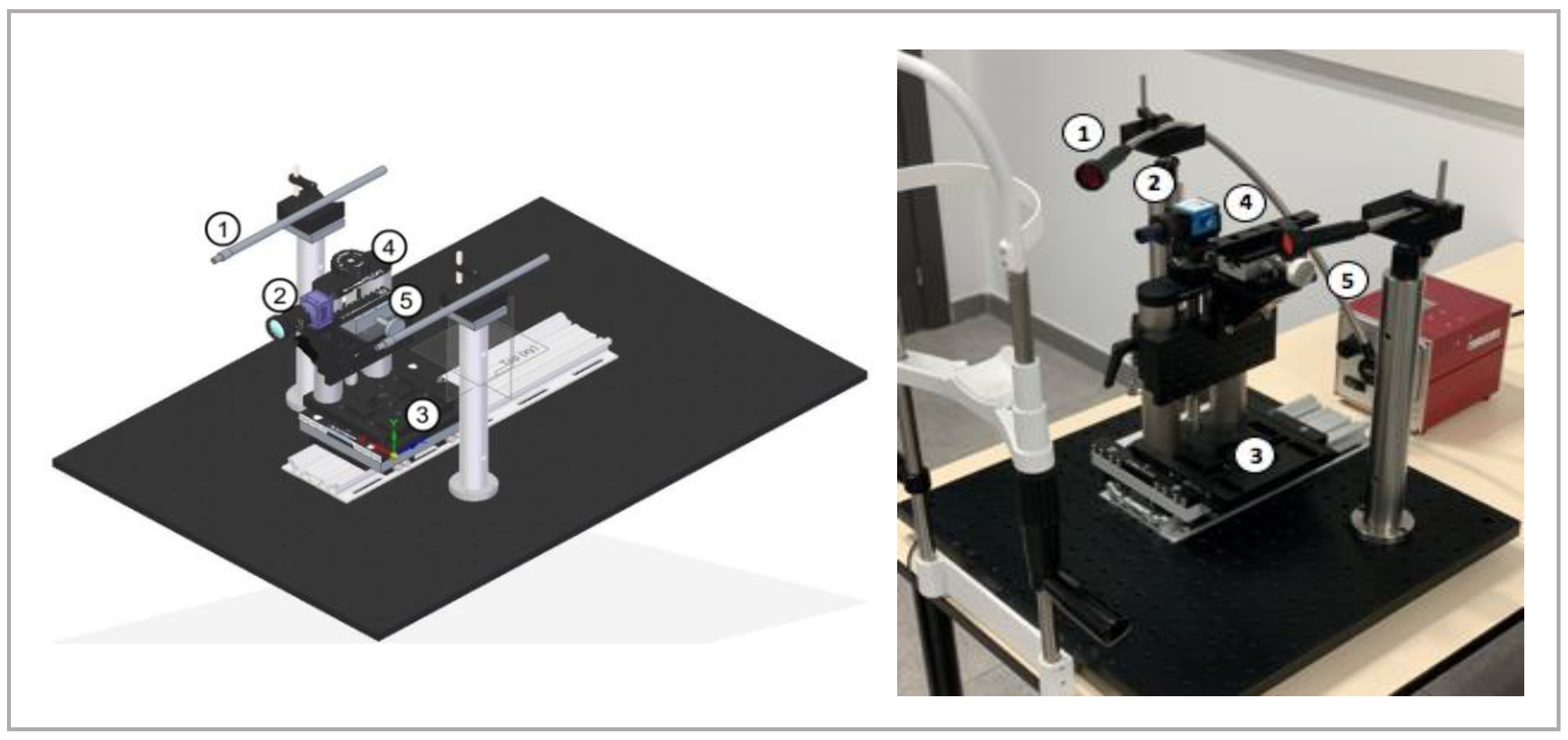
2.1. Meibography
2.2. Meibography Images and Subjective Grading
2.3. Statistical Analysis
3. Results
3.1. Clinical Symptoms and Signs
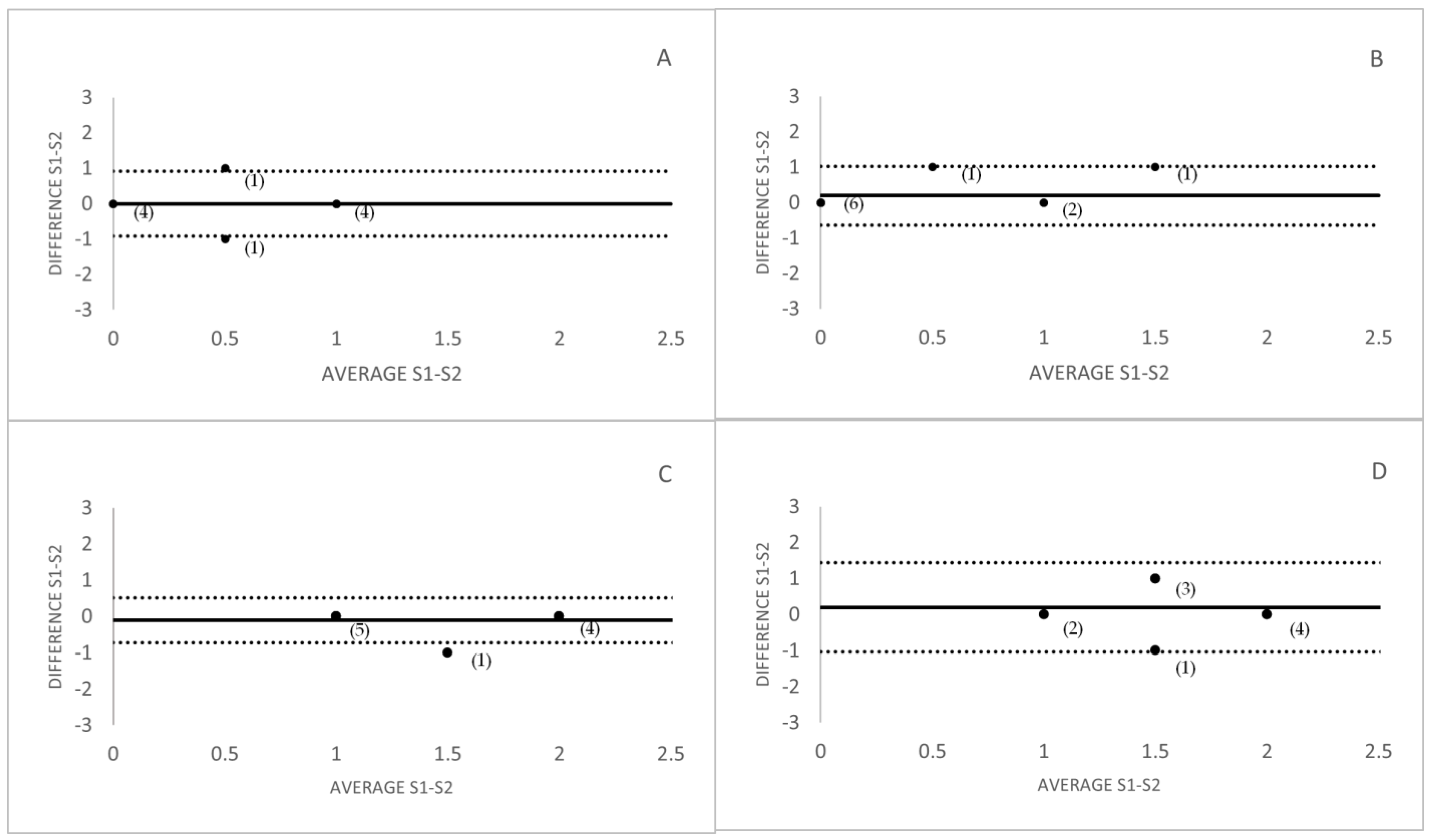
3.2. Inter-Observer and Intra-Observer Variability Analysis
3.3. Repeatability Analysis
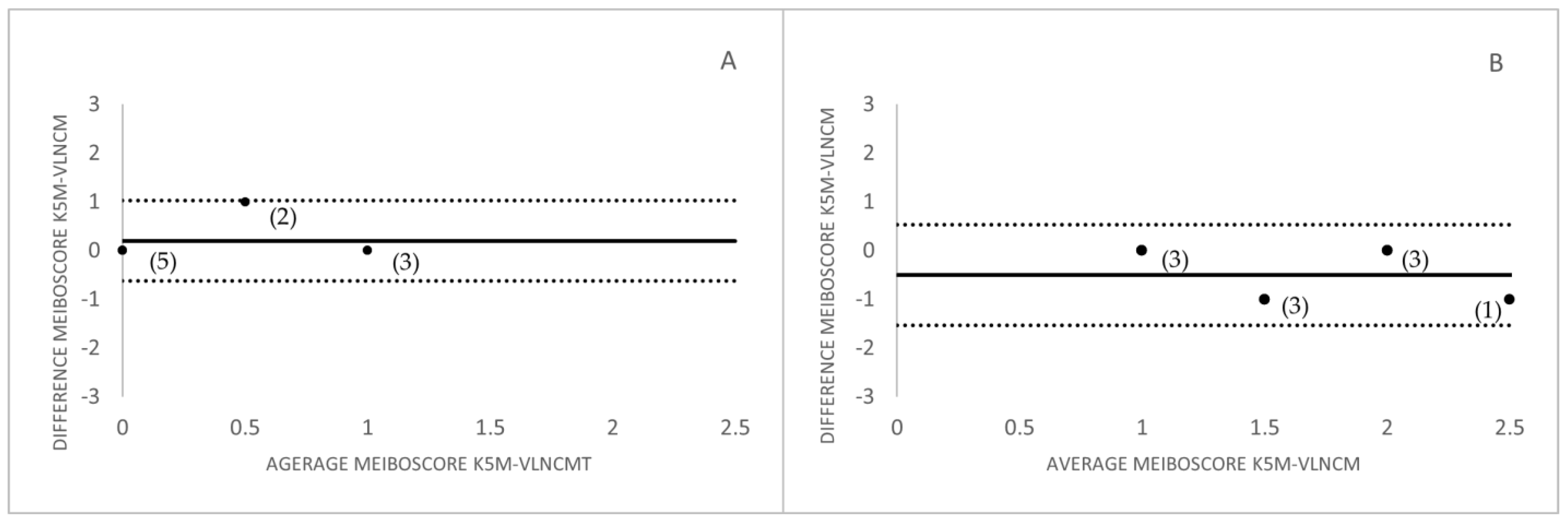
3.4. Comparison between Meibographers
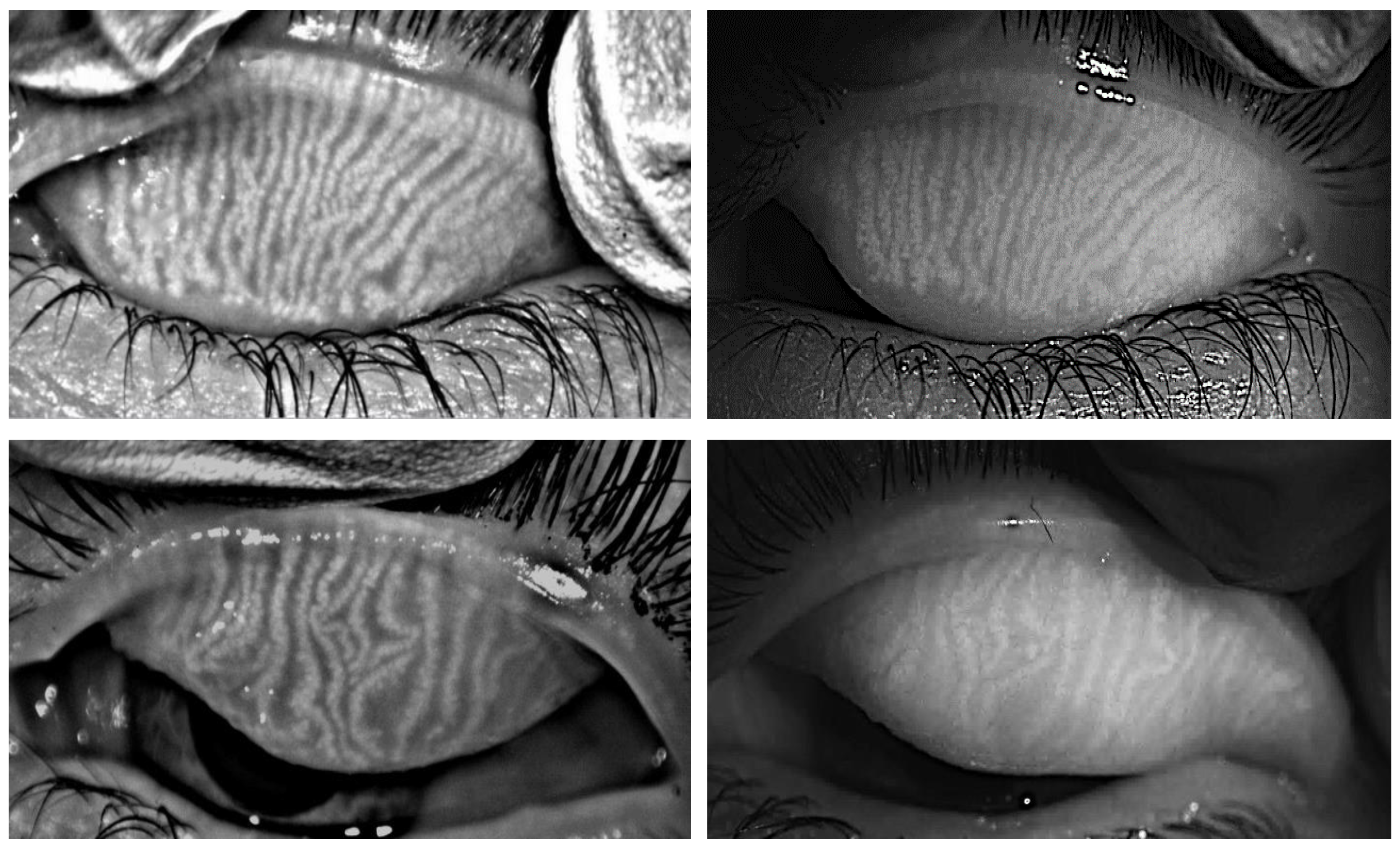
3.5. MG Images Captured with the Two Instruments
4. Discussion
Author Contributions
Funding
Institutional Review Board Statement
Informed Consent Statement
Data Availability Statement
Acknowledgments
Conflicts of Interest
References
- Knop, E.; Knop, N.; Millar, T.; Obata, H.; Sullivan, D.A. The International Workshop on Meibomian Gland Dysfunction: Report of the Subcommittee on Anatomy, Physiology, and Pathophysiology of the Meibomian Gland. Investig. Ophthalmol. Vis. Sci. 2011, 52, 1938–1978. [Google Scholar] [CrossRef] [PubMed]
- Nichols, K.K.; Foulks, G.N.; Bron, A.J.; Glasgow, B.J.; Dogru, M.; Tsubota, K.; Lemp, M.A.; Sullivan, D.A. The International Workshop on Meibomian Gland Dysfunction: Executive Summary. Investig. Ophthalmol. Vis. Sci. 2011, 52, 1922–1929. [Google Scholar] [CrossRef]
- Arita, R.; Itoh, K.; Inoue, K.; Kuchiba, A.; Yamaguchi, T.; Amano, S. Contact Lens Wear is Associated With Decrease of Meibomian Glands. Ophthalmology 2009, 116, 379–384. [Google Scholar] [CrossRef] [PubMed]
- Viso, E.; Rodríguez-Ares, M.T.; Abelenda, D.; Oubiña, B.; Gude, F. Prevalence of Asymptomatic and Symptomatic Meibomian Gland Dysfunction in the General Population of Spain. Investig. Ophthalmol. Vis. Sci. 2012, 53, 2601–2606. [Google Scholar] [CrossRef] [PubMed]
- Yeotikar, N.S.; Zhu, H.; Markoulli, M.; Nichols, K.K.; Naduvilath, T.; Papas, E.B. Functional and Morphologic Changes of Meibomian Glands in an Asymptomatic Adult Population. Investig. Ophthalmol. Vis. Sci. 2016, 57, 3996–4007. [Google Scholar] [CrossRef]
- Nichols, K.K. The International Workshop on Meibomian Gland Dysfunction: Introduction. Investig. Ophthalmol. Vis. Sci. 2011, 52, 1917–1921. [Google Scholar] [CrossRef]
- Arita, R.; Minoura, I.; Morishige, N.; Shirakawa, R.; Fukuoka, S.; Asai, K.; Goto, T.; Imanaka, T.; Nakamura, M. Development of Definitive and Reliable Grading Scales for Meibomian Gland Dysfunction. Am. J. Ophthalmol. 2016, 169, 125–137. [Google Scholar] [CrossRef]
- Arita, R.; Suehiro, J.; Haraguchi, T.; Shirakawa, R.; Tokoro, H.; Amano, S. Objective Image Analysis of the Meibomian Gland Area. Br. J. Ophthalmol. 2014, 98, 746–755. [Google Scholar] [CrossRef]
- Wise, R.J.; Sobel, R.K.; Allen, R.C. Meibography: A Review of Techniques and Technologies. Saudi J. Ophthalmol. 2012, 26, 349–356. [Google Scholar] [CrossRef]
- Peral, A.; Alonso, J.; Gomez-Pedrero, J.A. Effect of Illuminating Wavelength on the Contrast of Meibography Images. OSA Continuum. 2018, 1, 1041–1054. [Google Scholar] [CrossRef]
- Lee, S.M.; Park, I.; Goo, Y.H.; Choi, D.; Shin, M.C.; Kim, E.C.; Alkwikbi, H.F.; Kim, M.S.; Hwang, H.S. Validation of Alternative Methods for Detecting Meibomian Gland Dropout without an Infrared Light System: Red Filter for Simple and Effective Meibography. Cornea 2019, 38, 574. [Google Scholar] [CrossRef] [PubMed]
- Abdelfattah, N.S.; Dastiridou, A.; Sadda, S.R.; Lee, O.L. Noninvasive Imaging of Tear Film Dynamics in Eyes with Ocular Surface Disease. Cornea 2015, 34, S48–S52. [Google Scholar] [CrossRef] [PubMed]
- Geerling, G.; Baudouin, C.; Aragona, P.; Rolando, M.; Boboridis, K.G.; Benítez-del-Castillo, J.M.; Akova, Y.A.; Merayo-Lloves, J.; Labetoulle, M.; Steinhoff, M.; et al. Emerging Strategies for the Diagnosis and Treatment of Meibomian Gland Dysfunction: Proceedings of the OCEAN Group Meeting. Ocul. Surf. 2017, 15, 179–192. [Google Scholar] [CrossRef] [PubMed]
- Schiffman, R.M.; Christianson, M.D.; Jacobsen, G.; Hirsch, J.D.; Reis, B.L. Reliability and Validity of the Ocular Surface Disease Index. Arch. Ophthalmol. 2000, 118, 615–621. [Google Scholar] [CrossRef]
- Price, D.D.; McGrath, P.A.; Rafii, A.; Buckingham, B. The Validation of Visual Analogue Scales As Ratio Scale Measures for Chronic and Experimental Pain. Pain 1983, 17, 45–56. [Google Scholar] [CrossRef]
- Burckhardt, C.S.; Jones, K.D. Adult Measures of Pain: The McGill Pain Questionnaire (MPQ). Arthritis Care Res. 2003, 49, S96–S97. [Google Scholar] [CrossRef]
- Ferris, F.L., III; Kassoff, A.; Bresnick, G.H.; Bailey, I. New Visual Acuity Charts for Clinical Research. Am. J. Ophthalmol. 1982, 94, 91–96. [Google Scholar] [CrossRef]
- Khurana, A.K.; Chaudhary, R.; Ahluwalia, B.K.; Gupta, S. Tear Film Profile in Dry Eye. Acta Ophthalmol. 1991, 69, 79–86. [Google Scholar] [CrossRef]
- Lemp, A. Report of the National Eye Institute/Industry Workshop on Clinical Trials in Dry Eyes. Eye Contact Lens. 1995, 21, 221–232. [Google Scholar]
- OCULUS Optikgeräte GmbH. OCULUS, Wetzlar, Germany. 2021. Available online: https://www.oculus.de (accessed on 18 July 2022).
- Pult, H.; Riede-Pult, B.H. Non-Contact Meibography: Keep It Simple but Effective. Contact Lens Anterior Eye 2012, 35, 77–80. [Google Scholar] [CrossRef]
- Pult, H.; Nichols, J.J. A Review of Meibography. Optom. Vis. Sci. 2012, 89, E760–E769. [Google Scholar] [CrossRef] [PubMed]
- Bland, J.M.; Altman, D. Statistical Methods for Assessing Agreement Between Two Methods of Clinical Measurement. Lancet 1986, 327, 307–310. [Google Scholar] [CrossRef]
- Ban, Y.; Shimazaki-Den, S.; Tsubota, K.; Shimazaki, J. Morphological Evaluation of Meibomian Glands Using Noncontact Infrared Meibography. Ocul. Surf. 2013, 11, 47–53. [Google Scholar] [CrossRef] [PubMed]
- Wong, S.; Srinivasan, S.; Murphy, P.J.; Jones, L. Comparison of Meibomian Gland Dropout Using Two Infrared Imaging Devices. Contact Lens Anterior Eye 2019, 42, 311–317. [Google Scholar] [CrossRef]
- Cuevas, M.; González-García, M.J.; Castellanos, E.; Quispaya, R.; De la Parra, P.; Fernández, I.; Calonge, M. Correlations among Symptoms, Signs, and Clinical Tests in Evaporative-Type Dry Eye Disease Caused by Meibomian Gland Dysfunction (MGD). Curr. Eye Res. 2012, 37, 855–863. [Google Scholar] [CrossRef]
- Srinivasan, S.; Menzies, K.; Sorbara, L.; Jones, L. Infrared Imaging of Meibomian Gland Structure Using a Novel Keratograph. Optom. Vis. Sci. 2012, 89, 788–794. [Google Scholar] [CrossRef]
- Pult, H.; Riede-Pult, B. Comparison of Subjective Grading and Objective Assessment in Meibography. Contact Lens Anterior Eye 2013, 36, 22–27. [Google Scholar] [CrossRef]


| Clinical Tests | Grading Scales |
|---|---|
| Visual Acuity (ETDRS) | LogMar |
| Tear meniscus height | Millimetres |
| Tear film break up time (TBUT) | Seconds |
| Corneal integrity assessment (0–15) (Divided into five regions) | 0: No staining 1: Trace/mild 2: Moderate 3: Severe |
| Bulbar Conjunctival integrity (0–18) (Divided into six regions) | 0: No staining 1: Trace/Mild 2: Moderate 3: Severe |
| Lid Margin Irregularity | 0: No lid margin irregularity 1: Lid margin irregularity in one eyelid 2: Lid margin irregularity in both eyelids |
| Telangiectasia | 0: No Telangiectasia 1: Telangiectasia in one eyelid 2: Telangiectasia in both eyelids |
| Plugging of Gland Orifices | 0: No Plugging 1: Plugging in one eyelid 2: Plugging in both eyelids |
| Meibum Quality (Evaluated in the 8 central glands of both eyelids) | 0: Clear 1: Cloudy 2: Granular 3: Toothpaste 4: No secretion |
| Meibomian Gland (MG) Expression (Evaluated in the 8 central glands of both eyelids) | 0: Light 1: Moderate 2: Strong 3: No expressiondata |
| Clinical Test | Control (n = 10) Mean ± SD | Pathology (n = 10) Mean ± SD | p-Value |
|---|---|---|---|
| Ocular Surface disease index (OSDI) | |||
| Visual analogue scale (VAS) | 6.9 ± 5.2 | 36.3 ± 15.0 | <0.001 * |
| Pain | 0.2 ± 0.6 | 3.9 ± 5.9 | 0.05 * |
| Dry Eye Sensation | 1.2 ± 1.2 | 42.8 ± 26.1 | <0.001 * |
| Irritation | 0.3 ± 0.7 | 35.8 ± 27.6 | <0.001 * |
| Burning | 0.1 ± 0.3 | 14.2 ± 19.1 | <0.001 * |
| Itching | 0.8 ± 1.2 | 26.5 ± 25.3 | 0.01 * |
| Photophobia | 0.6 ± 1.0 | 37.7 ± 27.4 | <0.001 * |
| Foreign Body Sensation | 0.5 ± 0.7 | 27.5 ± 23.6 | 0.01 * |
| Visual Acuity | −0.0 ± 0.1 | 0.0 ± 0.1 | >0.05 |
| Meniscus | 0.3 ± 0.1 | 0.2 ± 0.1 | >0.05 |
| Tear film break up time (TBUT) | 6.3 ± 2.2 | 3.5 ± 1.6 | 0.01 * |
| Corneal integrity assessment (0–15) | 0.4 ± 0.7 | 0.6 ± 0.7 | >0.05 |
| Bulbar Conjunctival integrity (0–18) | 0.2 ± 0.4 | 4.4 ± 3.7 | <0.001 * |
| Lid Margin Irregularity (0–2) | 0.1 ± 0.3 | 1.1 ± 0.7 | <0.001 * |
| Telangiectasia (0–2) | 0.4 ± 0.7 | 1.6 ± 0.5 | <0.001 * |
| Plugging (0–2) | 0.2 ± 0.6 | 0.4 ± 0.5 | >0.05 |
| Meibum Quality (0–4) | 0.0 ± 0.0 | 0.4 ± 0.5 | >0.05 |
| MG Expression (0–3) | 0.2 ± 0.4 | 0.6 ± 1.0 | >0.05 |
| Control | Pathology | |||||||
|---|---|---|---|---|---|---|---|---|
| Title 1 | K5M | VLNCM | K5M | VLNCM | ||||
| (A) | Mean ± SD | p-Value | Mean ± SD | p-Value | Mean ± SD | p-Value | Mean ± SD | p-Value |
| OI-S1 | 0.5 ± 0.53 | >0.05 ◊ | 0.3 ± 0.48 | >0.05 ◊ | 1.5 ± 0.53 | >0.05 ◊ | 1.5 ± 0.53 | >0.05 ◊ |
| OI-S2 | 0.5 ± 0.53 | 0.5 ± 0.71 | 1.5 ± 0.52 | 1.8 ± 0.63 | ||||
| OII-S1 | 0.7 ± 0.48 | >0.05 ◊ | 0.8 ± 0.63 | >0.05 ◊ | 1.7 ± 0.82 | >0.05 ◊ | 1.7 ± 0.82 | >0.05 ◊ |
| OII-S2 | 0.7 ± 0.67 | 0.4 ± 0.52 | 1.8 ± 0.92 | 1.9 ± 0.74 | ||||
| OIII-S1 | 1.2 ± 0.63 | >0.05 ◊ | 0.7 ± 0.67 | >0.05 ◊ | 1.7 ± 0.82 | >0.05 ◊ | 1.7 ± 0.82 | >0.05 ◊ |
| OIII-S2 | 1.2 ± 0.63 | 0.5 ± 0.53 | 1.8 ± 0.92 | 1.7 ± 0.82 | ||||
| (B) | ||||||||
| SI: OI-OII-OIII | 0.01 *◊◊ | >0.05 ◊◊ | 0.04 *◊◊ | >0.05 ◊◊ | ||||
| SII: OI-OII-OIII | <0.001 *◊◊ | >0.05 ◊◊ | >0.05 ◊◊ | >0.05 ◊◊ | ||||
Disclaimer/Publisher’s Note: The statements, opinions and data contained in all publications are solely those of the individual author(s) and contributor(s) and not of MDPI and/or the editor(s). MDPI and/or the editor(s) disclaim responsibility for any injury to people or property resulting from any ideas, methods, instructions or products referred to in the content. |
© 2023 by the authors. Licensee MDPI, Basel, Switzerland. This article is an open access article distributed under the terms and conditions of the Creative Commons Attribution (CC BY) license (https://creativecommons.org/licenses/by/4.0/).
Share and Cite
Diz-Arias, E.; Fernández-Jiménez, E.; Peral, A.; Gomez-Pedrero, J.A. A Comparative Study of Two Imaging Techniques of Meibomian Glands. Life 2023, 13, 791. https://doi.org/10.3390/life13030791
Diz-Arias E, Fernández-Jiménez E, Peral A, Gomez-Pedrero JA. A Comparative Study of Two Imaging Techniques of Meibomian Glands. Life. 2023; 13(3):791. https://doi.org/10.3390/life13030791
Chicago/Turabian StyleDiz-Arias, Elena, Elena Fernández-Jiménez, Assumpta Peral, and Jose A. Gomez-Pedrero. 2023. "A Comparative Study of Two Imaging Techniques of Meibomian Glands" Life 13, no. 3: 791. https://doi.org/10.3390/life13030791
APA StyleDiz-Arias, E., Fernández-Jiménez, E., Peral, A., & Gomez-Pedrero, J. A. (2023). A Comparative Study of Two Imaging Techniques of Meibomian Glands. Life, 13(3), 791. https://doi.org/10.3390/life13030791