Technical Note

Simple and Effective Squash-PCR for Rapid Genotyping of Industrial Microalgae

1
Chemical and Biological Processes Development Group, Pacific Northwest National Laboratory, Richland, WA 99352, USA
2
Marine and Coastal Research Laboratory, Pacific Northwest National Laboratory, Sequim, WA 98382, USA
*
Author to whom correspondence should be addressed.
Life 2024, 14(1), 115; https://doi.org/10.3390/life14010115
Submission received: 17 November 2023 / Revised: 6 January 2024 / Accepted: 8 January 2024 / Published: 12 January 2024
(This article belongs to the Section Microbiology)

Abstract

:
Microalgae are recognized for their versatility in providing renewable energy, biopharmaceuticals, and nutraceuticals, attributed to their sustainable, renewable, and cost-effective nature. Genetic engineering has proven highly effective in enhancing microalgae production. PCR-based genotyping is the primary method for screening genetically transformed microalgae cells. Recently, we developed a novel PCR method, namely Squash-PCR, and employed it for the molecular analysis of industrially important fungi and yeasts. In this study, we successfully implemented the Squash-PCR technique in 12 industrially significant algae species. This approach offers a quick and reliable means of obtaining DNA templates directly from squashed algal cells, eliminating the need for time-consuming and labor-intensive cultivation and genomic DNA extraction steps. Our results demonstrate the effectiveness of Squash-PCR in detecting and characterizing target genes of interest in 12 different algae species. Overall, this study establishes the Squash-PCR method as a valuable tool for molecular studies in algae, enabling researchers to rapidly screen and manipulate genetic traits in diverse algal species.

1. Introduction

Microalgae have gained substantial interest from industrial sectors due to their capability to synthesize diverse biologically active compounds, exhibit rapid biomass growth, and adjust their biochemical composition in response to cultivation conditions [1]. These characteristics make microalgae an exceptionally versatile resource with applications spanning numerous sectors. As an example, microalgae have played multiple roles in pharmaceuticals, food production, wastewater treatment, and biofuel production [2,3]. Thus, these multifaceted microorganisms hold a pivotal position in advancing diverse industries, contributing to the development of sustainable and environmentally friendly technologies. Genetic engineering plays a crucial role in enhancing the traits of these microorganisms, offering opportunities for improved productivity and resource utilization [2]. Genotyping PCR techniques are particularly invaluable tools that enable rapid and precise screening of mutants to select strains with desired characteristics.
Colony PCR with microalgae is challenging due to their complex cell wall structure, which can hinder efficient genetic material extraction and results in a more labor-intensive and time-consuming PCR process. This is particularly problematic when screening a large number of transformants [4]. Boiling algae with a buffer has the potential to disrupt the cell wall, allowing for the release of genomic DNA and thereby facilitating colony PCR detection. Hence, the practice of boiling microalgal cells has been commonly employed to aid in the liberation of genomic DNA, enabling its subsequent PCR amplification [4,5,6]. Nonetheless, research has shown that the effectiveness of colony PCR in microalgal cells after boiling can vary depending on their growth conditions. Notably, cells thriving under favorable growth conditions, such as exponential growth and early stationary phases, tend to yield more amplification products compared to those in less favorable conditions, like during stationary phase and under nitrogen deprivation [5].
In contrast, we have recently developed a novel approach known as Squash-PCR for microbial spore/colony PCR [7]. This method is built upon the “squash” technique, where whole cells are flattened on a glass surface and examined under a microscope. The effectiveness of Squash-PCR has been demonstrated across distinct filamentous fungal and yeast strains, and has consistently yielded high efficiency [7].
In the current study, our primary objective is to expand the utility of the Squash-PCR method to the realm of microalgae. To facilitate a clear understanding of the typical procedures involved in squash preparation for Squash-PCR, we have provided a detailed illustration in Figure 1. In general, the pretreatment of microalgae takes approximately 1 min per sample. We believe that applying the Squash-PCR technique to microalgae will significantly improve our ability to screen for mutants and assess their genotypes efficiently.

2. Materials and Methods

2.1. Strains

The microalgae strains used for Squash-PCR were ordered from UTEX Culture Collection of Algae at UT-Austin and have been listed in Table 1.

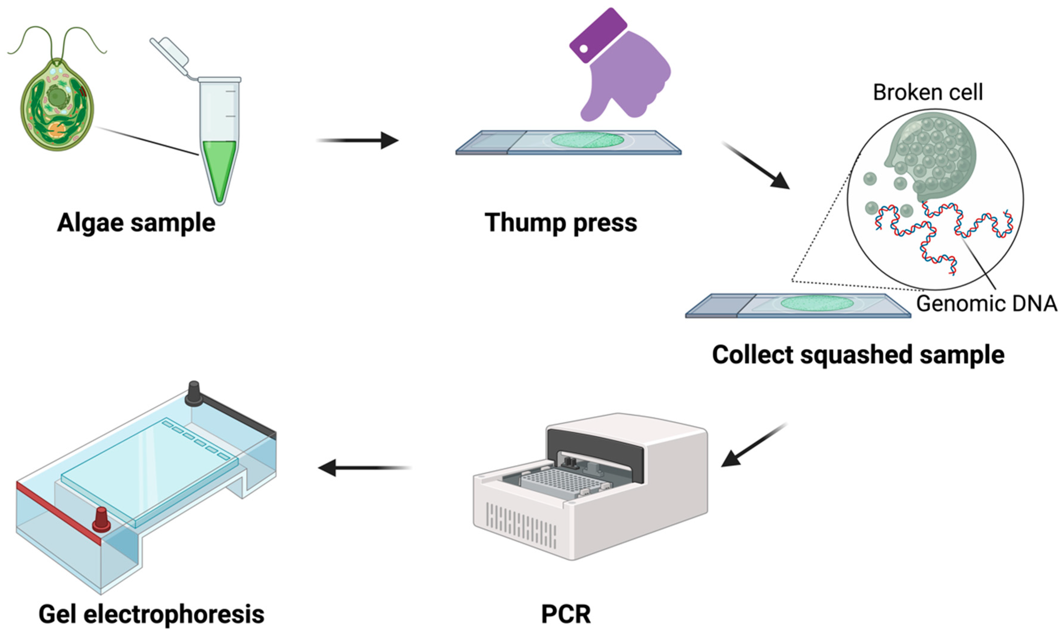
2.2. Squash Preparation

Squash preparations were performed as follows: (1) Dispense ~ 4–5 μL of the microalgae cell solution onto a glass slide and cover it with a coverslip. (2) Apply firm, horizontal pressure to the coverslip using your thumb. (3) Remove the coverslip and add 10 μL of ultrapure water or PBS–Tween 20 solution (v/v, 0.05%). (4) Collect the cell solution with a pipette and dilute it with 10 μL of ultrapure water or PBS–Tween 20 as required. For more detailed instructions, refer to Yuan et al. [7].

2.3. Genomic DNA Extraction

A rapid plant DNA extraction method was employed to extract genomic DNA from all tested microalgal strains [8]. The protocol was as follows: collect a 200–500 μL microalgae sample in a 1.5 mL centrifuge tube and subject it to centrifugation at maximum speed (12,000–16,000× g). Discard the supernatant and proceed by grinding the sample using liquid nitrogen and a pestle. Subsequently, add 400 μL of the extraction buffer to the ground sample and incubate it at 60 °C for 15 min. The extraction buffer consists of a composition of 200 mM Tris-HCl, 200 mM NaCl, 25 mM EDTA, and 0.5% SDS (sodium dodecyl sulfate). Centrifuge the mixture at 4000 rpm to pellet the tissue. Transfer 300 μL of the supernatant to a new 1.5 mL centrifuge tube and add an equal volume of isopropanol. Recap the tubes and gently invert them several times to ensure thorough mixing. Allow the mixture to incubate at room temperature for 5 min. Following this, centrifuge the sample at 4000 rpm to pellet the DNA. Carefully pour off the supernatant without disturbing the pelleted DNA. Next, add 500 μL of 70% ethanol to each sample. Cover the tubes with clean caps and gently invert them to facilitate washing of the DNA. Centrifuge the samples at 4000 rpm for a washing cycle. Place the tubes flat on a paper towel and cover the open tubes with a KimWipe. Once the pellet is dry and no residual liquid remains in the tubes, proceed to re-suspend the pellets using 50 µL of TE buffer or nanopure H2O.

2.4. PCR Procedure

Water is utilized as a negative control during PCR. Genomic DNA extracted using SDS-method (as described in Section 2.3) was used as a positive control for each algae strain. Direct-PCR utilizes the algae solution (1 μL/reaction, ranging from 1 × 10⁵ to 1 × 10⁷ cells/mL) as the DNA template for PCR without any pre-treatment prior to the reaction. The DNA template of Squash-PCR was prepared using squash preparation described in Section 2.2. Dilution is applied for some of the tested samples after squashing.
For the PCR reactions in this study, we used GoTaq Green Master Mix from Promega. The PCR cycling conditions were as follows: an initial denaturation at 95 °C for 2 min, followed by 35 cycles of denaturation at 95 °C for 30 s, annealing at 55 °C for 30 s, and extension at 72 °C for 1 min, with a final elongation step at 72 °C for 5 min. Each reaction employed 1 μL of cell solution in a 20 μL reaction.

2.5. Primers for PCR

All primers used for microalgal Squash-PCR are listed in Table 2. The primers have been manually designed utilizing SnapGene software (https://www.snapgene.com/) and had a melting temperature (Tm) range between 55 and 60 °C.

2.6. Microscopy

Microscopic examination was employed to evaluate the condition of the microalgal cells, both prior to and following the squashing procedure. An Olympus BX60 microscope was utilized to examine samples and collect images. Initially, a volume of 5 to 10 μL of the microalgae sample was pipetted onto a microscope slide, followed by the placement of a coverslip. The slide was examined under a 40X objective lens to capture the initial cellular state. Subsequently, the sample underwent a squashing process by firmly pressing the coverslip using a thumb. This action was performed to disrupt the cell wall, facilitating the release of genomic DNA into the solution. The slide was once again examined under the same 40X objective lens setting to observe and document any discernible alterations in cellular morphology resulting from the squashing procedure.

2.7. Cell Counting

Cell counts were determined using a hemocytometer and a microscope. Cell concentrations were determined as previously described [9]. Briefly, the procedure was as follows: Start by vortexing the target cell suspension to ensure homogeneity. Next, pipette 5 μL of the cell sample into 995 μL of water and thoroughly mix it by vortexing. Place a glass coverslip over the counting chambers of a hemocytometer. Following this, pipette 10 μL of the cell sample into the hemocytometer and allow 30 s for the cells to settle. Subsequently, position the hemocytometer under a microscope using a 40X objective lens. Manually count the cells in the sample. Note that if cells touch the perimeter sides of a corner square, count cells only on two sides, either the two outer sides or the two inner sides, to ensure accuracy. To calculate cell concentration (cells/mL), multiply the total number of cells by the dilution factor and then by 104. Finally, clean the hemocytometer and glass coverslip using 70% ethanol to ensure proper sterilization and preparation for subsequent use.

3. Results

3.1. DNA Template Preparation via Squash Technique

To evaluate the versatility and applicability of the Squash-PCR method across various microalgae species, we carefully selected a diverse set of 12 microalgae strains for our testing (Table 1). Each strain was chosen due to its importance to or potential utility in various industrial applications. For colony PCR to be effective, it is crucial that the genomic DNA from microalgae cells is thoroughly released and extracted before advancing to the PCR amplification step. This initial DNA extraction is a pivotal prerequisite, as the effectiveness of DNA release significantly influences the accuracy and reliability of downstream genetic analyses and manipulations. Following the procedures described in Figure 1, we subjected 12 distinct microalgae strains to the squashing process. To assess the efficacy of the squash preparation, we conducted a comparative analysis of cell samples both before and after squashing under a microscope (Figure 2). The microalgae strains we examined exhibited characteristics typical of single-celled, green microalgae and often presented a spherical or ellipsoidal shape before squashing (Figure 2). Notably, these cells spanned a diverse size and shape, with diameters ranging from 2 to 20 µm. Furthermore, this size variability was not confined to distinct populations but could even be observed within a single group of microalgae. This considerable diversity underscores the importance of employing a versatile and effective technique like squash technique, which can be applied to microalgae of varying shapes and sizes, enhancing its utility in genetic research across different species. Following the squashing process, all tested algae samples exhibited complete cell disruption, with no intact cells remaining observable under the microscope (Figure 2). This outcome indicates the effectiveness of the squash technique in achieving comprehensive cell disruption, a critical step in genetic research and colony PCR.

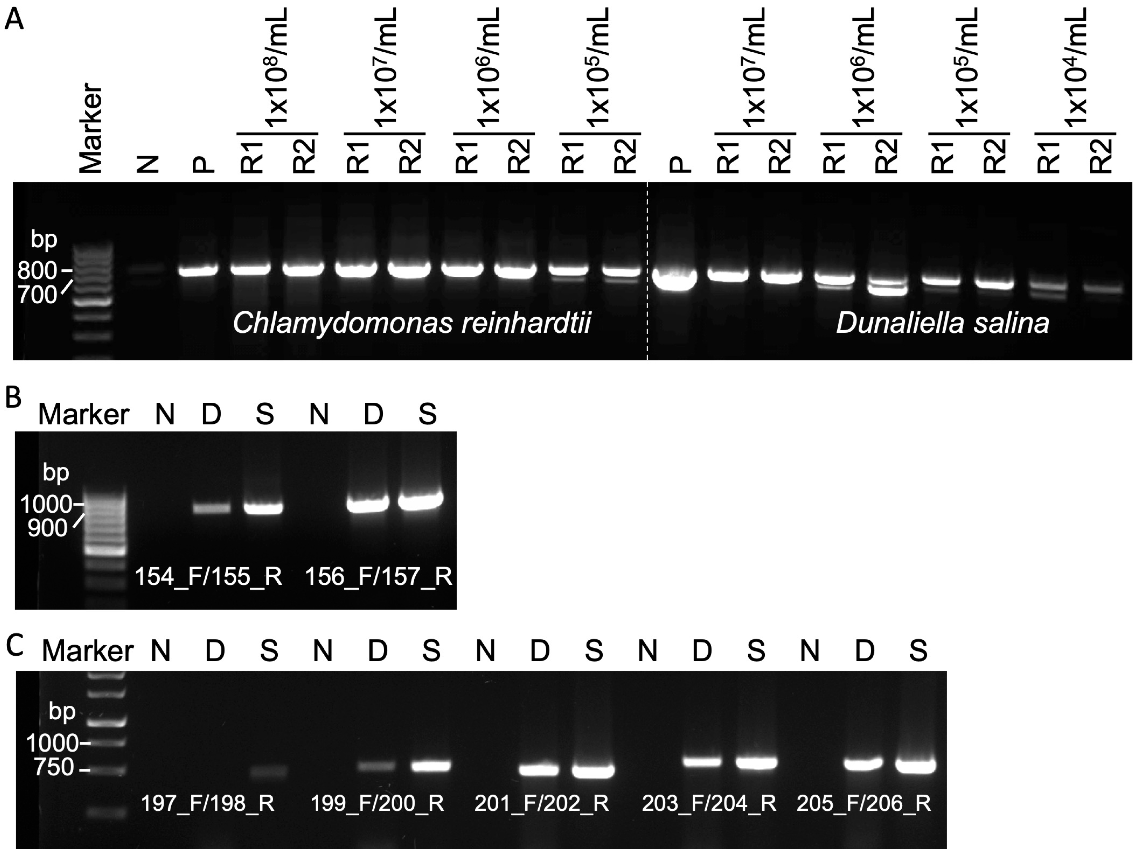
3.2. Efficacy of Squash-PCR in Diverse Microalgae Strains

To assess the effectiveness of Squash-PCR, we employed a specific primer pair (102_F and 103_R) designed for the amplification of the internal transcribed spacer 2 (ITS2) region [10]. The ITS2 region, which is a highly conserved segment of ribosomal DNA but exhibits variability between different species, serves as a valuable genetic marker for distinguishing and identifying various microorganisms, including microalgae [11]. To rigorously evaluate Squash-PCR’s effectiveness, genomic DNA was initially extracted from all tested strains using a widely employed SDS-based DNA extraction method, serving as a positive control in our study. In the direct PCR of strains 1 to 5, strains 1 and 5 produced sharp PCR bands, while strains 2 and 4 showed relatively weaker bands, and strain 3 did not show any band (Figure 3A). In contrast, all five tested strains displayed sharp bands, comparable to the positive control samples, which were visibly more distinct than the results obtained through direct PCR (Figure 3A). In a similar manner, we conducted direct PCR for strains 6 to 12. Among these strains, strain 6, 8, 9, 10, and 11 exhibited sharp PCR bands, indicating successful amplification (Figure 3B). However, we observed relatively weak bands for strain 7 and no bands for strain 12 in the direct PCR reactions. In contrast, employing the Squash-PCR method for strains 6 to 12 resulted in consistently sharp bands, equivalent to those of the positive control samples (Figure 3B). In line with the findings observed in strains 1 to 5, the results obtained through Squash-PCR for strains 6 to 12 exhibited clearer and more distinct outcomes compared to the direct PCR approach. Overall, Squash-PCR consistently outperformed direct PCR in producing clear and distinct bands across all tested strains.

3.3. Evaluation of Squash-PCR in Microalgae Using Different Cell Numbers and Primer Pairs

The performance of Squash-PCR may be influenced by factors such as cell number and the specific primer pairs used. In this study, we evaluated the Squash-PCR technique (with primers 102_F and 103_R) using different cell concentrations for two microalgae species, Chlamydomonas reinhardtii (ranging from 1 × 10⁵ to 108 cells/mL) and Dunaliella salina (ranging from 1 × 10⁴ to 107 cells/mL). In the case of Chlamydomonas reinhardtii, we observed sharp bands across all tested concentrations. Particularly, the concentration of 1 × 10⁷ cells/mL exhibited the highest abundance of PCR products, followed by 1 × 10⁸, 1 × 10⁶, and 1 × 10⁵ cells/mL, in decreasing order (Figure 4A). In Dunaliella salina, the intensity of PCR bands gradually decreased from the 1 × 10⁷ sample to the 1 × 10⁴ sample, with only very faint bands observed at 1 × 10⁴ cells/mL (Figure 4A). Taken together, effective PCR amplification is expected to be achieved when working with cell concentrations ranging from 1 × 10⁵ to 108 cells/mL.
Furthermore, we conducted experiments with different primer pairs in both Chlamydomonas reinhardtii and Dunaliella salina. In Chlamydomonas reinhardtii, sharp bands were obtained using primers 154_F/155_R in the Squash-PCR method, while only a faint band was detected in direct PCR. However, both direct PCR and Squash-PCR using primers 156_F/157_R resulted in sharp bands (Figure 4B). Subsequently, five different primer pairs (197_F/198_R, 199_F/200_R, 201_F/202_R, 203_F/204_R, and 205_F/206_R) were employed to test Squash-PCR in Dunaliella salina (Figure 4C). Sharp PCR bands were observed in both direct PCR and Squash-PCR when using primer pairs 201_F/202_R, 203_F/204_R, and 205_F/206_R. In contrast, sharp bands were exclusively seen in Squash-PCR but not in direct PCR with primer pair 199_F/200_R. However, no sharp bands were detected in either direct PCR or Squash-PCR when employing primers 197_F/198_R. Notably, Squash-PCR consistently yielded higher amounts of PCR products compared to direct PCR. The increased PCR product yield in Squash-PCR further emphasizes the technique’s utility.

4. Discussion

PCR is a fundamental technique in microbiology and molecular biology with applications spanning genetic transformation validation, strain profiling, mutation screening, microalgal isolate selection, and quantitative analyses. PCR is a firmly established tool used to advance microalgae research [4,12,13,14,15,16,17]. Therefore, developing a more streamlined, efficient, and robust PCR method is vital for advancing and accelerating microalgae research. Having an adequate amount of microalgae DNA as the requisite template is the first step for successful PCR amplification. Various cetyltrimethylammonium bromide (CTAB)/SDS-based methods have been frequently reported and modified to extract microalgae DNA [18,19]. Although these methods can yield large quantities of high-quality genomic DNA, they often require several days for extended cell culturing steps and the need for whole-cell lysis methods, including mechanical homogenization, organic solvents, enzymatic digestion, or a combination of these methods [18].
Colony PCR is a well-established method primarily utilized for the rapid screening of genetically transformed colonies that is more commonly applied in bacteria or yeast research. Unlike typical colony PCR used with bacteria or yeast, the process used with microalgae colonies usually involves additional treatments before PCR can be performed. Multiple colony PCR methods have been specifically adapted for some microalgae. These methods generally involve the resuspension of cells using different lysis buffers, such as 10 mM Ethylenediaminetetraacetic acid (EDTA), 5–6% Chelex-100, Tris/EDTA (TE), 0.2 % SDS, 0.2% Triton X-100 or yeast protein extraction buffer (Y-PER), followed by a critical boiling step to promote cell lysis [14,20,21]. Among the various lysis buffers assessed, Y-PER has demonstrated superior effectiveness compared to other methods [21]. Furthermore, the boiling time has been proven to be a critical factor for successful colony PCR. The optimal boiling time for Haematococcus pluvialis, Nannochloropsis salina, and Pseudochlorococcum sp. is 10 min, whereas for Nannochloropsis oculata, 20 min is needed [5]. It is worth noting that algal cells cultivated under unfavorable conditions often develop a thicker secondary cell wall, which can potentially reduce the efficiency of genomic DNA release through boiling and subsequent PCR amplification [5,22]. Therefore, the selection of an appropriate lysis buffer and the optimal boiling time must be based on the characteristics of the specific microalgal strain under investigation.
In contrast, the squash preparation method detailed in this study has proven its versatility in effectively disrupting microalgal cells and enabling the release of genomic DNA. The utilization of Squash-PCR in microalgae strains presents several noteworthy benefits and advantages when compared to previously reported methods. This technique showcases its utility across a diverse array of microalgae species, regardless of variations in cell size, shape, or the complexity of the cell wall. Microalgal cells, with sizes ranging from as small as 2 μm to as large as 20 μm, can be effectively disrupted through squash preparation (Figure 2). Compared to traditional DNA extraction methods that typically necessitate a certain amount of biomass, often starting from a colony [18], Squash-PCR offers a substantial reduction in both labor and turnaround time, transforming the process from several days to just a matter of minutes (approximately 1 min per sample). This time-saving feature makes Squash-PCR a compelling choice for expediting microalgal research and streamlining genetic analysis. In contrast to colony PCR methods that rely on lysis buffers and boiling steps, Squash-PCR eliminates the influence of various lysis buffers, boiling times, and the choice of microalgal species on PCR amplification. This simplification of the process enhances the reliability and versatility of Squash-PCR in microalgal research. The present format of Squash-PCR is highly effective for processing a small number of samples. Recognizing the growing demand for screening large numbers of samples in the field of metabolic engineering, our forthcoming endeavor focuses on adapting this robust method for high-throughput screening. This adaptation will be achieved with the collaboration of mechanical engineers, who will contribute their expertise to enhance the method’s scalability and efficiency.
Interestingly, direct PCR using untreated microalgae cells yielded promising PCR amplification in some of the tested strains such as Monoraphidium minutum 26B-AM, Scenedesmus obliquus UTEX 393, Chlorella vulgaris UTEX 395, Chlamydomonas reinhardtii UTEX 90, and Botryococcus braunii UTEX 572 (Table 1). This outcome differs from previous reports where none of the tested strains displayed any PCR bands when samples were not boiled [5]. This variation may be attributed to the utilization of distinct strains, variations in culture conditions, lysed cells due to stress development, and differences in PCR enzymes and buffers. Regardless of whether direct PCR was successful, Squash-PCR consistently showed a significant increase in PCR amplification in all tested strains. This enhanced sensitivity ensures reliable mutant screening. Squash-PCR is particularly useful when working with specific microalgae strains in which obtaining PCR-quality DNA samples can be challenging due to their complex cell wall structures.
Notably, the squash preparation is effective for releasing and extracting PCR-quality genomic DNA. Nevertheless, it may not be the best choice when large quantities of high-quality DNA are required, such as for Next-Generation Sequencing (NGS) applications.

5. Conclusions

In this study, we introduce Squash-PCR, an innovative method that enhances microalgal research by simplifying DNA extraction and improving PCR efficiency. It has versatility and applicability to various microalgal species and simplifies strain engineering by providing an efficient and reliable genomic DNA release method. With improved PCR product detection and adaptability to different conditions, Squash-PCR is valuable for industrial applications like biofuels, pharmaceuticals, and biotechnology. Overall, this adaptability makes it a versatile technique for a broad range of microalgal strains in both industry and research.

Author Contributions

S.D. and G.Y. conceived the research. G.Y. conducted the experiments. S.G. prepared the microalgae strains. G.Y. wrote the paper. J.J.C., Z.D., K.R.P., R.D.D. and B.A.H. revised the manuscript. S.D. supervised the research. All authors have read and agreed to the published version of the manuscript.

Funding

This research was supported by the Pacific Northwest National Laboratory (PNNL) Directed Research and Development Program. A portion of this work was part of the Agile Bio Foundry (agilebiofoundry.org) supported by the U.S. Department of Energy, Energy Efficiency and Renewable Energy, Bioenergy Technologies Office through contract DE-NL0030038. A portion of this work was part of the DISCOVER national laboratory consortium project sponsored by the U.S. Department of Energy (DOE) Office of Energy Efficiency and Renewable Energy (EERE), Bioenergy Technology Office, under Award No. NL0032208. PNNL is a multi-program national laboratory operated by Battelle Memorial Institute for the U.S. Department of Energy under contract DE-AC05-76RL01830.

Institutional Review Board Statement

Not applicable.

Informed Consent Statement

Not applicable.

Data Availability Statement

Data are contained within the article.

Conflicts of Interest

The authors declare no conflicts of interests.

References

  1. Dolganyuk, V.; Belova, D.; Babich, O.; Prosekov, A.; Ivanova, S.; Katserov, D.; Patyukov, N.; Sukhikh, S. Microalgae: A Promising Source of Valuable Bioproducts. Biomolecules 2020, 10, 1153. [Google Scholar] [CrossRef] [PubMed]
  2. Lin, H.; Wang, Q.; Shen, Q.; Zhan, J.; Zhao, Y. Genetic engineering of microorganisms for biodiesel production. Bioengineered 2013, 4, 292–304. [Google Scholar] [CrossRef] [PubMed]
  3. Radakovits, R.; Jinkerson, R.E.; Darzins, A.; Posewitz, M.C. Genetic engineering of algae for enhanced biofuel production. Eukaryot Cell 2010, 9, 486–501. [Google Scholar] [CrossRef] [PubMed]
  4. Nouemssi, S.B.; Ghribi, M.; Beauchemin, R.; Meddeb-Mouelhi, F.; Germain, H.; Desgagné-Penix, I. Rapid and Efficient Colony-PCR for High Throughput Screening of Genetically Transformed Chlamydomonas reinhardtii. Life 2020, 10, 186. [Google Scholar] [CrossRef] [PubMed]
  5. Liu, J.; Gerken, H.; Li, Y. Single-tube colony PCR for DNA amplification and transformant screening of oleaginous microalgae. J. Appl. Phycol. 2014, 26, 1719–1726. [Google Scholar] [CrossRef]
  6. Tear, C.J.; Lim, C.; Wu, J.; Zhao, H. Accumulated lipids rather than the rigid cell walls impede the extraction of genetic materials for effective colony PCRs in Chlorella vulgaris. Microb. Cell Fact. 2013, 12, 106. [Google Scholar] [CrossRef]
  7. Yuan, G.; Czajka, J.J.; Dai, Z.; Hu, D.; Pomraning, K.R.; Hofstad, B.A.; Kim, J.; Robles, A.L.; Deng, S.; Magnuson, J.K. Rapid and robust squashed spore/colony PCR of industrially important fungi. Fungal Biol. Biotechnol. 2023, 10, 15. [Google Scholar] [CrossRef]
  8. Kasajima, I.; Ide, Y.; Ohkama-Ohtsu, N.; Hayashi, H.; Yoneyama, T.; Fujiwara, T. A protocol for rapid DNA extraction from Arabidopsis thaliana for PCR analysis. Plant Mol. Biol. Report. 2004, 22, 49–52. [Google Scholar] [CrossRef]
  9. Thesseling, F.A.; Bircham, P.W.; Mertens, S.; Voordeckers, K.; Verstrepen, K.J. A Hands-On Guide to Brewing and Analyzing Beer in the Laboratory. Curr. Protoc. Microbiol. 2019, 54, e91. [Google Scholar] [CrossRef] [PubMed]
  10. Radha, S.; Fathima, A.A.; Iyappan, S.; Ramya, M. Direct colony PCR for rapid identification of varied microalgae from freshwater environment. J. Appl. Phycol. 2013, 25, 609–613. [Google Scholar] [CrossRef]
  11. Chen, S.; Yao, H.; Han, J.; Liu, C.; Song, J.; Shi, L.; Zhu, Y.; Ma, X.; Gao, T.; Pang, X.; et al. Validation of the ITS2 Region as a Novel DNA Barcode for Identifying Medicinal Plant Species. PLoS ONE 2010, 5, e8613. [Google Scholar] [CrossRef] [PubMed]
  12. Kira, N.; Ohnishi, K.; Miyagawa-Yamaguchi, A.; Kadono, T.; Adachi, M. Nuclear transformation of the diatom Phaeodactylum tricornutum using PCR-amplified DNA fragments by microparticle bombardment. Mar. Genom. 2016, 25, 49–56. [Google Scholar] [CrossRef]
  13. Fawley, M.W.; Fawley, K.P. Identification of Eukaryotic Microalgal Strains. J. Appl. Phycol. 2020, 32, 2699–2709. [Google Scholar] [CrossRef] [PubMed]
  14. Wan, M.; Rosenberg, J.N.; Faruq, J.; Betenbaugh, M.J.; Xia, J. An improved colony PCR procedure for genetic screening of Chlorella and related microalgae. Biotechnol. Lett. 2011, 33, 1615–1619. [Google Scholar] [CrossRef]
  15. Godhe, A.; Anderson, D.M.; Rehnstam-Holm, A.-S. PCR amplification of microalgal DNA for sequencing and species identification: Studies on fixatives and algal growth stages. Harmful Algae 2002, 1, 375–382. [Google Scholar] [CrossRef]
  16. Penna, A.; Galluzzi, L. The quantitative real-time PCR applications in the monitoring of marine harmful algal bloom (HAB) species. Environ. Sci. Pollut. Res. Int. 2013, 20, 6851–6862. [Google Scholar] [CrossRef]
  17. Duong, V.T.; Li, Y.; Nowak, E.; Schenk, P.M. Microalgae Isolation and Selection for Prospective Biodiesel Production. Energies 2012, 5, 1835–1849. [Google Scholar] [CrossRef]
  18. Jagielski, T.; Gawor, J.; Bakuła, Z.; Zuchniewicz, K.; Żak, I.; Gromadka, R. An optimized method for high quality DNA extraction from microalga Prototheca wickerhamii for genome sequencing. Plant Methods 2017, 13, 77. [Google Scholar] [CrossRef]
  19. Stark, J.R.; Cardon, Z.G.; Peredo, E.L. Extraction of high-quality, high-molecular-weight DNA depends heavily on cell homogenization methods in green microalgae. Appl. Plant Sci. 2020, 8, e11333. [Google Scholar] [CrossRef]
  20. Cao, M.; Fu, Y.; Guo, Y.; Pan, J. Chlamydomonas (Chlorophyceae) colony PCR. Protoplasma 2009, 235, 107–110. [Google Scholar] [CrossRef]
  21. Packeiser, H.; Lim, C.; Balagurunathan, B.; Wu, J.; Zhao, H. An extremely simple and effective colony PCR procedure for bacteria, yeasts, and microalgae. Appl. Biochem. Biotechnol. 2013, 169, 695–700. [Google Scholar] [CrossRef] [PubMed]
  22. Wang, S.-B.; Hu, Q.; Sommerfeld, M.; Chen, F. Cell wall proteomics of the green alga Haematococcus pluvialis (Chlorophyceae). Proteomics 2004, 4, 692–708. [Google Scholar] [CrossRef] [PubMed]
Figure 1. The workflow of Squash-PCR in microalgae.
Life 14 00115 g001
Figure 2. Microscopic observation of various microalgae strains in squash preparations. (A) Chlamydomonas reinhardtii UTEX 89. (B) Scenedesmus sp. UTEX 1589. (C) Haematococcus pluvialis UTEX 2505. (D) Chlorella vulgaris UTEX 395. (E) Chlamydomonas reinhardtii UTEX 90. (F) Botryococcus braunii UTEX 572. (G) Dunaliella salina UTEX LB 200. (H) Monoraphidium minutum 26B-AM. (I) Chlorella sorokiniana DOE1412. (J) Tetraselmis striata LANL1001. (K) Picochlorum celeri TG2. (L) Scenedesmus obliquus UTEX 393. Scale bar = 20 µm.
Life 14 00115 g002
Figure 3. Squash-PCR of different microalgae strains using primers targeting ITS2 region. (A) Direct PCR using the cell suspensions of strain 1 to 5 without any treatment. P, the genomic DNA of strain 1 was extracted using SDS, serving as a positive control. (B) Squash-PCR of strains 1 to 5. (C) Direct PCR using the cell suspensions of strains 6 to 12 without any treatment. (D) Squash-PCR of strains 6 to 12. N, negative control; *, the samples underwent a 5-fold dilution following the squashing process; Squash-PCR was performed with two replicates, labeled as R1 and R2.
Life 14 00115 g003
Figure 4. Investigation of potential limiting factors affecting Squash-PCR in microalgae. (A) The effective range of cell concentration for Squash-PCR in Chlamydomonas reinhardtii and Dunaliella salina. Squash-PCR was performed with two replicates, labeled as R1 and R2. (B) The examination of Squash-PCR in Chlamydomonas reinhardtii with different primers. (C) The examination of Squash-PCR in Dunaliella salina with different primers. N, negative control; P, positive control; D, direct PCR; S, Squash-PCR.
Life 14 00115 g004
Table 1. Results of Squash-PCR in different microalgae strains.
No.Microalgae Species1 Direct-PCRSquash-PCR
1Monoraphidium minutum 26B-AM+++
2Chlorella sorokiniana DOE1412+ *++
3Tetraselmis striata LANL1001++
4Picochlorum celeri TG2+ *++
5Scenedesmus obliquus UTEX 393+++
6Chlamydomonas reinhardtii UTEX 89+++
7Scenedesmus sp. UTEX 1589+ *++
8Haematococcus pluvialis UTEX 2505+++
9Chlorella vulgaris UTEX 395+++
10Chlamydomonas reinhardtii UTEX 90+++
11Botryococcus braunii UTEX 572+++
12Dunaliella salina UTEX LB 200++
+: Indicates a positive PCR band. −: Indicates a negative PCR band. ++: Represents a brighter PCR band. *: Indicates a faint PCR band. 1 Direct-PCR: PCR utilizing the algae solution directly as the DNA template without any pre-treatment before the PCR reaction.
Table 2. The oligos used for PCR in this study.
PrimerSequenceMicroalgae SpeciesGeneSize (bp)
102_FAGGAGAAGTCGTAACAAGGTStrains 1 to 12ITS2700–900
103_RTCCTCCGCTTATTGATATGC
154_FTTGCACACAAGAACGCATGAChlamydomonas
reinhardtii
FTSY815
155_RGGCATTGGAGTAAACGACCC
156_FCTACGTCGCCTTACTGTGTGChlamydomonas
reinhardtii
ZEP809
157_RGATTGCCTACTCACCACTCG
197_FATGGTTCCACAAACTGAAACGDunaliella salinarbcL480
198_RGTCACGCTCAACTTGAATACC
199_FCGTAGACTGTGTAGAAGCTGDunaliella salinaorf121518
200_RGTGGAACTACACCTTCAGGT
201_FATGCCAATTGGTGTTCCACDunaliella salinaclpP496
202_RGTGCATTAAAATCGTAAGGCG
203_FCAATGCGCACACCAGAAGDunaliella salinaatpA551
204_RGCATATAGTTTCCTGCGTCC
205_FATGGCACGTGCTAAATTTGADunaliella salinatufA524
206_RGCAGAACCTGAAACGATAGG
Disclaimer/Publisher’s Note: The statements, opinions and data contained in all publications are solely those of the individual author(s) and contributor(s) and not of MDPI and/or the editor(s). MDPI and/or the editor(s) disclaim responsibility for any injury to people or property resulting from any ideas, methods, instructions or products referred to in the content.

