Abstract
Enterococci are a group of microorganisms that have a controversial position from some scientific points of view. The species of the greatest clinical importance are E. faecalis and E. faecium, which are common agents of nosocomial infections. However, enterococci also have important applications in the dairy industry, as they are used as non-starter lactic acid bacteria (NSLAB) in a variety of cheeses, especially artisanal cheeses. The aim of this study was to determine the presence of representatives from the Enterococcus genus using PCR and MALDI-TOF MS methods on samples of raw milk, processing environment swabs, and cheese from four different artisanal dairy plants in Slovakia. Among the 136 isolates of enterococci, 9 species of genus Enterococci (E. faecalis, E. faecium, E. durans, E. devriesi, E. hirae, E. italicus, E. casseliflavus, E. malodoratus, and E. gallinarum) were identified and were tested for their antimicrobial resistance (AMR) to 8 antibiotics (amoxicillin, penicillin, ampicillin, erythromycin, levofloxacin, vancomycin, rifampicin, and tetracycline); most of them were resistant to rifampicin (35.3%), ampicillin (22.8%), and tetracycline (19.9%). A PCR analysis of vanA (4.41%) and tetM (14.71%) revealed that antimicrobial resistance genes were present in not only phenotypic resistant isolates of enterococci but also susceptible isolates. The investigation of antimicrobial resistance in enterococci during the cheesemaking process can be a source of valuable information for public health in the concept of “One Health”.
1. Introduction
Enterococci can be concisely described as Gram-positive ovoid cocci occurring singly or in pairs, both in short chains and in clusters [1]. They are non-sporular and they can produce yellow pigment. Enterococci are catalase-negative, but when cultured on blood media, they can show a positive catalase reaction [1,2].
Enterococci occur and grow in a variety of cheeses, especially those made from raw or pasteurized goat’s, ewe’s, or cow’s milk. In these types of milk, the most commonly detected Enterococcus spp. are E. faecium, E. faecalis, E. hirae, and E. durans [3]. Within this genus, there is a specific group of bacteria that can play a role in cheesemaking; these representatives are generally referred to as controversial microorganisms due to their simultaneous positive (producing bioactive peptides) and negative (biogenic amine producer) influences in the process [3,4,5]. Enterococci have an important role in the production of certain types of cheese and fermented sausages produced in the Mediterranean basin. Through their lipolytic and proteolytic activity, they contribute to the development of desirable organoleptic characteristics [6,7]. In many artisanal cheeses, enterococci are part of the non-starter lactic acid bacteria, and they have, in some instances, been used as components of experimental starter cultures [8].
The impact of the enterococcal microbiota of artisanal cheeses on the health of their hosts is still the object of debate, despite the wealth of knowledge gathered in recent years on their presence, technological properties, potential health benefits, antibiotic resistance, and carriage of virulence factors [9]. There are possible risks associated with each method of cheese production, specifically through the transfer of microorganisms that may pose a risk to the health of the final consumer. Therefore, it is necessary to ensure adequate hygiene conditions from the time when the milk is obtained to the time when the product is unpacked by the consumer. The European Union has established a maximum limit for the presence of coliforms and Escherichia coli in some dairy products, both of which are considered indicators of hygiene, while no limit has been set for enterococci. Enterococcus spp. do not have a generally recognized as safe (GRAS) status nor are they included on the qualified presumption of safety (QPS) list [10,11,12].
Antibiotic resistance is a global health challenge, involving the transfer of bacteria and genes between humans, animals, and the environment. A set of genetic determinants allows enterococci to colonize their host efficiently, and they can exchange genes with other bacteria, promoting their adaptation to the seemingly inhospitable conditions they face [13]. The acquisition of antimicrobial resistance during the development of enterococci is one of the factors that explain their success as nosocomial pathogens [14]. Enterococci demonstrate these abilities to acquire antimicrobial resistance through the exchange of resistance-coding genes transmitted by transposons and plasmids. Plasmids are abundant in enterococci and entail another important contribution to their genomic plasticity [8,15]. Species of the genus Enterococcus are naturally resistant to cephalosporins and low concentrations of aminoglycosides, sulphonamides, clindamycin, quinupristine, dalfopristin, and most beta-lactams. In addition, E. casseliflavus and E. gallinarum have natural resistance to low vancomycin concentrations [16]. Acquired enterococcal resistance includes resistance to chloramphenicol, erythromycin, tetracycline, fluoroquinolones, glycopeptides, and high concentrations of clindamycin, aminoglycosides, and beta-lactams [13,16]. Despite the profusion of studies on antibiotic resistance and its genetic determinants of clinical enterococci, few studies are available that focus on enterococcal species carried by traditional cheeses [8].
Based on the above, the aim of our study was to determine the occurrence of Enterococcus spp. and identify them in raw bulk milk, milk after pasteurization, the environment of cheesemaking, and cheese made from pasteurized milk in Slovakia and subsequently detect antimicrobial resistance and the presence of antimicrobial resistance genes.
In addition, most studies have addressed the occurrence of enterococci and their antimicrobial resistance in raw milk or cheeses made from raw milk, but few studies are focused on the cheesemaking environment and the occurrence of enterococci in cheeses, even though they are made from pasteurized milk. Also, evaluation of the two standard systems for assessing AMR may show differences between them. This study may contribute to the acquisition of knowledge in this area.
2. Materials and Methods
2.1. Isolation and Identification of Strain
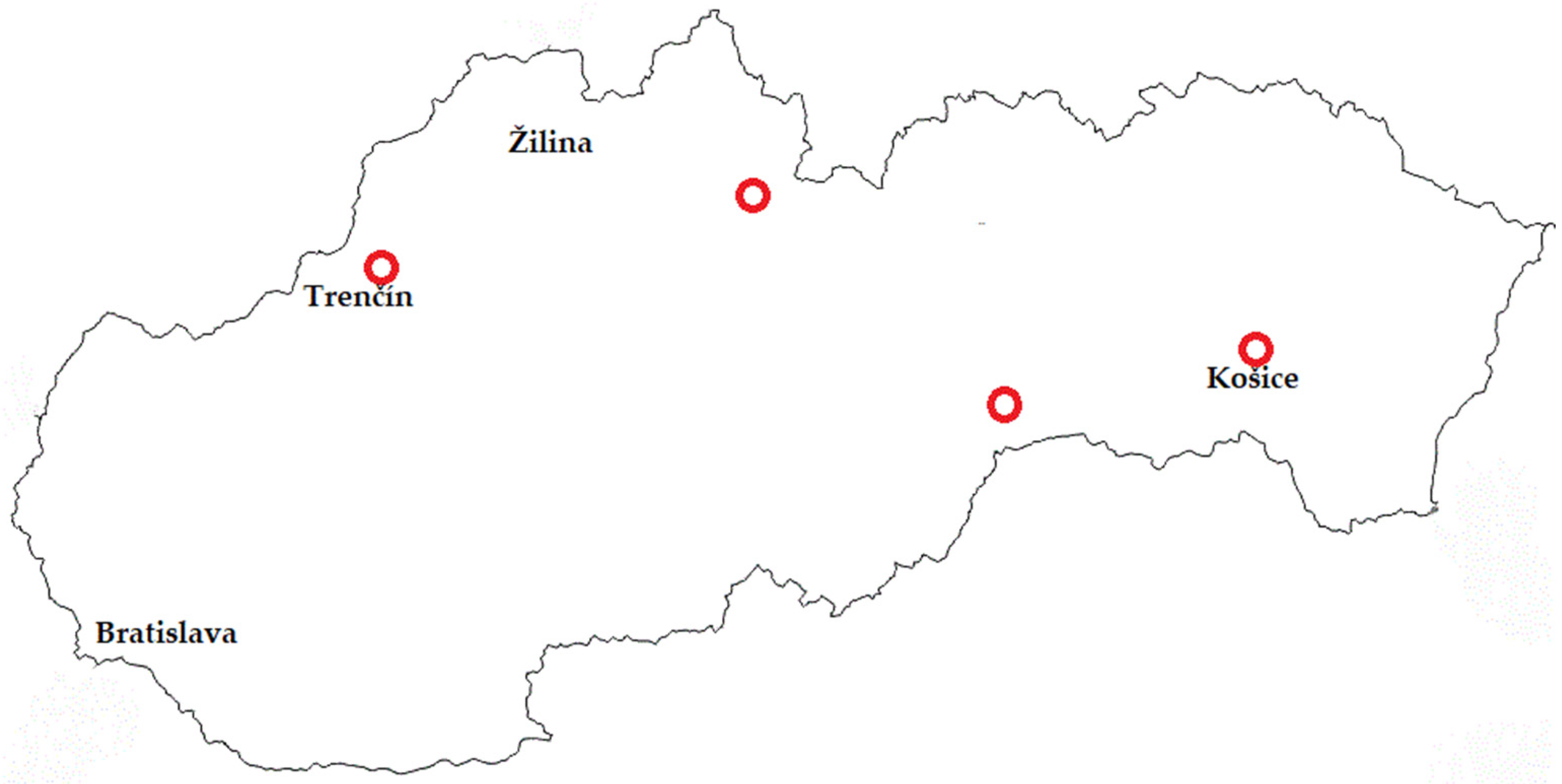
A total of 127 samples were taken disposably from four different dairy plants in Slovakia (Figure 1) that are focused only on artisanal cheesemaking. The samples (Table S1) included raw bulk milk (n = 14), milk after pasteurization (n = 14), swabs from the cheesemaking environment (n = 75), and produced cheeses (n = 24). Cheeses are made from pasteurized milk through the addition of starter cultures and it is possible to buy them in the local shop as well as in supermarkets in Slovakia.

Figure 1.
Map of Slovakia with places (red circles), where all samples were taken for the study.
The swab samples, samples of raw milk, and pasteurized milk were taken in the dairy plants. Cheeses that had aged for a maximum of three weeks were selected, and samples were taken from dairy plants immediately after packaging. The raw bulk milk and milk after pasteurization was taken in volumes of 500 mL per sample. The moistened swabs were taken from various places (Table S2) from the areas where it was possible to take swabs using sterile swab templates with a capacity of 100 cm2 according to ISO 18593 [17]. The other swabs were taken according to availability (molds, hands, switches, knives) without using the corresponding area size. The cheeses taken were packaged by producers in a quantity of at least 150 g per sample. Milk samples were placed in sterile sample containers and swabs in sterile tubes with 0.1% (wt/vol) peptone water, and all samples were transported at 4 °C without the addition of preservatives to the laboratory at the University of Veterinary Medicine and Pharmacy in Košice for analysis. Within 4 h of collection, all samples were subjected to microbiological analysis.
All the samples underwent decimal dilution according to ISO 6887-5 [18]. Then, the samples in volume 0.1 mL were streaked on Slanetz and Bartley agar (Merck, Germany) plates and incubated at 37 °C for 24–48 h. After incubation, the bacterial count was evaluated. Subsequently, based on characteristic appearance and typical colonies in numbers of three per plate were taken and purified on Brain Heart Infusion Agar (Merck, Germany) and stored at −18 °C for further investigation.
The DNA was extracted from the 156 enterococcal strains according to Hein et al. [19]. PCR was used to identify the genus of Enterococcus according to Ke et al. [20] and Martineau et al. [21]. The primers derived from the tuf gene were used to obtain a specific sequence for the genus Enterococcus spp. Each primer was synthesized in Genomed (Table 1; Warszawa, Poland). The PCR protocol was optimized as follows: initial denaturation at 95 °C for 13 min, followed by 30 cycles (denaturation at 95 °C for 20 s, annealing at 52 °C for 30 s, and extension at 72 °C for 2 min). The final extension was performed at 72 °C for 10 min. The HotFirepol® Mastermix (Solis BioDyne, Tart, Estonia) was used in the PCR. The PCR products were visualized in a 1.5% agarose gel with Goldview™ nucleic acid staining (Beijing SBS Genetech Co., Ltd., Beijing, China) by using the MiniBIS Pro® (DNR Bio-Imaging system Ltd., Neve Yamin, Israel).

Table 1.
The primers used in this study for the identification of Enterococcus spp.
After PCR identification, the 136 isolates belonging to the Enterococcus genus (Figure S2) were streaked on Columbia Blood Agar (Merck, Darmstadt, Germany) to prepare for MALDI-TOF MS examination and incubated at 37 °C for 24 h. To make the analysis as accurate as possible, individual samples were prepared via an extraction procedure using ethanol and formic acid [23]. Then, the colonies were used for identification using MALDI-TOF MS (Bruker Daltonics, Billerica, MA, USA).
2.2. Antimicrobial Susceptibility Testing
The identified Enterococcus spp. isolates were tested for antibiotic susceptibility using the disc diffusion method (DDM) according to the procedure described in the CLSI document [24]. Eight antibiotics were tested: amoxicillin (AML, 25 µg), penicillin (P, 10 µg), ampicillin (AMP, 10 µg), erythromycin (E, 10 µg), levofloxacin (LEV, 5 µg), vancomycin (VAN, 30 µg), rifampicin (RD, 5 µg), and tetracycline (TET, 30 µg). Antibiotics were selected based on the most common antimicrobial agents used in veterinary and human medicine in Slovakia for treatment [25,26]. Enterococcus faecalis ATCC 29212 was used as a reference strain. The results were evaluated according to two systems, namely the European Committee on Antimicrobial Susceptibility Testing [27] and the Clinical and Laboratory Standards Institute guidelines [24]. The disks used for susceptibility testing were manufactured by HIMEDIA, Mumbai, India. The diameters of the zone of inhibition were recorded in millimeters (mm) and interpreted as susceptible or resistant.
2.3. Detection of Antimicrobial Resistant Genes (ARG)
Genes that can confer antimicrobial resistance (AMR) to glycopeptide, vanA, and tetracycline, tetM, were detected in a PCR using the gene-specific primers shown in Table 2. The PCR protocol was optimized as follows: initial denaturation at 95 °C for 13 min, followed by 30 cycles of denaturation at 95 °C for 20 s, annealing at different temperatures depending on the gene for 30 s (Table 2), and extension at 72 °C for 2 min. The final extension was performed at 72 °C for 10 min. PCR amplicons were run on 1.5% agarose gel. The expected sizes of PCR products differed for each gene (Table 2).
Table 2.
The primers used in this study for the detection of resistance genes using a PCR-based method.
2.4. Data Analysis
Data were entered, cleaned, and validated in a Microsoft TM Excel spreadsheet (MS Office Excel® 2021, Wanchai, Hong Kong). The average number of enterococci in the raw milk and cheese was recalculated to log10 transformation. The distribution of each species in the raw milk was determined by calculating the percentage of each species out of the total number of enterococci isolated. The PCR results (positive or negative) were reference variables for descriptive analyses. Univariate analyses were conducted for descriptive statistics and the data were presented as percentages.
3. Results
3.1. Isolation and Enumeration of Enterococci
Enterococci were presented in 52 (40.94%) of all 127 tested samples. In total, 11 (78.57%) of 14 raw bulk milk samples, 31 (41.33%) of 75 environmental samples, and 10 (41.67%) of 24 cheese samples harboring the presence of enterococci. The average enumerations of enterococci in raw milk and cheese were 3.38 log10 ± 0.39 CFU/mL and 3.88 log10 ± 0.79 CFU/g, respectively (Table 3). In all the samples of milk, after batch pasteurization at a low temperature, no enterococci were detected. The milk prepared via pasteurization at each dairy plant was used for cheesemaking.
Table 3.
Enumeration of enterococci in different samples from artisanal dairy plants in Slovakia.
The samples from the environment were divided into nine groups (Table S2). The highest number of positive samples were from the samples from ripening rooms, work aprons, and vats for cheesemaking. Only one swab was evaluated for the work apron; this is because the workers of the examined dairy plants use washable work aprons. No enterococci were detected in swabs from pasteurization equipment. The evaluation of enterococcal counts in the environment was not included in the study, considering it was not possible to take samples with the same area from some types of swabs, such as, for instance, hands, molds, handles, and switches, but the presence of enterococci in these parts was of interest to our investigation.
In the raw milk, environment, and cheese samples, experimental analysis confirmed the presence of seven, five, and five species of enterococci, respectively (Table 4). Only in raw milk were E. malodoratus and E. hirae found, and only in cheeses was E. gallinarum detected. Overall, the most common species were E. faecalis (38.97%) and E. faecium (34.56%), but only one isolate of E. faecium was detected in raw milk. The majority of E. faecium isolates were found in the environment and cheeses.
Table 4.
Distribution and number of isolates among Enterococcus spp. identified using MALDI-TOF MS isolated from different samples associated with cheesemaking.
3.2. Antimicrobial Susceptibility Test Results for Enterococci
Antimicrobial susceptibility results were evaluated separately according to EUCAST 2023 and CLSI 2023 documents because of the differences between these two systems. Evaluation using the EUCAST document was possible for only four of the selected antimicrobial agents (AML, AMP, VAN, and LEV). The zone diameter breaking points defined by EUCAST are different compared to values defined by CLSI. The results according to EUCAST are presented in Table 5 and Table 6.
Table 5.
Antimicrobial-resistant phenotypes of enterococci isolated from different samples associated with cheesemaking according to EUCAST (2023).
Table 6.
Antimicrobial-resistant phenotypes of enterococci identified by MALDI-TOF MS according to EUCAST (2023).
No similarities can be found in the evaluation results between EUCAST and CLSI documents (Table 7 and Table 8) because the final interpretation guidelines are different for all agents except amoxicillin. In particular, the percentage of resistant isolates is lower according to the evaluation of AMR according to EUCAST compared to CLSI for similar agents. Evaluation by CLSI (Table 7) showed that all the isolates of enterococci were only fully susceptible to amoxicillin. For penicillin, erythromycin, vancomycin, levofloxacin, and tetracycline, the rates of resistance among isolates were in the range of 2.9–19.9%. Higher resistance to rifampicin (35.3%) and ampicillin (22.8%) were observed. Isolates from raw milk showed the highest resistance to ampicillin (56.8%) and rifampicin (34.1%) and isolates from the environment showed the highest resistance to rifampicin (29.2%). Isolates from cheeses showed the highest resistance to rifampicin (43.2%) and vancomycin (31.8%). The distribution of enterococci by species showed that in all isolates of E. faecalis, the highest resistance to rifampicin (62.3%) and vancomycin (33.9%) was observed. Furthermore, in isolates of E. faecium, the highest resistance to erythromycin (21.3%) was observed. The resistance levels of other species are listed in Table 8.
Table 7.
Antimicrobial-resistant phenotypes of enterococci isolated from different samples associated with cheesemaking according to CLSI (2023).
Table 8.
Antimicrobial-resistant phenotypes of enterococci identified by MALDI-TOF MS according to CLSI (2023).
Multidrug resistance (MDR) is defined as resistance to three or more classes of antibiotics. In this study, overall, thirty-five isolates evaluated using the CLSI 2023 document were multidrug resistant. Four isolates (milk and cheeses) were resistant to five antibiotics. Isolates from the environment were the most resistant to three antibiotics; however, this group contained the most isolates that were not resistant to any antibiotics. The most resistant isolates were E. faecalis, all of which were resistant to at least one to five antibiotics, except one isolate. On the other hand, thirty-two isolates of E. faecium were reported as sensitive to all the tested antibiotics. Overall, only five isolates from the other species of enterococci, including E. devriesi (2), E. durae (1), E. hirae (1), and E. gallinarum (1), were multidrug resistant to a maximum of four antibiotics.
3.3. Detection of Resistance Genes among All Isolates Genus Enterococcus
The evaluation of enterococci safety through the in vitro expression of virulence traits does not always reflect the real hazard in these microorganisms because of the presence of silent genes, which could potentially be activated by environmental conditions, thus transforming these bacteria into pathogens or enhancing their pathogenicity [30]. The isolates of enterococci were tested for the resistance genes vanA (Figure S2A) and tetM (Figure S2B). We focused on only two genes, but they were studied not only in isolates with detected resistance but also in those that were evaluated as sensitive to vancomycin and tetracycline.
Among all the identified isolates that belonged to the Enterococcus genus, there were five E. faecalis isolates and one E. devriesi isolate with the resistance gene vanA. However, only two isolates with vanA were evaluated as resistant according to CLSI, while four isolates with vanA were susceptible.
Overall, 20 isolates of enterococci carried tetM, with most of them being E. faecalis (12) and the others being E. durans (3), E. devriesi (1), E. italicus (3), and E. gallinarum (1). There were no E. faecium isolates with identified tetM genes. According to CLSI, out of the 20 isolates with confirmed tetM, 14 and 6 were evaluated as resistant and susceptible, respectively. The occurrence of the gene in all enterococcal isolates, regardless of phenotypic manifestation, was 14.7%. Multidrug resistance was detected in ten isolates with a confirmed tetM gene; nine isolates were E. faecalis and one isolate was E. gallinarum. No enterococcal isolate carried both genes studied. Raw milk samples contained the highest occurrence of both vanA (6.8%) and tetM (29.6%). Enterococcal isolates from the environment of cheesemaking revealed two isolates with the vanA gene and four with the tetM gene. Isolates from the environment, which included both studied genes originated in molds, aprons, and tables for draining.
4. Discussion
This study indicates that the occurrence of representatives from the Enterococcus genus can be expected in each stage of cheesemaking without exception, including their presence in raw milk, which is the basic raw material for cheesemaking. In spite of the fact that all the milk used for cheesemaking was pasteurized before processing, enterococci were found in the final products. The occurrence of enterococci was in 78.6% of the raw milk samples, which is higher than that found in the study from Nigeria (12%) [31]. The percentage of positive samples of enterococci in a study in Iraq [32] was 31% in raw milk. In the studies in Slovakia, the number of enterococci determined in the raw bulk milk ranged from 3.11 to 3.46 log10 CFU/mL and 3.32 to 4.51 log10 CFU/mL in the samples from the tank. The average number in raw milk was 3.92 log10 CFU/mL. Authors of the study noted the presence of enterococci in milk after pasteurization in the number of less than 1 log10 CFU/mL [33]. These bacterial counts are only slightly higher than in our findings except for the absence of enterococci in pasteurized milk in our case.
According to our study, the species that were the most commonly isolated from raw milk were E. faecalis (63.64%), E. hirae (11.36%), E. durans (9.09%), E. devriesi (6.82%), and E. malodoratus (4.54%). E. faecium was detected in only one case. Similar results were observed by Bouymajane et al. [34] in their study, where E. faecalis (64.7%), E. durans (11.8%), and E. hirae (5.9%) were the most common enterococcal isolates. Another study carried out in Australia reported a similar distribution of enterococci in raw milk, with a majority of isolates being E. faecalis (74.3%) and a minority of isolates being E. faecium (8.6%) [35]. The presence of Enterococcus spp. in food in general and particularly in consumed raw milk increases the risks to public health. Infants, the elderly, and immunodeficient patients are the populations most vulnerable to poisoning [34]. Therefore, it is worth considering the safety of the consumption of raw milk and raw milk products by these groups.
In general, it is known that there are many sources of enterococci in different types of environments. In our investigation, we predicted that we would find enterococcal representatives in the cheesemaking environment. Many studies focus on hospital environments and are oriented toward clinical isolates. In our study, swab samples were taken from the cheesemaking environment only after regular disinfection in dairy plants by focusing on places of possible food contamination. The occurrence of enterococci in the environment of cheesemaking was in 41.33% of tested samples, and the isolated species were E. faecium (50.0%), E. faecalis (25%), E. durans (14.58%), E. italicus (8.33), and E. casseliflavus (2.08%). This suggests that the enterococcal diversity in the environment was lower than that in raw milk. The greatest difference was in the presence of E. faecium, which had a similar presence in the environment and cheese (50.0%) and a lower presence in raw milk (2.27%). Gaglio et al. [36] described their study as the first report on the presence, antimicrobial resistance, and virulence genes of enterococci isolated during different steps of cheesemaking in Sicilia, including raw milk, equipment surfaces, and cheese. They worked with a total of 40 enterococcal isolates. There were 13 samples from the environment (only wooden vat surfaces), mostly including E. faecalis (53.85%) and E. faecium (38.46%), and only 1 isolate of E. gallinarum. As already mentioned, in many non-clinical contexts, the presence of enterococci is presumptively underestimated because most studies focus only on clinical isolates [37].
From a food safety perspective, cheeses are a potential source of bacteria, which can affect public health because of the presence of resistance genes and virulence factors. The third group investigated for the occurrence of enterococci in this study was cheeses produced from milk after heat treatment according to the relevant legislation of Slovakia. There are currently no legislative requirements for the monitoring of enterococcal presence and numbers in cheeses made from raw or pasteurized milk. A higher quantity of them in the cheeses, especially those made from pasteurized milk, may be a sign of poorer hygienic conditions, mainly if they originate from the processing environment. Enterococci may be present in large numbers in dairy products (up to 8 log10 CFU/g) [8]. In a study from Iran [38], the prevalence of enterococci ranged from 4.99 to 5.041 log10 CFU/g and 3.04 to 3.99 log10 CFU/g in Urmia and Tabriz cheese samples, respectively. Most studies focus attention on determining the numbers of enterococci in cheeses made from raw milk. For instance, a study with typical cheese from Slovakia, lump sheep cheese made from unpasteurized milk, showed that the total enterococcal count reached 3.93 log10 CFU/g ± 1.98, on average [3,5]. In our study, the number of enterococci determined in the cheeses ranged from 2.78 log10 CFU/g to 5.48 log10 CFU/g, and the total enterococcal count reached 3.88 log10 CFU/g ± 0.79, on average.
Enterococci were presented in 41.67% of all tested cheese samples. The isolated species were E. faecium (50.0%), E. faecalis (29.55%), E. devriesi (11.36%), E. durans (6.82%), and E. gallinarum (2.27%). The percentages of E. faecalis and E. faecium in cheeses were very similar to those in the environment. The most frequent species detected in artisanal cheeses produced with ewe’s, goat’s, buffalo’s, and cow’s milk, both pasteurized and raw, were also E. faecalis and E. faecium [39,40]. Studies show that, in cheeses produced from cow’s pasteurized milk ripened for up to three weeks, it is possible to find other enterococcal species such as E. hirae, E. ratti, and E. villorum. Similar results compared to our research were observed in studies from the UK and Italy in cheeses such as Stilton, Talegio, and Toma Piemontese, where the dominant representatives of enterococci were E. faecium, E. faecalis, and E. durans [8,41,42].
In this study, antimicrobial resistance was investigated, and the results show phenotypic variability across individual stages of cheesemaking. Overall, isolates of enterococci were the most resistant to rifampicin (35.3%), both in the environment (29.2%) and in cheeses (43.2%). Rifampicin is a member of the antibiotic group referred to as reserve antibiotics according to the ESAC Net 2021 report [25]. It is commonly used in the hospital sector in Slovakia, and its consumption has been increasing for the last ten years. Importantly, rifampicin is used to treat tuberculosis in human medicine. Jahansepas et al. [38] investigated resistance to rifampicin in enterococcal isolates in two groups, namely clinical isolates and traditional cheeses. The results showed a high level of resistance in both groups, with 76.25% resistance in clinical isolates and 79.17% resistance in cheeses. The increase in resistance to reserve antibiotics is a globally relevant threat to public health.
In enterococcal isolates from raw milk, the greatest resistance was to ampicillin (56.8%), rifampicin (34.1%), and tetracycline (29.6%). According to ESVAC 2023 [26], antibiotic use in Slovakia in food-producing animals is primarily based on the penicillin group and tetracycline antibiotics, which are mainly used for treatment in veterinary medicine. The trend of antibiotic consumption has been stable in Slovakia for the last four years. It is important to consider country-specific data and treatment patterns when assessing AMR because, while AMR is a global problem that evolves over time, trends in antibiotic use are country-specific, and resistance outcomes may vary from country to country. A study in Slovakia investigated AMR in cow’s milk; the Enterococcus genus was detected in 3.08% of isolates, but AMR was found to be more predominant in other species of bacteria [43]. According to Hammad et al. [44], Enterococcus spp. isolates from raw cow’s milk were resistant to erythromycin (87.5%) and tetracycline (29.1%). The results of a study from Morocco showed that the resistance rate in raw cow’s milk was remarkably high for ampicillin (82.4%), tetracycline (70.6%), and erythromycin (23.5%) and moderate for penicillin (17.6%). As can be seen from the results of the cited authors, these acquired resistances reflect the frequency and importance of antibiotics used in veterinary medicine and animal breeding [34].
Gaglio et al. [36] found that isolates of Enterococcus spp. from artisanal cheeses were resistant to erythromycin (52.5%), ciprofloxacin (35.0%), and tetracycline (17.5%); this is consistent with our results only for tetracycline (18.2%). A study related to the investigation of enterococci in dairy products in Poland, including most cheeses, showed similar resistance to erythromycin and tetracycline compared to our study, but the resistance to rifampicin was lower (8.7%) [45]. Our study demonstrated resistance to vancomycin (31.8%) in isolates from cheeses; this contradicts the outcomes of other studies, in which resistance to vancomycin was very low or absent [38,45].
Our study aimed to detect only two resistance genes. In this study, vanA was detected in 4.41% of all enterococcal isolates. In cheeses, only 2.27% of isolates had the vanA gene; this is consistent with results from the study by Domingo-Lopes et al. [46], who reported that a small number (<5%) of enterococcal isolates obtained from cheeses harbored vanA resistance genes. Also, according to results from a similar study by Chajęcka-Wierzchowska et al. [45], the vanA gene was absent, even though two isolates showed phenotypic expression. Oruc et al. [47] found that 13.63% of strains of Enterococcus spp. isolated from traditional white cheese harbored the vanA gene. In our study, overall, four enterococcal isolates did not have a phenotypic manifestation of vancomycin resistance, yet the presence of one of a group of genes associated with resistance to vancomycin was demonstrated. Gaglio et al. [36] published the results of a study with a similar design to the present study (i.e., investigating milk, environment, and cheeses), but all the tested enterococci (40) were free of phenotypic resistance, and no isolates with the vanA gene were found. Outputs from studies investigating the presence of resistance genes in the cheesemaking environment are limited. VRE genotypes vanA and vanB are the most prevalent in Europe, and vanA genotypes in E. faecium are the most problematic for humans [8]. This contradicts our finding of the presence of the vanA gene only in E. faecalis isolates and one E. devriesi isolate.
Chajęcka-Wierzchowska et al. [45] showed that resistance to tetracycline in dairy products in their examination was most often conferred by tetM (14.3%), while in our outputs from cheeses, genotypic resistance only occurred in 6.81% of isolates. Gaglio et al. [36] described the presence of the tetM gene in all enterococcal isolates with resistance phenotype from milk, the environment, and cheeses. They found resistance to tetracycline in 87.5% of strains (8); in our study, the overall presence of tetM was 51.85% in resistant isolates (27). Meanwhile, in their study, only one isolate from milk was resistant to tetracycline in contrast to thirteen resistant isolates from raw milk in our results, in which tetM was detected in ten strains. In addition, our study revealed the presence of tetM in six isolates without phenotypic manifestation. A study in Italy published this year examined the biodiversity and antimicrobial resistance profile of enterococci in raw cow’s milk and detected the presence of the tetM gene in 75.76% of enterococcal isolates that were resistant to tetracycline [48]. An investigation of the presence of resistance genes in a study on raw goat’s milk showed that tetM was the most commonly detected resistance gene [30]. As reported by many authors, tetracycline and erythromycin resistance are widespread in the dairy environment [49,50].
As the results of our study showed, the cheesemaking environment may be the source of some resistance genes in enterococci, even though no phenotypic expression in some isolates was observed. It should also be emphasized that all swabs from the processing environment were taken after the process of cleaning and disinfection in all dairy plants. An important outcome of our study is the detection of resistance of enterococcal isolates to the reserve antibiotic rifampicin, both in the production environment and in raw milk, but especially an almost 50% resistance in cheese isolates. As our study showed, resistance at the level of phenotypic expression, but also the presence of two genes associated with AMR, does not only concern the species E. faecium and E. faecalis, but also other enterococcal species. However, the aim was to offer a complex perspective on this issue. In the future, it would be necessary to examine the relationships between the raw material, the environment, and the final product in one line, as well as to focus attention on cheeses that are made from pasteurized milk, because some undesirable bacteria can be transferred during their handling. The limitations of our study include the small number of resistance genes tested and the lack of confirmation of these genes by sequencing, which represents a stronger level of evidence for the presence of resistance genes.
5. Conclusions
This study showed the presence of nine enterococcal species in raw milk, the environment, and cheeses during the cheesemaking, but overall, the most common species were E. faecalis (38.97%) and E. faecium (34.56%). The phenotypic manifestation of antimicrobial resistance to some of the eight tested antibiotics in enterococcal isolates was demonstrated, regardless of whether it was raw milk, the environment of cheesemaking, or cheese made from pasteurized milk. The finding of resistance to rifampicin in all tested isolates at the level of 35.3% and in cheeses at the level of 43.2% is important due to the use of this antibiotic as a reserve in Slovakia with increasing consumption in the hospital sector. Also, the detection of the highest resistance to ampicillin in enterococcal isolates from raw milk in 56.8% is relevant, given that it is one of the most widely used antibiotics in clinical practice in Slovakia. In this study, thirty-five isolates evaluated were multidrug resistant. Overall, 20 isolates of enterococci carried tetM, with most of them being E. faecalis, and there were 5 E. faecalis isolates and 1 E. devriesi isolate with the resistance gene vanA. No enterococcal isolate carried both of the genes studied.
Food safety is important in every step of processing from raw materials to final product preparation for consumers. Scientists are responsible for providing a constant supply of current information regarding all areas of antimicrobial resistance, including not only human medicine but also veterinary medicine, as well as their cooperation.
Supplementary Materials
The following supporting information can be downloaded at: https://www.mdpi.com/article/10.3390/life14070890/s1, Figure S1: agarose gel electrophoresis of PCR product of tuf gene title; Figure S2: agarose gel electrophoresis of PCR product of vanA and tetM; Table S1: the origin and numbers of all samples; Table S2: the origin of samples from cheesemaking environment and their frequency of enterococci.
Author Contributions
Conceptualization, Z.H. and E.D.; methodology, Z.H. and V.L.; software, V.L.; formal analysis, J.V.; investigation, I.R.; resources, K.B.; writing—original draft preparation, Z.H.; writing—review and editing, E.D. and F.Z.; funding acquisition, F.Z. All authors have read and agreed to the published version of the manuscript.
Funding
This study was supported by the Slovak Research and Development Agency under Contract No. APVV-22-0457 and VEGA No. 1/0162/23.
Institutional Review Board Statement
Not applicable.
Informed Consent Statement
Not applicable.
Data Availability Statement
The data presented in this study are available within the article.
Conflicts of Interest
The authors declare no conflicts of interest.
References
- Švec, P.; Franz, C.M.A.P. Lactic Acid Bacteria: Biodiversity and Taxonomy. In The Genus Enterococcus; Holzapfel, W.H., Wood, B.J.B., Eds.; John Wiley & Sons, Ltd.: Chichester, UK, 2014; 632p. [Google Scholar]
- Pilipčinec, E.; Pistl, J.; Žilka, N.; Dorko, E.; Koščová, J.; Tkáčiková, Ľ.; Nemcová, R.; Segurado, B.I. Špeciálna Bakteriológia, Gram-Pozitívne Baktérie, 1st ed.; Univerzita Veterinárskeho Lekárstva a Farmácie: Košice, Slovakia, 2019; 328p. [Google Scholar]
- Lauková, A.; Tomáška, M.; Kmeť, V.; Strompfová, V.; Pogány Simonová, M.; Dvorožňáková, E. Slovak local ewe’s milk lump cheese, a source of beneficial Enterococcus durans strain. Food 2021, 10, 3091. [Google Scholar] [CrossRef] [PubMed]
- Lauková, R.; Szabóová, P.; Pleva, P.; Buňková, L.; Chrastinová, L. Decarboxylase-positive Enterococcus faecium strains isolated from rabbit meat and their sensitivity to enterocins. Food Sci. Nutr. 2017, 5, 31–37. [Google Scholar] [CrossRef] [PubMed]
- Bondi, M.; Laukova, A.; De Niederhausern, S.; Messi, P.; Papadopoulou, C.; Economou, V. Controversial aspects displayed by enterococci: Probiotics or pathogens? Biomed. Res. Int. 2020, 20, e9816185. [Google Scholar] [CrossRef] [PubMed]
- Silva, N.; Igrejas, G.; Gonçalves, A.; Poeta, P. Commensal gut bacteria: Distribution of Enterococcus species and prevalence of Escherichia coli phylogenetic groups in animals and humans in Portugal. Ann. Microbiol. 2012, 62, 449–459. [Google Scholar] [CrossRef]
- Silvetti, T.; Morandi, S.; Brasca, M. Does Enterococcus faecalis from traditional raw milk cheeses serve as a reservoir of antibiotic resistance and pathogenic traits? Foodborne Pathog Dis. 2019, 16, 359–367. [Google Scholar] [CrossRef] [PubMed]
- Dapkevicius, M.D.L.E.; Sgardioli, B.; Câmara, S.P.A.; Poeta, P.; Malcata, F.X. Current trends of enterococci in dairy products: A comprehensive review of their multiple roles. Foods 2021, 10, 821. [Google Scholar] [CrossRef] [PubMed]
- Graham, K.; Stack, H.; Rea, R. Safety, beneficial and technological properties of enterococci for use in functional food applications—A review. Crit. Rev. Food Sci. Nutr. 2020, 60, 3836–3861. [Google Scholar] [CrossRef] [PubMed]
- Ogier, J.-C.; Serror, P. Safety assessment of dairy microorganisms: The Enterococcus genus. Int. J. Food Microbiol. 2008, 126, 291–301. [Google Scholar] [CrossRef] [PubMed]
- Hanchi, H.; Mottawea, W.; Sebei, K.; Hammami, R. The genus Enterococcus: Between probiotic potential and safety concerns-an update. Front. Microbiol. 2018, 9, 1791. [Google Scholar] [CrossRef]
- Terzić-Vidojević, A.; Veljović, K.; Popović, N.; Tolinački, M.; Golić, N. Enterococci from raw-milk cheeses: Current knowledge on safety, technological, and probiotic concerns. Foods 2021, 11, 2753. [Google Scholar] [CrossRef]
- García-Solache, M.; Rice, L.B. The Enterococcus: A model of adaptability to its environment. Clin. Microbiol. Rev. 2019, 32, e00058-18. [Google Scholar] [CrossRef] [PubMed]
- Fiore, E.; Van Tyne, D.; Gilmore, M.S. Pathogenicity of enterococci. Microbiol. Spectr. 2019, 7. [Google Scholar] [CrossRef] [PubMed]
- Różańska, H.; Lewtak-Piłat, A.; Osek, J. Enterococci—Multifaceted microorganisms. Życie Weter. 2013, 88, 562–564. [Google Scholar]
- Hollenbeck, B.L.; Rice, L.B. Intrinsic and acquired resistance mechanisms in Enterococcus. Virulence 2012, 3, 421–569. [Google Scholar] [CrossRef] [PubMed]
- ISO 6887-5:2010; Microbiology of Food and Animal Feeding Stuffs. Preparation of Test Samples, Initial Suspension and Decimal Dilutions for Microbiological Examination. Part 5 Specific Rules for the Preparation of Milk and Milk Products. Slovak Standards Institute: Bratislava, Slovakia, 2010.
- ISO 18593:2018; Microbiology of the Food Chain—Horizontal Methods for Surface Sampling. Slovak Standards Institute: Bratislava, Slovakia, 2018.
- Hein, I.; Jorgensen, H.J.; Loncarevic, S.; Wagner, M. Quantification of Staphylococcus aureus in unpasteurised bovine and caprine milk by real-time PCR. Res. Microbiol. 2005, 156, 554–563. [Google Scholar] [CrossRef]
- Ke, D.; Picard, F.J.; Martineau, F.; Ménard, C.; Roy, P.H.; Ouellette, M.; Bergeron, M.G. Development of a PCR assay for rapid detection of enterococci. J. Clin. Microbiol. 1999, 37, 3497–3503. [Google Scholar] [CrossRef] [PubMed]
- Martineau, F.; Picard, F.J.; Roy, P.H.; Ouellette, M.; Bergeron, M.G. Species-specific and ubiquitous DNA-based assays for rapid identification of Staphylococcus epidermidis. J. Clin. Microbiol. 1996, 34, 2888–2893. [Google Scholar] [CrossRef] [PubMed]
- Mwikuma, G.; Kainga, H.; Kallu, S.A.; Nakajima, C.; Suzuki, Y.; Hang’ombe, B.M. Determination of the Prevalence and Antimicrobial Resistance of Enterococcus faecalis and Enterococcus faecium Associated with Poultry in Four Districts in Zambia. Antibiotics 2023, 12, 657. [Google Scholar] [CrossRef] [PubMed]
- Brucker Daltonics. MALDI Biotyper 2.0. Software for Microorganism Identification and Classification User Manual; Bruker Scientific LLC: Billerica, MA, USA, 2008. [Google Scholar]
- CLSI document M100–S33. In Performance Standards for Antimicrobial Susceptibility Testing; Thirty-third Informational Supplement; Clinical and Laboratory Standards Institute: Wayne, PA, USA, 2023.
- European Centre for Disease Prevention and Control. European Surveillance of Antimicrobial Consumption Network (ESAC-Net). Available online: https://www.ecdc.europa.eu/en/about-us/partnerships-and-networks/disease-and-laboratory-networks/esac-net (accessed on 31 May 2024).
- European Surveillance of Veterinary Antimicrobial Consumption (ESVAC): 2009–2023. Available online: https://www.ema.europa.eu/en/veterinary-regulatory-overview/antimicrobial-resistance-veterinary-medicine/european-surveillance-veterinary-antimicrobial-consumption-esvac-2009-2023 (accessed on 31 May 2024).
- European Committee on Antimicrobial Susceptibility Testing. EUCAST Clinical Breakpoint Table. 2023. Available online: https://www.eucast.org/fileadmin/src/media/PDFs/EUCAST_files/Breakpoint_tables/v_14.0_Breakpoint_Tables.pdf (accessed on 31 May 2024).
- Výrostková, J.; Regecová, I.; Dudriková, E.; Marcinčák, S.; Vargová, M.; Kováčová, M.; Maľová, J. Antimicrobial Resistance of Enterococcus sp. Isolated from Sheep and Goat Cheeses. Foods 2021, 10, 1844. [Google Scholar] [CrossRef]
- Dutka-Malen, S.; Evers, S.; Courvalin, P. Detection of glycopeptide resistance genotypes and identification to the species level of clinically relevant enterococci by PCR. J. Clin. Microbiol. 1995, 33, 24–27. [Google Scholar] [CrossRef]
- Gołaś-Prądzyńska, M.; Rola, J.G. Occurrence and antimicrobial resistance of enterococci isolated from goat’s milk. J. Vet. Res. 2021, 65, 449–455. [Google Scholar] [CrossRef] [PubMed]
- Nafadi, H.K.M.; Ahmed, S.A.; Rashwan, R.S.; Galal, S.M.; Abd El-Kareem, D.M.A.; Hamid, R.F.A. Molecular Characterization of Antibiotics Resistance Genes of Enterococci Isolated from Raw Milk in Assiut City. Assiut Vet. Med. J. 2023, 10, 148–159. [Google Scholar] [CrossRef]
- Hamzah, A.M.; Kadim, H.K. Isolation and identification of Enterococcus faecalis from cow milk samples and vaginal swabs from human. J. Entomol. Zool. Stud. 2018, 6, 218–222. [Google Scholar]
- Fabianová, J.; Ducková, V.; Čanigová, M.; Kročko, M. Presence of enterococci in cow milk and their antibiotic resistance. Potravinárstvo 2010, 4, 17–21. [Google Scholar] [CrossRef]
- Bouymajane, A.; Rhazi, F.F.; Oulghazi, S.; Eddra, A.; Benhallam, F.; El Allaoui, A.; Anissi, J.; Sendide, K.; Ouhmidou, B.; Moumni, M. Occurrence, molecular and antimicrobial resistance of Enterococcus spp. isolated from raw cow’s milk trade by street trading in Meknes city, Morocco. GERMS 2018, 8, 77–84. [Google Scholar] [CrossRef] [PubMed]
- Jamet, E.; Akary, E.; Poisson, M.A.; Chamba, J.F.; Bertrand, X.; Serror, P. Prevalence and characterization of antibiotic resistant Enterococcus faecalis in French cheeses. Food Microbiol. 2012, 31, 191–198. [Google Scholar] [CrossRef] [PubMed]
- Gaglio, R.; Couto, N.; Marques, C.; De Fatima Silva Lopes, M.; Moschetti, G.; Pomba, C.; Settanni, L. Evaluation of antimicrobial resistance and virulence of enterococci from equipment surfaces, raw materials, and traditional cheeses. Int. J. Food Microbiol. 2016, 236, 107–114. [Google Scholar] [CrossRef] [PubMed]
- Cattoir, V. The multifaceted lifestyle of enterococci: Genetic diversity, ecology and risks for public health. Curr. Opin. Microbiol. 2022, 65, 73–80. [Google Scholar] [CrossRef]
- Jahansepas, A.; Sharifi, Y.; Aghazadeh, M.; Ahangarzadeh Rezaee, M. Comparative analysis of Enterococcus faecalis and Enterococcus faecium strains isolated from clinical samples and traditional cheese types in the Northwest of Iran: Antimicrobial susceptibility and virulence traits. Arch. Microbiol. 2020, 202, 765–772. [Google Scholar] [CrossRef]
- Prichula, J.; Zvoboda, D.; Pereira, R.; Medeiros, N.; Motta, A.; Azevedo, P.; Giordani, A.; Frazzon, A. Antimicrobial susceptibility profile and diversity of Enterococci species isolated from raw milk of buffalo in South Brazil. Periódicos Bras. Em Med. Veterinária Zootec. 2013, 20, 104–109. [Google Scholar]
- Souza, B.D.; Pereira, R.I.; Endres, C.M.; Frazzon, J.; Prichula, J.; Frazzon, A.P.G. Resistant enterococci isolated from raw sheep’s milk and cheeses from South region of Brazil. Ciência Rural. 2023, 53, e20220288. [Google Scholar] [CrossRef]
- Ercolini, D.; Hill, P.J.; Dodd, C.E.R. Bacterial community structure and location in Stilton cheese. Appl. Environ. Microbiol. 2003, 69, 3540–3548. [Google Scholar] [CrossRef] [PubMed]
- Muruzović, M.Ž.; Mladenović, K.G.; Žugić-Petrović, T.D.; Comić, L.R. Characterization of lactic acid bacteria isolated from traditionally made Serbian Cheese and evaluation of their antagonistic potential against Enterobacteriaceae. J. Food Process. Preserv. 2018, 42, e13577. [Google Scholar] [CrossRef]
- Idriss, S.E.; Foltýs, V.; Tančin, V.; Kirchnerová, K.; Tancinova, D.; Zaujec, K. Mastitis Pathogens and Their Resistance Against Antimicrobial Agents in Dairy Cows in Nitra, Slovakia. Slovak J. Anim. Sci. 2014, 47, 33–38. [Google Scholar]
- Hammad, A.M.; Aly, S.S.; Hassan, H.A.; Abbas, N.H.; Eltahan, A.; Khalifa, E.; Shimamoto, T. Occurrence, phenotypic and molecular characteristics of vancomycin-resistant enterococci isolated from retail raw milk in Egypt. Foodborne Pathog. Dis. 2021, 19, 192–198. [Google Scholar] [CrossRef] [PubMed]
- Chajęcka-Wierzchowska, W.; Zadernowska, A.; Zarzecka, U.; Zakrzewski, A.; Gajewska, J. Enterococci from ready-to-eat food—Horizontal gene transfer of antibiotic resistance genes and genotypic characterization by PCR melting profile. J. Sci. Food Agric. 2019, 99, 1172–1179. [Google Scholar] [CrossRef] [PubMed]
- Domingos-Lopes, M.F.P.; Stanton, C.; Ross, P.R.; Dapkevicius, M.L.E.; Silva, C.C.G. Genetic diversity, safety and technological characterization of lactic acid bacteria isolated from artisanal Pico cheese. J. Food Microbiol. 2017, 63, 178–190. [Google Scholar] [CrossRef]
- Oruc, O.; Cetin, O.; Darilmaz, D.O.; Yüsekdag, Z.N. Determination of the biosafety of potential probiotic Enterococcus faecalis and Enterococcus faecium strains isolated from traditional white cheeses. LWT-Food Sci. Technol. 2021, 148, 111741. [Google Scholar] [CrossRef]
- Morandi, S.; Silvetti, T.; Lopreiato, V.; Piccioli-Cappelli, F.; Trevisi, E.; Brasca, M. Biodiversity and antibiotic resistance profile provide new evidence for a different origin of enterococci in bovine raw milk and feces. Food Microbiol. 2024, 120, 104492. [Google Scholar] [CrossRef]
- Florez, A.B.; Vazquez, L.; Rodríguez, J.; Mayo, B. Directed recovery and molecular characterization of antibiotic resistance plasmids from cheese bacteria. Int. J. Mol. Sci. 2021, 22, 7801. [Google Scholar] [CrossRef]
- Al-Sahlany, S.T. Production of biodegradable film from soy protein and essential oil of lemon peel and use it as cheese preservative. Basrah J. Agric. Sci. 2017, 30, 27–35. [Google Scholar] [CrossRef]
Disclaimer/Publisher’s Note: The statements, opinions and data contained in all publications are solely those of the individual author(s) and contributor(s) and not of MDPI and/or the editor(s). MDPI and/or the editor(s) disclaim responsibility for any injury to people or property resulting from any ideas, methods, instructions or products referred to in the content. |
© 2024 by the authors. Licensee MDPI, Basel, Switzerland. This article is an open access article distributed under the terms and conditions of the Creative Commons Attribution (CC BY) license (https://creativecommons.org/licenses/by/4.0/).