Review

Post-Translational Modifications of Proteins Orchestrate All Hallmarks of Cancer

1
Biochemistry & Proteomics Laboratories, Department of Chemistry and Biochemistry, Clarkson University, Potsdam, NY 13699-5810, USA
2
Advanced Research and Development Center for Experimental Medicine (CEMEX), “Grigore T. Popa” University of Medicine and Pharmacy, University Street No. 16, 700115 Iasi, Romania
3
Laboratory of Animal Histology, Faculty of Biology, “Alexandru Ioan Cuza” University of Iași, Carol I bvd. 20A, 700505 Iasi, Romania
*
Authors to whom correspondence should be addressed.
Life 2025, 15(1), 126; https://doi.org/10.3390/life15010126
Submission received: 23 December 2024 / Revised: 14 January 2025 / Accepted: 16 January 2025 / Published: 18 January 2025
(This article belongs to the Section Physiology and Pathology)

Abstract

:
Post-translational modifications (PTMs) of proteins dynamically build the buffering and adapting interface between oncogenic mutations and environmental stressors, on the one hand, and cancer cell structure, functioning, and behavior. Aberrant PTMs can be considered as enabling characteristics of cancer as long as they orchestrate all malignant modifications and variability in the proteome of cancer cells, cancer-associated cells, and tumor microenvironment (TME). On the other hand, PTMs of proteins can enhance anticancer mechanisms in the tumoral ecosystem or sustain the beneficial effects of oncologic therapies through degradation or inactivation of carcinogenic proteins or/and activation of tumor-suppressor proteins. In this review, we summarized and analyzed a wide spectrum of PTMs of proteins involved in all regulatory mechanisms that drive tumorigenesis, genetic instability, epigenetic reprogramming, all events of the metastatic cascade, cytoskeleton and extracellular matrix (ECM) remodeling, angiogenesis, immune response, tumor-associated microbiome, and metabolism rewiring as the most important hallmarks of cancer. All cancer hallmarks develop due to PTMs of proteins, which modulate gene transcription, intracellular and extracellular signaling, protein size, activity, stability and localization, trafficking, secretion, intracellular protein degradation or half-life, and protein–protein interactions (PPIs). PTMs associated with cancer can be exploited to better understand the underlying molecular mechanisms of this heterogeneous and chameleonic disease, find new biomarkers of cancer progression and prognosis, personalize oncotherapies, and discover new targets for drug development.

1. Introduction

Even if cancer is considered a genetic disease, it may have its origin in epigenetic processes and could be a consequence of stress-buffering mechanisms that allow cells to survive [1]. Post-translational modifications (PTMs) of proteins occur through the addition of various chemical groups, such as phosphoryl, acetyl, or methyl, or small proteins/carbohydrates/lipids, to one or more amino acid residues or result from the conversion of a residue to another, and are essential to maintain homeostasis of cells and tissues, but are also common in human cancer [2]. PTMs modulate protein stability, localization, activity, protein–protein interactions (PPIs), and half-life to cope with changing environmental conditions and cellular stress [3,4,5]. Thus, PTMs increase proteomic diversity, so the characterization of various functions of PTMs of proteins, as well as the understanding of modification dynamic and cooperation, is crucial in cancer as well as in any other disease [3,6]. Mitosis, DNA damage response (DDR), gene expression, cell cycle, cell signaling pathways, energy, apoptosis, and metabolic pathways are regulated by PTMs of histones and non-histone proteins [7]. Induced by stressful conditions, autophagy, a process that allows for the survival of cancer cells, causes PTMs of proteins, which are recognized by the immune system through a mechanism that can be used to enhance immune defense [8]. PTMs change the physical and chemical characteristics of proteins through conformational alterations, modify their stability and activity, subcellular localization, and protein–protein interactions (PPIs) [2]. Hundreds of PTMs of thousands of proteins have been reported [9,10], including chemical modifications (phosphorylation, acetylation, methylation, and oxidation), polypeptide modifications (including ubiquitination, SUMOylation, and NEDDylation), modifications by complex molecules, such as glycans, lipids and glycosaminoglycans, and modifications of the amino acids or the polypeptide backbone [11]. PTMs can be transient or stable in the cell [5].
The most studied functions of PTMs in cancer cells and TME are summarized in Table 1. Phosphorylation, one of the most important PTM of proteins, occurs by the attachment of a phosphate group to specific amino acid residues of a protein [12,13]. Ubiquitination (also named ubiquitylation or ubiquitinylation), the second most common PTM of proteins, covalently modifies substrate proteins with ubiquitin [14], mediating protein degradation and turnover [15]. SUMOylation involves the covalent attachment of a small ubiquitin-like modifier (SUMO) molecule to a substrate protein, regulating numerous cellular processes [16]. Protein acylation, especially responsive to metabolic changes in cancer, is also involved in protein stability, subcellular localization, enzyme activity, transcription, PPIs, and protein-DNA interactions, including acetylation [17,18] and novel forms of acylation [19], such as malonylation [20], crotonylation [21], succinylation [22], glutarylation, β-hydroxybutyrylation [23,24], dihidroxyisobutyrylation, benzoylation, lactylation, myristoylation, prenylation, and palmitoylation [25]. Acetylation, a PTM that participates in gene expression, cell cycle progress, DNA repair, and cancer cell proliferation, occurs by the transfer of acetyl groups from acetyl-coenzyme A to a specific site on a polypeptide chain [18,26]. Recently, Qin et al. (2024) showed that lysine β-hydroxybutyrylation is a PTM that can be induced by the ketogenic diet, reshaping cancer metabolism [23]. Glycosylation is a glucosyltransferase-dependent enzymatic reaction that involves the addition of individual carbohydrates or whole oligosaccharides (glycans) to corresponding protein or lipid [27]. NEDDylation consists of the attachment of a ubiquitin-like protein, the neural precursor cell expressed developmentally downregulated protein 8 (NEDD8), to a lysine residue of a protein substrate represented by cullins and non-cullins proteins [28]. S-nitrosylation, a PTM that consists of the addition of a nitrosyl group to the reactive thiol group of cysteine to form S-nitrosothiol, is involved in protein stability and turnover, steroid synthesis, transcription regulation, DDR, cellular growth, apoptosis, and redox regulation [29]. Protein citrullination/citrullinome is a rich source of neo-antigens in BC with applications for diagnosis and therapy [30].

2. PTMs’ Role in Cancer Hallmarks and Enabling Characteristics

Hanahan (2022) proposed an extended list of cancer hallmarks and enabling characteristics, including sustaining proliferative signaling, evading growth suppressors, nonmutational epigenetic reprogramming, avoiding immune destruction, enabling replicative immortality, tumor-promoting inflammation, polymorphic microbes, activating invasion and metastasis, inducing or accessing vasculature, senescent cells, genome instability and mutation, resisting cell death, deregulating cellular metabolism, and unlocking phenotypic plasticity [58]. In this review, we summarized and analyzed the most studied PTMs of proteins that orchestrate all cancer hallmarks.

2.1. PTMs and Cancer Hallmarks

2.1.1. Sustaining Proliferative Signaling

Proliferation, a crucial process in cancer development and progression, is widely associated with deregulation of cell cycle-associated proteins [59]. Hypoxia is also a hallmark of cancer [60] and a driving force for cancer cell proliferation and progression of tumors [61]. As a common hallmark of cancer, tumor cell populations maintain cell proliferation potential even in oxygen-deprivation conditions, mainly for the maintenance of cancer stem cells (CSCs) [62]. For various types of tumors, different patterns of PTMs of proteins, including phosphorylation, glycosylation, methylation, acetylation, ubiquitination, SUMOylation, lactylation, and palmitoylation, can modulate the cancer stemness (Figure 1), known as the ability of tumor cells to self-renew and differentiate, initiating tumor relapse and therapy resistance [63]. It is known that phosphorylation cascades play a central role in pluripotent cells, occurring in pluripotency factors such as octamer-binding transcription factor 4 (OCT4), SRY-box transcription factor 2 (SOX2), and NANOG [64]. Cluster of differentiation 24 (CD24) is a highly glycosylated cell surface adhesion protein and signaling molecule known to regulate CSC-like characteristics, emerging as a crucial CSC biomarker, overexpressed in various cancers such as lung, prostate, colon, pancreatic, and bladder tumors, among others, or contrarily, downregulated in breast cancer, leading to BCSCs proliferation and metastasis [65,66,67]. CD24 and CD44 surface markers regulate phosphorylation and acetylation of signal transducer and activator of transcription-3 (STAT3), maintaining stemness and EMT of cancer cells [68]. In addition, it was demonstrated that changes in CD44 glycosylation regulate its binding to hyaluronic acid, fibronectin, collagen, and podoplanin, and modulates c-Src/STAT3/TWIST1, PI3K/AKT/mTOR, ERK/NF-kB/NANOG and other signaling pathways, modulating TME traits and tumor cells behavior [69]. NANOG, a homeobox TF, is also a biomarker of tumor pluripotency in various cancers, such as ovarian, breast, and prostate cancers, with NANOG stability being assured through phosphorylation by ERK and cyclin-dependent kinase 1 (CDK1) [64,70]. The level, stability, and functions of NANOG are regulated by the ubiquitination [71].
It is known that cell cycle progression involves global changes in protein phosphorylation catalyzed by cyclin-dependent kinases (CDKs) [72], a class of serine/threonine kinases that regulate the cell cycle and modulate the development of diverse malignancies [73]. CDK1 is an oncogenic kinase necessary for mitotic entry, thus playing an important role in cell cycle regulation [74,75]. Phosphorylation is the most important PTM to control the activity of CDK1 in addition to its binding to cyclin B1, forming together the cyclin B1-CDK1 complex, also called the pre-mitosis promoting factor (pre-MPF) [76]. This pre-MPF is maintained in a cytosolic inactive state by phosphorylation of CDK1 at inhibitory Tyr14/15 residues [76]. The MPF becomes activated at the end of the G2 phase of the cell cycle by CDK1 dephosphorylation of Tyr14/15 and subsequent phosphorylation of the activator Thr161 residue [76]. Recently, Bu et al. (2024) found a novel peptide derived from the C-terminus of forkhead box M1 (FOXM1), named M1-20, that enhances the ubiquitination and degradation of CDK1 through proteasome pathway, with potential inhibitory effects for tumor cells [74]. FOXM1, an oncogenic TF, acts as an important regulator of G2/M-phase transition, mitosis, and DNA damage response [77]. FOXM1 is highly upregulated in BC, promoting tumorigenesis, cancer growth, and progression, and phosphorylation regulates FOXM1 subcellular distribution, PPIs, and partners of gene regulation [78]. Lin et al. (2024) have shown that CDK1 is highly overexpressed in endometrial carcinoma, enhancing cell survival, proliferation, invasion, and migration, inhibiting cell apoptosis, and inducing DNA damage dependent on the CDK1-mediated phosphorylation of kinesin family member C1 (KIFC1) and subsequent activation of PI3K/AKT pathway, known to be involved in cell division and proliferation related to tumor development [79].
Encoded by the MKI67 gene, the expression of Ki-67, one of the most reliable immunohistochemical nuclear biomarkers used by histopathologists to identify proliferating cells throughout the cell cycle, is also a prognostic biomarker in the clinic, as in the case of invasive BCs among other malignancies [75,80,81]. Ki-67 is inactive in resting cells [82]. PTMs of Ki-67 are involved in the bidirectional redistribution of the protein from the interior of the nucleus to the periphery of the condensed chromosomes [82]. Ki-67 has many consensus target sequences for CDK1 phosphorylation and remains phosphorylated throughout mitosis, while dephosphorylation by protein phosphatase 1 (PP1) assures the reversal of CDK1 phosphorylation during mitotic exit [75]. In the nucleus of non-small cell lung cancer cells, Ki-67 and ubiquitin-specific processing protease (USP7) have been found to be highly expressed [81]. Thus, the overexpression of USP7 can promote cell proliferation by deubiquitinating Ki-67 protein [81]. The specificity protein 1 (SP1) is a TF that binds to the MKI67 promoter region to regulate the expression of genes that promote cell cycle progression, including E2F that stimulates Ki-67 transcription [75]. SP1 stability and activity are modulated by diverse PTMs, such as phosphorylation, acetylation, SUMOylation, ubiquitination, and glycosylation [83]. SP1 is degraded in normal cells, but it is overexpressed in cancer cells, contributing to the proliferation and survival of cancer cells [84]. SP1 is phosphorylated by CDK1 at Thr 739, and p-SP1 reduces its DNA-binding ability and facilitates the chromatin condensation process during mitosis [84]. SP1 is dephosphorylated by protein phosphatase 2A (PP2A) at the end of mitosis and the beginning of interphase and returned to the chromatin [84]. Thus, cancer cell proliferation is modulated by SP1 translocation during the cell cycle progression, orchestrated by CDK1 and PP2A [84].

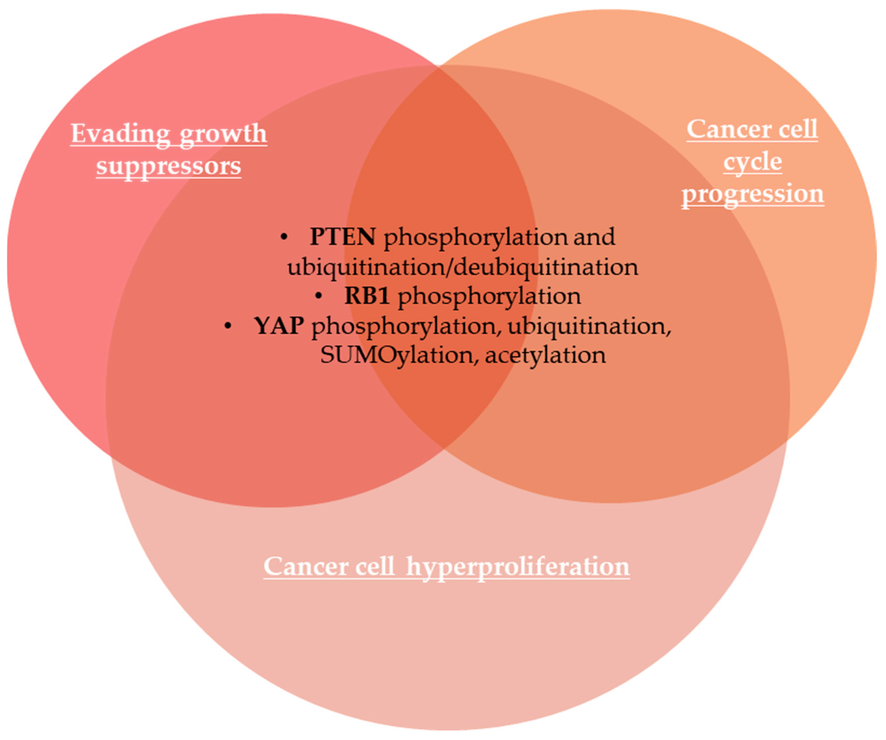
2.1.2. Evading Growth Suppressors

The evasion of anti-growth signaling is an important hallmark of cancer cells [85]. Ruhul Amin et al. (2015) summarized the most important pathways involved in growth signaling, such as p53, phosphatase and tensin homolog (PTEN), retinoblastoma protein (RB/p16 pathway), Hippo, growth differentiation factor 15 (GDF15), Notch, insulin-like growth factor (IGF), and Krűppel-like factor 5 (KLF5) [85].
The tumor-suppressor PTEN, a phosphatidylinositol-3,4,5-triphosphate phospholipid phosphatase that catalyzes the dephosphorylation of phosphatidylinositol 3,4,5-triphosphate (PIP3) to phosphatidylinositol 4,5-diphosphate (PIP2) into the plasma membrane, is important for cell growth signaling, being usually mutated or silenced in cancer cells [86]. PTEN inactivation leads to the activation of phosphoinositide 3-kinase (PI3K) and subsequent downstream AKT and mammalian target of rapamycin (PI3K-AKT-mTOR) signaling pathway that regulates genomic stability, cell growth, survival, metabolism, and proliferation [87,88]. PTEN protein levels are regulated by various PTMs, including phosphorylation, ubiquitination, and oxidation [88]. Dempsey et al. (2021) showed that multi-site phosphorylation of PTEN at Ser380/385 and Thr382/383 drives conformational changes in PTEN, inducing the dissociation of phospho-PTEN from the plasma membrane, reduction of catalytic activity and its subsequent ubiquitination [86]. Thus, cell growth control is modulated by ubiquitination-mediated degradation or dysregulated subcellular localization of PTEN [88,89]. USP13, a PTEN deubiquitinating enzyme, can restore PTEN reactivation and stability in tumors [88]. The retinoblastoma protein 1 (RB1) is an important tumor suppressor that acts as a regulator of cell cycle progression via E2F TFs [90,91]. RB1 protein prevents excessive cell growth by suppressing cell proliferation by inhibition of E2F target genes [92]. Cyclin D/CDK4/6-dependent phosphorylation inactivates RB protein, leading to cell cycle progression [92]. The tumor-suppressor cyclin-dependent kinase inhibitor p16 inhibits cyclin D/CDK4/6 activity, enhancing RB activity and suppressing cell growth [92]. The methylation of p16 at Arg138 and the phosphorylation at Ser140 are critical for the modulation of cell proliferation and apoptosis [93]. The Hippo pathway is a key regulator of cell/tissue growth and organ size, and its dysregulation is involved in many cancers [94,95]. The Yes-associated protein (YAP) and transcriptional coactivator with PDZ-binding motif (TAZ) are effectors of the Hippo pathway and are also aberrantly expressed in cancer cells [96]. YAP protein stability, abundance, transcriptional activity, and subcellular localization are modulated by its PTMs, including phosphorylation, ubiquitination, SUMOylation, and acetylation [96]. YAP phosphorylation at Ser127 suppresses YAP activity due to the cytoplasmic retention of YAP that becomes isolated from its nuclear target TFs like transcriptional enhanced associate domain (TEAD) family members, and YAP phosphorylation at Ser381 facilitates its subsequent phosphorylation by CK1 kinase and recognition by E3 ubiquitin ligase, inducing its proteasomal degradation [97]. Conversely, YAP/TAZ accumulation into the nucleus is followed by downstream transcription of genes involved in cell hyperproliferation [97]. The most important PTMs occurring in cancer biomarkers involved in evading growth suppressors are illustrated in Figure 2.

2.1.3. Avoiding Immune Destruction

Cancer progression requires immune evasion, the ability of tumor cells to activate immunosuppressive pathways to avoid an immune response from the host [98]. The programmed cell death protein 1 (PD-1/CD279)/programmed cell death ligand 1 (PD-L1/CD274) signaling pathway is responsible for cancer immune escape [99], suppressing T-cell-mediated anticancer immune responses [100]. Blocking the PD-1/PD-L1 axis is effective in preventing tumor evasion [101]. PD-1 is a transmembrane protein expressed on activated T cells that acts as an important immune checkpoint receptor, which negatively impacts the response to antigens [102]. PD-L1 is a transmembrane protein expressed on the surface of tumor cells that interacts with PD-1 to reduce the proliferation of PD-1-positive cells, inhibit their cytokine secretion, and activate apoptosis of antigen-specific T cells [99].
Ubiquitination and deubiquitination of PD-1 and PD-L1 play key roles in PD-1 regulation and PD-L1 stabilization and dynamics [102]. In addition, glycosylation, phosphorylation, palmitoylation, SUMOylation, and acetylation also play a crucial role in protein stabilization and protein–protein interactions (PPIs) on the PD-1/PD-L1 signaling pathway [102]. PD-1/PD-L1 signaling pathways are negatively regulated by phosphorylation, ubiquitination, ubiquitin-like modification, and methylation, while deubiquitination, glycosylation, palmitoylation, ADP-ribosylation, and deacetylation assure its positive regulation [101]. N-linked glycosylation of PD-L1, found in melanoma, BC, lung, and colon cancers, assures protein stability by a slower turnover rate, PD-1 and PD-L1 interaction, and promotes evasion of T-cell immunity [100,103]. PD-L1 N192, N200, and N219 glycosylation prevents PD-L1 degradation through GSK3-mediated 26S proteasome [104]. In addition, PD-L1 glycosylation impedes antibody recognition of PD-L1 in fixed samples, leading to inaccurate immunohistochemical (IHC) readout in some patient samples and resulting in an inadequate result of anti-PD-1/PD-L1 immunotherapy in clinic [104]. Consequently, deglycosylation of PD-L1 strengthens the signal intensity in PD-L1 IHC assays [100]. Recently, it was demonstrated that ionizing radiation induces translocation of PD-L1 from the membrane into the nucleus after the deglycosylation of PD-L1; thus, PD-L1 deglycosylation accelerates DNA repair in cancer and produces radioresistance [105]. Wang et al. (2021) showed that inhibition of the NEDDylation pathway upregulates PD-L1 expression in gliomas, suppressing cancer-associated immunity [106].

2.1.4. Enabling Replicative Immortality

Telomeres, the repetitive ends of chromosomes, maintain genomic integrity, being major players in enabling replicative immortality, a crucial hallmark of cancer cells [107,108]. Normal human cells undergo a limited number of successive cell divisions and then enter replicative senescence characterized by irreversible cell cycle arrest [109]. Telomere length has been associated with cell proliferation [110]. Over time, telomere shortening occurs through each division of somatic cells and has a dual function in cancer progression [111,112]. The tumor suppressive effect of telomere shortening leads to cell cycle arrest, senescence, and apoptosis, or conversely, loss of telomere can promote genome instability and cancer progression [111]. Telomerase is an enzyme that elongates telomeres, and its abnormal activation occurs in the majority of cancer cells [2,110]. Consequently, cancer cells must reactivate telomerase to extend their telomeres, escape from cellular senescence, and achieve cell immortalization or unlimited proliferative potential [112]. Conversely, in most differentiated human normal cells, telomerase expression is silenced due to the transcriptional repression of human telomerase reverse transcriptase (hTERT), the most important catalytic component of the telomerase enzyme [2,113]. Detectable in up to 90% of human primary cancers [113], TERT is regulated by TFs and PTMs, including phosphorylation, ubiquitination, and SUMOylation, which regulate telomerase integrity and activity, telomere elongation, and tumorigenesis [2]. Sanyal et al. (2020) reported that SUMOylation of hTERT positively regulates telomerase activity, while seSUMOylation inhibits telomerase activity [114]. Moreover, these authors showed that hTERT SUMOylation leads to repression of E-cadherin expression and initiation of the EMT program, promoting invasion and migration in BC cells [114].

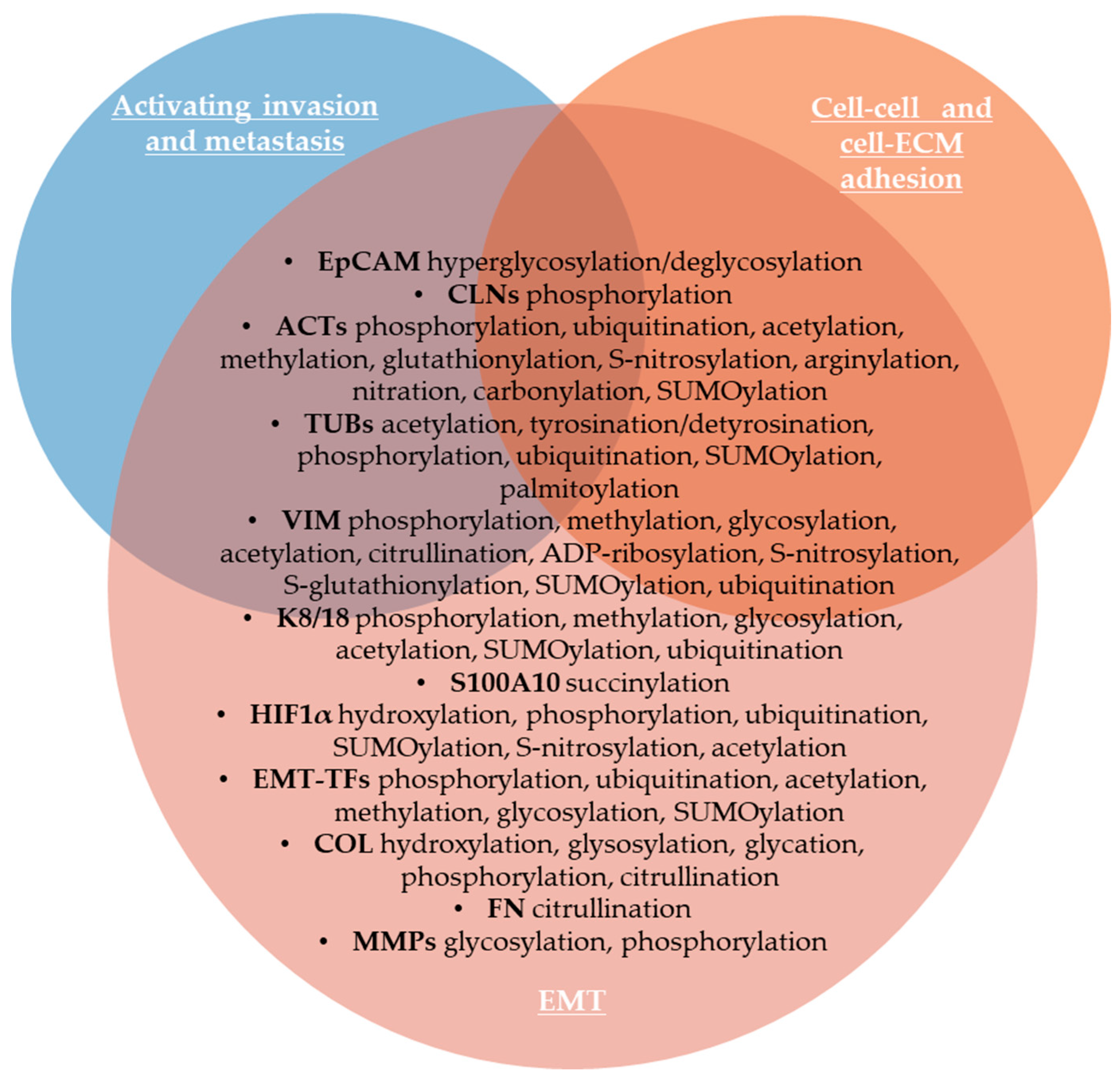
2.1.5. Activating Invasion and Metastasis

Invasion of single or clusters of cancer cells into the surrounding stroma is the first step of metastasis, the major cause of death among cancer patients [115,116]. The metastatic cascade, defined as the development of secondary tumors far from the original primary cancer, is also a crucial hallmark of malignancy [117]. Intrinsic characteristics of tumor cells associated with their communication with the tumor microenvironment (TME) contribute to the shaping of this cancer hallmark [117,118]. EMT is an essential step in cancer progression characterized by loss of cell junctions accompanied by downregulation of epithelial cell adhesion molecules, loss of apical-basal polarity, and cytoskeletal remodeling [119].
Circulating tumor cells (CTCs) are released by the primary tumor or metastatic lesions into the blood or the lymphatic system and ensure the intermediate stage of metastasis [118]. Analyzing the expression of the phosphorylation state of circulating tumor cells (CTCs), Payne et al. (2023) identified differences between epithelial, early EMT, and advanced EMT groups of cells [3]. The most important biomarker of CTCs is the epithelial cell adhesion molecule (EpCAM/CD326), a single transmembrane glycoprotein on the cell surface that mediates intercellular adhesion, is overexpressed in the majority of invasive epithelial cancers [120,121]. In BC, EpCAM was found to be hyperglycosylated, whereas deglycosylated EpCAM promoted autophagy and apoptosis, and inhibits proliferation in BC cells through suppressing PI3K/AKT/mTOR pathway [122,123]. Moreover, N-glycosylation mutation of EpCAM inhibits EMT, invasion and metastasis [124]. EpCAM regulates claudins’ dynamic and tight junctions [125]. Claudin-1 (CLN1) is important in tight junction formation, and protein kinase A (PKA) and protein kinase C (PKC) can phosphorylate claudins [126]. French et al., (2009) showed that the subcellular localization of CLN1 can be controlled by phosphorylation that dictates the effects on metastatic ability [126]. Thus, cytoplasmic expression of CLN1 in metastatic melanoma cells was correlated with increased migration, while nuclear claudin-1 expression was found in benign lesions [126].
Cytoskeleton remodeling has a crucial role in cancer cell invasion and migration [127]. Cytoskeletal proteins undergo a broad spectrum of PTMs, which increase the diversity of the cytoskeletal proteome and modulate the function of the cytoskeletal proteins [127]. Thus, phosphorylation of cytoskeletal proteins regulates dynamics of polymerization/filamentation of a plethora of monomers, including actin, myosin, tubulin, keratins, desmins, glial fibrillary acidic protein, peripherin, vimentin, anexins, nestins and others, modulating stability and disassembly of various cytoskeletal polymers, so altered phosphorylation of cytoskeletal proteins was observed in many cancers [128]. MacTaggart and Kashina (2021) summarized the most important PTMs of cytoskeletal proteins [127]. Cancer cell movement involves the formation and elongation of invadopodia, lamellipodia, and filopodia, so the dynamic of actin and actin-binding proteins is essential for cancer cell invasion [129]. Thus, actin (ACT), which represents 1–5% of all non-muscle cell proteins [130], undergoes various PTMs, such as acetylation, arginylation, methylation, oxidation, glutathionylation, S-nitrosylation, nitration, carbonylation, phosphorylation at multiple amino acid residues, ubiquitination, and SUMOylation [127]. Tubulin isoforms undergo acetylation, detyrosination/tyrosination, glutamylation/glycynation, phosphorylation, ubiquitination, SUMOylation, polyamination, and methylation [127]. The intermediate filament vimentin (VIM) protein is upregulated during EMT to enhance the invasiveness of carcinoma cells [131]. Thus, VIM is a biomarker expressed in metastatic cancers, where it is required for tumor progression and metastasis [132]. VIM protein undergoes phosphorylation, methylation, glycosylation, acetylation, citrullination, ADP-ribosylation, S-nitrosylation, S-glutathionylation, SUMOylation, and ubiquitination [127,133]. VIM phosphorylation is critical for its structure and function [131]. Thus, Kuburich et al. (2023) demonstrated that stabilizing VIM phosphorylation inhibits stem-like characteristics and metastasis of hybrid epithelial/mesenchymal carcinoma cells [131]. It was also demonstrated that VIM controls collagen deposition and extracellular matrix structure (ECM) via posttranscriptional regulation of ECM-related genes [133]. In gastric cancer cell lines, deubiquitination stabilizes VIM to contribute to malignancy [134]. VIM is frequently citrullinated in cells during EMT of metastasizing epithelial tumor cells [8]. Loss of keratins 8 and 18 (K8/18) is also a hallmark of EMT, increasing collective cell migration [135]. K8 and K18 also undergo phosphorylation, methylation, glycosylation, acetylation, SUMOylation, and ubiquitination [127]. In addition, PTMs of EMT-inducing transcription factors (EMT-TFs), such as phosphorylation, ubiquitination, acetylation, methylation, glycosylation, SUMOylation of SNAIL, SLUG, ZEB1/2, and TWIST1 induce their activation or destabilization, nuclear/cytoplasmic shuttle, promoting EMT or, contrarily, mediating their destruction via proteasomal degradation [136]. On the other hand, succinylation of S100A10, a member of S100 calcium-binding cytosolic proteins involved in invasion and migration of tumor cells, increases human gastric cancer invasion, by suppression of its ubiquitination and subsequent proteasomal degradation [137]. Hypoxia-inducible factor 1α (HIF1α) activation is also a factor that can induce EMT in tumor cells [138]. HIF1α activity is modulated by multiple PTMs that affect its stability and transcriptional activity, including acetylation, ubiquitination, and propionylation [46].
The extracellular matrix (ECM), mainly consisting of proteoglycans, glycoproteins, and matrisome proteins, also plays an important role in tumor development [139]. Evidence suggests that EMT can be induced in cancer cells by adhesion to type I collagen [140]. Collagen family members, the most abundant proteins in ECM, undergo extensive PTMs, affecting receptor binding, cell migration, and tumor stiffness [141]. Borst et al. (2024) showed that collagen PTMs include proline and lysine hydroxylation that takes place in the endoplasmic reticulum (ER), glycosylation and glycation occur in the ER and Golgi apparatus, and collagen can also be modified by phosphorylation and citrullination [141]. Thus, targeting intracellular PTMs can inhibit collagen secretion and deposition, remodeling ECM to improve immune cell migration [141]. Citrullination of other matrisome proteins, including fibrinogen and fibronectin (FN), as well as matrix metalloproteinases (MMPs), has been reported in cancer [57]. MMPs, their substrates, and ECM-binding partners are modified by PTMs, including glycosylation and phosphorylation [11]. The most important PTMs of cancer biomarkers involved in EMT, as well as in activating invasion and metastasis, are listed in Figure 3.

2.1.6. Inducing or Accessing Vasculature

The previous cancer hallmark, “inducing angiogenesis”, has been more recently replaced by ”inducing or accessing vasculature” due to the discovery of non-angiogenic tumors able to exploit normal vessels [142]. Sonic hedgehog (SHH) and its signaling pathway promote neovascularization by capillary morphogenesis and induce endothelial cell migration, contributing to the development and progression of many cancers [143,144]. Zinc-finger protein glioma-associated oncogene 1 (GLI1) is a transcriptional effector of hedgehog signaling, and its aberrant expression was linked to many hallmarks of cancer, including angiogenesis, cell proliferation, survival, metastasis, metabolic reprogramming, and chemotherapeutic resistance [145]. Recently, Lei et al. (2024) showed that GLI1 overexpression enhanced the endothelial cells and pericytes’ motility required for angiogenesis in non-small cell lung cancer [146]. GLI1 proteins are modified by PTMs, including phosphorylation, ubiquitination, SUMOylation, acetylation, and O-GlcNAcylation, which regulate its intracellular trafficking and transcriptional activity of the SHH signaling pathway [147]. GLI1 translocates from the cytoplasm to the nucleus, targeting the transcription of various downstream genes [145,146]. The nuclear GLI1 is acetylated by p300 or CBP at K518, the AcGLI1 being temporarily inactive, while deacetylation allows GLI1 to initiate transcription of target genes [148].

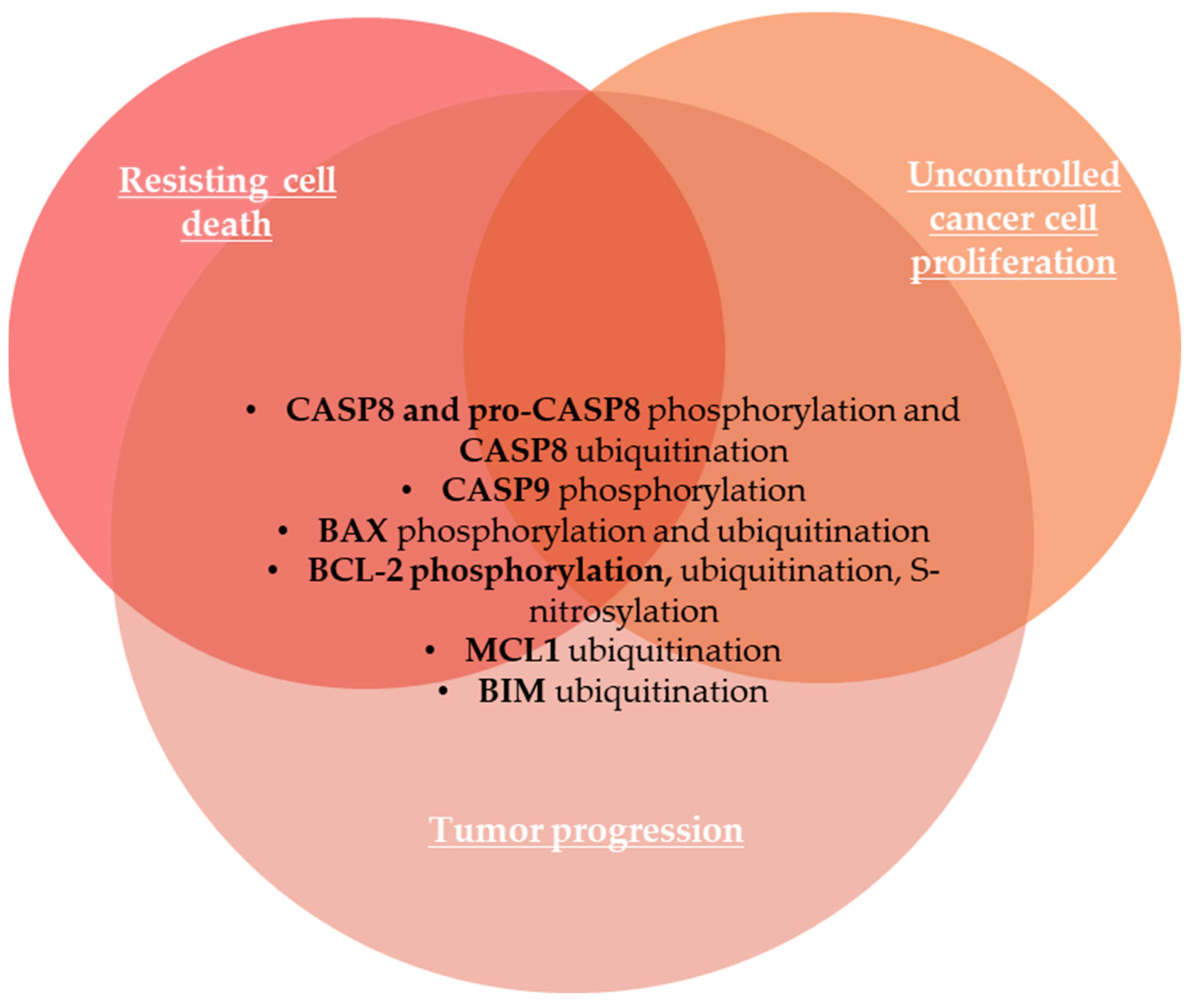
2.1.7. Resisting Cell Death

Apoptosis is a major form of programmed cell death or cell suicide, and evasion of apoptosis or aberrant apoptosis is a hallmark of cancer that results in uncontrolled cell proliferation in tumors [149,150,151]. The extrinsic or death receptor-mediated apoptotic pathway and the intrinsic or mitochondrial-mediated pathway are two distinct core branches of apoptosis [152]. Caspases are crucial players in apoptosis [153].
The extrinsic pathway is initiated by cell surface death receptors like tumor necrosis factor receptor (TNFR) superfamily, FasR (CD95), and TRAIL R1/2 (also called death receptor DR4/5), which bind with tumor necrosis factor α (TNFα), Fas ligand (FasL/CD95L), and TNF-related apoptosis-inducing ligand (TRAIL/APO2) [154,155]. Death receptor activation leads to the activation of pro-caspases 8 and 10 into active caspases 8 and 10 [154]. The intrinsic apoptosis pathway is regulated by the B-cell lymphoma 2 (Bcl-2) family proteins [155]. Cancer cells prevent apoptosis through modulation of Bcl-2 proteins, via upregulation of anti-apoptotic and pro-survival proteins, such as BCL-2, BCL-XL, and MCL1, or/and downregulation of pro-apoptotic proteins, such as BAX, BAK, and BOK [150]. Internal and external stresses lead to the release of cytochrome c from mitochondria during the intrinsic or stress-mediated apoptotic pathway that activates pro-caspase 9 in caspase 9 that activates caspase 3 [154].
Both apoptotic pathways are strongly regulated by PTMs, mainly by ubiquitination [156]. Phosphorylation is also a key process in the TNFR1-mediated signaling pathway [157]. Both phosphorylation and ubiquitination highly regulate caspase 8 activity [158]. Thus, caspase 8 is phosphorylated at Tyr380 by Src kinase upon EGF receptor stimulation, which suppresses apoptosis and participates in tumor progression [153], while pro-caspase 8 can be phosphorylated at Ser387 by pERK1/2 in a cell cycle specific manner, also inhibiting caspase 8 induced apoptosis in ovarian and BC cell lines [154]. Caspase 8 is modified with ubiquitin, which mainly inhibits its pro-apoptotic functions [156].
Pro-apoptotic BAX protein is mainly distributed in the cytoplasm in the inactive form [152]. Upon apoptotic stimuli, BAX proteins are activated, undergo a conformational change, and then are translocated to the mitochondria’s outer membrane [152]. BAX activation induces mitochondrial outer membrane permeabilization, followed by the release of cytochrome c and other pro-apoptotic factors, leading to cancer cell death [152,159]. In the intrinsic apoptotic pathway, ubiquitination targets both anti-apoptotic and pro-apoptotic proteins [156]. For example, PTMs of BAX, including phosphorylation and ubiquitination, play key roles in the regulation and stabilization of BAX proteins [160]. Thus, phosphorylation of BAX at Ser184 by protein kinase B (PKB), a serine/threonine kinase also known as AKT, converts pro-apoptotic BAX protein into an anti-apoptotic protein, thus inhibiting apoptosis in cancer cell lines [161]. Moreover, BAX can undergo degradation through the ubiquitin–proteasome pathway [152]. IBRDC1, an IBR-type RING-finger E3 ubiquitin ligase, regulates BAX levels through increased BAX ubiquitination, degradation, and downregulation, while IBRDC1 downregulation leads to accumulation of activated BAX and apoptosis [162]. The oncogenic beclin-2 (BCL-2) is a key anti-apoptotic protein of the mitochondrial pathway, so BCL-2 PTM, such as ubiquitination and proteasomal degradation, are known as regulators of BCL-2 function [163]. BCL-2 undergoes S-nitrosylation by endogenous nitric oxide (NO) that inhibits its ubiquitin-proteasomal degradation, thus preventing BCL-2 downregulation and apoptosis [163]. Phosphorylated BCL-2 at Ser70 exhibits enhanced binding to BIM and BAK pro-apoptotic proteins during mitosis, leading to increased sequestration of BAK and BIM [164]. The chemotherapeutic drug cisplatin initiates an apoptotic pathway by phosphorylation of anti-apoptotic BCL-XL by cyclin-dependent kinase 2 (CDK2) [165]. Thus, phosphorylated BCL-XL translocates to the outer mitochondrial membrane, forms pores, and increases membrane permeabilization, cytochrome c release, and caspase activation, leading to cell death [165]. Ubiquitination targets both anti-apoptotic proteins, like MCL1, as well as pro-apoptotic proteins like BIM [156]. Caspase 9, an important player in the initiation of the intrinsic apoptosis pathway, is frequently phosphorylated at Thr125 by the mitogen-activated protein kinase (MAPK) pathway, leading to inhibition of caspase 9 activation and caspase 9-mediated apoptosis in gastric carcinoma [166]. PTMs of proteins involved in resisting cell death are summarized in Figure 4.

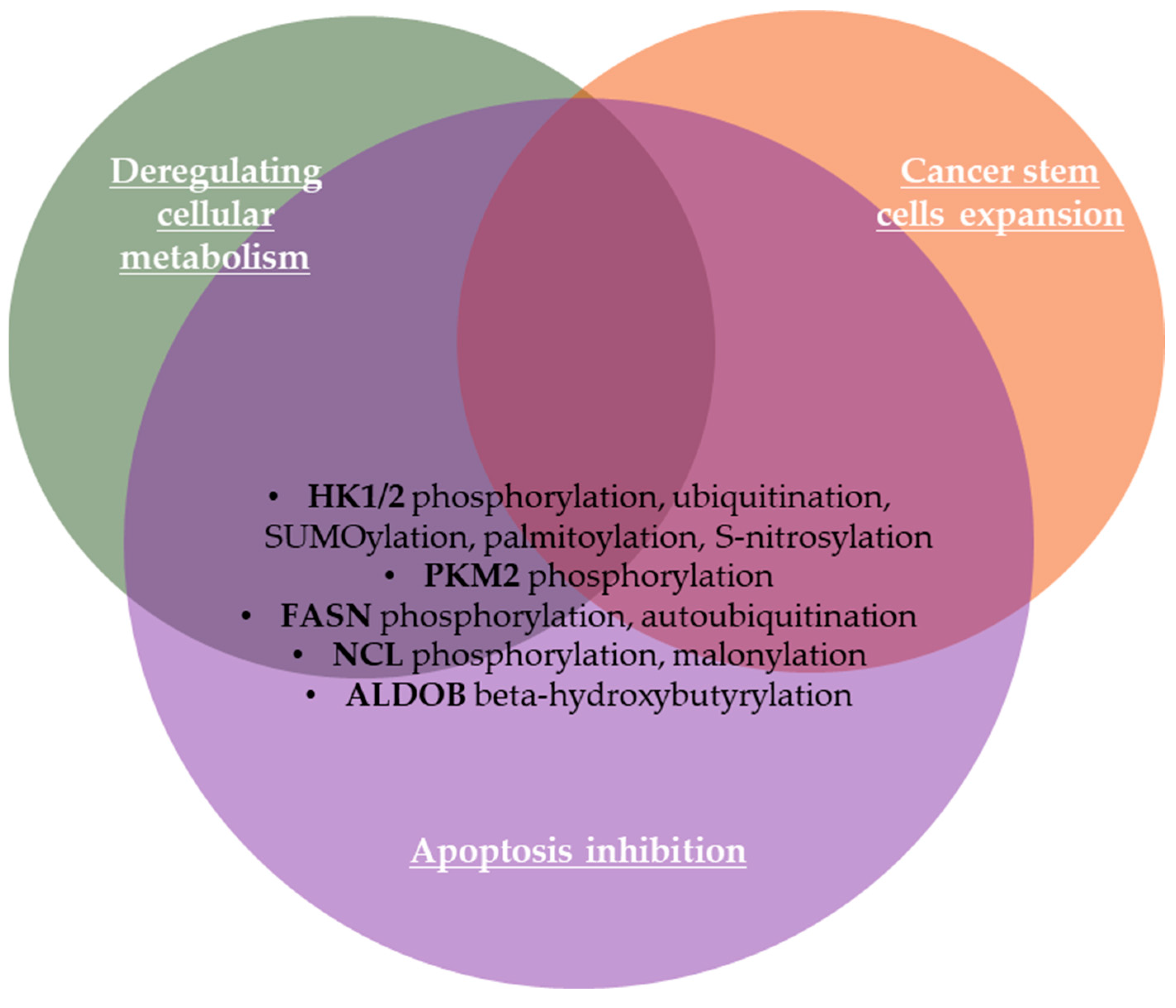
2.1.8. Deregulating Cellular Metabolism

Cancer can be characterized as a metabolic disease and metabolic reprogramming is considered to be the most important emerging hallmark of tumor cells [20,167]. Malignant cells emphasize increased aerobic glycolysis, glutaminolysis, and de novo lipogenesis to assure their survival and increased proliferation [168,169]. Thus, malignant cells manage the ability to acquire necessary nutrients from a nutrient-poor environment and develop reprogrammed glucose metabolism that is essential for cell growth and survival [170,171]. The most studied altered metabolism in cancer is aerobic glycolysis or the Warburg metabolism/effect, which refers to the switch from the normal respiratory pathway to aerobic glycolysis, resulting in a high amount of lactate in the cytosol and ECM [167,172].
Tyrosine phosphorylation influences the activity of some metabolic enzymes, being essential for reprogramming the energy metabolism in cancer cells [173], as well as ubiquitination and deubiquitination [169]. The rate-limiting glycolytic hexokinase (HK) isoenzymes, including HK1 with many isoforms [174], HK2, HK3, HK4 (also named glucokinase (GK)), and hexokinase domain-containing protein 1 (HKDC1), phosphorylate glucose to produce glucose-6-phosphate [170] that then initiate major pathways of glucose utilization including glycolysis, pentose phosphate pathway (PPP) and oxidative phosphorylation (OXPHOS) [175]. Hexokinase activity is regulated through protein expression as well as PTMs of HKs [170]. Thus, HK2 phosphorylation by AKT2 at the threonine 473 residue (T473) promotes its binding to mitochondria, which increases HK2 catalytic activity and enhances glycolysis, promoting tumorigenesis in colon cancer cells and lung metastasis in nude mice through upregulation of nuclear factor-kB (NF-kB), hypoxia-inducible factor 1α (HIF1α), matrix metalloproteinase 2 (MMP2), and matrix metalloproteinase 9 (MMP9) upregulation [176]. Both HK1 and HK2 phosphorylation, especially HK1 phosphorylation at tyrosine 732 (Tyr732/Y732) by c-Src, increases their catalytic activities and enhances glycolysis in various cancer cell lines and tumor types so that the phosphorylation level of HK1-Y732 may be used as a novel biomarker to predict metastasis risk of primary tumors [177]. HK2 shuttles from the cytoplasm to the mitochondria in response to environmental and metabolic stress, so HK2 ubiquitination by HectH9 E3 ligase at lysine K63 residue regulates its mitochondrial localization, regulating cancer stem-cell (CSC) expansion, promoting glycolysis, inhibiting apoptosis, and enhancing CSC-associated drug resistance [175]. Consequently, K63-linked ubiquitination activates kinases and TFs, targets transmembrane proteins for lysosomal degradation, and modulates protein trafficking [175]. Guo et al. (2023) also summarized other PTMs of hexokinases, such as SUMOylation of HK2 in prostate cancer cells, HK1 palmitoylation in hepatic stellate cells, and S-nitrosylation of HK4 [170]. Pyruvate kinase plays an essential role in regulating cell metabolism and contributes to tumorigenesis [167].
Pyruvate kinase M2 (PKM2) has an upregulated expression in most cancer cells, where it tends to exist as a dimer and drives the Warburg effect for tumor progression [167,178]. Zhou et al. (2024) demonstrated that PKM2 phosphorylation at Ser333 antagonizes O-GlcNAcylation, promotes its tetramer formation, decreasing its nuclear localization, and thus inhibiting c-Myc expression [178]. Consequently, PKM2 phosphorylation and tetramer formation attenuate the Warburg effect in cancer cells by downregulation of glucose consumption and lactate production, whereas phospho-PKM2 deficiency promotes tumor growth in vivo [167,178].
The fatty acid synthase (FASN), an enzyme involved in de novo fatty acid synthesis, is overexpressed in many cancers [179], including colorectal cancer [168]. FASN promotes cell proliferation through membrane biosynthesis, energy storage, generation of signal mediators, resistance to apoptosis, cell invasion, immune escape, metastasis, and angiogenesis [168,179]. Jin et al. (2010) showed that FASN phosphorylation by human epidermal growth factor receptor 2 (HER2) plays a key role in HER2-overexpressing BC progression [180]. FASN cytoplasmic degradation can be prevented by autoubiquitination and degradation of enhanced F-box and WD repeat domain-containing 7β (FBXW7β), a cytoplasmic isoform of FBXW7 component of ubiquitin E3 ligase complex involved in FBXW7β/GSK3β-mediated FASN ubiquitination, resulting in FASN-mediated lipogenesis and promotion of colorectal cancer growth [168]. Nucleolin (NCL) is a multifunctional protein distributed in the nucleoli, nucleoplasm, cytoplasm, and plasma membrane. NCL is also overexpressed in many cancers, including BC, gastric cancer, leukemia, and glioblastoma, due to its roles in regulating proliferation, survival, and metastasis of cancer cells through its involvement in ribosome biogenesis, mRNA translation, and chromatin remodeling [20,181]. Recently, Sun et al. (2024) showed that PTMs, in particular phosphorylation, play an important role in the subcellular localization and function of NCL [20]. These authors also found that NCL is highly malonylated in hepatocellular carcinoma [20]. Thus, malonylated NCL translocates from nucleolus to nucleoplasm, binds to AKT mRNA, and facilitates AKT translation, promoting cell proliferation [20]. These findings suggest malonylation as a novel mechanism controlling cancer cell proliferation based on the potential communication between lipid metabolism and protein function regulated by PTMs [20]. Succinylation also has a key relationship with energy metabolism, participating in fatty acid synthesis, amino acid degradation, electron chain transmission, ketone body formation, the TCA cycle, and other metabolic processes [22]. Cancer cell metabolism can be reshaped by aldolase B (ALDOB) β-hydroxybutyrylation at lysine 108 (Lys108) induced by a ketogenic diet, inhibiting its enzymatic activity [23]. These authors demonstrated that attenuation of ALDOB activity inhibits mammalian/mechanistic target of rapamycin (mTOR) signaling and glycolysis, suppressing hepatocellular carcinoma cell proliferation [23]. PTMs of proteins involved in deregulating cellular metabolism in cancer are summarized in Figure 5.

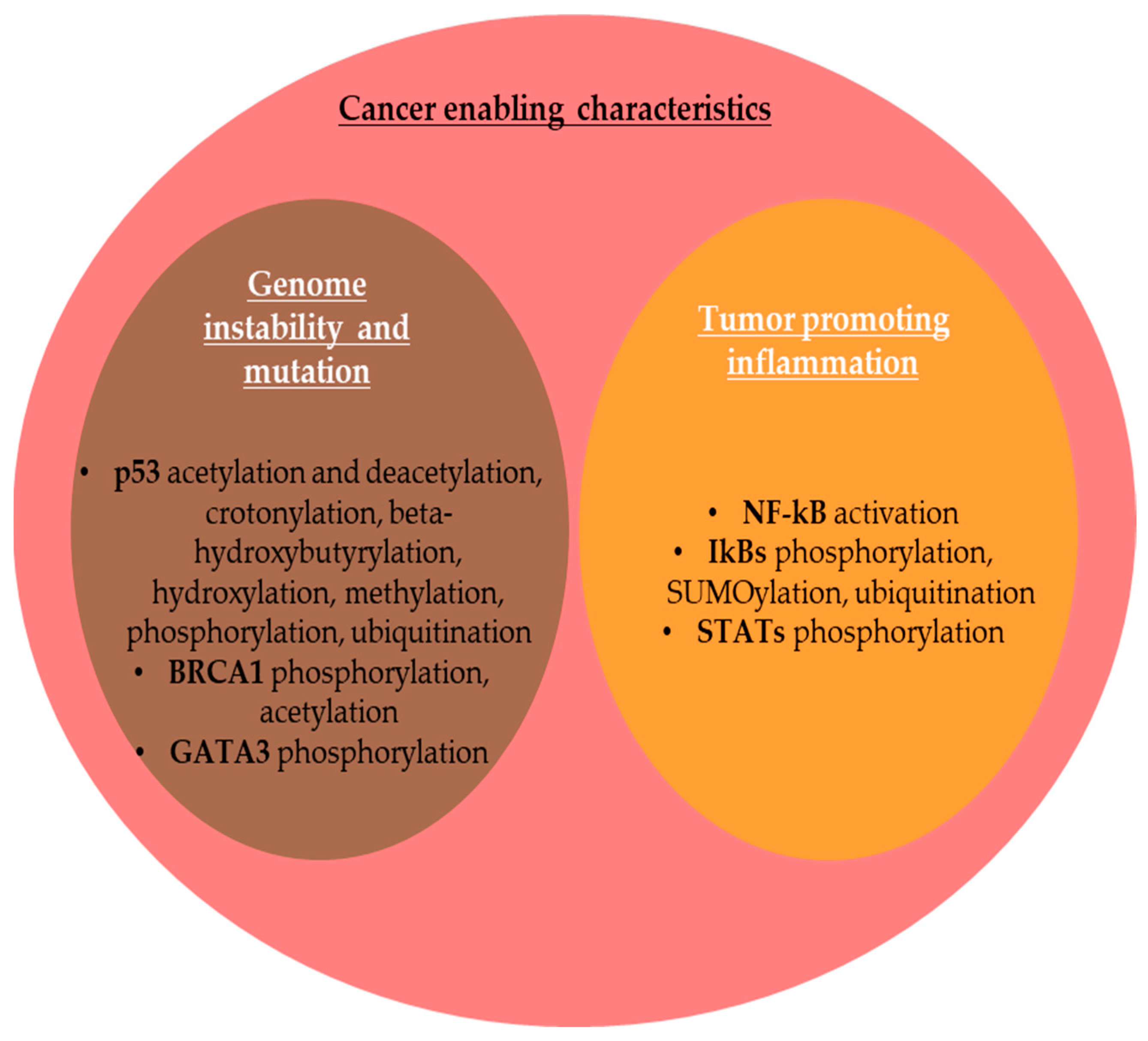
2.2. PTMs and Cancer Enabling Characteristics

2.2.1. Genome Instability and Mutation

The integrity of the genome is controlled by DNA damage signaling and DNA damage repair (DDR) [182]. Genomic instability (GI) is a hallmark of most cancer cells during cell division due to mutations in DNA repair genes or mitotic checkpoint genes, which predispose cells to malignant transformation [183]. GI is a direct consequence of the inactivation of the tumor-suppressor protein p53, as well as the p53 pathway, which is inactive in the majority of cancer cells [184]. p53, encoded by the TP53 gene, is known as the ”guardian of the genome” for its role in the preservation of genomic stability [185]. p53 undergoes multiple PTMs that modulate its diverse functions in specific biological contexts [186]. Wen and Wang (2021) highlighted the potential of ”PTMomic” studies of p53 for cancer therapy [186]. In response to DNA damage, p53 undergoes acetylation and deacetylation with consequences on sequence-specific DNA-binding activities [186]. Crotonylation, β-hydroxybutyrylation, hydroxylation, and methylation have also been cited as PTMs that occur in p53 [186]. Thus, under cellular stress, p53 undergoes phosphorylation via a series of kinases, including ataxia telangiectasia-mutated serine/threonine kinase (ATM), ATM and RAD3-related serine/threonine kinase (ATR), and checkpoint kinase 1/2 (CHK1/CHK2) [186], phosphorylation stabilizing p53 by disrupting its interactions with mouse double minute 2 (MDM2) oncoprotein, an E3-ubiquitin ligase that triggers p53 degradation through ubiquitin–proteasome-dependent pathway [187]. Consequently, p53 is translocated into the nucleus, where it contributes to transcription of target genes. Moreover, in cells where the p53 was activated by stress, p53 polyubiquitination is switched to p53 monoubiquitination, maintaining its nuclear localization and DNA-binding activity [14]. p53 is also modified by β-hydroxybutyrylation at lysine K120/319/370 residues, which attenuates its activity and explains the link between ketone bodies and cancer [188].
The germline mutations in breast cancer susceptibility genes BRCA1 and BRCA2 have been linked to genomic instability [182]. In addition, the function of breast cancer tumor-suppressor 1 (BRCA1) protein is regulated by phosphorylation and acetylation [189]. BRCA1 is a nuclear phosphoprotein that plays several roles in genome stability, including checkpoint promotion, DDR, replication fork stability, and DNA double-strand break repair [190]. BRCA1 regulates transcription loss, leading to genome instability and malignant transformation [191]. Thus, CHKs phosphorylate specific residues of BRCA1 under conditions of DNA damage, modulating the response of cells to various stresses [191]. BRCA1 is part of an obligate multifunctional heterodimer with BRCA1-associated RING domain 1 (BARD1), acting together as a tumor-suppressor E3 ubiquitin ligase involved in repairing double-strand DNA breaks through homologous recombination, maintaining genome stability [190,192]. Lahusen et al. (2018) emphasized the role of BRCA1 acetylation at K830 in the activation of BRCA1 function at the intra-S checkpoint after DNA damage [189]. GATA binding protein 3 (GATA3) is a nuclear zinc-finger TF that functions downstream of BRCA1 to promote DDR and suppress dedifferentiation in BC, acting as a tumor suppressor [193]. PTMs of GATA3 are crucial for its function [194]. The progestin-activated progesterone receptor (PR) reduces GATA3 expression by GATA3 phosphorylation at Ser308, followed by its proteasome-mediated degradation [195]. Thus, PR activation downregulates GATA3 by transcriptional repression and increases protein turnover, promoting BC growth [195].

2.2.2. Tumor-Promoting Inflammation

Inflammation either promotes or suppresses tumor progression [196]. Thus, chronic inflammation promotes tumor progression and treatment resistance, while induction of acute inflammation leads to anti-tumor immune responses [196]. The nuclear factor of a k-light chain of enhancer-activated B cells (NF-kB) is a pro-inflammatory family of TFs that activate the transcription of key genes that connect tumorigenesis and inflammation [197,198]. In the cytoplasm, NF-kB binds to the inhibitors of kappa B (IkBs) responsible for maintaining NF-kB in an inactive state [199,200]. The activation of the NF-kB pathway involves phosphorylation of IkBs [200]. SUMOylation of IkBa was also recognized as a regulator mechanism for the NF-kB signaling pathway [60]. As a consequence of the phosphorylation and ubiquitination of IkB proteins, NF-kB is dissociated and translocated into the nucleus to bind with DNA for inducing the transcription of immune and inflammatory cytokines [197,198]. The Janus kinase (JAK)/signal transducer and activator of transcription (STAT) signaling is a key pathway involved in tumorigenesis by mediating the cellular response to inflammation [201]. STAT proteins are inactive in the cytoplasm, but phosphorylation leads to conformational changes in the STATs, which migrate into the nucleus and regulate the expression of target genes upon activation by several cytokines, such as interleukin 6 (IL-6), interleukin 10 (IL-10), and interleukin 11 (IL-11) [202,203]. JAKs phosphorylate mainly STAT1 and STAT3, activating their nuclear translocation [203,204,205]. PTMs of cancer biomarkers involved in tumor-enabling characteristics are summarized in Figure 6.

2.3. PTMs Involved in Additional Emerging Hallmarks and Enabling Characteristics of Cancer

In 2022, Hanahan incorporated four additional proposed emerging hallmarks and enabling characteristics involving unlocking phenotypic plasticity, nonmutational epigenetic reprogramming, polymorphic microbiomes, and senescent cells. PTMs of protein also orchestrate these characteristics of cancer (Figure 7).

2.3.1. Unlocking Phenotypic Plasticity

Cell plasticity refers to the ability of cells to be reprogrammed, enabling them to acquire novel molecular, phenotypic, and functional traits that allow for tumor initiation, progression, metastasis, and therapy resistance [206,207]. The epithelial-to-mesenchymal transition (EMT) is a reversible form of cellular plasticity by which cancer cells undergo molecular, phenotypic plasticity, and behavioral transition from an epithelial to a mesenchymal state, increasing their migratory and invasive features [208]. Dysregulation of cadherin-catenin complexes, crucial components of the adherens junctions, was linked to cancer development [209]. The hallmark of EMT is the upregulation of neural cadherin (N-cadherin) followed by loss of functional epithelial cadherin (E-cadherin), a switch orchestrated by various signaling pathways and transcription factors (TFs) [210]. Thus, PTMs of cadhesome proteins, including phosphorylation at serine, threonine, and tyrosine residues, play a key role in the regulation of adherens junctions [210]. In addition, activation of EMT-inducing transcription factors (EMT-TFs), such as zinc-finger protein SNAI1 (SNAIL), zinc-finger protein SNAI2 (SLUG), zinc-finger E-box binding homeobox 1/2 (ZEB1/ZEB2), and twist-related protein 1/2 (TWIST1/2), leads to transcription of genes that are involved in mesenchymal programs and inhibition of genes that drive the epithelial differentiation [211]. These EMT-TFs mediate the transcriptional repression of E-cadherin and upregulation of vimentin [132,212] and undergo PTMs to regulate metastasis phosphorylation, mainly affecting the flexibility and reversibility of EMT [213]. The silencing of E-cadherin expression, a tumor-suppressor protein from epithelial adherens junctions, has been associated with the destruction of intercellular adhesion mechanisms and EMT, significantly occurring during tumor metastasis [214]. Phosphorylation of E-cadherin at Thr790 by protein kinase C (PKC) suppresses the function of E-cadherin by diminishing its interaction with β-catenin in cervical cancer tissues [215]. Moreover, β-catenin itself can emphasize various subcellular localizations and functions orchestrated by several PTMs. Thus, β-catenin is regulated by WNT signaling, so in the absence of extracellular WNT signals, the expression of β-catenin is kept at a low level in the cytoplasm through the ubiquitin–proteasome system [216]. For this purpose, β-catenin is phosphorylated by casein kinase 1 (CK1) and then by glycogen synthase kinase 3 beta (GSK3β), leading to its subsequent ubiquitination and proteasomal degradation [216]. The activation of WNT signaling pathway, conversely, leads to β-catenin stabilization through inhibition of phosphorylation or inactivation of ubiquitination, followed by its accumulation in the cytoplasm and successive translocation into the nucleus, where it initiates the transcription of WNT target genes, such as c-Myc, Jun, and cyclinD-1 oncogenes [216]. Evidence suggests that protein lysine acetylation is crucial for WNT/β-catenin signaling activation [217]. You et al., (2022) concluded that β-catenin acetylation improves its stability by inhibiting the ubiquitin-mediated degradation and induces its nuclear translocation to enhance the transcription of WNT target genes [217]. In addition, β-catenin SUMOylation in melanoma cell lines stabilizes β-catenin and inhibits its degradation by the proteasomal system [218]. The intermediate filament VIM protein is highly overexpressed in metastatic cancers, where it facilitates the formation and elongation of the invadopodia that facilitate cancer cell invasion and migration [132]. The phosphorylation of VIM at Ser39 enhances the ability of VIM to induce motility and invasion, also protecting VIM from caspase-induced proteolysis [219]. SNAIL, SLUG and TWIST1 are major EMT-TFs that undergo nuclear-cytoplasmic shuttling [220] and activate genes associated with the mesenchymal-migratory phenotype of cancer cells [221]. EMT-TFs degradation is initiated by specific phosphorylation, followed by subsequent ubiquitination and destruction in the ubiquitin–proteasome system [211]. Conversely, deubiquitination stabilize TFs, accelerating the EMT program [221]. SUMOylation of SNAIL in a prostate carcinoma cell line is essential for the EMT-activating function of SNAIL [211,222]. SLUG acetylation increases its stability in BC cells [223] as well as SNAIL acetylation, which promotes invasion and metastasis in lung cancer cells [224]. SUMOylation of TWIST2 through ligase activity of ZNF451 stabilizes TWIST2 and promotes EMT [225].

2.3.2. Nonmutational Epigenetic Reprogramming

Dysregulation of the epigenetic program is a hallmark of cancer [226]. Reversible epigenetics modifications, including DNA hypo- or/and hypermethylation [227], PTMs of proteins including histone and non-histone protein modifications [228], and noncoding RNAs (ncRNAs) affect gene expression in cancer cells by alteration of chromatin and DNA accessibility without changes on DNA sequences [19,229]. Epigenetic changes lead to activation of oncogenes and silencing of tumor-suppressor genes, thereby encouraging tumorigenesis and cancer progression [226].
DNA methylation is catalyzed by DNMT1 as well as de novo DNMT3A and DNMR3B methyltransferases aberrantly expressed in cancer [227,230]. Regulation of chromatin biology and gene transcription is modulated by DNMT3A binding with many proteins [231]. DNMT3A enzyme is modified by SUMO1, and SUMOylation disrupts DNMT3A-histone deacetylases (HDAC) interaction, thus changing histone acetylation pattern and eliminating the ability of DNMT3A to repress transcription [231,232]. Thus, SUMOylation was recognized as an important mechanism to regulate gene transcription [232]. In addition, O-GlcNAcylation impacts DNA methylation through modulation of DNMT1 activity and ten-eleven translocation 1/2/3 (TET1/2/3) stability and DNA binding [233].
Histone proteins play a central role in gene regulation [56]. Aberrant histone modification affects genome stability and disrupts gene expression, leading to cancer and other diseases [228]. Histone PTMs are covalent changes in histone tails, including H2A, H2B, H3, and H4 [228]. PTMs of histones, as well as other chromosomal proteins, regulate chromatin conformation and gene transcription [234]. Histone PTMs, including methylation, acetylation, phosphorylation, ubiquitination, SUMOylation, NEDDylation [235], acylation [236], crotonylation [237], lysine lactylation [238], borylation and propionylation [239], succinylation [240], citrullination [55,56], malonylation, glutarylation, hydroxybutyrylation, benzoylation, O-GlcNAcylation [233], biotinylation [241,242], tyrosine hydroxylation, ADP-ribosylation [243], N-formylation at multiple lysyl residues [234], proline isomerization, and can contribute to chromatin remodeling, nucleosome dynamics, and transcription of key oncogenes, tumor-suppressor genes, and TFs [6,228]. Abnormal histone methylation or demethylation in the corresponding cancer-related genes contributes to carcinogenesis, proliferation, metabolic rewiring, EMT, invasion, and migration in digestive cancers, including gastric, liver cancer, pancreatic cancer, and colorectal cancer [244]. Histone acetylation and deacetylation are essential for genome integrity, DNA replication, DDR, and RNA transcription [228,245]. Li et al. (2014) showed that H2A NEDDylation antagonizes its ubiquitination [235]. Thus, NEDD8 negatively regulates DDR by suppressing the ubiquitination of H1A, also blocking the recruitment of damage-response protein BRCA1 [235]. In addition, aberrant histone phosphorylation can also mediate tumor development and metastasis [228]. Histone formylation is involved in nucleosome organization and DNA binding [234]. Biotinylation of histones plays a role in cell proliferation, gene silencing, and DDR [242]. Kebede et al. (2017) emphasized that histone acetylation, butyrylation, and propionylation act in a combinatorial mode to promote elevated transcription and to associate cellular metabolism with the structure and function of chromatin [239]. In cancerous tissue, histone lysine lactylation (KIa) has been associated with metabolic reprogramming, mainly glucose and glutamine metabolism, thus contributing to tumorigenesis, immune evasion, and angiogenesis [238]. The poly(ADP-ribose) polymerase 1 (PARP1) is activated by DNA damage and is able to recruit other proteins to repair DNA single-strand breaks, and PARP inhibitors (PARPi) are an important class of anticancer therapies that target tumors with BRCA1/2 mutations, such as ovarian, BC, lung and pancreatic cancer [246]. Histone ADP-ribosylation is essential for PARP1 release from DNA lesions, thus contributing to resistance to PARP inhibitors [243]. Histone citrullination also plays functions in tumors [56].

2.3.3. Polymorphic Microbiomes

Recently, updated definitions for the term microbiome have been proposed. Thus, the term microbiome is actually used to define a complex microbial ecosystem [247], containing both the community of microorganisms, known as microbiota, which includes bacteria, archaea, fungi, protozoa, and viruses [248], and their milieu, including microbial structural elements, such as proteins/peptides, lipids, polysaccharides and nucleic acids, microbial metabolites, including signaling molecules, toxins and other organic and inorganic molecules, as well as other microenvironmental conditions [249]. The most studied in association with cancer is the gut microbiome, but other microbiomes from different tissues and organs, such as lung, genitourinary, skin, and breast, as well as the intratumoral microbiome, emphasized an increased interest in cancer research [247].
Polymorphic microbiomes are involved in tumorigenesis and oncotherapy response [247,250]. Recently, dysbiotic microbiota was recognized as an important emerging hallmark of cancer [58,251]. Small molecule metabolites produced by tumor microbiota diffuse into tissues and cells, remodeling signaling pathways of cancer and cancer-associated cells [252]. For example, it is known that gut microbiota may promote colon tumor formation [253] and can be considered to be a new prognostic biomarker and a putative therapeutic target [254]. Gut bacteria emphasize estrogen metabolizing properties and it was also suggested that breast microbiota could originate in the gut microbiota via bacterial spreading through mesenteric lymph nodes [250]. Thus, Li et al. (2012) demonstrated that gut microbiome, through lipopolysaccharide, a major cell-wall component of Gram-negative bacteria that promotes colon cancer metastasis [255], accelerates tumor growth through phosphorylation of c-Jun/Janus kinase (JNK) and STAT3 [256]. STAT3 regulates the transcriptional activation of several anti-apoptotic and pro-proliferative genes, such as BCL-2, so attenuation of STAT3 phosphorylation can promote apoptosis in human osteosarcoma [257]. The human enterotoxigenic Bacteroides fragilis secretes a specific toxin that enhances STAT3 phosphorylation and nuclear translocation [253]. Helicobacter pylori, a class 1 human carcinogen considered to be one of the most important risk factors in gastric cancer, produces cytotoxin-associated gene A (CAGA) [258], which is first translocated into the epithelial cells and then is tyrosine phosphorylated by Src and tyrosine-protein kinase ABL, resulting in significant elongation and motility of infected cells [259]. In addition, Piao et al., (2020) demonstrated that H. pylori infection induces STAT3 phosphorylation at Ser727, localization of pSTAT3 mainly in mitochondria, and autophagy in human gastric epithelial cells [260]. Mitochondrial localized pSTAT3 controls the activity of the electron transport chain and also promotes BC growth [261]. Viral genomes can be integrated into the host cellular genome [262,263]. Consequently, in human papillomavirus (HPV)-induced cervical cancer, two major viral oncogenes/oncoproteins, HPV E6 and E7, are able to destabilize tumor suppressors p53 and retinoblastoma protein (RB), inducing all hallmarks of cancer, mainly blocking apoptosis and initiating cell division, which result in tumor formation [263,264]. Recently, Malone et al., (2024) showed that the protein E7 from high-risk HPV is serine phosphorylated by the casein kinase 2 (CK2) to a higher degree compared to low-risk HPV, this phosphorylation event increasing the binding activity for tumor-suppressor pRB that leads to an increased ability of E7 to enhance cellular proliferation and reduce senescence [264].

2.3.4. Senescent Cells

Senescence is a hallmark of cancer [265]. Cellular senescence acts as a tumor-suppressor mechanism by preventing the growth of damaged cells, but it can also play, under certain conditions, a tumor-promoting role by building an inflammatory microenvironment [265,266,267]. This cellular state involves a permanent cell cycle arrest, altered metabolism, a senescence-associated secretory phenotype (SASP), and molecular damage [268]. Cancer cells emphasize senescence escape by which cancer cells regain their proliferative potential [269]. p53-p16 and p21-RB, mTOR, MAPK, and PI3K are major pathways involved in senescence [265,269]. p21 is a cyclin-dependent kinase inhibitor, which plays an important role in senescence [270]. In many human tumor cells, proteins involved in the ubiquitin-dependent proteolysis of p21 are upregulated [271]. Some authors showed that the oncogenic potential of p21 is activated when it accumulates in the cytoplasm, while the tumor suppressive functions were associated with its nuclear localization [272]. Thus, p21 is phosphorylated by AKT in the nucleus and then translocates into the cytoplasm, where it accumulates and exerts anti-apoptotic effects [272]. In gastric cancer, nuclear p21 inhibits, while cytoplasmic p21 promotes cancer cell migration and invasion [270]. In BC patients, cytoplasmic overexpression of phospho-p21, HER2/neu receptor tyrosine kinase, and phospho-AKT have been associated with worse overall survival [273].

3. Discussion

Post-translational modifications (PTMs) of proteins assure a dynamic and adapting interface between oncogenic mutations and environmental stressors, on the one hand, and cancer cell structure, functioning, and behavior. Aberrant PTMs can be considered as a specific group of enabling characteristics of cancer as long as they orchestrate all malignant modifications and variability in the proteome of cancer cells, cancer-associated cells, and TME. In this review, we summarized and analyzed a wide spectrum of PTMs of proteins involved in all regulatory mechanisms that drive tumorigenesis, genetic instability, epigenetic reprogramming, all events of the metastatic cascade, angiogenesis, immune response, tumor-associated microbiome, and metabolism rewiring.
Sustaining proliferative signaling is a crucial process for carcinogenesis and tumor progression. We analyzed PTMs that occur in the most important biomarkers of cell proliferation, such as octamer-binding transcription factor 4 (OCT4), SRY-box transcription factor 2 (SOX2), and NANOG, cluster of differentiation CD24/44, cyclin-dependent kinases (CDKs), Ki-67 nuclear biomarker used by histopathologists to identify proliferating cells by immunohistochemical techniques, and specificity protein 1 (SP1), known to modulate cancer cell proliferation through its translocation during the cell cycle progression. The evading of anti-growth signaling is also an important hallmark of cancer cells, and PTMs of PTEN, RB1 protein, and YAP/TAZ are important in the modulation of growth signaling. Reversible epigenetics modifications sustain nonmutational epigenetic reprogramming, including DNA hypo- or/and hypermethylation, PTMs of proteins, including histone and non-histone proteins, and noncoding RNAs (ncRNAs) affect gene expression in cancer cells by alteration of chromatin and DNA accessibility without changes on DNA sequences. We analyzed the PTM of DNA methyltransferases aberrantly expressed in cancer, as well as aberrant histone PTMs. Avoiding immune destruction is required for cancer progression. PTMs of the PD-1/PD-L1 axis, including ubiquitination/deubiquitination, glycosylation, phosphorylation, palmitoylation, SUMOylation, NEDDylation, and acetylation, play a crucial role in avoiding immune destruction signaling pathway. Enabling replicative immortality is also a crucial hallmark of cancer cells, based on telomeres activity to maintain genomic integrity. Phosphorylation, ubiquitination, and SUMOylation have been identified as important PTMs that modulate the activity of human telomerase reverse transcriptase (hTERT) that maintain telomerase integrity and activity, as well as telomere elongation and tumorigenesis. Chronic inflammation promotes tumor progression and treatment resistance, while induction of acute inflammation leads to anti-tumor immune responses. Thus, PTMs of NF-kB and STAT family members induce their nuclear translocation and activation of transcription of target genes. Recently, dysbiotic microbiota was recognized as an important hallmark of cancer. Phosphorylation of c-Jun N-terminal kinase (JNK) and signal transducer and activator of transcription 3 (STAT3) induced by dysbiotic gut microbiome can accelerate tumor growth.
The development of the metastatic cascade is also a crucial hallmark of malignancy. PTMs of tight junction proteins, including epithelial cell adhesion molecule (EpCAM) and claudins, cytoskeletal proteins (vimentin, actins, and tubulins), HIF1α, EMT-TFs, and matrisome proteins (collagen, fibronectin, and metalloproteinases) modulate invasion and metastasis of cancer. PTMs of GLI1 protein regulate its intracellular trafficking and the transcriptional activity of the sonic hedgehog signaling pathway that promotes angiogenesis. Cellular senescence acts as a tumor-suppressor mechanism by preventing the growth of damaged cells, but it can also play, under certain conditions, a tumor-promoting role by building an inflammatory microenvironment. p21 and p16 PTMs are involved in the modulation of pathways related to cellular senescence. Genomic instability is also a hallmark of most cancer cells during cell division due to mutations in DNA damage repair genes or mitotic checkpoint genes, which predispose cells to malignant transformation. PTMs of tumor-suppressor p53, BRCA1, and GATA3 are involved in genome instability and mutation. Apoptosis is a major form of programmed cell death or cell suicide, and evasion of apoptosis or aberrant apoptosis is a hallmark of cancer that results in uncontrolled cell proliferation in tumors. PTMs of caspases, the oncogenic Bcl-2 family proteins, and pro-apoptotic BAX proteins are involved in resisting cell death. Cancer can be characterized as a metabolic disease and metabolic reprogramming is considered the most important emerging hallmark of tumor cells. We emphasized the role of several PTMs of hexokinases, pyruvate kinase M2, fatty acid synthase, nucleolin, and aldolase B involved in deregulating cellular metabolism. Finally, cell plasticity refers to the ability of cells to be reprogrammed, enabling them to acquire novel molecular, phenotypic, and functional traits that allow for tumor progression, metastasis, and therapy resistance. We emphasized that PTMs of proteins involved in activating invasion and metastasis also play a key role in unlocking phenotypic plasticity in cancer cells.

4. Conclusions

All hallmarks and enabling characteristics of cancer can develop due to aberrant PTMs of proteins, which modulate gene transcription, intracellular and extracellular signaling, protein size, activity, stability and localization, trafficking, secretion, intracellular protein degradation or half-life, and protein–protein interactions (PPIs). PTMs associated with cancer can be exploited to better understand the underlying molecular mechanisms of this heterogeneous and chameleonic disease, find new biomarkers of cancer progression and prognosis, personalize oncotherapies, and discover new targets for drug development. PTMs are involved in the dynamics of protein localization and can explain the false results that could be obtained by classic immunohistochemistry. Moreover, PTM quantification is also necessary to explain the dynamic and integrative nature of signaling pathways, as well as for understanding the functional role of these transient modifications of proteins in cancer cells.
Recent advances in mass spectrometry-based proteomics, especially based on mass spectrometry imaging (MSI), could be useful to deeply understand the role of PTMs of proteins in patient tumor tissue samples in order to translate the results obtained through cultured cell lines and animal models. In conclusion, aberrant PTMs of proteins can be considered as determinants or/and enabling characteristics of cancer. On the other hand, PTMs of proteins can enhance anticancer mechanisms in the tumoral ecosystem or sustain the beneficial effects of oncologic therapies by destruction or inactivation of carcinogenic proteins or activation of tumor-suppressor proteins.

Author Contributions

Conceptualization, A.-N.N., P.S.B. and C.C.D.; literature search, P.S.B., A.A., M.-R.G., N.W., R.F., K.D., C.C.D. and A.-N.N.; writing-original draft preparation: P.S.B., A.A., M.-R.G., N.W., R.F., K.D., C.C.D. and A.-N.N.; writing-review and editing: P.S.B., A.A., M.-R.G., N.W., R.F., K.D., C.C.D. and A.-N.N.; project administration: C.C.D.; funding acquisition: C.C.D. All authors have read and agreed to the published version of the manuscript.

Funding

This publication was supported in part by the National Cancer Institute of the National Institutes of Health under Award Number R15CA260126. The content is solely the responsibility of the authors and does not necessarily represent the official views of the National Institutes of Health. P.B., A.A., and C.C.D. were in part supported through the Erasmus+ exchange program granted to “Al. I. Cuza” University of Iasi, Romania.

Acknowledgments

The authors thank the members of the Biochemistry & Proteomics Laboratories for the pleasant working environment. Research reported in this publication was supported in part by the National Cancer Institute of the National Institutes of Health under Award Number R15CA260126. The content is solely the responsibility of the authors and does not necessarily represent the official views of the National Institutes of Health.

Conflicts of Interest

The authors declare no conflicts of interest.

References

  1. Aranda-Anzaldo, A.; Dent, M.A.R. Is cancer a disease set up by cellular stress responses? Cell Stress Chaperones 2021, 26, 597–609. [Google Scholar] [CrossRef] [PubMed]
  2. Liu, M.; Zhang, Y.; Jian, Y.; Gu, L.; Zhang, D.; Zhou, H.; Wang, Y.; Xu, Z.-X. The regulations of telomerase reverse transcriptase (TERT) in cancer. Cell Death Dis. 2024, 15, 90. [Google Scholar] [CrossRef] [PubMed]
  3. Payne, K.; Brooks, J.; Batis, N.; Khan, N.; El-Asrag, M.; Nankivell, P.; Mehanna, H.; Taylor, G. Feasibility of mass cytometry proteomic characterisation of circulating tumour cells in head and neck squamous cell carcinoma for deep phenotyping. Br. J. Cancer 2023, 129, 1590–1598. [Google Scholar] [CrossRef] [PubMed]
  4. Rein, T. Post-translational modifications and stress adaptation: The paradigm of FKBP51. Biochem. Soc. Trans. 2020, 48, 441–449. [Google Scholar] [CrossRef]
  5. Suskiewicz, M.J. The logic of protein post-translational modifications (PTMs): Chemistry, mechanisms and evolution of protein regulation through covalent attachments. BioEssays 2024, 46, 2300178. [Google Scholar] [CrossRef]
  6. Zhao, Z.; Shilatifard, A. Epigenetic modifications of histones in cancer. Genome Biol. 2019, 20, 245. [Google Scholar] [CrossRef]
  7. Shanmugam, M.K.; Arfuso, F.; Arumugam, S.; Chinnathambi, A.; Jinsong, B.; Warrier, S.; Wang, L.Z.; Prem Kumar, A.; Seok Ahn, K.; Sethi, G.; et al. Role of novel histone modifications in cancer. Oncotarget 2017, 9, 11414–11426. [Google Scholar] [CrossRef]
  8. Brentville, V.A.; Metheringham, R.L.; Gunn, B.; Symonds, P.; Daniels, I.; Gijon, M.; Cook, K.; Xue, W.; Durrant, L.G. Citrullinated Vimentin Presented on MHC-II in Tumor Cells Is a Target for CD4+ T-Cell–Mediated Antitumor Immunity. Cancer Res. 2016, 76, 548–560. [Google Scholar] [CrossRef]
  9. Dutta, H.; Jain, N. Post-translational modifications and their implications in cancer. Front. Oncol. 2023, 13, 1240115. [Google Scholar] [CrossRef]
  10. Ko, P.J.; Dixon, S.J. Protein palmitoylation and cancer. EMBO Rep. 2018, 19, e46666. [Google Scholar] [CrossRef]
  11. Madzharova, E.; Kastl, P.; Sabino, F.; auf dem Keller, U. Post-Translational Modification-Dependent Activity of Matrix Metalloproteinases. Int. J. Mol. Sci. 2019, 20, 3077. [Google Scholar] [CrossRef] [PubMed]
  12. Ghosh, R.; Ahmed, R.; Ahmed, H.; Chatterjee, B.P. Phosphorylated Proteins from Serum: A Promising Potential Diagnostic Biomarker of Cancer. Int. J. Mol. Sci. 2022, 23, 12359. [Google Scholar] [CrossRef] [PubMed]
  13. Dong, Q.; Shen, D.; Ye, J.; Chen, J.; Li, J. PhosCancer: A comprehensive database for investigating protein phosphorylation in human cancer. iScience 2024, 27, 111060. [Google Scholar] [CrossRef] [PubMed]
  14. Landré, V.; Revi, B.; Mir, M.G.; Verma, C.; Hupp, T.R.; Gilbert, N.; Ball, K.L. Regulation of transcriptional activators by DNA-binding domain ubiquitination. Cell Death Differ. 2017, 24, 903–916. [Google Scholar] [CrossRef]
  15. Tang, X.; Sui, X.; Weng, L.; Liu, Y. SNAIL1: Linking Tumor Metastasis to Immune Evasion. Front. Immunol. 2021, 12, 724200. [Google Scholar] [CrossRef]
  16. Rosonina, E.; Akhter, A.; Dou, Y.; Babu, J.; Sri Theivakadadcham, V.S. Regulation of transcription factors by sumoylation. Transcription 2017, 8, 220–231. [Google Scholar] [CrossRef]
  17. Yang, J.; Song, C.; Zhan, X. The role of protein acetylation in carcinogenesis and targeted drug discovery. Front. Endocrinol. 2022, 13, 972312. [Google Scholar] [CrossRef]
  18. López-Bañuelos, L.; Vega, L. Inhibition of acetylation, is it enough to fight cancer? Crit. Rev. Oncol./Hematol. 2022, 176, 103752. [Google Scholar] [CrossRef]
  19. Shi, H.; Cui, W.; Qin, Y.; Chen, L.; Yu, T.; Lv, J. A glimpse into novel acylations and their emerging role in regulating cancer metastasis. Cell. Mol. Life Sci. 2024, 81, 76. [Google Scholar] [CrossRef]
  20. Sun, L.; Meng, H.; Liu, T.; Zhao, Q.; Xia, M.; Zhao, Z.; Qian, Y.; Cui, H.; Zhong, X.; Chai, K.; et al. Nucleolin malonylation as a nuclear-cytosol signal exchange mechanism to drive cell proliferation in Hepatocarcinoma by enhancing AKT translation. J. Biol. Chem. 2024, 300, 107785. [Google Scholar] [CrossRef]
  21. Yang, P.; Qin, Y.; Zeng, L.; He, Y.; Xie, Y.; Cheng, X.; Huang, W.; Cao, L. Crotonylation and disease: Current progress and future perspectives. Biomed. Pharmacother. 2023, 165, 115108. [Google Scholar] [CrossRef] [PubMed]
  22. Lu, K.; Han, D. A review of the mechanism of succinylation in cancer. Medicine 2022, 101, e31493. [Google Scholar] [CrossRef] [PubMed]
  23. Qin, J.; Huang, X.; Gou, S.; Zhang, S.; Gou, Y.; Zhang, Q.; Chen, H.; Sun, L.; Chen, M.; Liu, D.; et al. Ketogenic diet reshapes cancer metabolism through lysine β-hydroxybutyrylation. Nat. Metab. 2024, 6, 1505–1528. [Google Scholar] [CrossRef] [PubMed]
  24. Huang, H.; Zhang, D.; Weng, Y.; Delaney, K.; Tang, Z.; Yan, C.; Qi, S.; Peng, C.; Cole, P.A.; Roeder, R.G.; et al. The regulatory enzymes and protein substrates for the lysine β-hydroxybutyrylation pathway. Sci. Adv. 2021, 7, eabe2771. [Google Scholar] [CrossRef]
  25. Shang, S.; Liu, J.; Hua, F. Protein acylation: Mechanisms, biological functions and therapeutic targets. Signal Transduct. Target. Ther. 2022, 7, 396. [Google Scholar] [CrossRef]
  26. Drazic, A.; Myklebust, L.M.; Ree, R.; Arnesen, T. The world of protein acetylation. Biochim. Biophys. Acta (BBA)-Proteins Proteom. 2016, 1864, 1372–1401. [Google Scholar] [CrossRef]
  27. Liang, D.; Gao, Q.; Meng, Z.; Li, W.; Song, J.; Xue, K. Glycosylation in breast cancer progression and mammary development: Molecular connections and malignant transformations. Life Sci. 2023, 326, 121781. [Google Scholar] [CrossRef]
  28. Zhang, S.; Yu, Q.; Li, Z.; Zhao, Y.; Sun, Y. Protein neddylation and its role in health and diseases. Signal Transduct. Target. Ther. 2024, 9, 85. [Google Scholar] [CrossRef]
  29. Fernando, V.; Zheng, X.; Walia, Y.; Sharma, V.; Letson, J.; Furuta, S. S-Nitrosylation: An Emerging Paradigm of Redox Signaling. Antioxidants 2019, 8, 404. [Google Scholar] [CrossRef]
  30. Katayama, H.; Kobayashi, M.; Irajizad, E.; Sevillano, A.M.; Patel, N.; Mao, X.; Rusling, L.; Vykoukal, J.; Cai, Y.; Hsiao, F.; et al. Protein citrullination as a source of cancer neoantigens. J. Immunother. Cancer 2021, 9, e002549. [Google Scholar] [CrossRef]
  31. Singh, V.; Ram, M.; Kumar, R.; Prasad, R.; Roy, B.K.; Singh, K.K. Phosphorylation: Implications in Cancer. Protein J. 2017, 36, 1–6. [Google Scholar] [CrossRef] [PubMed]
  32. Liu, X.; Zhang, Y.; Wang, Y.; Yang, M.; Hong, F.; Yang, S. Protein Phosphorylation in Cancer: Role of Nitric Oxide Signaling Pathway. Biomolecules 2021, 11, 1009. [Google Scholar] [CrossRef] [PubMed]
  33. Coopman, P. Protein Phosphorylation in Cancer: Unraveling the Signaling Pathways. Biomolecules 2022, 12, 1036. [Google Scholar] [CrossRef] [PubMed]
  34. Dai, X.; Ren, T.; Zhang, Y.; Nan, N. Methylation multiplicity and its clinical values in cancer. Expert Rev. Mol. Med. 2021, 23, e2. [Google Scholar] [CrossRef]
  35. Deng, L.; Meng, T.; Chen, L.; Wei, W.; Wang, P. The role of ubiquitination in tumorigenesis and targeted drug discovery. Signal Transduct. Target. Ther. 2020, 5, 11. [Google Scholar] [CrossRef]
  36. Liu, F.; Chen, J.; Li, K.; Li, H.; Zhu, Y.; Zhai, Y.; Lu, B.; Fan, Y.; Liu, Z.; Chen, X.; et al. Ubiquitination and deubiquitination in cancer: From mechanisms to novel therapeutic approaches. Mol. Cancer 2024, 23, 148. [Google Scholar] [CrossRef]
  37. Dagar, G.; Kumar, R.; Yadav, K.K.; Singh, M.; Pandita, T.K. Ubiquitination and deubiquitination: Implications on cancer therapy. Biochim. Biophys. Acta (BBA)-Gene Regul. Mech. 2023, 1866, 194979. [Google Scholar] [CrossRef]
  38. Sampson, C.; Wang, Q.; Otkur, W.; Zhao, H.; Lu, Y.; Liu, X.; Piao, H.-l. The roles of E3 ubiquitin ligases in cancer progression and targeted therapy. Clin. Transl. Med. 2023, 13, e1204. [Google Scholar] [CrossRef]
  39. Gu, Y.; Fang, Y.; Wu, X.; Xu, T.; Hu, T.; Xu, Y.; Ma, P.; Wang, Q.; Shu, Y. The emerging roles of SUMOylation in the tumor microenvironment and therapeutic implications. Exp. Hematol. Oncol. 2023, 12, 58. [Google Scholar] [CrossRef]
  40. Han, Z.-J.; Feng, Y.-H.; Gu, B.-H.; Li, Y.-M.; Chen, H. The post-translational modification, SUMOylation, and cancer (Review). Int. J. Oncol. 2018, 52, 1081–1094. [Google Scholar] [CrossRef]
  41. Liu, D.; Che, X.; Wu, G. Deciphering the role of neddylation in tumor microenvironment modulation: Common outcome of multiple signaling pathways. Biomark. Res. 2024, 12, 5. [Google Scholar] [CrossRef] [PubMed]
  42. Wang, X.; Best, S.; Danilov, A.V. Neddylation and anti-tumor immunity. Oncotarget 2021, 12, 2227–2230. [Google Scholar] [CrossRef] [PubMed]
  43. Thomas, D.; Rathinavel, A.K.; Radhakrishnan, P. Altered glycosylation in cancer: A promising target for biomarkers and therapeutics. Biochim. Biophys. Acta (BBA)-Rev. Cancer 2021, 1875, 188464. [Google Scholar] [CrossRef] [PubMed]
  44. Ouyang, M.; Yu, C.; Deng, X.; Zhang, Y.; Zhang, X.; Duan, F. O-GlcNAcylation and Its Role in Cancer-Associated Inflammation. Front. Immunol. 2022, 13, 861559. [Google Scholar] [CrossRef]
  45. Cao, D.; Sun, W.; Li, X.; Jian, L.; Zhou, X.; Bode, A.M.; Luo, X. The role of novel protein acylations in cancer. Eur. J. Pharmacol. 2024, 979, 176841. [Google Scholar] [CrossRef]
  46. Jeong, K.; Choi, J.; Choi, A.; Shim, J.; Kim, Y.; Oh, C.; Youn, H.-D.; Cho, E.-J. Determination of HIF-1α degradation pathways via modulation of the propionyl mark. BMB Rep. 2023, 56, 252–257. [Google Scholar] [CrossRef]
  47. Yang, S.; Fan, X.; Yu, W. Regulatory Mechanism of Protein Crotonylation and Its Relationship with Cancer. Cells 2024, 13, 1812. [Google Scholar] [CrossRef]
  48. Zhang, Z.; Wang, Y.; Liang, Z.; Meng, Z.; Zhang, X.; Ma, G.; Chen, Y.; Zhang, M.; Su, Y.; Li, Z.; et al. Modification of lysine-260 2-hydroxyisobutyrylation destabilizes ALDH1A1 expression to regulate bladder cancer progression. iScience 2023, 26, 108142. [Google Scholar] [CrossRef]
  49. Hong, G.; Su, X.; Xu, K.; Liu, B.; Wang, G.; Li, J.; Wang, R.; Zhu, M.; Li, G. Salt stress downregulates 2-hydroxybutyrylation in Arabidopsis siliques. J. Proteom. 2022, 250, 104383. [Google Scholar] [CrossRef]
  50. Zheng, H.; Mei, H.; Li, X.; Li, D.; Liu, W. Proteome-Wide Analysis of Lysine 2-Hydroxyisobutyrylation in Aspergillus fumigatus. Curr. Microbiol. 2024, 81, 74. [Google Scholar] [CrossRef]
  51. Li, H.; Sun, L.; Gao, P.; Hu, H. Lactylation in cancer: Current understanding and challenges. Cancer Cell 2024, 42, 1803–1807. [Google Scholar] [CrossRef] [PubMed]
  52. Zha, J.; Zhang, J.; Lu, J.; Zhang, G.; Hua, M.; Guo, W.; Yang, J.; Fan, G. A review of lactate-lactylation in malignancy: Its potential in immunotherapy. Front. Immunol. 2024, 15, 1384948. [Google Scholar] [CrossRef] [PubMed]
  53. Ren, X.; Zhou, Y.; Xue, Z.; Hao, N.; Li, Y.; Guo, X.; Wang, D.; Shi, X.; Li, H. Histone benzoylation serves as an epigenetic mark for DPF and YEATS family proteins. Nucleic Acids Res. 2020, 49, 114–126. [Google Scholar] [CrossRef] [PubMed]
  54. Greaves, J.; Chamberlain, L.H. New links between S-acylation and cancer. J. Pathol. 2014, 233, 4–6. [Google Scholar] [CrossRef] [PubMed]
  55. Yuzhalin, A.E. Citrullination in Cancer. Cancer Res. 2019, 79, 1274–1284. [Google Scholar] [CrossRef] [PubMed]
  56. Zhu, D.; Zhang, Y.; Wang, S. Histone citrullination: A new target for tumors. Mol. Cancer 2021, 20, 90. [Google Scholar] [CrossRef]
  57. Rappu, P.; Suwal, U.; Siljamäki, E.; Heino, J. Inflammation-related citrullination of matrisome proteins in human cancer. Front. Oncol. 2022, 12, 1035188. [Google Scholar] [CrossRef]
  58. Hanahan, D. Hallmarks of Cancer: New Dimensions. Cancer Discov. 2022, 12, 31–46. [Google Scholar] [CrossRef]
  59. Feitelson, M.A.; Arzumanyan, A.; Kulathinal, R.J.; Blain, S.W.; Holcombe, R.F.; Mahajna, J.; Marino, M.; Martinez-Chantar, M.L.; Nawroth, R.; Sanchez-Garcia, I.; et al. Sustained proliferation in cancer: Mechanisms and novel therapeutic targets. Semin. Cancer Biol. 2015, 35, S25–S54. [Google Scholar] [CrossRef]
  60. Filippopoulou, C.; Simos, G.; Chachami, G. The Role of Sumoylation in the Response to Hypoxia: An Overview. Cells 2020, 9, 2359. [Google Scholar] [CrossRef]
  61. Semenza, G.L. The hypoxic tumor microenvironment: A driving force for breast cancer progression. Biochim. Biophys. Acta (BBA)-Mol. Cell Res. 2016, 1863, 382–391. [Google Scholar] [CrossRef] [PubMed]
  62. Hubbi, M.E.; Semenza, G.L. Regulation of cell proliferation by hypoxia-inducible factors. Am. J. Physiol. Cell Physiol. 2015, 309, C775–C782. [Google Scholar] [CrossRef] [PubMed]
  63. Wang, Y.; Tong, M. Protein Posttranslational Modification in Stemness Remodeling and Its Emerging Role as a Novel Therapeutic Target in Gastrointestinal Cancers. Int. J. Mol. Sci. 2023, 24, 9173. [Google Scholar] [CrossRef] [PubMed]
  64. Brumbaugh, J.; Russell, J.D.; Yu, P.; Westphall, M.S.; Coon, J.J.; Thomson, J.A. NANOG Is Multiply Phosphorylated and Directly Modified by ERK2 and CDK1 In Vitro. Stem Cell Rep. 2014, 2, 18–25. [Google Scholar] [CrossRef]
  65. Ooki, A.; VandenBussche, C.J.; Kates, M.; Hahn, N.M.; Matoso, A.; McConkey, D.J.; Bivalacqua, T.J.; Hoque, M.O. CD24 regulates cancer stem cell (CSC)-like traits and a panel of CSC-related molecules serves as a non-invasive urinary biomarker for the detection of bladder cancer. Br. J. Cancer 2018, 119, 961–970. [Google Scholar] [CrossRef]
  66. Pallegar, N.K.; Ayre, D.C.; Christian, S.L. Repression of CD24 surface protein expression by oncogenic Ras is relieved by inhibition of Raf but not MEK or PI3K. Front. Cell Dev. Biol. 2015, 3, 47. [Google Scholar] [CrossRef]
  67. Skowron, M.A.; Becker, T.K.; Kurz, L.; Jostes, S.; Bremmer, F.; Fronhoffs, F.; Funke, K.; Wakileh, G.A.; Müller, M.R.; Burmeister, A.; et al. The signal transducer CD24 suppresses the germ cell program and promotes an ectodermal rather than mesodermal cell fate in embryonal carcinomas. Mol. Oncol. 2022, 16, 982–1008. [Google Scholar] [CrossRef]
  68. Shen, Y.-A.; Wang, C.-Y.; Chuang, H.-Y.; Hwang, J.J.-J.; Chi, W.-H.; Shu, C.-H.; Ho, C.-Y.; Li, W.-Y.; Chen, Y.-J. CD44 and CD24 coordinate the reprogramming of nasopharyngeal carcinoma cells towards a cancer stem cell phenotype through STAT3 activation. Oncotarget 2016, 7, 58351–58366. [Google Scholar] [CrossRef]
  69. Liao, C.; Wang, Q.; An, J.; Chen, J.; Li, X.; Long, Q.; Xiao, L.; Guan, X.; Liu, J. CD44 Glycosylation as a Therapeutic Target in Oncology. Front. Oncol. 2022, 12, 883831. [Google Scholar] [CrossRef]
  70. Deng, L.; Zhang, X.; Xiang, X.; Xiong, R.; Xiao, D.; Chen, Z.; Liu, K.; Feng, G. NANOG Promotes Cell Proliferation, Invasion, and Stemness via IL-6/STAT3 Signaling in Esophageal Squamous Carcinoma. Technol. Cancer Res. Treat. 2021, 20, 153303382110384. [Google Scholar] [CrossRef]
  71. Liu, X.; Yao, Y.; Ding, H.; Han, C.; Chen, Y.; Zhang, Y.; Wang, C.; Zhang, X.; Zhang, Y.; Zhai, Y.; et al. USP21 deubiquitylates Nanog to regulate protein stability and stem cell pluripotency. Signal Transduct. Target. Ther. 2016, 1, 16024. [Google Scholar] [CrossRef] [PubMed]
  72. Valverde, J.M.; Dubra, G.; Phillips, M.; Haider, A.; Elena-Real, C.; Fournet, A.; Alghoul, E.; Chahar, D.; Andrés-Sanchez, N.; Paloni, M.; et al. A cyclin-dependent kinase-mediated phosphorylation switch of disordered protein condensation. Nat. Commun. 2023, 14, 6316. [Google Scholar] [CrossRef] [PubMed]
  73. Ghafouri-Fard, S.; Khoshbakht, T.; Hussen, B.M.; Dong, P.; Gassler, N.; Taheri, M.; Baniahmad, A.; Dilmaghani, N.A. A review on the role of cyclin dependent kinases in cancers. Cancer Cell Int. 2022, 22, 325. [Google Scholar] [CrossRef] [PubMed]
  74. Bu, H.; Pei, C.; Ouyang, M.; Chen, Y.; Yu, L.; Huang, X.; Tan, Y. The antitumor peptide M1-20 induced the degradation of CDK1 through CUL4-DDB1-DCAF1-involved ubiquitination. Cancer Gene Ther. 2024. [Google Scholar] [CrossRef]
  75. Remnant, L.; Kochanova, N.Y.; Reid, C.; Cisneros-Soberanis, F.; Earnshaw, W.C. The intrinsically disorderly story of Ki-67. Open Biol. 2021, 11, 210120. [Google Scholar] [CrossRef]
  76. Massacci, G.; Perfetto, L.; Sacco, F. The Cyclin-dependent kinase 1: More than a cell cycle regulator. Br. J. Cancer 2023, 129, 1707–1716. [Google Scholar] [CrossRef]
  77. Myatt, S.S.; Kongsema, M.; Man, C.W.Y.; Kelly, D.J.; Gomes, A.R.; Khongkow, P.; Karunarathna, U.; Zona, S.; Langer, J.K.; Dunsby, C.W.; et al. SUMOylation inhibits FOXM1 activity and delays mitotic transition. Oncogene 2014, 33, 4316–4329. [Google Scholar] [CrossRef]
  78. Katzenellenbogen, B.S.; Guillen, V.S.; Katzenellenbogen, J.A. Targeting the oncogenic transcription factor FOXM1 to improve outcomes in all subtypes of breast cancer. Breast Cancer Res. 2023, 25, 76. [Google Scholar] [CrossRef]
  79. Lin, X.; He, Y.; Liu, Y.; Zhou, H.; Xu, X.; Xu, J.; Zhou, K. CDK1 promotes the phosphorylation of KIFC1 to regulate the tumorgenicity of endometrial carcinoma. J. Gynecol. Oncol. 2024, 35, e68. [Google Scholar] [CrossRef]
  80. Davey, M.G.; Hynes, S.O.; Kerin, M.J.; Miller, N.; Lowery, A.J. Ki-67 as a Prognostic Biomarker in Invasive Breast Cancer. Cancers 2021, 13, 4455. [Google Scholar] [CrossRef]
  81. Zhang, C.; Lu, J.; Zhang, Q.-W.; Zhao, W.; Guo, J.-H.; Liu, S.-L.; Wu, Y.-L.; Jiang, B.; Gao, F.-H. USP7 promotes cell proliferation through the stabilization of Ki-67 protein in non-small cell lung cancer cells. Int. J. Biochem. Cell Biol. 2016, 79, 209–221. [Google Scholar] [CrossRef] [PubMed]
  82. Endl, E.; Gerdes, J. Posttranslational modifications of the KI-67 protein coincide with two major checkpoints during mitosis. J. Cell. Physiol. 2000, 182, 371–380. [Google Scholar] [CrossRef]
  83. Tan, N.Y.; Khachigian, L.M. Sp1 Phosphorylation and Its Regulation of Gene Transcription. Mol. Cell. Biol. 2009, 29, 2483–2488. [Google Scholar] [CrossRef] [PubMed]
  84. Chuang, J.Y.; Wang, S.A.; Yang, W.B.; Yang, H.C.; Hung, C.Y.; Su, T.P.; Chang, W.C.; Hung, J.J. Sp1 phosphorylation by cyclin-dependent kinase 1/cyclin B1 represses its DNA-binding activity during mitosis in cancer cells. Oncogene 2012, 31, 4946–4959. [Google Scholar] [CrossRef] [PubMed]
  85. Amin, A.R.M.R.; Karpowicz, P.A.; Carey, T.E.; Arbiser, J.; Nahta, R.; Chen, Z.G.; Dong, J.-T.; Kucuk, O.; Khan, G.N.; Huang, G.S.; et al. Evasion of anti-growth signaling: A key step in tumorigenesis and potential target for treatment and prophylaxis by natural compounds. Semin. Cancer Biol. 2015, 35, S55–S77. [Google Scholar] [CrossRef]
  86. Dempsey, D.R.; Viennet, T.; Iwase, R.; Park, E.; Henriquez, S.; Chen, Z.; Jeliazkov, J.R.; Palanski, B.A.; Phan, K.L.; Coote, P.; et al. The structural basis of PTEN regulation by multi-site phosphorylation. Nat. Struct. Mol. Biol. 2021, 28, 858–868. [Google Scholar] [CrossRef]
  87. Vidotto, T.; Melo, C.M.; Castelli, E.; Koti, M.; dos Reis, R.B.; Squire, J.A. Emerging role of PTEN loss in evasion of the immune response to tumours. Br. J. Cancer 2020, 122, 1732–1743. [Google Scholar] [CrossRef]
  88. Xia, Q.; Ali, S.; Liu, L.; Li, Y.; Liu, X.; Zhang, L.; Dong, L. Role of Ubiquitination in PTEN Cellular Homeostasis and Its Implications in GB Drug Resistance. Front. Oncol. 2020, 10, 1569. [Google Scholar] [CrossRef]
  89. Wang, K.; Liu, J.; Li, Y.-L.; Li, J.-P.; Zhang, R. Ubiquitination/de-ubiquitination: A promising therapeutic target for PTEN reactivation in cancer. Biochim. Biophys. Acta (BBA)-Rev. Cancer 2022, 1877, 188723. [Google Scholar] [CrossRef]
  90. Vélez-Cruz, R.; Johnson, D.G. The Retinoblastoma (RB) Tumor Suppressor: Pushing Back against Genome Instability on Multiple Fronts. Int. J. Mol. Sci. 2017, 18, 1776. [Google Scholar] [CrossRef]
  91. Matsui, T.; Nieto-Estévez, V.; Kyrychenko, S.; Schneider, J.W.; Hsieh, J. Retinoblastoma protein controls growth, survival and neuronal migration in human cerebral organoids. Development 2017, 144, 1025–1034. [Google Scholar] [CrossRef] [PubMed]
  92. Simpson, D.S.; Mason-Richie, N.A.; Gettler, C.A.; Wikenheiser-Brokamp, K.A. Retinoblastoma Family Proteins Have Distinct Functions in Pulmonary Epithelial Cells In vivo Critical for Suppressing Cell Growth and Tumorigenesis. Cancer Res. 2009, 69, 8733–8741. [Google Scholar] [CrossRef] [PubMed]
  93. Lu, Y.; Ma, W.; Li, Z.; Lu, J.; Wang, X. The interplay between p16 serine phosphorylation and arginine methylation determines its function in modulating cellular apoptosis and senescence. Sci. Rep. 2017, 7, 41390. [Google Scholar] [CrossRef] [PubMed]
  94. Nita, A.; Moroishi, T. Hippo pathway in cell–cell communication: Emerging roles in development and regeneration. Inflamm. Regen. 2024, 44, 18. [Google Scholar] [CrossRef]
  95. Xiao, Y.; Dong, J. The Hippo Signaling Pathway in Cancer: A Cell Cycle Perspective. Cancers 2021, 13, 6214. [Google Scholar] [CrossRef]
  96. Yan, F.; Qian, M.; He, Q.; Zhu, H.; Yang, B. The posttranslational modifications of Hippo-YAP pathway in cancer. Biochim. Biophys. Acta (BBA)-Gen. Subj. 2020, 1864, 129397. [Google Scholar] [CrossRef]
  97. Luo, J.; Zou, H.; Guo, Y.; Tong, T.; Chen, Y.; Xiao, Y.; Pan, Y.; Li, P. The oncogenic roles and clinical implications of YAP/TAZ in breast cancer. Br. J. Cancer 2023, 128, 1611–1624. [Google Scholar] [CrossRef]
  98. Lin, X.; Kang, K.; Chen, P.; Zeng, Z.; Li, G.; Xiong, W.; Yi, M.; Xiang, B. Regulatory mechanisms of PD-1/PD-L1 in cancers. Mol. Cancer 2024, 23, 108. [Google Scholar] [CrossRef]
  99. Han, Y.; Liu, D.; Li, L. PD-1/PD-L1 pathway: Current researches in cancer. Am. J. Cancer Res. 2020, 10, 727–742. [Google Scholar]
  100. Duan, Z.; Shi, R.; Gao, B.; Cai, J. N-linked glycosylation of PD-L1/PD-1: An emerging target for cancer diagnosis and treatment. J. Transl. Med. 2024, 22, 705. [Google Scholar] [CrossRef]
  101. Wang, R.; He, S.; Long, J.; Wang, Y.; Jiang, X.; Chen, M.; Wang, J. Emerging therapeutic frontiers in cancer: Insights into posttranslational modifications of PD-1/PD-L1 and regulatory pathways. Exp. Hematol. Oncol. 2024, 13, 46. [Google Scholar] [CrossRef] [PubMed]
  102. Hu, X.; Wang, J.; Chu, M.; Liu, Y.; Wang, Z.-W.; Zhu, X. Emerging Role of Ubiquitination in the Regulation of PD-1/PD-L1 in Cancer Immunotherapy. Mol. Ther. 2021, 29, 908–919. [Google Scholar] [CrossRef] [PubMed]
  103. Wang, Y.-N.; Lee, H.-H.; Hsu, J.L.; Yu, D.; Hung, M.-C. The impact of PD-L1 N-linked glycosylation on cancer therapy and clinical diagnosis. J. Biomed. Sci. 2020, 27, 77. [Google Scholar] [CrossRef] [PubMed]
  104. Li, C.-W.; Lim, S.-O.; Xia, W.; Lee, H.-H.; Chan, L.-C.; Kuo, C.-W.; Khoo, K.-H.; Chang, S.-S.; Cha, J.-H.; Kim, T.; et al. Glycosylation and stabilization of programmed death ligand-1 suppresses T-cell activity. Nat. Commun. 2016, 7, 12632. [Google Scholar] [CrossRef]
  105. Shu, Z.; Dwivedi, B.; Switchenko, J.M.; Yu, D.S.; Deng, X. PD-L1 deglycosylation promotes its nuclear translocation and accelerates DNA double-strand-break repair in cancer. Nat. Commun. 2024, 15, 6830. [Google Scholar] [CrossRef]
  106. Zhou, S.; Zhao, X.; Yang, Z.; Yang, R.; Chen, C.; Zhao, K.; Wang, W.; Ma, Y.; Zhang, Q.; Wang, X. Neddylation inhibition upregulates PD-L1 expression and enhances the efficacy of immune checkpoint blockade in glioblastoma. Int. J. Cancer 2019, 145, 763–774. [Google Scholar] [CrossRef]
  107. Bailey, S.M. Editorial: Hallmark of cancer: Replicative immortality. Front. Oncol. 2023, 13, 1204094. [Google Scholar] [CrossRef]
  108. Jafri, M.A.; Ansari, S.A.; Alqahtani, M.H.; Shay, J.W. Roles of telomeres and telomerase in cancer, and advances in telomerase-targeted therapies. Genome Med. 2016, 8, 69. [Google Scholar] [CrossRef]
  109. Razgonova, M.P.; Zakharenko, A.M.; Golokhvast, K.S.; Thanasoula, M.; Sarandi, E.; Nikolouzakis, K.; Fragkiadaki, P.; Tsoukalas, D.; Spandidos, D.A.; Tsatsakis, A. Telomerase and telomeres in aging theory and chronographic aging theory (Review). Mol. Med. Rep. 2020, 22, 1679–1694. [Google Scholar] [CrossRef]
  110. Shepelev, N.; Dontsova, O.; Rubtsova, M. Post-Transcriptional and Post-Translational Modifications in Telomerase Biogenesis and Recruitment to Telomeres. Int. J. Mol. Sci. 2023, 24, 5027. [Google Scholar] [CrossRef]
  111. Maciejowski, J.; de Lange, T. Telomeres in cancer: Tumour suppression and genome instability. Nat. Rev. Mol. Cell Biol. 2017, 18, 175–186. [Google Scholar] [CrossRef] [PubMed]
  112. Robinson, N.J.; Schiemann, W.P. Telomerase in Cancer: Function, Regulation, and Clinical Translation. Cancers 2022, 14, 808. [Google Scholar] [CrossRef] [PubMed]
  113. Yuan, X.; Larsson, C.; Xu, D. Mechanisms underlying the activation of TERT transcription and telomerase activity in human cancer: Old actors and new players. Oncogene 2019, 38, 6172–6183. [Google Scholar] [CrossRef] [PubMed]
  114. Sanyal, S.; Mondal, P.; Sen, S.; Sengupta, S.; Das, C. SUMO E3 ligase CBX4 regulates hTERT-mediated transcription of CDH1 and promotes breast cancer cell migration and invasion. Biochem. J. 2020, 477, 3803–3818. [Google Scholar] [CrossRef]
  115. Xiao, D.; Xiong, M.; Wang, X.; Lyu, M.; Sun, H.; Cui, Y.; Chen, C.; Jiang, Z.; Sun, F. Regulation of the Function and Expression of EpCAM. Biomedicines 2024, 12, 1129. [Google Scholar] [CrossRef]
  116. Clark, A.G.; Vignjevic, D.M. Modes of cancer cell invasion and the role of the microenvironment. Curr. Opin. Cell Biol. 2015, 36, 13–22. [Google Scholar] [CrossRef]
  117. Fares, J.; Fares, M.Y.; Khachfe, H.H.; Salhab, H.A.; Fares, Y. Molecular principles of metastasis: A hallmark of cancer revisited. Signal Transduct. Target. Ther. 2020, 5, 28. [Google Scholar] [CrossRef]
  118. Zhan, Q.; Liu, B.; Situ, X.; Luo, Y.; Fu, T.; Wang, Y.; Xie, Z.; Ren, L.; Zhu, Y.; He, W.; et al. New insights into the correlations between circulating tumor cells and target organ metastasis. Signal Transduct. Target. Ther. 2023, 8, 465. [Google Scholar] [CrossRef]
  119. Kyuno, D.; Takasawa, A.; Kikuchi, S.; Takemasa, I.; Osanai, M.; Kojima, T. Role of tight junctions in the epithelial-to-mesenchymal transition of cancer cells. Biochim. Biophys. Acta (BBA)-Biomembr. 2021, 1863, 183503. [Google Scholar] [CrossRef]
  120. Sankpal, N.V.; Mayfield, J.D.; Willman, M.W.; Fleming, T.P.; Gillanders, W.E. Activator protein 1 (AP-1) contributes to EpCAM-dependent breast cancer invasion. Breast Cancer Res. 2011, 13, R124. [Google Scholar] [CrossRef]
  121. Gu, X.; Wei, S.; Lv, X. Circulating tumor cells: From new biological insights to clinical practice. Signal Transduct. Target. Ther. 2024, 9, 226. [Google Scholar] [CrossRef] [PubMed]
  122. Yang, L.; Wang, Q.; Zhao, Q.; Yang, F.; Liu, T.; Huang, X.; Yan, Q.; Yang, X. Deglycosylated EpCAM regulates proliferation by enhancing autophagy of breast cancer cells via PI3K/Akt/mTOR pathway. Aging 2022, 14, 316–329. [Google Scholar] [CrossRef] [PubMed]
  123. Liu, Y.; Wang, Y.; Sun, S.; Chen, Z.; Xiang, S.; Ding, Z.; Huang, Z.; Zhang, B. Understanding the versatile roles and applications of EpCAM in cancers: From bench to bedside. Exp. Hematol. Oncol. 2022, 11, 97. [Google Scholar] [CrossRef] [PubMed]
  124. Liu, X.; Yang, L.; Zhang, D.; Liu, T.; Yan, Q.; Yang, X. Deglycosylation of epithelial cell adhesion molecule affects epithelial to mesenchymal transition in breast cancer cells. J. Cell. Physiol. 2019, 234, 4504–4514. [Google Scholar] [CrossRef]
  125. Fagotto, F.; Aslemarz, A. EpCAM cellular functions in adhesion and migration, and potential impact on invasion: A critical review. Biochim. Biophys. Acta (BBA)-Rev. Cancer 2020, 1874, 188436. [Google Scholar] [CrossRef]
  126. French, A.D.; Fiori, J.L.; Camilli, T.C.; Leotlela, P.D.; O’Connell, M.P.; Frank, B.P.; Subaran, S.; Indig, F.E.; Taub, D.D.; Weeraratna, A.T. PKC and PKA Phosphorylation Affect the Subcellular Localization of Claudin-1 in Melanoma Cells. Int. J. Med. Sci. 2009, 6, 93–101. [Google Scholar] [CrossRef]
  127. MacTaggart, B.; Kashina, A. Posttranslational modifications of the cytoskeleton. Cytoskeleton 2021, 78, 142–173. [Google Scholar] [CrossRef]
  128. Llorente-González, C.; González-Rodríguez, M.; Vicente-Manzanares, M. Targeting cytoskeletal phosphorylation in cancer. Explor. Target. Anti-Tumor Ther. 2021, 2, 292–308. [Google Scholar] [CrossRef]
  129. Augoff, K.; Hryniewicz-Jankowska, A.; Tabola, R. Invadopodia: Clearing the way for cancer cell invasion. Ann. Transl. Med. 2020, 8, 902. [Google Scholar] [CrossRef]
  130. Izdebska, M.; Zielińska, W.; Grzanka, D.; Gagat, M. The Role of Actin Dynamics and Actin-Binding Proteins Expression in Epithelial-to-Mesenchymal Transition and Its Association with Cancer Progression and Evaluation of Possible Therapeutic Targets. BioMed Res. Int. 2018, 2018, 4578373. [Google Scholar] [CrossRef]
  131. Kuburich, N.A.; den Hollander, P.; Castaneda, M.; Pietilä, M.; Tang, X.; Batra, H.; Martínez-Peña, F.; Visal, T.H.; Zhou, T.; Demestichas, B.R.; et al. Stabilizing vimentin phosphorylation inhibits stem-like cell properties and metastasis of hybrid epithelial/mesenchymal carcinomas. Cell Rep. 2023, 42, 113470. [Google Scholar] [CrossRef] [PubMed]
  132. Berr, A.L.; Wiese, K.; dos Santos, G.; Koch, C.M.; Anekalla, K.R.; Kidd, M.; Davis, J.M.; Cheng, Y.; Hu, Y.-S.; Ridge, K.M. Vimentin is required for tumor progression and metastasis in a mouse model of non–small cell lung cancer. Oncogene 2023, 42, 2074–2087. [Google Scholar] [CrossRef] [PubMed]
  133. Ostrowska-Podhorodecka, Z.; Ding, I.; Norouzi, M.; McCulloch, C.A. Impact of Vimentin on Regulation of Cell Signaling and Matrix Remodeling. Front. Cell Dev. Biol. 2022, 10, 869069. [Google Scholar] [CrossRef] [PubMed]
  134. Zhu, Y.; Zhang, Y.; Sui, Z.; Zhang, Y.; Liu, M.; Tang, H. USP14 de-ubiquitinates vimentin and miR-320a modulates USP14 and vimentin to contribute to malignancy in gastric cancer cells. Oncotarget 2016, 8, 48725–48736. [Google Scholar] [CrossRef]
  135. Fortier, A.-M.; Asselin, E.; Cadrin, M. Keratin 8 and 18 Loss in Epithelial Cancer Cells Increases Collective Cell Migration and Cisplatin Sensitivity through Claudin1 Up-regulation. J. Biol. Chem. 2013, 288, 11555–11571. [Google Scholar] [CrossRef]
  136. Neagu, A.-N.; Josan, C.-L.; Jayaweera, T.M.; Morrissiey, H.; Johnson, K.R.; Darie, C.C. Bio-Pathological Functions of Posttranslational Modifications of Histological Biomarkers in Breast Cancer. Molecules 2024, 29, 4156. [Google Scholar] [CrossRef]
  137. Wang, C.; Zhang, C.; Li, X.; Shen, J.; Xu, Y.; Shi, H.; Mu, X.; Pan, J.; Zhao, T.; Li, M.; et al. CPT1A-mediated succinylation of S100A10 increases human gastric cancer invasion. J. Cell. Mol. Med. 2019, 23, 293–305. [Google Scholar] [CrossRef]
  138. Shen, Z.; Yu, N.; Zhang, Y.; Jia, M.; Sun, Y.; Li, Y.; Zhao, L. The potential roles of HIF-1α in epithelial-mesenchymal transition and ferroptosis in tumor cells. Cell Signal. 2024, 122, 111345. [Google Scholar] [CrossRef]
  139. Yuan, Z.; Li, Y.; Zhang, S.; Wang, X.; Dou, H.; Yu, X.; Zhang, Z.; Yang, S.; Xiao, M. Extracellular matrix remodeling in tumor progression and immune escape: From mechanisms to treatments. Mol. Cancer 2023, 22, 48. [Google Scholar] [CrossRef]
  140. Fujisaki, H.; Futaki, S. Epithelial–Mesenchymal Transition Induced in Cancer Cells by Adhesion to Type I Collagen. Int. J. Mol. Sci. 2023, 24, 198. [Google Scholar] [CrossRef]
  141. Borst, R.; Meyaard, L.; Pascoal Ramos, M.I. Understanding the matrix: Collagen modifications in tumors and their implications for immunotherapy. J. Transl. Med. 2024, 22, 382. [Google Scholar] [CrossRef] [PubMed]
  142. Harris, A.L.; Kerr, D.J.; Pezzella, F.; Ribatti, D. Accessing the vasculature in cancer: Revising an old hallmark. Trends Cancer 2024, 10, 1038–1051. [Google Scholar] [CrossRef] [PubMed]
  143. Renault, M.-A.; Roncalli, J.; Tongers, J.; Thorne, T.; Klyachko, E.; Misener, S.; Volpert, O.V.; Mehta, S.; Burg, A.; Luedemann, C.; et al. Sonic hedgehog induces angiogenesis via Rho kinase-dependent signaling in endothelial cells. J. Mol. Cell. Cardiol. 2010, 49, 490–498. [Google Scholar] [CrossRef] [PubMed]
  144. Takabatake, K.; Shimo, T.; Murakami, J.; Anqi, C.; Kawai, H.; Yoshida, S.; Wathone Oo, M.; Haruka, O.; Sukegawa, S.; Tsujigiwa, H.; et al. The Role of Sonic Hedgehog Signaling in the Tumor Microenvironment of Oral Squamous Cell Carcinoma. Int. J. Mol. Sci. 2019, 20, 5779. [Google Scholar] [CrossRef]
  145. Avery, J.T.; Zhang, R.; Boohaker, R.J. GLI1: A Therapeutic Target for Cancer. Front. Oncol. 2021, 11, 673154. [Google Scholar] [CrossRef]
  146. Lei, X.; Li, Z.; Huang, M.; Huang, L.; Huang, Y.; Lv, S.; Zhang, W.; Chen, Z.; Ke, Y.; Li, S.; et al. Gli1-mediated tumor cell-derived bFGF promotes tumor angiogenesis and pericyte coverage in non-small cell lung cancer. J. Exp. Clin. Cancer Res. 2024, 43, 83. [Google Scholar] [CrossRef]
  147. Doheny, D.; Manore, S.G.; Wong, G.L.; Lo, H.-W. Hedgehog Signaling and Truncated GLI1 in Cancer. Cells 2020, 9, 2114. [Google Scholar] [CrossRef]
  148. Mirza, A.N.; McKellar, S.A.; Urman, N.M.; Brown, A.S.; Hollmig, T.; Aasi, S.Z.; Oro, A.E. LAP2 Proteins Chaperone GLI1 Movement between the Lamina and Chromatin to Regulate Transcription. Cell 2019, 176, 198–212.e115. [Google Scholar] [CrossRef]
  149. Tian, X.; Srinivasan, P.R.; Tajiknia, V.; Sanchez Sevilla Uruchurtu, A.F.; Seyhan, A.A.; Carneiro, B.A.; De La Cruz, A.; Pinho-Schwermann, M.; George, A.; Zhao, S.; et al. Targeting apoptotic pathways for cancer therapy. J. Clin. Investig. 2024, 134, e179570. [Google Scholar] [CrossRef]
  150. Lopez, A.; Reyna, D.E.; Gitego, N.; Kopp, F.; Zhou, H.; Miranda-Roman, M.A.; Nordstrøm, L.U.; Narayanagari, S.-R.; Chi, P.; Vilar, E.; et al. Co-targeting of BAX and BCL-XL proteins broadly overcomes resistance to apoptosis in cancer. Nat. Commun. 2022, 13, 1199. [Google Scholar] [CrossRef]
  151. Jan, R.; Chaudhry, G.-e.-S. Understanding Apoptosis and Apoptotic Pathways Targeted Cancer Therapeutics. Adv. Pharm. Bull. 2019, 9, 205–218. [Google Scholar] [CrossRef] [PubMed]
  152. Peng, R.; Zhu, J.; Deng, S.; Shi, H.; Xu, S.; Wu, H.; Zou, F. Targeting BAX ubiquitin-binding sites reveals that BAX activation is essential for its ubiquitin-dependent degradation. J. Cell. Biochem. 2020, 121, 2802–2810. [Google Scholar] [CrossRef] [PubMed]
  153. Cursi, S.; Rufini, A.; Stagni, V.; Condò, I.; Matafora, V.; Bachi, A.; Bonifazi, A.P.; Coppola, L.; Superti-Furga, G.; Testi, R.; et al. Src kinase phosphorylates Caspase-8 on Tyr380: A novel mechanism of apoptosis suppression. EMBO J. 2006, 25, 1895–1905. [Google Scholar] [CrossRef] [PubMed]
  154. Mandal, R.; Raab, M.; Matthess, Y.; Becker, S.; Knecht, R.; Strebhardt, K. pERK 1/2 inhibit Caspase-8 induced apoptosis in cancer cells by phosphorylating it in a cell cycle specific manner. Mol. Oncol. 2014, 8, 232–249. [Google Scholar] [CrossRef]
  155. Nair, P.; Lu, M.; Petersen, S.; Ashkenazi, A. Chapter Five—Apoptosis Initiation Through the Cell-Extrinsic Pathway. In Methods in Enzymology; Ashkenazi, A., Yuan, J., Wells, J.A., Eds.; Academic Press: Cambridge, MA, USA, 2014; Volume 544, pp. 99–128. [Google Scholar]
  156. Roberts, J.Z.; Crawford, N.; Longley, D.B. The role of Ubiquitination in Apoptosis and Necroptosis. Cell Death Differ. 2022, 29, 272–284. [Google Scholar] [CrossRef]
  157. Nam, Y.W.; Shin, J.-H.; Kim, S.; Hwang, C.H.; Lee, C.-S.; Hwang, G.; Kim, H.-R.; Roe, J.-S.; Song, J. EGFR inhibits TNF-α-mediated pathway by phosphorylating TNFR1 at tyrosine 360 and 401. Cell Death Differ. 2024, 31, 1318–1332. [Google Scholar] [CrossRef]
  158. Contadini, C.; Ferri, A.; Cirotti, C.; Stupack, D.; Barilà, D. Caspase-8 and Tyrosine Kinases: A Dangerous Liaison in Cancer. Cancers 2023, 15, 3271. [Google Scholar] [CrossRef]
  159. Liu, Z.; Ding, Y.; Ye, N.; Wild, C.; Chen, H.; Zhou, J. Direct Activation of Bax Protein for Cancer Therapy. Med. Res. Rev. 2016, 36, 313–341. [Google Scholar] [CrossRef]
  160. Yan, D.; Li, X.; Yang, Q.; Huang, Q.; Yao, L.; Zhang, P.; Sun, W.; Lin, S.; Dou, Q.P.; Liu, J.; et al. Regulation of Bax-dependent apoptosis by mitochondrial deubiquitinase USP30. Cell Death Discov. 2021, 7, 211. [Google Scholar] [CrossRef]
  161. Kale, J.; Kutuk, O.; Brito, G.C.; Andrews, T.S.; Leber, B.; Letai, A.; Andrews, D.W. Phosphorylation switches Bax from promoting to inhibiting apoptosis thereby increasing drug resistance. EMBO Rep. 2018, 19, e45235. [Google Scholar] [CrossRef]
  162. Benard, G.; Neutzner, A.; Peng, G.; Wang, C.; Livak, F.; Youle, R.J.; Karbowski, M. IBRDC2, an IBR-type E3 ubiquitin ligase, is a regulatory factor for Bax and apoptosis activation. EMBO J. 2010, 29, 1458–1471. [Google Scholar] [CrossRef] [PubMed]
  163. Azad, N.; Vallyathan, V.; Wang, L.; Tantishaiyakul, V.T.; Stehlik, C.; Leonard, S.; Rojanasakul, Y. S-nitrosylation of Bcl-2 inhibits its ubiquitin-proteasomal degradation. A novel antiapoptotic mechanism that suppresses apoptosis. J. Biol. Chem. 2006, 281, 34124–34134. [Google Scholar] [CrossRef] [PubMed]
  164. Dai, H.; Ding, H.; Meng, X.W.; Lee, S.-H.; Schneider, P.A.; Kaufmann, S.H. Contribution of Bcl-2 Phosphorylation to Bak Binding and Drug Resistance. Cancer Res. 2013, 73, 6998–7008. [Google Scholar] [CrossRef] [PubMed]
  165. Megyesi, J.; Tarcsafalvi, A.; Seng, N.; Hodeify, R.; Price, P.M. Cdk2 phosphorylation of Bcl-xL after stress converts it to a pro-apoptotic protein mimicking Bax/Bak. Cell Death Discov. 2016, 2, 15066. [Google Scholar] [CrossRef]
  166. Yoo, J.; Lee, S.; Jeong, E.-G.; Lee, S.; Yoo, N.J.; Lee, S.H.; Jeong, E.G. Expression of phosphorylated caspase-9 in gastric carcinomas. APMIS Acta Pathol. Microbiol. Immunol. Scand. 2007, 115, 354–359. [Google Scholar] [CrossRef]
  167. Zahra, K.; Dey, T.; Ashish; Mishra, S.P.; Pandey, U. Pyruvate Kinase M2 and Cancer: The Role of PKM2 in Promoting Tumorigenesis. Front. Oncol. 2020, 10, 159. [Google Scholar] [CrossRef]
  168. Wei, W.; Qin, B.; Wen, W.; Zhang, B.; Luo, H.; Wang, Y.; Xu, H.; Xie, X.; Liu, S.; Jiang, X.; et al. FBXW7β loss-of-function enhances FASN-mediated lipogenesis and promotes colorectal cancer growth. Signal Transduct. Target. Ther. 2023, 8, 187. [Google Scholar] [CrossRef]
  169. Sun, T.; Liu, Z.; Yang, Q. The role of ubiquitination and deubiquitination in cancer metabolism. Mol. Cancer 2020, 19, 146. [Google Scholar] [CrossRef]
  170. Guo, D.; Meng, Y.; Jiang, X.; Lu, Z. Hexokinases in cancer and other pathologies. Cell Insight 2023, 2, 100077. [Google Scholar] [CrossRef]
  171. Pavlova, N.N.; Thompson, C.B. The Emerging Hallmarks of Cancer Metabolism. Cell Metab. 2016, 23, 27–47. [Google Scholar] [CrossRef]
  172. Ciccarone, F.; Ciriolo, M.R. Editorial: Hallmark of cancer: Sustained proliferative signalling. Front. Oncol. 2023, 13, 1328827. [Google Scholar] [CrossRef] [PubMed]
  173. Taddei, M.L.; Pardella, E.; Pranzini, E.; Raugei, G.; Paoli, P. Role of tyrosine phosphorylation in modulating cancer cell metabolism. Biochim. Biophys. Acta (BBA)-Rev. Cancer 2020, 1874, 188442. [Google Scholar] [CrossRef] [PubMed]
  174. Yozgat, Y.; Karakoc, E.; Sahin, O.; Cimen, S.; Rabeh, W.M.; Aydin, M.S.; Mardinoglu, A.; Gursel, I.; Cakir, A.; Sensoy, O.; et al. Hexokinase 1b is a novel target for Non–small-cell lung cancer. bioRxiv 2022. [Google Scholar] [CrossRef]
  175. Lee, H.-J.; Li, C.-F.; Ruan, D.; He, J.; Montal, E.D.; Lorenz, S.; Girnun, G.D.; Chan, C.-H. Non-proteolytic ubiquitination of Hexokinase 2 by HectH9 controls tumor metabolism and cancer stem cell expansion. Nat. Commun. 2019, 10, 2625. [Google Scholar] [CrossRef]
  176. Li, H.; Lu, S.; Chen, Y.; Zheng, L.; Chen, L.; Ding, H.; Ding, J.; Lou, D.; Liu, F.; Zheng, B. AKT2 phosphorylation of hexokinase 2 at T473 promotes tumorigenesis and metastasis in colon cancer cells via NF-κB, HIF1α, MMP2, and MMP9 upregulation. Cell Signal. 2019, 58, 99–110. [Google Scholar] [CrossRef]
  177. Zhang, J.; Wang, S.; Jiang, B.; Huang, L.; Ji, Z.; Li, X.; Zhou, H.; Han, A.; Chen, A.; Wu, Y.; et al. c-Src phosphorylation and activation of hexokinase promotes tumorigenesis and metastasis. Nat. Commun. 2017, 8, 13732. [Google Scholar] [CrossRef]
  178. Zhou, Z.; Zheng, X.; Zhao, J.; Yuan, A.; Lv, Z.; Shao, G.; Peng, B.; Dong, M.-Q.; Xu, Q.; Xu, X.; et al. ULK1-dependent phosphorylation of PKM2 antagonizes O-GlcNAcylation and regulates the Warburg effect in breast cancer. Oncogene 2024, 43, 1769–1778. [Google Scholar] [CrossRef]
  179. Vanauberg, D.; Schulz, C.; Lefebvre, T. Involvement of the pro-oncogenic enzyme fatty acid synthase in the hallmarks of cancer: A promising target in anti-cancer therapies. Oncogenesis 2023, 12, 16. [Google Scholar] [CrossRef]
  180. Jin, Q.; Yuan, L.X.; Boulbes, D.; Baek, J.M.; Wang, Y.N.; Gomez-Cabello, D.; Hawke, D.H.; Yeung, S.C.; Lee, M.H.; Hortobagyi, G.N.; et al. Fatty acid synthase phosphorylation: A novel therapeutic target in HER2-overexpressing breast cancer cells. Breast Cancer Res. 2010, 12, R96. [Google Scholar] [CrossRef]
  181. Thongchot, S.; Aksonnam, K.; Thuwajit, P.; Yenchitsomanus, P.-T.; Thuwajit, C. Nucleolin-based targeting strategies in cancer treatment: Focus on cancer immunotherapy (Review). Int. J. Mol. Med. 2023, 52, 81. [Google Scholar] [CrossRef]
  182. Gorodetska, I.; Kozeretska, I.A.; Dubrovska, A. BRCA Genes: The Role in Genome Stability, Cancer Stemness and Therapy Resistance. J. Cancer 2019, 10, 2109–2127. [Google Scholar] [CrossRef] [PubMed]
  183. Negrini, S.; Gorgoulis, V.G.; Halazonetis, T.D. Genomic instability—An evolving hallmark of cancer. Nat. Rev. Mol. Cell Biol. 2010, 11, 220–228. [Google Scholar] [CrossRef] [PubMed]
  184. Janic, A.; Abad, E.; Amelio, I. Decoding p53 tumor suppression: A crosstalk between genomic stability and epigenetic control? Cell Death Differ. 2024. [Google Scholar] [CrossRef] [PubMed]
  185. Feroz, W.; Sheikh, A.M.A. Exploring the multiple roles of guardian of the genome: P53. Egypt. J. Med. Hum. Genet. 2020, 21, 49. [Google Scholar] [CrossRef]
  186. Wen, J.; Wang, D. Deciphering the PTM codes of the tumor suppressor p53. J. Mol. Cell Biol. 2021, 13, 774–785. [Google Scholar] [CrossRef]
  187. Marvalim, C.; Datta, A.; Lee, S.C. Role of p53 in breast cancer progression: An insight into p53 targeted therapy. Theranostics 2023, 13, 1421–1442. [Google Scholar] [CrossRef]
  188. Liu, K.; Li, F.; Sun, Q.; Lin, N.; Han, H.; You, K.; Tian, F.; Mao, Z.; Li, T.; Tong, T.; et al. p53 β-hydroxybutyrylation attenuates p53 activity. Cell Death Dis. 2019, 10, 243. [Google Scholar] [CrossRef]
  189. Lahusen, T.J.; Kim, S.-J.; Miao, K.; Huang, Z.; Xu, X.; Deng, C.-X. BRCA1 function in the intra-S checkpoint is activated by acetylation via a pCAF/SIRT1 axis. Oncogene 2018, 37, 2343–2350. [Google Scholar] [CrossRef]
  190. Densham, R.M.; Morris, J.R. The BRCA1 Ubiquitin ligase function sets a new trend for remodelling in DNA repair. Nucleus 2017, 8, 116–125. [Google Scholar] [CrossRef]
  191. Ouchi, T. BRCA1 phosphorylation: Biological consequences. Cancer Biol. Ther. 2006, 5, 470–475. [Google Scholar] [CrossRef]
  192. Salas-Lloret, D.; García-Rodríguez, N.; Soto-Hidalgo, E.; González-Vinceiro, L.; Espejo-Serrano, C.; Giebel, L.; Mateos-Martín, M.L.; de Ru, A.H.; van Veelen, P.A.; Huertas, P.; et al. BRCA1/BARD1 ubiquitinates PCNA in unperturbed conditions to promote continuous DNA synthesis. Nat. Commun. 2024, 15, 4292. [Google Scholar] [CrossRef] [PubMed]
  193. Wang, X.; Bai, F.; Liu, X.; Peng, B.; Xu, X.; Zhang, H.; Fu, L.; Zhu, W.-G.; Wang, B.; Pei, X.-H. GATA3 functions downstream of BRCA1 to promote DNA damage repair and suppress dedifferentiation in breast cancer. BMC Biol. 2024, 22, 85. [Google Scholar] [CrossRef] [PubMed]
  194. Li, X.; Jin, J.; Yang, S.; Xu, W.; Meng, X.; Deng, H.; Zhan, J.; Gao, S.; Zhang, H. GATA3 acetylation at K119 by CBP inhibits cell migration and invasion in lung adenocarcinoma. Biochem. Biophys. Res. Commun. 2018, 497, 633–638. [Google Scholar] [CrossRef] [PubMed]
  195. Izzo, F.; Mercogliano, F.; Venturutti, L.; Tkach, M.; Inurrigarro, G.; Schillaci, R.; Cerchietti, L.; Elizalde, P.V.; Proietti, C.J. Progesterone receptor activation downregulates GATA3 by transcriptional repression and increased protein turnover promoting breast tumor growth. Breast Cancer Res. 2014, 16, 491. [Google Scholar] [CrossRef] [PubMed]
  196. Zhao, H.; Wu, L.; Yan, G.; Chen, Y.; Zhou, M.; Wu, Y.; Li, Y. Inflammation and tumor progression: Signaling pathways and targeted intervention. Signal Transduct. Target. Ther. 2021, 6, 263. [Google Scholar] [CrossRef]
  197. Pavitra, E.; Kancharla, J.; Gupta, V.K.; Prasad, K.; Sung, J.Y.; Kim, J.; Tej, M.B.; Choi, R.; Lee, J.-H.; Han, Y.-K.; et al. The role of NF-κB in breast cancer initiation, growth, metastasis, and resistance to chemotherapy. Biomed. Pharmacother. 2023, 163, 114822. [Google Scholar] [CrossRef]
  198. Devanaboyina, M.; Kaur, J.; Whiteley, E.; Lin, L.; Einloth, K.; Morand, S.; Stanbery, L.; Hamouda, D.; Nemunaitis, J. NF-κB Signaling in Tumor Pathways Focusing on Breast and Ovarian Cancer. Oncol. Rev. 2022, 16, 10568. [Google Scholar] [CrossRef]
  199. Al-Mutairi, M.S.; Habashy, H.O. Nuclear Factor-κB Clinical Significance in Breast Cancer: An Immunohistochemical Study. Med. Princ. Pract. 2023, 32, 33–39. [Google Scholar] [CrossRef]
  200. Abdrabou, A.M. The Yin and Yang of IκB Kinases in Cancer. Kinases Phosphatases 2024, 2, 9–27. [Google Scholar] [CrossRef]
  201. Sabaawy, H.E.; Ryan, B.M.; Khiabanian, H.; Pine, S.R. JAK/STAT of all trades: Linking inflammation with cancer development, tumor progression and therapy resistance. Carcinogenesis 2021, 42, 1411–1419. [Google Scholar] [CrossRef]
  202. To, S.Q.; Dmello, R.S.; Richards, A.K.; Ernst, M.; Chand, A.L. STAT3 Signaling in Breast Cancer: Multicellular Actions and Therapeutic Potential. Cancers 2022, 14, 429. [Google Scholar] [CrossRef] [PubMed]
  203. Ma, J.-H.; Qin, L.; Li, X. Role of STAT3 signaling pathway in breast cancer. Cell. Commun. Signal. 2020, 18, 33. [Google Scholar] [CrossRef] [PubMed]
  204. Meissl, K.; Macho-Maschler, S.; Müller, M.; Strobl, B. The good and the bad faces of STAT1 in solid tumours. Cytokine 2017, 89, 12–20. [Google Scholar] [CrossRef] [PubMed]
  205. Bild, A.H.; Turkson, J.; Jove, R. Cytoplasmic transport of Stat3 by receptor-mediated endocytosis. EMBO J. 2002, 21, 3255–3263. [Google Scholar] [CrossRef]
  206. Pérez-González, A.; Bévant, K.; Blanpain, C. Cancer cell plasticity during tumor progression, metastasis and response to therapy. Nat. Cancer 2023, 4, 1063–1082. [Google Scholar] [CrossRef]
  207. Yuan, S.; Norgard, R.J.; Stanger, B.Z. Cellular Plasticity in Cancer. Cancer Discov. 2019, 9, 837–851. [Google Scholar] [CrossRef]
  208. Lüönd, F.; Sugiyama, N.; Bill, R.; Bornes, L.; Hager, C.; Tang, F.; Santacroce, N.; Beisel, C.; Ivanek, R.; Bürglin, T.; et al. Distinct contributions of partial and full EMT to breast cancer malignancy. Dev. Cell 2021, 56, 3203–3221.e3211. [Google Scholar] [CrossRef]
  209. Lin, W.-H.; Cooper, L.M.; Anastasiadis, P.Z. Cadherins and catenins in cancer: Connecting cancer pathways and tumor microenvironment. Front. Cell Dev. Biol. 2023, 11, 1137013. [Google Scholar] [CrossRef]
  210. Loh, C.-Y.; Chai, J.Y.; Tang, T.F.; Wong, W.F.; Sethi, G.; Shanmugam, M.K.; Chong, P.P.; Looi, C.Y. The E-Cadherin and N-Cadherin Switch in Epithelial-to-Mesenchymal Transition: Signaling, Therapeutic Implications, and Challenges. Cells 2019, 8, 1118. [Google Scholar] [CrossRef]
  211. Ye, X.; Weinberg, R. The SUMO guards for SNAIL. Oncotarget 2017, 8, 97701–97702. [Google Scholar] [CrossRef]
  212. Rodríguez-Alonso, A.; Casas-Pais, A.; Roca-Lema, D.; Graña, B.; Romay, G.; Figueroa, A. Regulation of Epithelial–Mesenchymal Plasticity by the E3 Ubiquitin-Ligases in Cancer. Cancers 2020, 12, 3093. [Google Scholar] [CrossRef] [PubMed]
  213. Xu, R.; Won, J.-Y.; Kim, C.-H.; Kim, D.-E.; Yim, H. Roles of the Phosphorylation of Transcriptional Factors in Epithelial-Mesenchymal Transition. J. Oncol. 2019, 2019, 5810465. [Google Scholar] [CrossRef] [PubMed]
  214. Na, T.-Y.; Schecterson, L.; Mendonsa, A.M.; Gumbiner, B.M. The functional activity of E-cadherin controls tumor cell metastasis at multiple steps. Proc. Natl. Acad. Sci. USA 2020, 117, 5931–5937. [Google Scholar] [CrossRef] [PubMed]
  215. Chen, C.l.; Wang, S.-H.; Chan, P.-C.; Shen, M.-R.; Chen, H.-C. Phosphorylation of E-cadherin at threonine 790 by protein kinase Cδ reduces β-catenin binding and suppresses the function of E-cadherin. Oncotarget 2016, 7, 37260–37276. [Google Scholar] [CrossRef] [PubMed]
  216. Shang, S.; Hua, F.; Hu, Z.-w. The regulation of β-catenin activity and function in cancer: Therapeutic opportunities. Oncotarget 2017, 8, 33972–33989. [Google Scholar] [CrossRef]
  217. You, H.; Li, Q.; Kong, D.; Liu, X.; Kong, F.; Zheng, K.; Tang, R. The interaction of canonical Wnt/β-catenin signaling with protein lysine acetylation. Cell. Mol. Biol. Lett. 2022, 27, 7. [Google Scholar] [CrossRef]
  218. Huang, H.-J.; Zhou, L.-L.; Fu, W.-J.; Zhang, C.-Y.; Jiang, H.; Du, J.; Hou, J. β-catenin SUMOylation is involved in the dysregulated proliferation of myeloma cells. Am. J. Cancer Res. 2015, 5, 309–320. [Google Scholar]
  219. Zhu, Q.S.; Rosenblatt, K.; Huang, K.L.; Lahat, G.; Brobey, R.; Bolshakov, S.; Nguyen, T.; Ding, Z.; Belousov, R.; Bill, K.; et al. Vimentin is a novel AKT1 target mediating motility and invasion. Oncogene 2011, 30, 457–470. [Google Scholar] [CrossRef]
  220. Muqbil, I.; Wu, J.; Aboukameel, A.; Mohammad, R.M.; Azmi, A.S. Snail nuclear transport: The gateways regulating epithelial-to-mesenchymal transition? Semin. Cancer Biol. 2014, 27, 39–45. [Google Scholar] [CrossRef]
  221. Sun, K.; Liao, S.; Yao, X.; Yao, F. USP30 promotes the progression of breast cancer by stabilising Snail. Cancer Gene Ther. 2024, 31, 472–483. [Google Scholar] [CrossRef]
  222. Gudey, S.K.; Sundar, R.; Mu, Y.; Wallenius, A.; Zang, G.; Bergh, A.; Heldin, C.-H.; Landström, M. TRAF6 Stimulates the Tumor-Promoting Effects of TGFβ Type I Receptor Through Polyubiquitination and Activation of Presenilin 1. Sci. Signal. 2014, 7, ra2. [Google Scholar] [CrossRef] [PubMed]
  223. Dai, X.; Xin, Y.; Xu, W.; Tian, X.; Wei, X.; Zhang, H. CBP-mediated Slug acetylation stabilizes Slug and promotes EMT and migration of breast cancer cells. Sci. China Life Sci. 2021, 64, 563–574. [Google Scholar] [CrossRef] [PubMed]
  224. Han, J.H.; Kim, Y.K.; Kim, H.; Lee, J.; Oh, M.J.; Kim, S.B.; Kim, M.; Kim, K.H.; Yoon, H.J.; Lee, M.-S.; et al. Snail acetylation by autophagy-derived acetyl-coenzyme A promotes invasion and metastasis of KRAS-LKB1 co-mutated lung cancer cells. Cancer Commun. 2022, 42, 716–749. [Google Scholar] [CrossRef] [PubMed]
  225. Zeng, W.; Gu, S.; Yu, Y.; Feng, Y.; Xiao, M.; Feng, X.-H. ZNF451 stabilizes TWIST2 through SUMOylation and promotes epithelial-mesenchymal transition. Am. J. Cancer Res. 2021, 11, 898–915. [Google Scholar]
  226. Costa, P.M.d.S.; Sales, S.L.A.; Pinheiro, D.P.; Pontes, L.Q.; Maranhão, S.S.A.; Pessoa, C.d.Ó.; Furtado, G.P.; Furtado, C.L.M. Epigenetic reprogramming in cancer: From diagnosis to treatment. Front. Cell Dev. Biol. 2023, 11, 1116805. [Google Scholar] [CrossRef]
  227. Nishiyama, A.; Nakanishi, M. Navigating the DNA methylation landscape of cancer. Trends Genet. 2021, 37, 1012–1027. [Google Scholar] [CrossRef]
  228. Yang, Y.; Zhang, M.; Wang, Y. The roles of histone modifications in tumorigenesis and associated inhibitors in cancer therapy. J. Natl. Cancer Cent. 2022, 2, 277–290. [Google Scholar] [CrossRef]
  229. Mravec, B. Nonmutational Epigenetic Reprogramming. In Neurobiology of Cancer: Role of the Nervous System in Cancer Etiopathogenesis, Treatment, and Prevention; Springer Nature: Cham, Switzerland, 2024; pp. 317–319. [Google Scholar]
  230. Zhang, J.; Zhao, Y.; Liang, R.; Zhou, X.; Wang, Z.; Yang, C.; Gao, L.; Zheng, Y.; Shao, H.; Su, Y.; et al. DNMT3A loss drives a HIF-1-dependent synthetic lethality to HDAC6 inhibition in non-small cell lung cancer. Acta Pharm. Sin. B 2024, 14, 5219–5234. [Google Scholar] [CrossRef]
  231. Seo, J.; Li, L.; Small, D. Dissociation of the DNMT3A-HDAC1 Repressor Complex Induces PD-L1 Expression. Blood 2019, 134, 3759. [Google Scholar] [CrossRef]
  232. Ling, Y.; Sankpal, U.T.; Robertson, A.K.; McNally, J.G.; Karpova, T.; Robertson, K.D. Modification of de novo DNA methyltransferase 3a (Dnmt3a) by SUMO-1 modulates its interaction with histone deacetylases (HDACs) and its capacity to repress transcription. Nucleic Acids Res. 2004, 32, 598–610. [Google Scholar] [CrossRef]
  233. Dupas, T.; Lauzier, B.; McGraw, S. O-GlcNAcylation: The sweet side of epigenetics. Epigenetics Chromatin 2023, 16, 49. [Google Scholar] [CrossRef] [PubMed]
  234. Wiśniewski, J.R.; Zougman, A.; Mann, M. N ε-Formylation of lysine is a widespread post-translational modification of nuclear proteins occurring at residues involved in regulation of chromatin function. Nucleic Acids Res. 2007, 36, 570–577. [Google Scholar] [CrossRef] [PubMed]
  235. Li, T.; Guan, J.; Huang, Z.; Hu, X.; Zheng, X. RNF168-mediated H2A neddylation antagonizes ubiquitylation of H2A and regulates DNA damage repair. J. Cell Sci. 2014, 127, 2238–2248. [Google Scholar] [CrossRef] [PubMed]
  236. Neja, S.; Dashwood, W.M.; Dashwood, R.H.; Rajendran, P. Histone Acyl Code in Precision Oncology: Mechanistic Insights from Dietary and Metabolic Factors. Nutrients 2024, 16, 396. [Google Scholar] [CrossRef]
  237. Ntorla, A.; Burgoyne, J.R. The Regulation and Function of Histone Crotonylation. Front. Cell Dev. Biol. 2021, 9, 624914. [Google Scholar] [CrossRef]
  238. Xu, K.; Zhang, K.; Wang, Y.; Gu, Y. Comprehensive review of histone lactylation: Structure, function, and therapeutic targets. Biochem. Pharmacol. 2024, 225, 116331. [Google Scholar] [CrossRef]
  239. Kebede, A.F.; Nieborak, A.; Shahidian, L.Z.; Le Gras, S.; Richter, F.; Gómez, D.A.; Baltissen, M.P.; Meszaros, G.; Magliarelli, H.d.F.; Taudt, A.; et al. Histone propionylation is a mark of active chromatin. Nat. Struct. Mol. Biol. 2017, 24, 1048–1056. [Google Scholar] [CrossRef]
  240. Liu, J.; Shangguan, Y.; Tang, D.; Dai, Y. Histone succinylation and its function on the nucleosome. J. Cell. Mol. Med. 2021, 25, 7101–7109. [Google Scholar] [CrossRef]
  241. Kuroishi, T.; Rios-Avila, L.; Pestinger, V.; Wijeratne, S.S.K.; Zempleni, J. Biotinylation is a natural, albeit rare, modification of human histones. Mol. Genet. Metab. 2011, 104, 537–545. [Google Scholar] [CrossRef]
  242. Kothapalli, N.; Camporeale, G.; Kueh, A.; Chew, Y.C.; Oommen, A.M.; Griffin, J.B.; Zempleni, J. Biological functions of biotinylated histones. J. Nutr. Biochem. 2005, 16, 446–448. [Google Scholar] [CrossRef]
  243. Zentout, S.; Imburchia, V.; Chapuis, C.; Duma, L.; Schützenhofer, K.; Prokhorova, E.; Ahel, I.; Smith, R.; Huet, S. Histone ADP-ribosylation promotes resistance to PARP inhibitors by facilitating PARP1 release from DNA lesions. Proc. Natl. Acad. Sci. USA 2024, 121, e2322689121. [Google Scholar] [CrossRef] [PubMed]
  244. Chen, Y.; Ren, B.; Yang, J.; Wang, H.; Yang, G.; Xu, R.; You, L.; Zhao, Y. The role of histone methylation in the development of digestive cancers: A potential direction for cancer management. Signal Transduct. Target. Ther. 2020, 5, 143. [Google Scholar] [CrossRef] [PubMed]
  245. Kim, J.J.; Lee, S.Y.; Choi, J.-H.; Woo, H.G.; Xhemalce, B.; Miller, K.M. PCAF-Mediated Histone Acetylation Promotes Replication Fork Degradation by MRE11 and EXO1 in BRCA-Deficient Cells. Mol. Cell 2020, 80, 327–344.e328. [Google Scholar] [CrossRef] [PubMed]
  246. Rose, M.; Burgess, J.T.; O’Byrne, K.; Richard, D.J.; Bolderson, E. PARP Inhibitors: Clinical Relevance, Mechanisms of Action and Tumor Resistance. Front. Cell Dev. Biol. 2020, 8, 564601. [Google Scholar] [CrossRef]
  247. Lythgoe, M.P.; Mullish, B.H.; Frampton, A.E.; Krell, J. Polymorphic microbes: A new emerging hallmark of cancer. Trends Microbiol. 2022, 30, 1131–1134. [Google Scholar] [CrossRef]
  248. Mafra, D.; Ribeiro, M.; Fonseca, L.; Regis, B.; Cardozo, L.F.M.F.; Fragoso dos Santos, H.; Emiliano de Jesus, H.; Schultz, J.; Shiels, P.G.; Stenvinkel, P.; et al. Archaea from the gut microbiota of humans: Could be linked to chronic diseases? Anaerobe 2022, 77, 102629. [Google Scholar] [CrossRef]
  249. Berg, G.; Rybakova, D.; Fischer, D.; Cernava, T.; Vergès, M.-C.C.; Charles, T.; Chen, X.; Cocolin, L.; Eversole, K.; Corral, G.H.; et al. Microbiome definition re-visited: Old concepts and new challenges. Microbiome 2020, 8, 103. [Google Scholar] [CrossRef]
  250. Papakonstantinou, A.; Nuciforo, P.; Borrell, M.; Zamora, E.; Pimentel, I.; Saura, C.; Oliveira, M. The conundrum of breast cancer and microbiome—A comprehensive review of the current evidence. Cancer Treat. Rev. 2022, 111, 102470. [Google Scholar] [CrossRef]
  251. Mendes, I.; Vale, N. How Can the Microbiome Induce Carcinogenesis and Modulate Drug Resistance in Cancer Therapy? Int. J. Mol. Sci. 2023, 24, 11855. [Google Scholar] [CrossRef]
  252. Zhou, X.; Kandalai, S.; Hossain, F.; Zheng, Q. Tumor microbiome metabolism: A game changer in cancer development and therapy. Front. Oncol. 2022, 12, 933407. [Google Scholar] [CrossRef]
  253. Wu, S.; Rhee, K.-J.; Albesiano, E.; Rabizadeh, S.; Wu, X.; Yen, H.-R.; Huso, D.L.; Brancati, F.L.; Wick, E.; McAllister, F.; et al. A human colonic commensal promotes colon tumorigenesis via activation of T helper type 17 T cell responses. Nat. Med. 2009, 15, 1016–1022. [Google Scholar] [CrossRef] [PubMed]
  254. Rahman, M.M.; Islam, M.R.; Shohag, S.; Ahasan, M.T.; Sarkar, N.; Khan, H.; Hasan, A.M.; Cavalu, S.; Rauf, A. Microbiome in cancer: Role in carcinogenesis and impact in therapeutic strategies. Biomed. Pharmacother. 2022, 149, 112898. [Google Scholar] [CrossRef] [PubMed]
  255. Cao, H. Bacterial endotoxin lipopolysaccharides regulate gene expression in human colon cancer cells. BMC Res. Notes 2023, 16, 216. [Google Scholar] [CrossRef] [PubMed]
  256. Li, Y.; Kundu, P.; Seow, S.W.; de Matos, C.T.; Aronsson, L.; Chin, K.C.; Kärre, K.; Pettersson, S.; Greicius, G. Gut microbiota accelerate tumor growth via c-jun and STAT3 phosphorylation in APC Min/+ mice. Carcinogenesis 2012, 33, 1231–1238. [Google Scholar] [CrossRef] [PubMed]
  257. Wang, Z.; Wang, C.; Zuo, D.; Zhang, T.; Yin, F.; Zhou, Z.; Wang, H.; Xu, J.; Mao, M.; Wang, G.; et al. Attenuation of STAT3 Phosphorylation Promotes Apoptosis and Chemosensitivity in Human Osteosarcoma Induced by Raddeanin A. Int. J. Biol. Sci. 2019, 15, 668–679. [Google Scholar] [CrossRef]
  258. Kouroumalis, E.; Tsomidis, I.; Voumvouraki, A. Helicobacter pylori and gastric cancer: A critical approach to who really needs eradication. Explor. Dig. Dis. 2024, 3, 107–142. [Google Scholar] [CrossRef]
  259. Krisch, L.M.; Posselt, G.; Hammerl, P.; Wessler, S. CagA Phosphorylation in Helicobacter pylori-Infected B Cells Is Mediated by the Nonreceptor Tyrosine Kinases of the Src and Abl Families. Infect. Immun. 2016, 84, 2671–2680. [Google Scholar] [CrossRef]
  260. Piao, J.-Y.; Kim, S.-J.; Kim, D.-H.; Park, J.H.; Park, S.-A.; Han, H.-j.; Na, H.-K.; Yoon, K.; Lee, H.-N.; Kim, N.; et al. Helicobacter pylori infection induces STAT3 phosphorylation on Ser727 and autophagy in human gastric epithelial cells and mouse stomach. Sci. Rep. 2020, 10, 15711. [Google Scholar] [CrossRef]
  261. Zhang, Q.; Raje, V.; Yakovlev, V.A.; Yacoub, A.; Szczepanek, K.; Meier, J.; Derecka, M.; Chen, Q.; Hu, Y.; Sisler, J.; et al. Mitochondrial Localized Stat3 Promotes Breast Cancer Growth via Phosphorylation of Serine 727 *. J. Biol. Chem. 2013, 288, 31280–31288. [Google Scholar] [CrossRef]
  262. Zheng, Z.-M. Viral Oncogenes, Noncoding RNAs, and RNA Splicing in Human Tumor Viruses. Int. J. Biol. Sci. 2010, 6, 730–755. [Google Scholar] [CrossRef]
  263. Pal, A.; Kundu, R. Human Papillomavirus E6 and E7: The Cervical Cancer Hallmarks and Targets for Therapy. Front. Microbiol. 2020, 10, 3116. [Google Scholar] [CrossRef]
  264. Malone, M.; Maeyama, A.; Ogden, N.; Perry, K.N.; Kramer, A.; Bates, C.; Marble, C.; Orlando, R.; Rausch, A.; Smeraldi, C.; et al. The effect of phosphorylation efficiency on the oncogenic properties of the protein E7 from high-risk HPV. Virus Res. 2024, 348, 199446. [Google Scholar] [CrossRef] [PubMed]
  265. Billimoria, R.; Bhatt, P. Senescence in cancer: Advances in detection and treatment modalities. Biochem. Pharmacol. 2023, 215, 115739. [Google Scholar] [CrossRef] [PubMed]
  266. Schmitt, C.A.; Wang, B.; Demaria, M. Senescence and cancer—Role and therapeutic opportunities. Nat. Rev. Clin. Oncol. 2022, 19, 619–636. [Google Scholar] [CrossRef] [PubMed]
  267. Nadeau, S.; Cheng, A.; Colmegna, I.; Rodier, F. Quantifying Senescence-Associated Phenotypes in Primary Multipotent Mesenchymal Stromal Cell Cultures. In Stem Cells and Aging: Methods and Protocols; Turksen, K., Ed.; Springer: New York, NY, USA, 2019; pp. 93–105. [Google Scholar]
  268. Domen, A.; Deben, C.; Verswyvel, J.; Flieswasser, T.; Prenen, H.; Peeters, M.; Lardon, F.; Wouters, A. Cellular senescence in cancer: Clinical detection and prognostic implications. J. Exp. Clin. Cancer Res. 2022, 41, 360. [Google Scholar] [CrossRef] [PubMed]
  269. O’Sullivan, E.A.; Wallis, R.; Mossa, F.; Bishop, C.L. The paradox of senescent-marker positive cancer cells: Challenges and opportunities. npj Aging 2024, 10, 41. [Google Scholar] [CrossRef]
  270. Huang, Y.; Wang, W.; Chen, Y.; Huang, Y.; Zhang, J.; He, S.; Tan, Y.; Qiang, F.; Li, A.; Røe, O.D.; et al. The opposite prognostic significance of nuclear and cytoplasmic p21 expression in resectable gastric cancer patients. J. Gastroenterol. 2014, 49, 1441–1452. [Google Scholar] [CrossRef]
  271. Abbas, T.; Dutta, A. p21 in cancer: Intricate networks and multiple activities. Nat. Rev. Cancer 2009, 9, 400–414. [Google Scholar] [CrossRef]
  272. Vincent, A.J.; Ren, S.; Harris, L.G.; Devine, D.J.; Samant, R.S.; Fodstad, O.; Shevde, L.A. Cytoplasmic translocation of p21 mediates NUPR1-induced chemoresistance. FEBS Lett. 2012, 586, 3429–3434. [Google Scholar] [CrossRef]
  273. Xia, W.; Chen, J.-S.; Zhou, X.; Sun, P.-R.; Lee, D.-F.; Liao, Y.; Zhou, B.P.; Hung, M.-C. Phosphorylation/Cytoplasmic Localization of p21Cip1/WAF1 Is Associated with HER2/neu Overexpression and Provides a Novel Combination Predictor for Poor Prognosis in Breast Cancer Patients. Clin. Cancer Res. 2004, 10, 3815–3824. [Google Scholar] [CrossRef]
Figure 1. PTMs of protein biomarkers involved in sustaining proliferative signaling in association with cancer cell stemness and metastasis. CDK1—cyclin-dependent kinase 1; EMT-TFs—epithelial-to-mesenchymal transition-related transcription factors; Ki-67—marker of proliferation Kiel 67; OCT4—octamer-binding transcription factor 4; SOX2—SRY-box transcription factor 2; SP1—specific protein 1 transcription factor.
Life 15 00126 g001
Figure 2. PTMs of biomarkers involved in evading growth suppressors in association with cancer cell cycle progression and cancer cell hyperproliferation. PTEN—phosphatase and tensin homolog deleted on chromosome 10; RB1—retinoblastoma protein 1; YAP1—YES-associated protein 1.
Life 15 00126 g002
Figure 3. PTMs of key biomarkers of cancer involved in activating invasion and metastasis. ACTs—actin proteins; CLNs—claudins; COL—collagen molecules; EMT-TFs-epithelial-to-mesenchymal transition-transcription factors; EpCAM—epithelial cell adhesion molecule; HIF1α—hypoxia-inducible factor 1-alpha; FN—fibronectin; K8/18—cytokeratin 8/18; MMPs—matrix metalloproteinases; S100A10—S100 calcium-binding protein A10; TUBs—tubulins; VIM—vimentin.
Life 15 00126 g003
Figure 4. PTMs of key biomarkers of cancer involved in resisting cell death in association with uncontrolled cancer cell proliferation and tumor progression. BAX—BCL-2-like protein 4; BCL-2—B-cell lymphoma2/leukemia protein; BIM—BCL-2-like protein 11/BCL-2 interacting mediator of cell death; CASP8—caspase 8; CASP9—caspase 9; MCL1—myeloid leukemia 1 protein.
Life 15 00126 g004
Figure 5. PTMs of protein biomarkers of cancer involved in deregulating cancer cell metabolism in association with cancer stem-cell expansion and apoptosis inhibition. ALDOB—aldolase B; FASN—fatty acid synthase; HK1/2—hexokinases 1/2; NCL-nucleolin; PKM2—pyruvate kinase M2.
Life 15 00126 g005
Figure 6. PTMs of protein biomarkers involved in cancer enabling characteristics. BRCA1—breast cancer type 1 susceptibility protein; IkBs—inhibitors of kappa B; NF-kB—nuclear factor kappa B; STAT—signal transducer and activator of transcription.
Life 15 00126 g006
Figure 7. PTMs of proteins involved in additional emerging hallmarks and enabling characteristics of tumors. DNMT1—DNA (cytosine-5)-methyltransferase 1; DNMT3A—DNA (cytosine-5)-methyltransferase 3A; EMT-TFs—epithelial-to-mesenchymal transition-related transcription factors; HPV E7—Human Papilloma Virus E7 oncoprotein; JNK—c-Jun N-terminal kinase; p-21—cyclin-dependent kinase inhibitor 1; STAT3—signal transducer and activator of transcription 3; VIM-vimentin.
Life 15 00126 g007
Table 1. Roles of the most common PTMs of proteins in cancer.
PTMs of ProteinsDefinitionRoles in CancerReferences
phosphorylationattachment of a phosphate group to specific amino acid residues of a proteinPPIs, cell growth, proliferation, division, differentiation, apoptosis, cell signaling, angiogenesis, metastasis[31,32,33]
methylationtransfer of active methyl group to target histone or non-histone proteinsprotein activity, localization, signaling, regulator of MAPK, WNT, JAK-STAT, Hippo, p53, NF-kB signaling[34]
ubiquitination/deubiquitinationtagging of specifically targeted proteins with ubiquitin for degradation, translocation, or activation/reverse process of ubiquitinationproteostasis (protein stability/activity/degradation/turnover), DDR, immune response, cancer cell stemness, proliferation via cell cycle progression, all cancer hallmarks (evading growth suppressors, evasion of apoptosis, reprogramming energy metabolism, unlocking phenotypic plasticity, polymorphic microbiomes, immune evasion, resisting cell death, tumor-promoting inflammation, senescent cells)[35,36,37,38]
SUMOylationcovalent attachment of a SUMO molecule to a substrate proteinchromatin organization, DDR, transcription, protein trafficking, signal transduction, TME formation and reprogramming, carcinogenesis, immune responses, cell cycle progression, apoptosis, metastasis[39,40]
NEDDylationattachment of NEDD8 to a lysine residue of a protein substrate (cullins and non-cullins proteins)tumor progression (proliferation), increased malignancy, cancer cell behavior, regulation of protein degradation, angiogenesis, modulation of ECM, regulation of T-cell functionality[28,41,42]
glycosylationaddition of individual carbohydrates or whole oligosaccharides (N-glycans, O-glycans, and proteoglycans) to corresponding protein or lipidprotein folding, secretion, cell adhesion, intra- and intercellular trafficking, cancer progression, cancer cell stemness, EMT, avoiding immune destruction[11,43]
O-linked-β-N-Acetylglucosaminylation
(O-GlcNAcylation)
addition of N-acetylglucosamine (GlcNAc) onto the hydroxyl groups of the Ser or Thr residues of proteinsdevelopment, maturation, and functions of immune cells, signal transduction, transcription, cell division, metabolism, and cytoskeletal regulation[44]
acylationtransfer of acyl groups from acyl-CoA donors to sidechain of lysine (glycine, cysteine, serine, and others) residues of proteinsprotein stability, subcellular localization, enzyme activity, transcriptional activity, PPIs, protein-DNA interactions[19,45]
acetylation (Kac)attachment of acetyl group from acetyl-coenzyme A to a specific site on a polypeptide chaingene expression, cell cycle progress, DDR, PPIs, protein-DNA interactions, cell proliferation[18]
propionylation (Kpr)addition of propionyl groups to specific amino acid residues in a proteinmodification of protein structure and functions, gene regulation, metabolic pathway, cellular signaling networks[46]
succinylation (ksucc)attachment of a succinyl group to a lysine residuetumorigenesis, transcriptional regulation of genes, energy metabolism[22]
crotonylation (Kcr)transfers of crotonyl group onto lysine residues using crotonyl-CoA as substrate under the action of crotonyltransferasecarcinogenesis, tumor progression[21,47]
glutarylation (Kglu)addition a five-carbon glutaryl group to lysine residue non-enzymatically driven by glutaryl-CoAregulation of mitochondrial proteins and metabolic enzymes, regulation of gene expression[19]
β-hydroxybutyrylation (Kbhb)addition of β-hydroxybutyrate to lysine residueattenuates ALDOB activity in ketogenic diet, inhibits mTOR, glycolysis, and suppresses cancer cell proliferation[23,24]
2-hydroxybutyrylation (Khib)addition of butanoate to lysine residueschromatin structure, gene transcription, protein subcellular localization, PPIs, signal transduction, glucose metabolism, amino acid synthesis, cell proliferation[48,49,50]
malonylation (Kmal)attachment of a malonyl group to a lysine residueprotein activity, localization, PPIs[20]
lactylation (Kla)covalent attachment of lactic acid moieties to protein lysine residuesgene transcription/expression regulation, cancer progression[51,52]
benzonylation (Kbz)introduction of benzoyl group by replacement of H- attached to O or N or to aromatic nucleus, stimulated by sodium benzoate through benzoyl-CoA generationgene transcription regulation[53]
S-palmitoylation/S-acylationreversible attachment of fatty acids onto cysteine residueprotein localization, membrane affinity, stability, accumulation, secretion, and function, PPIs; cell proliferation[10,54]
S-nitrosylationaddition of a nitrosyl group to the reactive thiol group of cysteine to form S-nitrosothiolprotein stability and turnover, steroid synthesis, transcription regulation, DDR, cellular growth, apoptosis, and redox regulation[29]
citrullination/arginine deiminationconverts arginine residues in proteins to citrulline; human citrullinome include VIM, ACTs, COL, FN, CKs, TUBs, and histonesprotein folding, PPIs, regulation of apoptosis and differentiation, promotion of EMT, and metastasis[8,55,56,57]
Abbreviations: ACTs—actins; ALDOB—aldolase B; CKs—cytokeratins; COL—collagen; ECM—extracellular matrix; EMT-epithelial-to-mesenchymal transition; DDR—DNA damage repair; FN—fibronectin; mTOR—mechanistic/mammalian target of rapamycin; NEDD8—neural precursor cell expressed developmentally downregulated protein 8; PPIs—protein–protein interactions; PTMs—post-translational modifications of proteins; SUMO—small ubiquitin-related modifier; TME—tumor microenvironment, TUBs—tubulins; VIM—vimentin.
Disclaimer/Publisher’s Note: The statements, opinions and data contained in all publications are solely those of the individual author(s) and contributor(s) and not of MDPI and/or the editor(s). MDPI and/or the editor(s) disclaim responsibility for any injury to people or property resulting from any ideas, methods, instructions or products referred to in the content.

