Article

Antimicrobial Investigation of Phthalimide and N-Phthaloylglycine Esters: Activity, Mechanism of Action, Synergism and Ecotoxicity

by
Francinara S. Alves
1,
Abraão P. Sousa
1,
Alexandre Almeida-Júnior
1,
Priscila S. V. Lima
2,
Marcelo F. R. Silva
3,
José L. F. M. Galvão
4,
Edeltrudes O. Lima
5,
Helivaldo D. S. Souza
1,
José A. S. Luis
6,
Petrônio F. Athayde-Filho
1 and
Gabriela F. Fiss
1,*
1
Laboratório de Pesquisa em Bioenergia e Síntese Orgânica (LPBS), Department of Chemistry, Federal University of Paraíba, João Pessoa 58051-900, Brazil
2
Núcleo de Química de Heterociclos (NUQUIMHE), Department of Chemistry, Federal University of Santa Maria, Santa Maria 97105-900, Brazil
3
Laboratório Multiusuário de Caracterização e Análises (LMCA), Institute of Research in Drugs and Medicines, Federal University of Paraíba, João Pessoa 58051-900, Brazil
4
Nova Esperança University Hospital, Nova Esperança College, João Pessoa 58067-695, Brazil
5
Department of Pharmaceutical Sciences, Federal University of Paraíba, João Pessoa 58051-900, Brazil
6
Center of Education and Health, Federal University of Campina Grande, Cuité 58429-600, Brazil
*
Author to whom correspondence should be addressed.
Life 2025, 15(4), 518; https://doi.org/10.3390/life15040518
Submission received: 26 February 2025 / Revised: 18 March 2025 / Accepted: 19 March 2025 / Published: 21 March 2025
(This article belongs to the Special Issue Structural Biology of Pathogens and Drug Development)

Abstract

:
Motivated by the search for novel antimicrobials against opportunistic resistant pathogens and based on the reported antimicrobial activity of phthalimides, two series of phthalimide and N-phthaloylglycine esters were designed to investigate whether the addition of butyl and aryl groups enhances their antimicrobial properties. Thus, in vitro antimicrobial activity, antifungal mechanism of action, effect combined with Chloramphenicol, in silico/vitro toxicity, and a docking molecular were studied. Phthalimide and N-phthaloylglycine aryl esters were obtained in yields of 75–98%. Phthalimide aryl ester 3b (R = Me) showed the best results against Gram-(+) and Gram-(−) bacteria, S. aureus and P. aeruginosa, respectively, and yeast fungi, C. tropicalis and C. albicans, with MIC values equal to 128 µg·mL−1. Regarding the antifungal mechanism of action on C. albicans, the MIC values of compound 3b changed from 128 to 1024 µg·mL−1 in the presence of ergosterol. Furthermore, compound 3b showed synergy with Chloramphenicol against P. aeruginosa, with a FICI value equal to 0.5. Finally, the four most promising compounds had their in silico/vitro toxicity evaluated, which showed moderate toxicity to non-toxicity on Artemia salina larvae. With the exception of Chloramphenicol, all selected compounds, including Fluconazole, are potentially hepatotoxic, but they were predicted not to cause skin sensitization, suggesting a potential application for topical use. Molecular docking revealed that compound 3b exhibits superior binding affinity and stability with the 50S ribosomal subunit (−92.69 kcal·mol−1) compared to Chloramphenicol, and a unique π–sulfur interaction with CYP51, suggesting its potential as a dual-action antibacterial and antifungal candidate against resistant pathogens.

Graphical Abstract

1. Introduction

It is estimated that the number of deaths per year related to resistant superbugs to traditional drugs will increase from 700 thousand to 1.2 million, warns the World Health Organization (WHO) in its report “Incentivising the development of new antibacterial treatments 2023” [1]. Considering that pharmaceutical companies have abandoned research into novel antibiotics, this report alerts the scientific community to the socioeconomic crisis posed by antimicrobial resistance, which causes more deaths than HIV or malaria. Just enough antibiotic for the necessary time needs to be prescribed to kill all bacteria in a diseased organism. When administered inappropriately, antibiotics kill only the most sensitive bacteria, and those that survive gain more resistance when exposed again to the same antibiotic [2].
Most antibiotics on the market are variations of drugs developed in the 1980s. According to the WHO report, only 77 novel treatments are in clinical development worldwide and are unlikely to reach the market. According to the organization, there is no viable market for novel antibiotics because the financial return does not cover the costs of their investment. It will be up to academia to finance new research. The goal is to place four novel antibiotics on the market by 2030 [1].
WHO priority-resistant microbial pathogens are especially dangerous because some are opportunistic, such as Candida albicans [3], a critical priority group [4], and Pseudomonas aeruginosa and Staphylococcus aureus [5,6]. Opportunistic infections occur when fungi, bacteria, viruses, or parasites damage immunocompromised patients, or when infections become persistently localized and/or disseminated [7]. Three main classes of antifungals generally treat candidiasis, an infection most commonly caused by C. albicans, polyenes, echinocandins, and azoles. Among azoles, Fluconazole is the most used due to its high effectiveness and low cost. However, recent studies have shown increased resistance of C. albicans to Fluconazole [8].
In this sense, the antimicrobial effects of N-substituted phthalimides have been investigated, especially against C. albicans [9,10]. Furthermore, N-phthaloyl amino acids have also demonstrated antimicrobial activity [11] (Figure 1). Phthalimide derivatives can also be found in commercial drugs such as Apremilast [12] and Pomalidomide [13], which are used in the treatment of psoriasis and multiple myeloma, respectively. Nevertheless, perhaps the best-known drugs with a phthalimide nucleus are (R) and (S)-thalidomide, famous for their sedative–hypnotic activity and tragic teratogenic activity, respectively. Nowadays, thalidomide is used for the treatment of leprosy, caused by infection with Mycobacterium leprae, as well as for multiple myeloma [14].
Our research group has been studying the antimicrobial properties of 2-phthalylethaneselenoate as a potential fungicidal against Cryptococcus gatti, with a Minimum Fungicidal Concentration (MFC) of 2.5 µg·mL−1 (Fluconazole, MFC = 2 µg·mL−1) [15], and N-phthaloylglycine alkyl esters, mainly against Candida species, whose best result showed a Minimum Inhibitory Concentration (MIC) of 64 µg·mL−1 against C. tropicalis (Amphotericin B, MIC = 100 µg·mL−1) [16]. Now, we are engaged in investigating the antimicrobial effects of phthalimide and N-phthaloylglycine aryl esters, from the determination of MIC values to the antifungal mechanism of action, and the combined effect with Chloramphenicol. Based on the reported antimicrobial activity of phthalimides [9,10,11], we want to verify whether adding butyl and aryl groups to phthalimide or N-phthaloylglycine improves the antimicrobial activity. Furthermore, for the most promising compounds, we intend to evaluate ecotoxicity on Artemia salina larvae as a possible indication of aquatic pollution [17], as well as conduct a molecular docking study to understand the mechanism of action at the molecular level.

2. Materials and Methods

2.1. Chemistry

All common reagents were purchased from commercial suppliers and used without further purification. Compounds were synthesized at LPBS-UFPB, and melting points were measured using QUIMIS equipment, model Q340S23 (Diadema, Brazil). 1H and 13C Nuclear Magnetic Resonance (NMR) spectra were acquired at LMCA-UFPB, using Bruker Ascend (Coventry, UK) and Bruker (Billerica, MA, USA) spectrometers at 400 and 500 MHz, respectively, for 1H, at 298 K, and 5 mm tubes. Infrared (IR) spectra were acquired on a Shimadzu spectrometer (Kyoto, Japan), model IRPrestige-21, at Laboratório de Síntese Orgânica Medicinal (LASOM-UFPB), using KBr pellets, and Núcleo de Pesquisa e Extensão de Combustíveis e de Materiais (NPE-LACOM-UFPB), using KBr pellets and attenuated total reflection (ATR) technique. High-Resolution Mass Spectrometry (HRMS) was performed at LMCA-UFPB, using a Shimadzu HPLC (Kyoto, Japan) coupled to a Bruker MicroTOF II (Billerica, MA, USA) with an electrospray ion (ESI) source, and reported as m/z (relative intensity) for molecular ion [M + Na]+; Acquisition Parameters: Ion Polarity Positive, Capillary 4500 V, End Plate Offset −500 V, Nebulizer 4.0 Bar, Dry Heater 200 °C, Dry Gas 8.0 L·min−1, Divert Valve Waste. Antimicrobial assays were performed at Laboratório de Atividades Antibacteriana e Antifúngica de Produtos Naturais e/ou Sintéticos Bioativos-UFPB. Ecotoxicity tests were performed at LPBS-UFPB.

2.1.1. General Procedure for Synthesis of Potassium Salts of Phthalimide and N-Phthaloylglycine

An ethanolic solution (40 mL) of potassium hydroxide (20 mmol) was slowly added to a hot ethanolic solution (40 mL) of phthalimide or N-phthaloylglycine (20 mmol). The reactional mixture was maintained under magnetic stirring at room temperature for 2 h. Afterward, the precipitate was filtered under reduced pressure and washed with excess cold ethanol. Potassium salts of phthalimide (80% yield) and N-phthaloylglycine (96% yield) were obtained as white solids with m.p. > 300 °C.

2.1.2. General Procedure for Synthesis of Esters (2ad)

Esters (2ad) were synthesized according to a method previously reported [18,19], with some adjustments. A mixture of aroyl chloride 1 (10 mmol), THF (10 mL), and 1 mol% ZnCl2 (0.1 mmol) was maintained under magnetic stirring at 65 °C for 2 h (Scheme 1). Afterward, the reactional mixture was cooled to room temperature and extracted with dichloromethane (20 mL), washed with ice water (3 × 20 mL), and saturated sodium bicarbonate solution (3 × 20 mL). Then, the organic layer was dried with anhydrous sodium sulfate, filtered, and the solvent was evaporated under reduced pressure. Esters (2ad) were purified by chromatographic column from hexane:ethyl acetate 97:3 and obtained in yields of 78–85%.
  • 4-Chlorobutyl benzoate (2a) in 85% yield; colorless liquid; full data were found to be identical with the ones described in Ref. [19].
  • 4-Chlorobutyl 4-methylbenzoate (2b) in 81% yield; colorless liquid; full data were found to be identical with the ones described in Ref. [19].
  • 4-Chlorobutyl 4-methoxybenzoate (2c) in 80% yield; yellow liquid; full data were found to be identical with the ones described in Ref. [18].
  • 4-Chlorobutyl 4-chlorobenzoate (2d) in 78% yield; yellow liquid; full data were found to be identical with the ones described in Ref. [19].

2.1.3. General Procedure for Synthesis of Phthalimide and N-Phthaloylglycine Esters (3ad and 4ad)

A mixture of ester 2 (1 mmol), 1 mol% NaI (0.01 mmol), anhydrous DMF (2 mL), and potassium salt of phthalimide or N-phthaloylglycine (1.4 mmol) was stirred at 100 °C for 20–24 h (Scheme 1), which was monitored by thin-layer chromatography (TLC). Afterward, the reactional mixture was cooled to room temperature and extracted with ethyl acetate (10 mL), washed with water (3 × 10 mL), and saturated sodium chloride solution (3 × 10 mL). Then, the organic layer was dried with anhydrous sodium sulfate, filtered, and the solvent was evaporated under reduced pressure. Phthalimide and N-phthaloylglycine esters (3ad and 4ad) were purified by crystallization from ethanol:water 4:1 and obtained in yields of 75–98%. Full NMR, IR, and HRMS spectra for phthalimide and N-phthaloylglycine esters (3ad and 4ad) are available in Figures S1–S32.
  • 4-(1,3-Dioxoisoindolin-2-yl)butyl benzoate (3a, C19H17NO4) in 91% yield; Rf: 0.43 (hexane:ethyl acetate 7:3); white solid; m.p.: 98–100 °C (Ref. [20] 97–98 °C); 1H NMR (400 MHz, CDCl3): δ = 8.02 (dd, J = 8.1, 1.0 Hz, 2H, HAr), 7.84 (dd, J = 5.5, 3.1 Hz, 2H, HAr), 7.72–7.69 (m, 2H, HAr), 7.54 (t, J = 7.5 Hz, 1H, HAr), 7.42 (t, J = 7.6 Hz, 2H, HAr), 4.35 (t, J = 6.8 Hz, 2H, CH2), 3.77 (t, J = 6.8 Hz, 2H, CH2), 1.87–1.84 (m, 4H, 2 × CH2) ppm; 13C NMR (100 MHz, CDCl3): δ = 168.5, 166.6 (2 × C=O), 134.4, 134.0, 133.0, 132.2, 130.3, 129.6, 128.4, 123.7, 123.3 (9 × CAr), 64.4, 37.7, 26.3, 25.4 (4 × CH2) ppm; IR (KBr): ν = 3068 (HAr), 2965, 2938, 2868 (Halkanic), 1771, 1705 (C=O), 1602, 1462 (C=C), 1273, 1105, 1040 (C–O/N), 868, 705 (Ar) cm−1; HRMS (ESI): calcd for C19H17NNaO4 ([M + Na]+) 346.1050, found 346.1052.
  • 4-(1,3-Dioxoisoindolin-2-yl)butyl 4-methylbenzoate (3b, C20H19NO4) in 89% yield; Rf: 0.48 (hexane:ethyl acetate 7:3); white solid; m.p.: 121–122 °C (Ref. [21] yellow liquid); 1H NMR (500 MHz, CDCl3): δ = 7.91 (d, J = 8.4 Hz, 2H, HAr), 7.84 (dd, J = 5.3, 3.1 Hz, 2H, HAr), 7.71 (dd, J = 5.5, 3.1 Hz, 2H, HAr), 7.21 (d, J = 8.1 Hz, 2H, HAr), 4.33 (t, J = 6.8 Hz, 2H, CH2), 3.77 (t, J = 6.8 Hz, 2H, CH2), 2.39 (s, 3H, CH3), 1.87–1.81 (m, 4H, 2 × CH2) ppm; 13C NMR (125 MHz, CDCl3): δ = 168.5, 166.7 (2 × C=O), 143.6, 134.0, 132.2, 129.7, 129.1, 127.6 (6 × CAr), 64.2, 37.7, 26.3, 25.5 (4 × CH2), 21.7 (CH3) ppm; IR (KBr): ν = 3062 (HAr), 2968, 2919, 2871 (Halkanic), 1770, 1710 (C=O), 1607, 1470 (C=C), 1280, 1122, 1059 (C–O/N), 755, 726 (Ar) cm−1; HRMS (ESI): calcd for C20H19NNaO4 ([M + Na]+) 360.1205, found 360.1206.
  • 4-(1,3-Dioxoisoindolin-2-yl)butyl 4-methoxybenzoate (3c, C20H19NO5) in 93% yield; Rf: 0.48 (hexane:ethyl acetate 7:3); white solid; m.p.: 110–112 °C; 1H NMR (500 MHz, CDCl3): δ = 7.97 (d, J = 6.9 Hz, 2H, HAr), 7.83 (d, 2H, J = 8.4 Hz, HAr), 7.71 (d, J = 3.1 Hz, 2H, HAr), 6.89 (d, J = 8.9 Hz, 2H, HAr), 4.31 (t, J = 6.1 Hz, 2H, CH2), 3.84 (s, 3H, CH3) 3.76 (t, J = 6.9 Hz, 2H, CH2), 1.85–1.83 (m, 4H, 2 × CH2) ppm; 13C NMR (125 MHz, CDCl3): δ = 168.5, 166.4 (2 × C=O), 163.4, 134.0, 132.2, 131.6, 123.3, 122.8, 113.6 (7 × CAr), 64.1 (CH2), 55.5 (CH3), 37.7 (CH2), 26.3 (CH2), 25.5 (CH2) ppm; IR (KBr): ν = 3019 (HAr), 2980, 2923, 2896 (Halkanic), 1773, 1701 (C=O), 1606, 1511 (C=C), 1253, 1161, 1127, 1055 (C–O/N), 852, 720 (Ar) cm−1; HRMS (ESI): calcd for C20H19NNaO5 ([M + Na]+) 376.1155, found 376.1157.
  • 4-(1,3-Dioxoisoindolin-2-yl)butyl 4-chlorobenzoate (3d, C19H16ClNO4) in 95% yield; Rf: 0.42 (hexane:ethyl acetate 7:3); white solid; m.p.: 98 °C; 1H NMR (400 MHz, CDCl3): δ = 7.95 (d, J = 8.8 Hz, 2H, HAr), 7.84 (dd, J = 5.5, 3.1 Hz, 2H, HAr), 7.71 (dd, J = 5.4, 3.1 Hz, 2H, HAr), 7.39 (d, J = 8.8 Hz, 2H, HAr), 4.34 (t, J = 6.1 Hz, 2H, CH2), 3.76 (t, 2H, J = 6.1 Hz, CH2), 1.84–1.83 (m, 4H, 2 × CH2) ppm; 13C NMR (100 MHz, CDCl3): δ = 168.5, 165.8 (2 × C=O), 139.4, 134.1, 132.2, 131.1, 128.8, 123.3 (6 × CAr), 64.6, 37.6, 26.2, 25.4 (4 × CH2) ppm; IR (KBr): ν = 3064 (HAr), 2968, 2923, 2868 (Halkanic), 1773, 1713 (C=O), 1607, 1462 (C=C), 1285, 1122, 1055 (C–O/N), 759, 722 (Ar) cm−1; HRMS (ESI): calcd for C19H16ClNNaO4 ([M + Na]+) 380.0660, found 380.0662.
  • 4-(2-(1,3-Dioxoisoindolin-2-yl)acetoxy)butyl benzoate (4a, C21H19NO6) in 83% yield; Rf: 0.43 (hexane:ethyl acetate 2:1); white solid; m.p.: 59–60 °C; 1H NMR (500 MHz, CDCl3): δ = 8.02 (d, J = 7.1 Hz, 2H, HAr), 7.88 (dd, J = 5.4, 3.0 Hz, 2H, HAr), 7.73 (dd, J = 5.5, 3.0 Hz, 2H, HAr), 7.56 (t, J = 7.4 Hz, 1H, HAr), 7.44 (t, J = 7.7 Hz, 2H, HAr), 4.45 (s, 2H, CH2), 4.33 (t, J = 5.9 Hz, 2H, CH2), 4.25 (t, J = 5.9 Hz, 2H, CH2), 1.85–1.81 (m, 4H, CH2) ppm; 13C NMR (125 MHz, CDCl3): δ = 167.6, 167.4, 166.6 (3 × C=O), 134.3, 133.0, 132.1, 129.6, 128.5, 123.7 (6 × CAr), 65.4, 64.4, 39.0, 25.4, 25.4 (5 × CH2) ppm; IR (ATR): ν = 3053, 3030 (HAr), 2964, 2943, 2897 (Halkanic), 1770, 1743, 1705 (C=O), 1600, 1467 (C=C), 1263, 1247, 1193, 1111, 1072, 1039 (C–O/N), 798 (Ar) cm−1; HRMS (ESI): calcd for C21H19NNaO6 ([M + Na]+) 404.1105, found 404.1100.
  • 4-(2-(1,3-Dioxoisoindolin-2-yl)acetoxy)butyl 4-methylbenzoate (4b, C22H21NO6) in 75% yield; Rf: 0.41 (hexane:ethyl acetate 2:1); beige solid; m.p.: 100–101 °C; 1H NMR (400 MHz, CDCl3): δ = 7.91 (d, J = 8.3 Hz, 2H, HAr), 7.88 (dd, J = 5.5, 3.1 Hz, 2H, HAr), 7.73 (dd, J = 5.5, 3.1 Hz, 2H, HAr), 7.23 (d, J = 8.3 Hz, 2H, HAr), 4.45 (s, 2H, CH2), 4.31 (t, J = 6.0 Hz, 2H, CH2), 4.24 (t, J = 6.0 Hz, 2H, CH2), 2.40 (s, 3H, CH3), 1.83–1.80 (m, 4H, CH2) ppm; 13C NMR (100 MHz, CDCl3): δ = 167.5, 167.3, 166.7 (3 × C=O), 143.7, 134.3, 132.1, 129.7, 129.2, 127.6, 123.7 (7 × CAr), 65.5, 64.2, 39.0, 25.4, 25.4 (5 × CH2), 21.8 (CH3) ppm; IR (KBr): ν = 3104 (HAr), 2953, 2934, 2853 (Halkanic), 1755, 1713 (C=O), 1607, 1417 (C=C), 1288, 1207, 1114, (C–O/N), 759, 719 (Ar) cm−1; HRMS (ESI): calcd for C21H19NNaO6 ([M + Na]+) 418.1261, found 418.1242.
  • 4-(2-(1,3-Dioxoisoindolin-2-yl)acetoxy)butyl 4-methoxybenzoate (4c, C22H21NO7) in 98% yield; Rf: 0.40 (hexane:ethyl acetate 2:1); white solid; m.p.: 98 °C; 1H NMR (400 MHz, CDCl3): δ = 7,98–7.96 (m, 2H, HAr), 7.88 (dd, J = 5.5, 3.0 Hz, 2H, HAr), 7.73 (dd, J = 5.5, 3.1 Hz, 2H, HAr), 6.93–6.90 (m, 2H, HAr), 4.45 (s, 2H, CH2), 4.30 (t, J = 5.5 Hz, 2H, CH2), 4.24 (t, J = 4.1 Hz, 2H, CH2), 3.86 (s, 3H, CH3), 1.81 (m, 4H, CH2) ppm; 13C NMR (100 MHz, CDCl3): δ = 167.6, 167.4, 166.4 (3 × C=O), 163.4, 134.3, 132.1, 131.7, 123.7, 122.7, 113.7 (7 × CAr), 65.5 (CH2), 64.1 (CH2), 55.5 (CH3), 39.0 (CH2), 25.4 (CH2) ppm; IR (KBr): ν = 2956, 2837 (Halkanic), 1771, 1721 (C=O), 1607, 1509 (C=C), 1262, 1231, 1162, 1121, 1029 (C–O/N), 771, 709 (Ar) cm−1; HRMS (ESI): calcd for C22H21NNaO7 ([M + Na]+) 434.1210, found 434.1193.
  • 4-(2-(1,3-Dioxoisoindolin-2-yl)acetoxy)butyl 4-chlorobenzoate (4d, C21H18ClNO6) in 85% yield; Rf: 0.43 (hexane:ethyl acetate 2:1); white solid; m.p.: 74–77 °C; 1H NMR (400 MHz, CDCl3): δ = 7.97–7.95 (m, 2H, HAr), 7.88 (dd, J = 5.5, 3.0 Hz, 2H, HAr), 7.74 (dd, J = 5.5, 3.0 Hz, 2H, HAr), 7.43–7.40 (m, 2H, HAr), 4.46 (s, 2H, CH2), 4.33 (t, J = 6.1 Hz, 2H, CH2), 4.25 (t, J = 6.1 Hz, 2H, CH2), 1.83 (m, 4H, CH2) ppm; 13C NMR (100 MHz, CDCl3): δ = 167.4, 167.2, 165.6 (3 × C=O), 139.4, 134.2, 132.0, 130.9, 128.7, 128.6, 123.6 (7 × CAr), 65.3, 64.5, 38.9, 25.3, 25.2 (5 × CH2) ppm; IR (ATR): ν = 3082, 3047 (HAr), 2974, 2951, 2891 (Halkanic), 1743, 1710 (C=O), 1591, 1467 (C=C), 1294, 1193, 1112, 1087, 1016 (C–O/N), 858, 798, 756, 732, 686 (Ar) cm−1; HRMS (ESI): calcd for C21H18ClNNaO6 ([M + Na]+) 438.0715, found 438.0718.

2.2. In Silico/Vitro Antimicrobial Evaluation

In silico antimicrobial activity for phthalimide and N-phthaloylglycine esters (3ad and 4ad) against Candida albicans was predicted through the open-access electronic site MolPredictX “https://www.molpredictx.ufpb.br (2 January 2025)” [22].
In vitro antimicrobial activity of phthalimide and N-phthaloylglycine esters (3ad and 4ad) against microorganisms from American Type Culture Collection (ATCC) and Laboratório de Micologia (LM)—Gram-(+) (Staphylococcus aureus ATCC 25923 and LM 01) and Gram-(−) bacteria (Escherichia coli ATCC 25922, and Pseudomonas aeruginosa ATCC 25853), yeast (C. albicans ATCC 76485 and LM 65, and C. tropicalis ATCC 13803 and LM 303), and filamentous fungi (Aspergillus flavus ATCC 4603, and A. niger ATCC 6275)—was evaluated using broth microdilution method to determine MIC values, according to Clinical and Laboratory Standards Institute (CLSI), documents M07-A10 [23] and M27-A3 [24]. Chloramphenicol and Fluconazole were used as antimicrobial controls against bacteria and fungi, respectively. All experiments were performed in triplicate.

2.3. Antifungal Mechanism of Action

The antifungal mechanism of action of compound 3b on strains of C. albicans ATCC 76485 was investigated by sorbitol and ergosterol assays [25,26].
The determination of MIC values of compound 3b in the presence of sorbitol or ergosterol was carried out using the broth microdilution method. In this assay, 100 µL of Sabouraud Dextrose Broth (SDB), supplemented with sorbitol (0.8 M) or ergosterol (400 µg·mL−1), was distributed into 96 “U”-shaped wells of a microtiter plate. Next, 100 µL of serial concentrations (1024, 512, 256, 128, 64, 32, 16, 8, 4, 2, 1, and 0.5 µg·mL−1) of compound 3b was added to the plate. Finally, 10 µL of fungal suspension was added to the plate. At the same time, fungal viability controls (100 µL of SDB with sorbitol or ergosterol, and 100 µL of sterile distilled water with 10 µL of fungal suspension) were conducted. Caspofungin and fluconazole were used as positive controls. Assays were incubated at 35 ± 2 °C for 24–48 h. All experiments were performed in duplicate.

2.4. Synergy Checkerboard Assay

The combined effect of compound 3b with Chloramphenicol was studied on strains of S. aureus ATCC 25923 and P. aeruginosa ATCC 25853.
In this assay, 100 µL of SDB was distributed into 96 “U”-shaped wells of a microtiter plate. Next, 50 µL of established concentrations (MIC × 8, MIC × 4, MIC × 2, MIC, MIC ÷ 2, MIC ÷ 4, MIC ÷ 8) of compound 3b (A) were added in a vertical direction, and 50 µL of established concentrations of Chloramphenicol (B) were added in a horizontal direction. Finally, 10 µL of bacterial suspension was added, previously adjusted to a tube of 0.5 McFarland standard [23,24]. Assays were incubated at 35 ± 2 °C for 3–5 days. Then, after incubation, Fractional Inhibitory Concentration (FIC) was calculated, where FICA = MICA+B/MICA and FICB = MICB+A/MICB. Next, Fractional Inhibitory Concentration Index (FICI) was calculated, where FICI = FICA + FICB [27,28].

2.5. In Silico/Vitro Toxicity Evaluation

In silico toxicity and lipophilicity for phthalimide and N-phthaloylglycine esters (3b, 3c, 4a, 4c) were predicted through the open-access electronic site pkCSM “https://biosig.lab.uq.edu.au/pkcsm/prediction (2 January 2025)” [29].
In vitro ecotoxicity of phthalimide and N-phthaloylglycine esters (3b, 3c, 4a, 4c) on Artemia salina larvae was evaluated according to the adapted method reported by Sousa et al. [30], with some adjustments.
In a rectangular aquarium of 3 L capacity and using 20 W light at a height of 20 cm from the water surface, 1 g of A. salina cysts was placed in 1 L of saline solution (30 g.L−1, pH ~ 8) and aerated at 27 °C for 48 h. The toxicity was evaluated at concentrations of 125, 250, and 375 µg·mL−1. To this end, stock solutions were prepared at a concentration of 12.5 mg.mL−1 for each compound, from which volumes of 50, 100, and 150 µL were captured, added to tubes and, when necessary, completed with DMSO q.s.p. 150 µL (3%). Next, 100 µL (2%) of Tween 80 was added and completed with saline solution q.s.p. 5 mL. Then, ten healthy A. salina nauplii were placed inside each tube. After 24 h, the number of alive nauplii was counted. Concentration series (125, 250, and 375 µg·mL−1) of potassium dichromate solution was used as a positive control. Two negative controls were also prepared, one containing only saline solution, and another containing saline solution with 3% DMSO and 2% Tween 80. All experiments were performed in triplicate. Data are expressed in terms of mortality percentage versus concentration sample. Median lethal concentration (LC50) was estimated using a linear regression equation (Table S1).

2.6. Molecular Docking Study

Molecular docking studies were conducted using the Molegro Virtual Docker (MVD) 6.0 (Molexus IVS, Rørth Ellevej 3, Odder, Denmark) [31]. Three-dimensional (3D) structures of two macromolecules were retrieved from Protein Data Bank (PDB) “https://www.rcsb.org (15 March 2025)”: 50S ribosomal subunit (PDB ID: 1NJI) (active site coordinates: x = 50.3, y = 60.3, z = 70.1) [32] and sterol 14-alpha demethylase (CYP51) (PDB ID: 5TZ1) (active site coordinates: x = 70.65, y = 66.23, z = 4.36) [33]. These macromolecules were selected because they serve as targets for the control drugs used in both in vitro assays and docking studies.
The ligand structures were drawn using PerkinElmer ChemDraw Professional 16.0 (Springfield, MA, USA) and subsequently converted into 3D structures in the .sdf format using Open Babel 3.1.0 [34]. Root-mean-square deviation (RMSD) was calculated to assess the distance between the ligand and the protein-binding site during redocking. An RMSD value between 0–2 Å indicates a valid study [35].
Compound 3b, Chloramphenicol and Fluconazole were subjected to molecular docking using MVD v. 6.0.1. All water molecules were removed from the enzyme structures, and both enzymes and compounds were prepared using predefined parameters. For the docking procedure (ligand–enzyme), a grid with a 15 Å radius and 0.30 Å resolution was applied, encompassing the binding site. A model was generated to evaluate the ligand–enzyme fit based on expected characteristics, using Moldock scoring (GRID) and search algorithms [31].
To visualize the interactions between compounds and active sites of the proteins, the Discovery Studio Visualizer 2017 17.2.0.16349 software was employed.

3. Results

3.1. Chemistry

Phthalimide and N-phthaloylglycine esters (3ad and 4ad) were obtained by the synthetic route shown in Scheme 1.
Full data of precursors (2ad) were found to be identical to the ones described previously [18,19]. Final products (3ad and 4ad) were characterized by spectroscopic techniques of IR, 1H and 13C NMR, and HRMS.

3.2. In Silico/Vitro Antimicrobial Evaluation

Phthalimide and N-phthaloylglycine esters (3ad and 4ad) had their in vitro antimicrobial activity evaluated against ten different strains, including Gram-(+) and Gram-(−) bacteria, yeast, and filamentous fungi, using the broth microdilution method to determine the MIC values, according to CLSI, documents M07-A10 [23] and M27-A3 [24]. Furthermore, in silico antimicrobial activity against C. albicans was acquired from the MolPredictX program [22]. In vitro assays and predicted data are available in Table 1 and Table 2.

3.3. Antifungal Mechanism of Action

Considering that compound 3b showed significant antimicrobial activity against strains of C. albicans, the antifungal mechanism of action was investigated by sorbitol and ergosterol assays [25,26]. MIC values of compound 3b in the absence and presence of sorbitol or ergosterol are available in Table 3.

3.4. Synergy Checkerboard Assay

Regarding antibacterial activity, compound 3b showed the best results against strains of S. aureus and P. aeruginosa. Thus, the combined effect of compound 3b with Chloramphenicol was studied, and FIC and FICI values are available in Table 4.

3.5. In Silico/Vitro Toxicity Evaluation

Considering compounds that showed significant antimicrobial activity, in silico/vitro toxicity evaluation was required for phthalimide and N-phthaloylglycine esters (3b, 3c, 4a, 4c). The toxicity and lipophilicity prediction were acquired from the pkCSM program [29]. In vitro ecotoxicity on Artemia salina larvae was evaluated according to the adapted method reported by Sousa et al. [30], with some adjustments. A saline solution and a saline solution containing 3% DMSO and 2% Tween 80 were used as negative controls, in which any nauplius died, and a potassium dichromate solution was used as positive control, in which all nauplii died. In silico/vitro toxicity data are available in Table 5.

3.6. Molecular Docking Study

The results of the molecular docking study are summarized in the tables below. Table 6 provides detailed information on the crystalline structures of the enzymes, including PDB ID, macromolecule, species, PDB ligand, resolution, and RMSD values for the poses obtained through redocking. Table 7 presents the MolDock scores for the compounds docked into the active sites of the enzymes studied. These results offer valuable insights into the binding interactions and potential efficacy of the ligands with their respective targets.

4. Discussion

Two series of four compounds each were planned through two synthetic steps (Scheme 1). Firstly, according to a method previously reported [18,19], with some adjustments, precursor 2 was prepared by opening the THF ring with aroyl chloride 1 catalyzed by ZnCl2. Esters 2ad were obtained in yields ranging from 78 to 85% after purification by chromatographic column. In parallel, potassium salts of phthalimide (80% yield) and N-phthaloylglycine (96% yield) were prepared by basic alcoholysis of phthalimide and N-phthaloylglycine, respectively. Finally, final compounds 3 and 4 were obtained by the alkylation reaction between ester 2 and potassium salt of phthalimide or N-phthaloylglycine catalyzed by NaI. Phthalimide and N-phthaloylglycine esters (3ad and 4ad) were obtained in yields ranging from 75 to 98% after purification by crystallization.
In NMR spectra of final compounds, the main signal that confirms the obtaining of products is that referring to the δ-position. In 1H NMR spectra, the triplet referring to Cl-CH2 appeared in the range of 3.58–3.62 ppm for precursors 2ad, while for final compounds 3ad and 4ad, the triplets referring to N-CH2 and O-CH2 appeared in the ranges of 3.77–3.84 and 4.24–4.25 ppm, respectively. This is due to the greater electronegativity of nitrogen/oxygen compared to chlorine, leading the shifts to a lower field. In IR spectra, the obtaining of final compounds was confirmed by the emergence of phthalimide carbonyl bands at 1770–1773 cm−1. Furthermore, an increase in symmetric/asymmetric stretching and in-plane/out-of-plane angular deformations relative to C–O/N at 1294–1016 cm−1 was observed.
With final compounds duly synthesized, purified, and characterized, an antimicrobial study was conducted. According to Table 1 and Table 2, with the exception of compound 3d (R = Cl), which was not active against any strain tested, phthalimide esters (3ac) showed activity against all ten different strains, including Gram-(+) and Gram-(−) bacteria, yeast, and filamentous fungi. On the other hand, with the exception of strains of filamentous fungi (A. flavus and A. niger), all N-phthaloylglycine esters (4ad) showed activity against strains of Gram-(+) and Gram-(−) bacteria, and yeast fungi.
The best results were observed for compound 3b (R = Me), especially against the Gram-(+) bacterium S. aureus, with MIC/MBC 128/512 µg·mL−1 (Chloramphenicol MIC/MBC 32/128 µg·mL−1), Gram-(−) bacterium P. aeruginosa, with MIC/MBC 128/512 µg·mL−1 (Chloramphenicol MIC/MBC 64/128 µg·mL−1), and yeast fungi C. tropicalis, with MIC/MFC 128/512 µg·mL−1 (Fluconazole MIC/MFC 256/512 µg·mL−1), and C. albicans, with MIC/MFC 128/512 µg·mL−1 (Fluconazole MIC/MFC 128/512 µg·mL−1). Furthermore, compared to N-phthaloylglycine pentyl ester previously reported by our research group [16], which presented MIC values equal to 256 and 1024 µg·mL−1 against strains of C. tropicalis ATCC 13803 and E. coli ATCC 25922, respectively, the antimicrobial activity seems to have been improved by adding butyl and aryl groups to phthalimide in the present work.
For C. albicans, the activity was evaluated from prediction to experiment. According to Table 2, there was a correlation between prediction and experiment only for compounds 3b, 3c, and 4d. In silico studies express a trend, a probability, but the experiments are irreplaceable. Considering that compound 3b was as or more active than Fluconazole against strains of C. albicans, the antifungal mechanism of action was investigated by sorbitol and ergosterol assays [25,26]. According to Table 3, the MIC values of compound 3b in the presence of sorbitol did not change, while in the presence of ergosterol, the MIC values changed from 128 to 1024 µg·mL−1. In order to investigate a possible effect of compound 3b on the integrity of the fungal cell wall, the culture medium was supplemented with sorbitol, an osmotic stabilizer for fungal protoplasts. The action of compound 3b on the fungal cell membrane was also evaluated using an assay with ergosterol, a lipid naturally present in the fungal cell membrane. Thus, it seems that the antifungal mechanism of action occurs via ergosterol, as well as Fluconazole, which inhibits ergosterol biosynthesis in the fungal cell membrane [8].
Regarding the best results for antibacterial activity, the combined effect of compound 3b with Chloramphenicol was studied against strains of S. aureus and P. aeruginosa. According to Table 4, a FICI value equal to 0.5 was found for P. aeruginosa, i.e., synergy, while for S. aureus was found a FICI value equal to 1.25, i.e., indifference. Therefore, the combination of compound 3b with Chloramphenicol eliminated strains of P. aeruginosa at a lower concentration, emerging as a possible option for the treatment of infections caused by multidrug-resistant (MDR) strains of P. aeruginosa. The reduced concentration of Chloramphenicol when combined with compound 3b may mitigate the common side effects of Chloramphenicol [39], which needs to be verified by further research, including clinical strain assays, cytotoxicity evaluation, in vivo assays, and clinical trials.
Finally, the four most promising compounds (with an MIC ≤ 512 µg·mL−1 against any strain) were selected to have their in silico/vitro toxicity evaluated. Thus, according to Table 5, with the exception of compound 4c (R = OMe) and Fluconazole, compounds 3b, 3c, and 4a and Chloramphenicol were predicted as mutagenic potential by the Ames test. With the exception of Chloramphenicol, all selected compounds (3b, 3c, 4a, 4c), including Fluconazole, are potentially hepatotoxic. On the other hand, they were predicted not to cause skin sensitization, suggesting a potential application for topical use.
The minnow test predicts the median lethal concentration (LC50) for flathead minnows, and log LC50 values below −0.3 are predicted as high acute toxicity [29]. Then, with the exception of compound 4a (R = H), compounds 3b, 3c, and 4c are potentially toxic on flathead minnows. With regard to the results on A. salina larvae, compounds 3b and 3c are moderately toxic, with LC50 values between 100 and 500 µg·mL−1, while compounds 4a and 4c are non-toxic, with LC50 values ≥ 1000 µg·mL−1. Thus, there was a trend correlation between prediction and experiment for compounds 3b, 3c, and 4a. Regarding ecotoxicity on A. salina larvae, there is no linear correlation, but it seems that more lipophilic compounds (log P ~ 3) tend to be more toxic. On the other hand, according to LC50 values from the literature (Table 5), Chloramphenicol and Fluconazole showed slight toxicity on A. salina larvae. Therefore, compounds 4a and 4b are less toxic on A. salina larvae than Chloramphenicol and Fluconazole.
In an attempt to understand the mechanism of action at the molecular level, a docking study was conducted with the most promising compound, 3b, against the targets 50S ribosomal subunit (1NJI) and Sterol 14-alpha demethylase (CYP51) (5TZ1), which are molecular targets for the drugs used as controls in both in vitro tests and this in silico study.
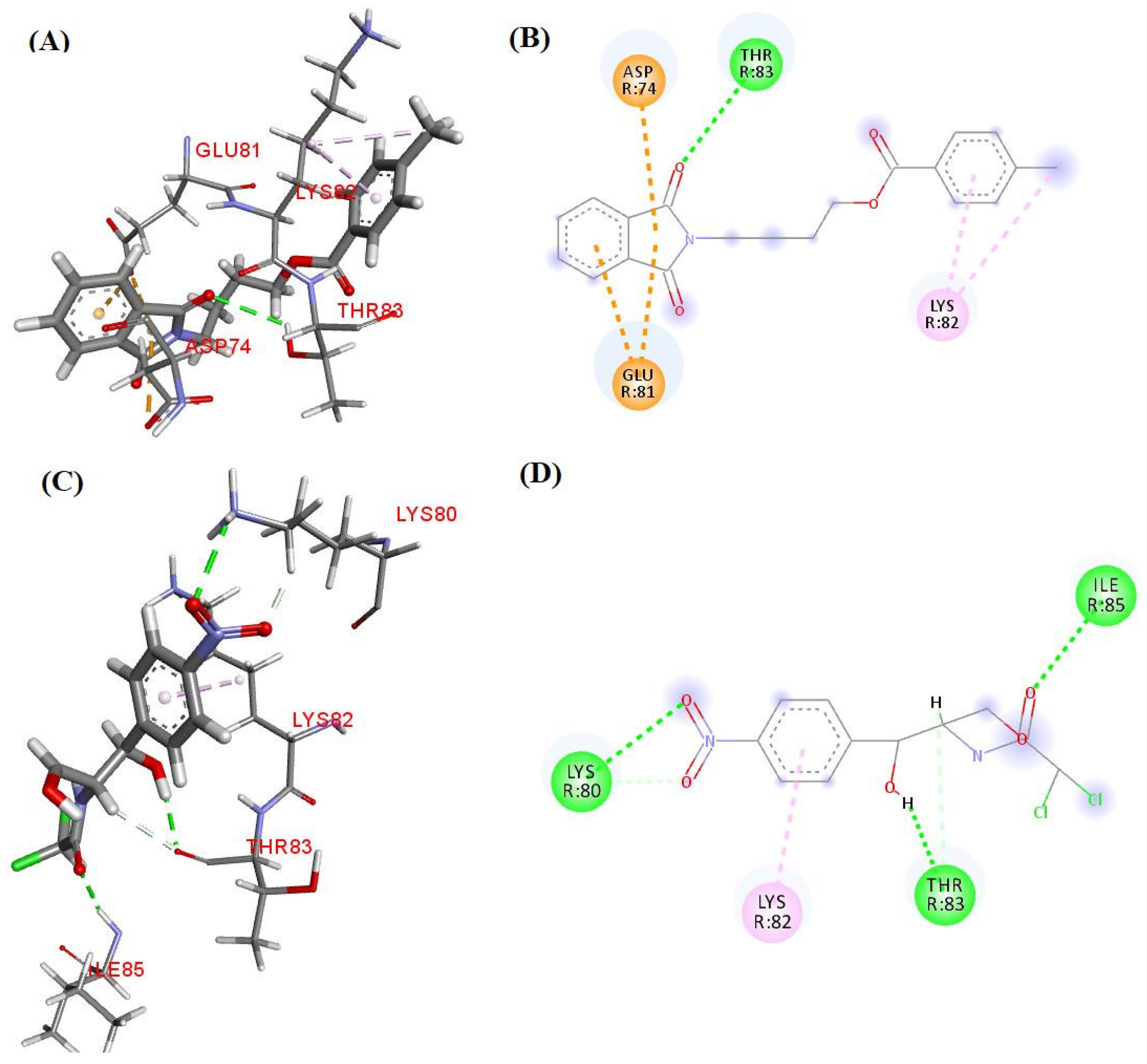
The results obtained from the molecular docking study between compound 3b and the target 1NJI, compared to Chloramphenicol, suggest that compound 3b has a more favorable antibacterial mechanism of action from a thermodynamic perspective. The interaction energy of compound 3b (−92.69 kcal·mol−1) was significantly more negative than that of Chloramphenicol (−73.13 kcal·mol−1), indicating greater stability in binding to the active site of the target protein (Table 7). This energy difference may be related to the interaction profile established by compound 3b, which includes π–anion interactions with residues ASP R:74 and GLU R:81, as well as hydrogen bonds with THR R:83, and alkyl interactions with LYS R:82 (Figure 2).
The π–anion interactions observed for compound 3b are particularly relevant, as they involve negatively charged residues (ASP R:74 and GLU R:81), which may contribute to greater specificity and affinity for the target protein. Additionally, the presence of alkyl interactions with LYS R:82 may further stabilize the protein–ligand complex. In contrast, Chloramphenicol exhibited hydrogen bonds with LYS R:80, THR R:83, and ILE R:85, as well as a π–alkyl interaction with LYS R:82, which, although relevant, appears to be less effective in terms of binding energy (Figure 2).
These results suggest that compound 3b may act as a promising candidate for the development of novel antibacterial agents, surpassing Chloramphenicol in terms of binding affinity and stability. The elucidation of the specific molecular interactions of compound 3b with the target 1NJI paves the way for future structural optimization and experimental validation studies, aiming at the discovery of more effective drugs against resistant pathogens.
The results of the molecular docking study between compound 3b and the target 5TZ1, compared to Fluconazole, reveal that compound 3b exhibits slightly less favorable interaction energy (−107.25 kcal·mol−1) than that of Fluconazole (−113.86 kcal·mol−1). However, the interaction profile established by compound 3b suggests a promising and potentially effective binding to the active site of the target protein, indicating a distinct and relevant antifungal mechanism of action (Table 7).
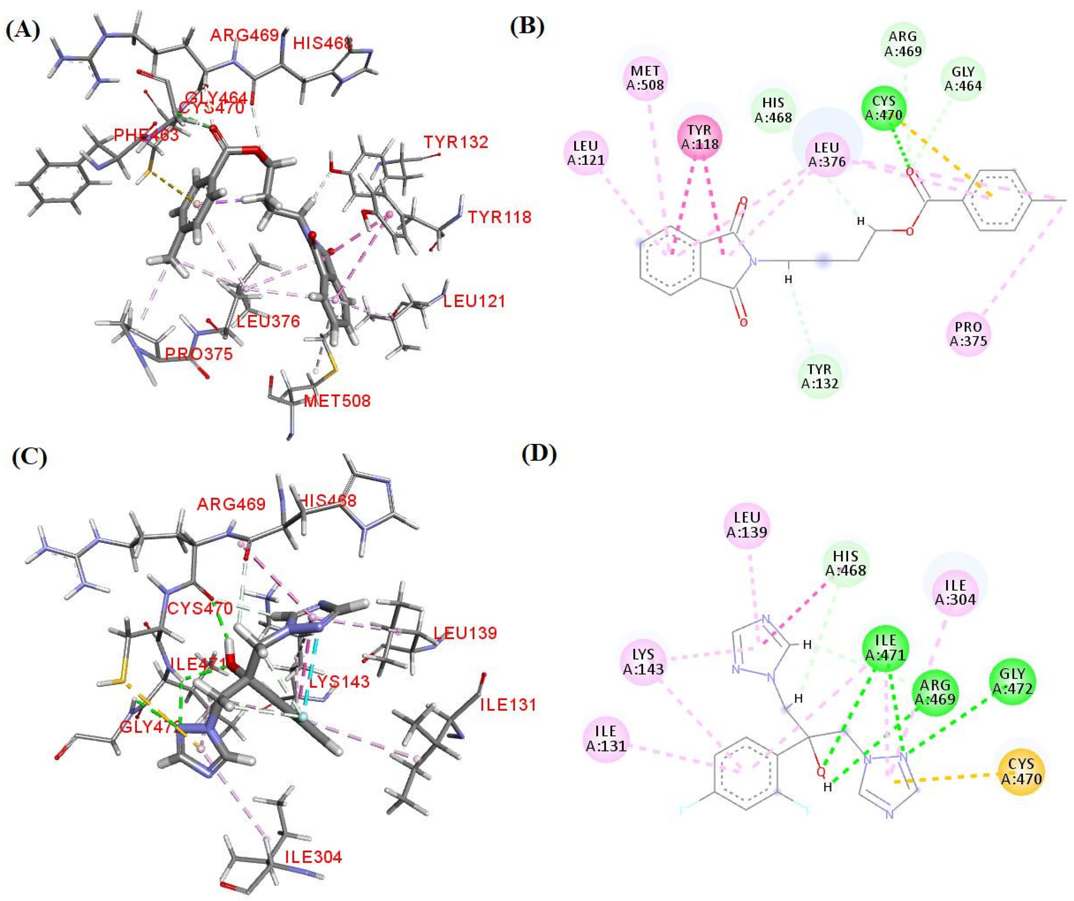
Fluconazole demonstrated hydrogen bonds with ILE A:471, ARG A:469, and GLY A:472, as well as a π–sulfur interaction with CYS A:470, and hydrophobic interactions with HIS A:468, ILE A:131, LYS A:143, LEU A:139, and ILE A:304. These interactions are characteristic of its binding to the target enzyme, contributing to its antifungal efficacy. On the other hand, compound 3b established hydrogen bonds and a π–sulfur interaction with CYS A:470, standing out for the formation of a π–sulfur bond, which is less common and may confer greater specificity to the protein–ligand complex. Additionally, compound 3b exhibited a broad profile of hydrophobic interactions with residues such as HIS A:468, ARG A:469, GLY A:464, TYR A:132, TYR A:118, LEU A:121, MET A:508, LEU A:376, and PRO A:375, suggesting possible additional stabilization of the complex (Figure 3).
Although the interaction energy of compound 3b is slightly less favorable than that of Fluconazole, the diversity and nature of the observed interactions suggest that compound 3b may act through a complementary or alternative mechanism, potentially useful against Fluconazole-resistant strains. The π–sulfur interaction with CYS A:470, in particular, is a notable highlight, as it may contribute to a more specific and stable binding (Figure 3). These results indicate that compound 3b is a viable candidate for future optimization and antifungal evaluation studies, potentially representing a new therapeutic strategy in the fight against fungal infections.

5. Conclusions

In conclusion, two series of phthalimide and N-phthaloylglycine esters were designed to investigate whether the addition of butyl and aryl groups enhances their antimicrobial properties. The best results were observed for phthalimide aryl ester 3b (R = Me), which showed significant antimicrobial activity against Gram-(+) and Gram-(−) bacteria, S. aureus and P. aeruginosa, respectively, and yeast fungi, C. tropicalis and C. albicans, with MIC values equal to 128 µg·mL−1. Considering that compound 3b was as or more active than Fluconazole against strains of C. albicans, its antifungal mechanism of action was studied, where MIC values changed from 128 to 1024 µg·mL−1 in the presence of ergosterol. Furthermore, compound 3b showed synergy with Chloramphenicol against P. aeruginosa, with a FICI value equal to 0.5. Finally, considering the potential disposal of drugs in the aquatic environment, the four most promising compounds had their ecotoxicity evaluated, which showed moderate toxicity (3b and 3c) to non-toxicity (4a and 4c) on A. salina larvae. The molecular docking results demonstrated that compound 3b exhibits enhanced binding affinity and stability with the 50S ribosomal subunit (−92.69 kcal·mol−1) compared to Chloramphenicol, along with a distinctive π–sulfur interaction with CYP51, highlighting its potential as a promising dual-action antibacterial and antifungal agent against resistant pathogens.

Supplementary Materials

The following supporting information can be downloaded at: https://www.mdpi.com/article/10.3390/life15040518/s1, Figure S1. 1H NMR spectrum (400 MHz, CDCl3) of compound 3a. Figure S2. 13C Attached Proton Test (APT) spectrum (100 MHz, CDCl3) of compound 3a. Figure S3. IR spectrum (KBr) of compound 3a. Figure S4. HRMS spectrum (ESI) of compound 3a. Figure S5. 1H NMR spectrum (500 MHz, CDCl3) of compound 3b. Figure S6. 13C NMR spectrum (125 MHz, CDCl3) of compound 3b. Figure S7. IR spectrum (KBr) of compound 3b. Figure S8. HRMS spectrum (ESI) of compound 3b. Figure S9. 1H NMR spectrum (500 MHz, CDCl3) of compound 3c. Figure S10. 13C NMR spectrum (125 MHz, CDCl3) of compound 3c. Figure S11. IR spectrum (KBr) of compound 3c. Figure S12. HRMS spectrum (ESI) of compound 3c. Figure S13. 1H NMR spectrum (400 MHz, CDCl3) of compound 3d. Figure S14. 13C NMR spectrum (100 MHz, CDCl3) of compound 3d. Figure S15. IR spectrum (KBr) of compound 3d. Figure S16. HRMS spectrum (ESI) of compound 3d. Figure S17. 1H NMR spectrum (500 MHz, CDCl3) of compound 4a. Figure S18. 13C APT spectrum (125 MHz, CDCl3) of compound 4a. Figure S19. IR spectrum (ATR) of compound 4a. Figure S20. HRMS spectrum (ESI) of compound 4a. Figure S21. 1H NMR spectrum (400 MHz, CDCl3) of compound 4b. Figure S22. 13C APT spectrum (100 MHz, CDCl3) of compound 4b. Figure S23. IR spectrum (KBr) of compound 4b. Figure S24. HRMS spectrum (ESI) of compound 4b. Figure S25. 1H NMR spectrum (400 MHz, CDCl3) of compound 4c. Figure S26. 13C NMR spectrum (100 MHz, CDCl3) of compound 4c. Figure S27. IR spectrum (KBr) of compound 4c. Figure S28. HRMS spectrum (ESI) of compound 4c. Figure S29. 1H NMR spectrum (400 MHz, CDCl3) of compound 4d. Figure S30. 13C NMR spectrum (100 MHz, CDCl3) of compound 4d. Figure S31. IR spectrum (ATR) of compound 4d. Figure S32. HRMS spectrum (ESI) of compound 4d. Table S1. Estimated median lethal concentration (LC50) for phthalimide and phthaloylglycine esters (3b, 3c, 4a, 4c) on Artemia salina larvae using linear regression equation.

Author Contributions

Conceptualization, F.S.A. and H.D.S.S.; Data curation, M.F.R.S.; Formal analysis, F.S.A., G.F.F., J.A.S.L., and J.L.F.M.G.; Funding acquisition, E.O.L., G.F.F., and P.F.A.-F.; Investigation, F.S.A. and J.L.F.M.G.; Methodology, F.S.A., J.A.S.L., and J.L.F.M.G.; Project administration, G.F.F.; Resources, E.O.L., G.F.F., and P.F.A.-F.; Supervision, G.F.F. and P.F.A.-F.; Validation, A.A.-J., A.P.S., and P.S.V.L.; Visualization, G.F.F.; Writing—original draft, F.S.A.; Writing—review and editing, G.F.F. and J.A.S.L. All authors have read and agreed to the published version of the manuscript.

Funding

This research was funded by Conselho Nacional de Desenvolvimento Científico e Tecnológico (CNPQ), grant number 404927/2023-8, and Coordenação de Aperfeiçoamento de Pessoal de Nível Superior (CAPES). Fellowships from CNPQ (E.O.L., M.F.R.S., P.F.A.-F.) and CAPES (A.P.S., F.S.A., H.D.S.S., P.S.V.L.) are also acknowledged.

Institutional Review Board Statement

Not applicable.

Informed Consent Statement

Not applicable.

Data Availability Statement

The original contributions presented in this study are included in the article/Supplementary Materials. Further inquiries can be directed to the corresponding author.

Conflicts of Interest

The authors declare no conflicts of interest.

References

  1. Global AMR R&D Hub. Available online: https://globalamrhub.org/publications/incentivising-the-development-of-new-antibacterial-treatments-2023/ (accessed on 2 January 2025).
  2. Rusic, D.; Vilovic, M.; Bukic, J.; Leskur, D.; Perisin, A.S.; Kumric, M.; Martinovic, D.; Petric, A.; Modun, D.; Bozic, J. Implications of COVID-19 pandemic on the emergence of antimicrobial resistance: Adjusting the response to future outbreaks. Life 2021, 11, 220. [Google Scholar] [CrossRef]
  3. Lu, H.; Hong, T.; Jiang, Y.; Whiteway, M.; Zhang, S. Candidiasis from cutaneous to systemic, new perspectives of potential targets and therapeutic strategies. Adv. Drug Deliv. Rev. 2023, 199, 114960. [Google Scholar] [CrossRef] [PubMed]
  4. WHO. WHO Fungal Priority Pathogens List to Guide Research, Development and Public Health Action; World Health Organization: Geneva, Switzerland, 2022; Available online: https://www.who.int/publications/i/item/9789240060241 (accessed on 2 January 2025).
  5. Bertagnolio, S.; Dobreva, Z.; Centner, C.M.; Olaru, I.D.; Donà, D.; Burzo, S.; Huttner, B.D.; Chaillon, A.; Gebreselassie, N.; Wi, T.; et al. WHO global research priorities for antimicrobial resistance in human health. Lancet Microbe 2024, 5, 100902. [Google Scholar] [CrossRef] [PubMed]
  6. Di Franco, S.; Alfieri, A.; Pace, M.C.; Sansone, P.; Pota, V.; Fittipaldi, C.; Fiore, M.; Passavanti, M.B. Blood stream infections from MDR bacteria. Life 2021, 11, 575. [Google Scholar] [CrossRef]
  7. Riccardi, N.; Rotulo, G.A.; Castagnola, E. Definition of opportunistic infections in immunocompromised children on the basis of etiologies and clinical features: A Summary for practical purposes. Curr. Pediatr. Rev. 2019, 15, 197–206. [Google Scholar] [CrossRef] [PubMed]
  8. Lu, H.; Shrivastava, M.; Whiteway, M.; Jiang, Y. Candida albicans targets that potentially synergize with fluconazole. Crit. Rev. Microbiol. 2021, 47, 323–337. [Google Scholar] [CrossRef]
  9. Shaik, S.; Lee, J.-H.; Kim, Y.-G.; Lee, J. Antifungal, anti-biofilm, and anti-hyphal properties of N-substituted phthalimide derivatives against Candida species. Front. Cell. Infect. Microbiol. 2024, 14, 1414618. [Google Scholar] [CrossRef]
  10. Jelali, H.; Mansour, L.; Deniau, E.; Sauthier, M.; Hamdi, N. An efficient synthesis of phthalimides and their biological activities. Polycycl. Aromat. Compd. 2020, 42, 1806–1813. [Google Scholar] [CrossRef]
  11. Homsi, A.; Kasideh, A. Synthesis of some N-phthalimide derivatives and evaluation their biological activity. Pharm. Chem. J. 2015, 2, 33–41. [Google Scholar]
  12. Padda, I.S.; Bhatt, R.; Parmar, M. Apremilast. In StatPearls [Internet]; StatPearls Publishing: Treasure Island, FL, USA, 2023. Available online: https://www.ncbi.nlm.nih.gov/books/NBK572078/ (accessed on 2 January 2025).
  13. Engelhardt, M.; Wäsch, R.; Reinhardt, H.; Kleber, M. Pomalidomide. In Small Molecules in Oncology; Martens, U., Ed.; Springer: Berlin/Heidelberg, Germany, 2014; Volume 201, pp. 359–372. [Google Scholar] [CrossRef]
  14. Kim, J.H.; Scialli, A.R. Thalidomide: The tragedy of birth defects and the effective treatment of disease. Toxicol. Sci. 2011, 122, 1–6. [Google Scholar] [CrossRef]
  15. Alves, F.; Oliveira, R.; Souza, H.; Lima, P.; Farias, F.; Marzari, B.; Coelho, H.; Luis, J.; Athayde-Filho, P.; Fiss, G. Cleaner approach and antimicrobial screening of 2-selenoacetanilides: Emergence of potential agents against co-infection. J. Sulfur Chem. 2025, 46, 212–225. [Google Scholar] [CrossRef]
  16. Oliveira, R.F.; Souza, H.D.S.; Alves, F.S.; Sousa, A.P.; Lima, P.S.V.; Huang, M.-F.N.; Cordeiro, L.V.; Diniz-Neto, H.; Lima, E.O.; Trindade, E.O.; et al. Synthesis, in silico study and antimicrobial evaluation of new diesters derived from phthaloylglycine. J. Braz. Chem. Soc. 2020, 31, 953–962. [Google Scholar] [CrossRef]
  17. Olmedo, D.A.; Vásquez, Y.; Morán, J.A.; De Léon, E.G.; Caballero-George, C.; Solís, P.N. Understanding the Artemia salina (brine shrimp) test: Pharmacological significance and global impact. Comb. Chem. High Throughput Screen. 2024, 27, 545–554. [Google Scholar] [CrossRef] [PubMed]
  18. Enthaler, S.; Weidauer, M. Synthesis of δ- and ε-cyanoesters by zinc-catalyzed ring-opening of cyclic ethers with acid chlorides and subsequent cyanation. Catal. Lett. 2012, 142, 168–175. [Google Scholar] [CrossRef]
  19. Trindade, E.O.; Dutra, T.F.; Brandão, M.C.R.; Diniz Neto, H.; Lima, E.O.; Lira, B.F.; Athayde-Filho, P.F.; Barbosa-Filho, J.M. Synthesis, in silico study and antimicrobial activity of new piperine derivatives containing substituted δ-esters. J. Braz. Chem. Soc. 2020, 31, 2590–2602. [Google Scholar] [CrossRef]
  20. Bergmann, E.D.; Kaluszyner, A. Reaction products of primary β-hydroxyamines with carbonyl compounds: XVI condensation of carbonyl compounds with 4-hydroxy- and 4-mercaptobutylamine. Rec. Trav. Chim. 1959, 78, 331–336. [Google Scholar] [CrossRef]
  21. Guan, W.; Lu, D.; Yang, X.; Deng, W.; Xiang, J.; Kambe, N.; Qiu, R. CF3SO2Na-mediated five-component carbonylation of triarylboroxines with TMSCF3 and THF/LiOH/NaI to give aroyloxyalkyl iodides. J. Org. Chem. 2022, 87, 9635–9644. [Google Scholar] [CrossRef]
  22. Scotti, M.T.; Herrera-Acevedo, C.; Menezes, R.P.B.; Martin, H.-J.; Muratov, E.N.; Silva, A.I.S.; Albuquerque, E.F.; Calado, L.F.; Coy-Barrera, E.; Scotti, L. MolPredictX: Online biological activity predictions by machine learning models. Mol. Inf. 2022, 41, 2200133. [Google Scholar] [CrossRef]
  23. CLSI. Methods for Dilution Antimicrobial Susceptibility Tests for Bacteria That Grow Aerobically; Approved Standard—Tenth Edition; CLSI Document M07-A10; Clinical and Laboratory Standards Institute: Wayne, PA, USA, 2015; Available online: https://clsi.org/media/1632/m07a10_sample.pdf (accessed on 2 January 2025).
  24. CLSI. Reference Method for Broth Dilution Antifungal Susceptibility Testing of Yeasts; Approved Standard—Third Edition; CLSI Document M27-A3; Clinical and Laboratory Standards Institute: Wayne, PA, USA, 2008; Available online: https://clsi.org/media/1461/m27a3_sample.pdf (accessed on 2 January 2025).
  25. Bhattacharya, S.; Sae-Tia, S.; Fries, B.C. Candidiasis and mechanisms of antifungal resistance. Antibiotics 2020, 9, 312. [Google Scholar] [CrossRef]
  26. Frost, D.J.; Brandt, K.D.; Cugier, D.; Goldman, R. A whole-cell Candida albicans assay for the detection of inhibitors towards fungal cell wall synthesis and assembly. J. Antibiot. 1995, 48, 306–310. [Google Scholar] [CrossRef]
  27. Balouiri, M.; Sadiki, M.; Ibnsouda, S.K. Methods for in vitro evaluating antimicrobial activity: A review. J. Pharm. Anal. 2016, 6, 71–79. [Google Scholar] [CrossRef] [PubMed]
  28. Bassolé, I.H.N.; Juliani, H.R. Essential oils in combination and their antimicrobial properties. Molecules 2012, 17, 3989–4006. [Google Scholar] [CrossRef] [PubMed]
  29. Pires, D.E.V.; Blundell, T.L.; Ascher, D.B. pkCSM: Predicting small-molecule pharmacokinetic and toxicity properties using graph-based signatures. J. Med. Chem. 2015, 58, 4066–4072. [Google Scholar] [CrossRef] [PubMed]
  30. Sousa, A.P.; Souza, H.D.S.; Almeida-Júnior, A.; Silva, M.F.R.; Cordeiro, L.V.; Lima, E.O.L.; Fiss, G.F.; Athayde-Filho, P.F. Novel esters derived from 4-hydroxychalcones as potential sunscreens with antimicrobial action. Synth. Commun. 2024, 54, 973–991. [Google Scholar] [CrossRef]
  31. Thomsen, R.; Christensen, M.H. MolDock:  a new technique for high-accuracy molecular docking. J. Med. Chem. 2006, 49, 3315–3321. [Google Scholar] [CrossRef]
  32. Hansen, J.L.; Moore, P.B.; Steitz, T.A. Structures of five antibiotics bound at the peptidyl transferase center of the large ribosomal subunit. J. Mol. Biol. 2003, 330, 1061–1075. [Google Scholar] [CrossRef]
  33. Hargrove, T.Y.; Friggeri, L.; Wawrzak, Z.; Qi, A.; Hoekstra, W.J.; Schotzinger, R.J.; York, J.D.; Guengerich, F.P.; Lepesheva, G.I. Structural analyses of Candida albicans sterol 14α-demethylase complexed with azole drugs address the molecular basis of azole-mediated inhibition of fungal sterol biosynthesis. J. Biol. Chem. 2017, 292, 6728–6743. [Google Scholar] [CrossRef]
  34. O’Boyle, N.M.; Banck, M.; James, C.A.; Morley, C.; Vandermeersch, T.; Hutchison, G.R. Open Babel: An open chemical toolbox. J. Cheminform. 2011, 3, 33. [Google Scholar] [CrossRef]
  35. Onodera, K.; Satou, K.; Hirota, H. Evaluations of molecular docking programs for virtual screening. J. Chem. Inf. Model. 2007, 47, 1609–1618. [Google Scholar] [CrossRef]
  36. Metcalf, J.S.; Linday, J.; Beattie, K.A.; Birmingham, S.; Saker, M.L.; Törökné, A.K.; Codd, G.A. Toxicity of cylindrospermopsin to the brine shrimp Artemia salina: Comparisons with protein synthesis inhibitors and microcystins. Toxicon 2002, 40, 1115–1120. [Google Scholar] [CrossRef]
  37. Silveira, B.M.; Novack, K.M.; Marcondes, H.C.; Santos, V.M.R. Preparation, characterization, and biological activity against Artemia salina of news copolymer PMMA-g-PEG derivatives incorporated with fluconazole. Macromol. Symp. 2018, 378, 1700066. [Google Scholar] [CrossRef]
  38. Nguta, J.M.; Mbaria, J.M.; Gakuya, D.W.; Gathumbi, P.K.; Kabasa, J.D.; Kiama, S.G. Biological screening of Kenyan medicinal plants using Artemia salina L. (Artemiidae). Pharmacologyonline 2011, 2, 458–478. [Google Scholar]
  39. Sedaghati, M.; Akbari, R.; Hagghi, L.L.; Yousefi, S.; Mesbahi, T.; Delfi, M. Survey of probable synergism between melittin and ciprofloxacin, rifampicin, and chloramphenicol against multidrug-resistant Pseudomonas aeruginosa. Front. Microbiol. 2024, 15, 1480299. [Google Scholar] [CrossRef]
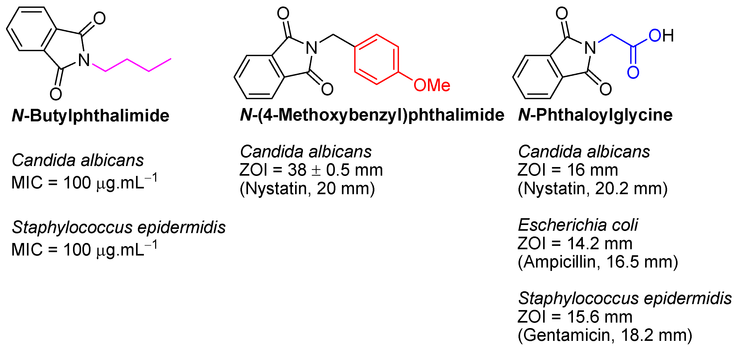
Figure 1. Best antimicrobial results for N-butylphthalimide [9], N-(4-methoxybenzyl)phthalimide [10], and N-phthaloylglycine [11]. MIC: Minimum Inhibitory Concentration; ZOI: Zone of Inhibition.
Life 15 00518 g001
Scheme 1. Reagents and reactional conditions to obtain esters (2ad): (i) THF, 1 mol% ZnCl2, 65 °C (reflux), 2 h; Reagents and reactional conditions to obtain phthalimide and N-phthaloylglycine esters (3ad and 4ad): (ii) 1 mol% NaI, DMF, 100 °C, 20–24 h.
Life 15 00518 sch001
Figure 2. (A) The 3D structure of compound 3b; (B) 2D structure of compound 3b; (C) 3D structure of Chloramphenicol; (D) 3D structure of Chloramphenicol. Interactions performed by compounds 3b and Chloramphenicol with 50S ribosomal subunit (1NJI) and the corresponding amino acid residues. Colors: Green (hydrogen bond), blue and pink (hydrophobic interaction), orange (π–Sulfur interaction). Residues: ASP (Aspartate), GLU (Glutamate), ILE (Isoleucine), LYS (Lysine), THR (Threonine).
Life 15 00518 g002
Figure 3. (A) The 3D structure of compound 3b; (B) 2D structure of compound 3b; (C) 3D structure of Fluconazole; (D) 2D structure of Fluconazole. Interactions performed by compounds 3b and Fluconazole with Sterol 14-alpha demethylase (CYP51) (5TZ1) and the corresponding amino acid residues. Colors: Green (hydrogen bond), blue and pink (hydrophobic interaction), orange (π–Sulfur interaction). Residues: ARG (Arginine), CYS (Cysteine), GLY (Glycine), HIS (Histidine), ILE (Isoleucine), LEU (Leucine), LYS (Lysine), MET (Methionine), PRO (Proline), TYR (Tyrosine).
Life 15 00518 g003
Table 1. In vitro antimicrobial evaluation of phthalimide and N-phthaloylglycine esters (3ad and 4ad).
CompoundRBacteria
MIC/MBC *
Fungi
MIC/MFC *
Gram-(+)Gram-(−)YeastFilamentous
S. aureusE. coliP. aeruginosaC. tropicalisA. flavusA. niger
ATCC 25923LM 01ATCC 25922ATCC 25853ATCC 13803LM 303ATCC 4603ATCC 6275
3aH1024/− 1024/−1024/−1024/−1024/−1024/−1024/−1024/−
3bMe128/512128/512256/512128/512128/512256/5121024/−1024/−
3cOMe512/−512/−512/−512/−256/1024256/10241024/10241024/1024
3dCl−/−−/−−/−−/−−/−−/−−/−−/−
4aH256/1024256/1024256/1024512/1024256/512256/512−/−−/−
4bMe1024/−1024/−1024/−1024/−1024/−1024/−−/−−/−
4cOMe512/−512/−512/−1024/−512/−512/−−/−−/−
4dCl1024/−1024/−1024/−1024/−1024/−1024/−−/−−/−
ChloramphenicolNA32/12832/12864/12864/128NANANANA
FluconazoleNANANANANA256/512256/512256/1024512/1024
Culture mediumNA−/−−/−−/−−/−−/−−/−−/−−/−
MIC: Minimum Inhibitory Concentration; MBC: Minimum Bactericidal Concentration; MFC: Minimum Fungicidal Concentration; NA: Not Applicable; (−): Not active in the concentration range of 1024–0.5 μg.mL−1. * µg·mL−1. All experiments were performed in triplicate.
Table 2. In silico/vitro antimicrobial evaluation of phthalimide and N-phthaloylglycine esters (3ad and 4ad).
CompoundRYeast Fungus C. albicans
In SilicoIn Vitro
MIC/MFC *
Predicted OutcomeProbability Active
(%)
ATCC 76485LM 65
3aHInactive401024/−1024/−
3bMeActive60128/512128/512
3cOMeActive100256/1024256/1024
3dClActive100−/−−/−
4aHInactive20256/512256/512
4bMeInactive201024/−1024/−
4cOMeInactive20512/−512/−
4dClActive601024/−1024/−
FluconazoleNAActive100128/512256/512
Culture mediumNANANA−/−−/−
MIC: Minimum Inhibitory Concentration; MFC: Minimum Fungicidal Concentration; NA: Not Applicable; (−): Not active in the concentration range of 1024–0.5 μg.mL−1. * µg·mL−1. In vitro assays were performed in triplicate.
Table 3. MIC * values of compound 3b in the absence and presence of sorbitol ** or ergosterol ***.
CompoundC. albicans ATCC 76485
SorbitolErgosterol
AbsencePresenceAbsencePresence
3b1281281281024
Caspofungin0.52NANA
FluconazoleNANA128512
Culture medium−/−−/−−/−−/−
MIC: Minimum Inhibitory Concentration; NA: Not Applicable; (−): Not active in the concentration range of 1024–0.5 μg.mL−1. * µg·mL−1. ** 0.8 M. *** 400 µg·mL−1. All experiments were performed in duplicate.
Table 4. FIC * and FICI values of compound 3b.
StrainsFICAFICBFICIOutcome
S. aureus
ATCC 25923
0.251.01.25Indifference
P. aeruginosa
ATCC 25853
0.250.250.5Synergy
FIC: Fractional Inhibitory Concentration; FICA: FIC of compound 3b; FICB: FIC of Chloramphenicol; FICI: Fractional Inhibitory Concentration Index. * µg·mL−1. All experiments were performed in duplicate. Reference for FICI: synergy ≤ 0.5 < addition ≤ 1 < indifference ≤ 4 < antagonism [27,28].
Table 5. In silico/vitro toxicity evaluation of phthalimide and N-phthaloylglycine esters (3b, 3c, 4a, 4c).
CompoundRToxicityLipophilicity
In SilicoIn VitroIn Silico
AmesHepatoSkin SensitizationMinnow
Log LC50
Artemia salina
LC50 *
Log P
3bMeYesYesNo−1.895239.0003.228
3cOMeYesYesNo−1.784224.0952.928
4aHYesYesNo0.4551028.332.463
4cOMeNoYesNo−0.497>>10002.471
ChloramphenicolNAYesNoNo1.892>20 [36]0.909
FluconazoleNANoYesNo3.872802.28 [37]0.735
LC50: Median Lethal Concentration; Log P: octanol/water partition coefficient; NA: Not Applicable. * µg·mL−1. Note: categorical (Yes/No) and numeric (log LC50) unit. Reference for LC50: highly toxic < 100 ≤ moderately toxic < 500 ≤ slightly toxic < 1000 ≤ non-toxic [38].
Table 6. Information on the crystalline structures of enzymes and root-mean-square deviation (RMSD) values for the poses obtained by redocking.
PDB IDMacromoleculeSpeciesPDB LigandResolutionRMSD
1NJI50S ribosomal subunitHaloarcula marismortuiCLM_90013.00 Å2.21 Å
5TZ1Sterol 14-alpha demethylase (CYP51)Candida albicansVT1_6022.00 Å1.87 Å
Table 7. MolDock scores (kcal·mol−1) for enzymes studied.
Compound50S Ribosomal SubunitSterol 14-Alpha Demethylase (CYP51)
3b−92.69−107.25
Chloramphenicol−73.13
Fluconazole−113.86
CLM_9001−73.13
VT1_602−112.54
Disclaimer/Publisher’s Note: The statements, opinions and data contained in all publications are solely those of the individual author(s) and contributor(s) and not of MDPI and/or the editor(s). MDPI and/or the editor(s) disclaim responsibility for any injury to people or property resulting from any ideas, methods, instructions or products referred to in the content.