
Metacarpophalangeal Joint Reconstruction of a Complex Hand Injury with a Vascularized Lateral Femoral Condyle Flap Using an Individualized 3D Printed Model—A Case Report
 , ,
, ,
Abstract
:1. Introduction
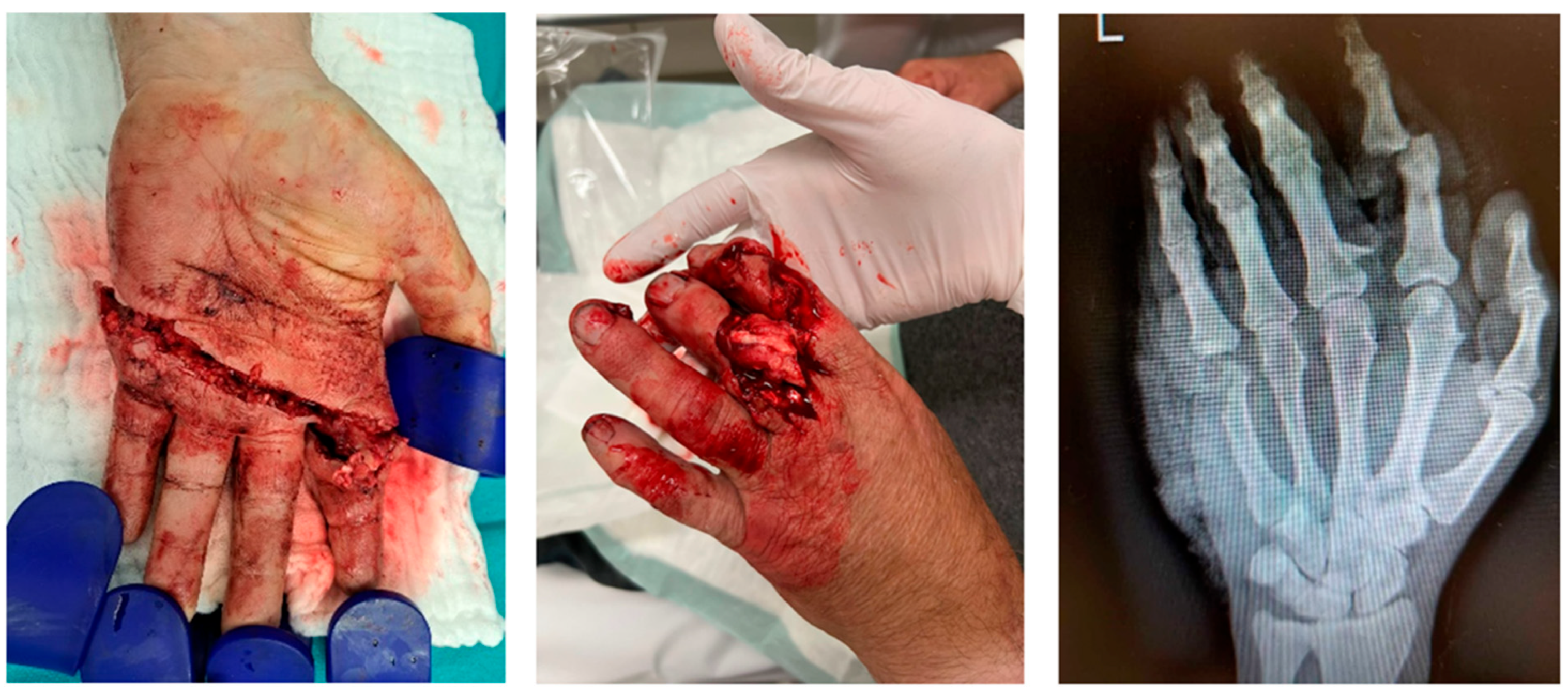
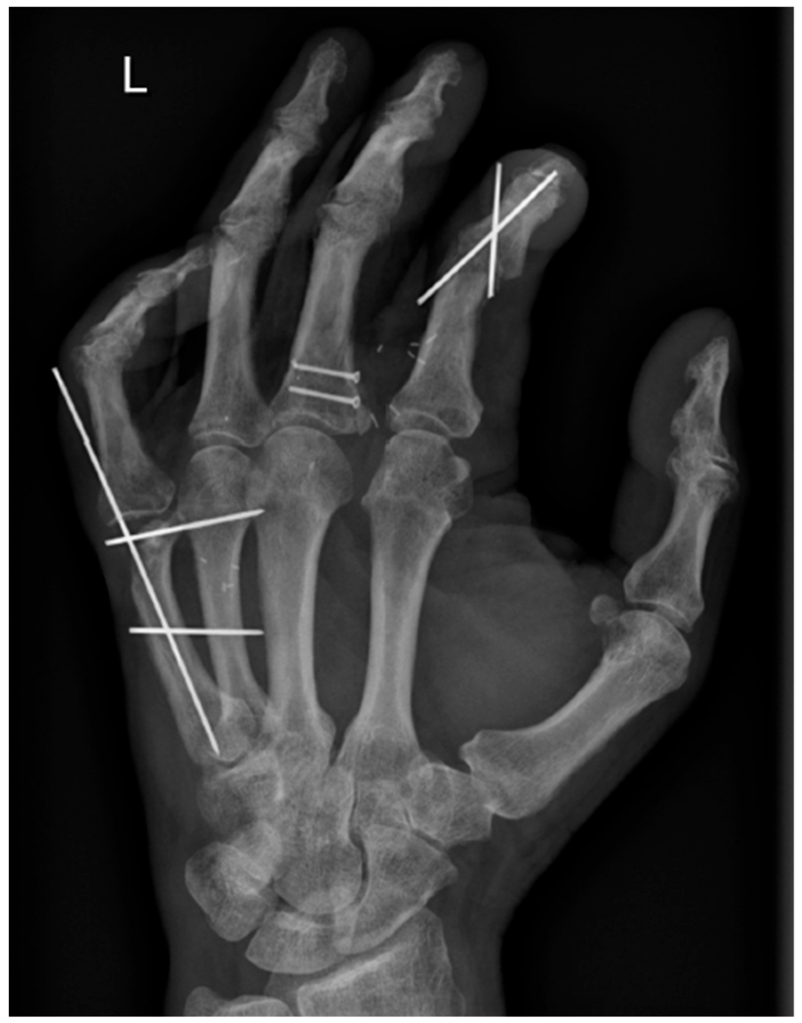
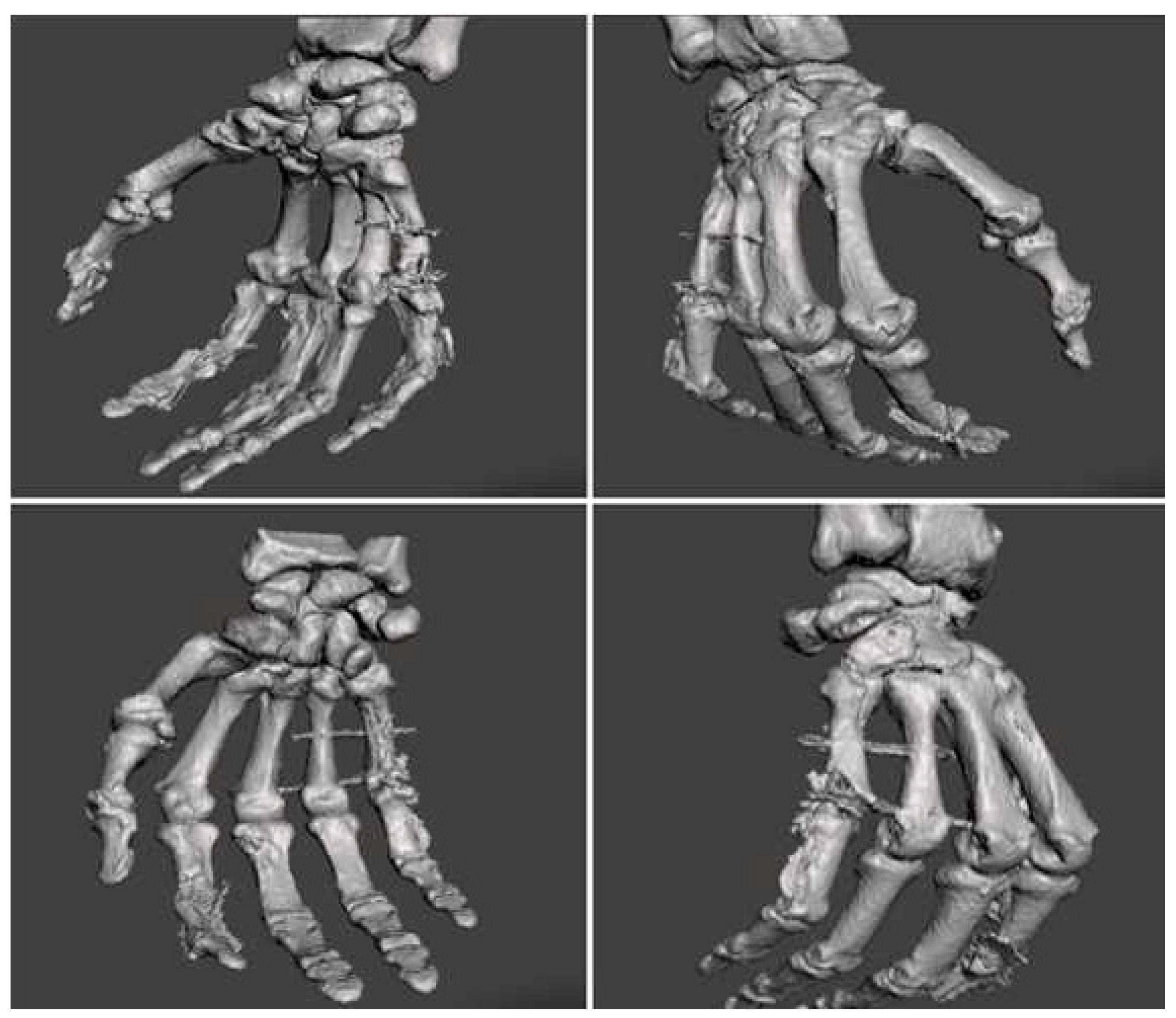
2. Case Report
3. Discussion
Author Contributions
Funding
Institutional Review Board Statement
Informed Consent Statement
Data Availability Statement
Conflicts of Interest
References
- Trybus, M.; Lorkowski, J.; Brongel, L.; Hľadki, W. Causes and consequences of hand injuries. Am. J. Surg. 2006, 192, 52–57. [Google Scholar] [CrossRef] [PubMed]
- Zyluk, A.; Janowski, P. Results of the treatment of major, complex hand injuries. Pol. Przegl. Chir. Pol. J. Surg. 2011, 83, 87–94. [Google Scholar] [CrossRef]
- Zhang, D.; Bauer, A.S.; Blazar, P.; Earp, B.E. Three-Dimensional Printing in Hand Surgery. J. Hand Surg. Am. 2021, 46, 1016–1022. [Google Scholar] [CrossRef] [PubMed]
- Hoang, D.; Perrault, D.; Stevanovic, M.; Ghiassi, A. Today surgical applications of three-dimensional printing: A review of the current literature & how to get started. Ann. Transl. Med. 2016, 4, 456. [Google Scholar] [CrossRef]
- Chung, K.C.; Pillsbury, M.S.; Walters, M.R.; Hayward, R.A. Reliability and validity testing of the Michigan Hand Outcomes Questionnaire. J. Hand Surg. Am. 1998, 23, 575–587. [Google Scholar] [CrossRef] [PubMed]
- Osagie, L.; Shaunak, S.; Murtaza, A.; Cerovac, S.; Umarji, S. Advances in 3D Modeling: Preoperative Templating for Revision Wrist Surgery. Hand 2017, 12, NP68–NP72. [Google Scholar] [CrossRef] [PubMed]
- Ten Berg, P.W.L.; Dobbe, J.G.G.; Streekstra, G.J. Three-dimensional printed anatomical models in scaphoid surgery. J. Hand Surg. Eur. Vol. 2018, 43, 101–102. [Google Scholar] [CrossRef] [PubMed]
- Bizzotto, N.; Tami, I.; Tami, A.; Spiegel, A.; Romani, D.; Corain, M.; Adani, R.; Magnan, B. 3D Printed models of distal radius fractures. Injury 2016, 47, 976–978. [Google Scholar] [CrossRef]
- Jew, N.; Lipman, J.D.; Carlson, M.G. The Use of Three-Dimensional Printing for Complex Scaphoid Fractures. J. Hand Surg. Am. 2019, 44, 165.e1–165.e6. [Google Scholar] [CrossRef]
- Parvizi, D.; Vasilyeva, A.; Wurzer, P.; Tuca, A.; Lebo, P.; Winter, R.; Clayton, R.P.; Rappl, T.; Schintler, M.V.; Kamolz, L.-P.; et al. Anatomy of the Vascularized Lateral Femoral Condyle Flap. Plast. Reconstr. Surg. 2016, 137, 1024e–1032e. [Google Scholar] [CrossRef]
- Higgins, J.P.; Bürger, H.K. Medial Femoral Trochlea Osteochondral Flap Applications for Scaphoid and Lunate Reconstruction. Clin. Plast. Surg. 2020, 47, 21218. [Google Scholar] [CrossRef]
- Wong, V.W.; Bürger, H.K.; Iorio, M.L.; Higgins, J.P. Lateral femoral condyle flap: An alternative source of vascularized bone from the distal femur. J. Hand Surg. Am. 2015, 40, 1972–1980. [Google Scholar] [CrossRef] [PubMed]
- Windhofer, C.M.; Anoshina, M.; Ivusits, P.; Bürger, H.P. The free vascularized lateral femoral trochlea osteochondral graft: A reliable alternative for Stage III Kienböck’s disease. J. Hand Surg. Eur. Vol. 2021, 46, 1032–1041. [Google Scholar] [CrossRef] [PubMed]
- Higgins, J.P.; Bürger, H.K. The use of osteochondral flaps in the treatment of carpal disorders. J. Hand Surg. Eur. Vol. 2018, 43, 48–56. [Google Scholar] [CrossRef] [PubMed]
- Bürger, H.K.; Windhofer, C.; Gaggl, A.J.; Higgins, J.P. Vascularized medial femoral trochlea osteochondral flap reconstruction of advanced Kienböck disease. J. Hand Surg. Am. 2014, 39, 1313–1322. [Google Scholar] [CrossRef] [PubMed]
- Bürger, H.K.; Windhofer, C.; Gaggl, A.J.; Higgins, J.P. Vascularized medial femoral trochlea osteocartilaginous flap reconstruction of proximal pole scaphoid nonunions. J. Hand Surg. Am. 2013, 38, 690–700. [Google Scholar] [CrossRef] [PubMed]
- Del Piñal, F.; García-Bernal, F.J.; Regalado, J.; Ayala, H.; Cagigal, L.; Studer, A. Vascularised corticoperiosteal grafts from the medial femoral condyle for difficult non-unions of the upper limb. J. Hand Surg. Am. 2007, 32, 135–142. [Google Scholar] [CrossRef]
- Willemot, L.; Stewart, D.; Lawson, R. Reconstruction of an infected midshaft radius and ulna nonunion using a free vascularized fibula and medial femoral condyle flap. Microsurgery 2021, 41, 666–670. [Google Scholar] [CrossRef]
- Henry, M. Vascularized Medial Femoral Condyle Bone Graft for Resistant Nonunion of the Distal Radius. J. Hand Surg. Asian-Pac. Vol. 2017, 22, 23–28. [Google Scholar] [CrossRef]
- Belyea, C.M.; Lansford, J.L.; Golden, J.B.; Shin, E.H.; Gumboc, R.D.L. Medial Femoral Condyle Vascularized Bone Graft for Treatment of Midshaft Clavicle Recalcitrant Nonunion with Use of the Transverse Cervical Artery as an Anastomosis. J. Am. Acad. Orthop. Surg. Glob. Res. Rev. 2020, 4, e19.00049. [Google Scholar] [CrossRef]
- Fuchs, B.; Steinmann, S.P.; Bishop, A.T. Free vascularized corticoperiosteal bone graft for the treatment of persistent nonunion of the clavicle. J. Shoulder Elb. Surg. 2005, 14, 264–268. [Google Scholar] [CrossRef] [PubMed]
- Jaloux, C.; Bettex, Q.; Levadoux, M.; Cerlier, A.; Iniesta, A.; Legre, R.; Mayoly, A.; Gay, A. Free vascularized medial femoral condyle corticoperiosteal flap with non-vascularized iliac crest graft for the treatment of recalcitrant clavicle non-union. J. Plast. Reconstr. Aesthetic Surg. 2020, 73, 1232–1238. [Google Scholar] [CrossRef] [PubMed]
- Schalamon, G.; Bürger, H.; Anoshina, M.; Mattiassich, G. Free vascularised osteochondral flap for the treatment of osteochondritis dissecans of the talus. BMJ Case Rep. 2022, 15, 10–13. [Google Scholar] [CrossRef] [PubMed]
- Windhofer, C.M.; Orthner, E.; Bürger, H.K. Vascularized osteochondral free flaps from the femoral trochlea as versatile procedure for reconstruction of osteochondral lesions of the talus. Foot Ankle Surg. 2022, 28, 935–943. [Google Scholar] [CrossRef] [PubMed]
- Jorge, A.C.S.R.G.; Estler, A.; Wahler, T.; Grözinger, G.; Stahl, S. Reconstruction of the Fourth Metacarpal Using a Chimeric Medial Femoral Condyle Vascularized Osteochondral Cutaneous Graft: Case Report. Ann. Plast. Surg. 2022, 89, E1–E4. [Google Scholar] [CrossRef] [PubMed]
- Hsu, C.-C.; Tseng, J.; Lin, Y.-T. Chimeric Medial Femoral Condyle Osteocutaneous Flap for Reconstruction of Multiple Metacarpal Defects. J. Hand Surg. Am. 2018, 43, 781.e1–781.e9. [Google Scholar] [CrossRef] [PubMed]
- Wong, V.W.; Higgins, J.P.; Katz, R.D. Functional reconstruction of subtotal thumb metacarpal defect with a vascularized medial femoral condyle flap: Case report. J. Hand Surg. Am. 2014, 39, 2005–2008. [Google Scholar] [CrossRef]
- Sammer, D.M.; Bishop, A.T.; Shin, A.Y. Vascularized Medial Femoral Condyle Graft for Thumb Metacarpal Reconstruction: Case Report. J. Hand Surg. Am. 2009, 34, 715–718. [Google Scholar] [CrossRef]
- Marks, M. Which patient-reported outcomes shall we use in hand surgery? J. Hand Surg. Eur. Vol. 2020, 45, 5–11. [Google Scholar] [CrossRef]


| Fingertip-to-Palm-Distance | |
|---|---|
| Finger I | 0 cm |
| Finger II | 3.5 cm |
| Finger III | 0 cm |
| Finger IV | 0 cm |
| Finger V | 1.5 cm |
| Scales | Score | |
|---|---|---|
| Left Hand | Right Hand | |
| Overall hand function | 95 | 100 |
| Activities of daily living | 100 | 100 |
| Work performance | 85 * | |
| Pain | 30 | 0 |
| Aesthetics | 100 | 100 |
| Satisfaction | 95 | 100 |
Disclaimer/Publisher’s Note: The statements, opinions and data contained in all publications are solely those of the individual author(s) and contributor(s) and not of MDPI and/or the editor(s). MDPI and/or the editor(s) disclaim responsibility for any injury to people or property resulting from any ideas, methods, instructions or products referred to in the content. |
© 2023 by the authors. Licensee MDPI, Basel, Switzerland. This article is an open access article distributed under the terms and conditions of the Creative Commons Attribution (CC BY) license (https://creativecommons.org/licenses/by/4.0/).
Share and Cite
Kohlhauser, M.; Vasilyeva, A.; Kamolz, L.-P.; Bürger, H.K.; Schintler, M. Metacarpophalangeal Joint Reconstruction of a Complex Hand Injury with a Vascularized Lateral Femoral Condyle Flap Using an Individualized 3D Printed Model—A Case Report. J. Pers. Med. 2023, 13, 1570. https://doi.org/10.3390/jpm13111570
Kohlhauser M, Vasilyeva A, Kamolz L-P, Bürger HK, Schintler M. Metacarpophalangeal Joint Reconstruction of a Complex Hand Injury with a Vascularized Lateral Femoral Condyle Flap Using an Individualized 3D Printed Model—A Case Report. Journal of Personalized Medicine. 2023; 13(11):1570. https://doi.org/10.3390/jpm13111570
Chicago/Turabian StyleKohlhauser, Michael, Anna Vasilyeva, Lars-Peter Kamolz, Heinz K. Bürger, and Michael Schintler. 2023. "Metacarpophalangeal Joint Reconstruction of a Complex Hand Injury with a Vascularized Lateral Femoral Condyle Flap Using an Individualized 3D Printed Model—A Case Report" Journal of Personalized Medicine 13, no. 11: 1570. https://doi.org/10.3390/jpm13111570