Article

Ultramorphological Comparison of Proboscis and Associated Sensilla of Scotogramma trifolii and Protoschinia scutosa (Lepidoptera: Noctuidae)

Institute of Biodiversity and Ecology, School of Life Sciences, Zhengzhou University, Zhengzhou 450001, China
*
Authors to whom correspondence should be addressed.
Insects 2021, 12(11), 992; https://doi.org/10.3390/insects12110992
Submission received: 11 October 2021 / Revised: 31 October 2021 / Accepted: 2 November 2021 / Published: 4 November 2021

Abstract

:

Simple Summary

The clover cutworm, Scotogramma trifolii Rottemberg, and the spotted clover moth, Protoschinia scutosa (Denis & Schiffermuller), are worldwide polyphagous pests, and the larvae feed mainly on the leaf backs of many agricultural crops. However, the food sources and feeding habits of the adults are still poorly known. We investigated the ultramorphology of the proboscis and associated sensilla of S. trifolii and P. scutosa using scanning electron microscopy. The results show that the proboscises of S. trifolii and P. scutosa are structurally similar, both including three sensillum types and three zones (Zone 1–3). The sensillum chaeticum is non-porous hair-like, the sensillum basiconicum is a short smooth cone with a sensory pore on the blunt tip, and each sensillum styloconicum is composed of a uniporous sensory cone inserted into a ribbed stylus. In addition, the movement and fluid uptake mechanisms of the proboscis and the possible function of sensilla are briefly discussed.

Abstract

The proboscis is an important feeding organ for the glossatan moths, mainly adapted to the flower and non-flower visiting habits. The clover cutworm, Scotogramma trifolii Rottemberg, and the spotted clover moth, Protoschinia scutosa (Denis & Schiffermuller), are serious polyphagous pests, attacking numerous vegetables and crops, resulting in huge economic losses. However, the feeding behavior and mechanisms of the adult stage remain unsatisfactorily explored. In this study, the proboscis morphology of S. trifolii and P. scutosa are described in detail using scanning electron microscopy, with the aim of investigating the morphological differences and feeding behavior of these two species. The proboscises of S. trifolii and P. scutosa are similar in morphology and structure and are divided into three zones (Zone 1–3) based on the morphological changes of the dorsal legulae. Three sensillum types are located on the proboscises of both species, sensilla chaetica, sensilla basiconica, and sensilla styloconica. Significant differences were observed in the length of the proboscis and each zone between these two species, as well as in sensilla size and number. Based on the morphology of the proboscis and associated sensilla, S. trifolii and P. scutosa are potential flower visitors, which was also reinforced by the pollen observed at the proboscis tip. These results will strengthen our understanding of the structure of the proboscis related to the feeding behavior of Noctuidae.

1. Introduction

Insect mouthparts are modified appendages of head segments, bearing various types of sensory organs and adapted to exploit different food resources [1]. Due to host selection and optimized feeding techniques, the structure of the mouthparts has undergone significant differentiation in the long evolutionary process [2,3]. Characteristic adaptations of mouthparts have resulted in feeding specialization and enhanced functional performance [3]. For example, the chewing mouthparts in Orthoptera [4] and Coleoptera [5,6,7] have been adapted for grinding, chewing, pinching, or crushing bits of solid food, whereas the piercing–sucking mouthparts in Hemiptera [8,9,10] have evolved to feed on the tissue of the host plants [11]. In Lepidoptera, a siphoning type of proboscis has evolved only once, and is an autapomorphy of the Glossata [1]. The proboscis is well-suited to feed on nectar, pollen, fruit, blood and tears, mainly adapted to flower and non-flower visiting habits [1,12].
In adult Glossata, the proboscis has a relatively simple morphology, but it is unique in its coiled resting position. The proboscis consists of two extended concave maxillary galeae and a hollow food tube, which are joined by dorsal and ventral cuticular projections, i.e., legulae [13,14]. The proboscis with specialized structural organization probably has co-evolved in context with the flowering plant in the Cretaceous [1], thus facilitating the diversification of feeding habits in Glossata [15]. An array of mechano- and chemosensilla on the proboscis of Glossatan species, such as sensilla chaetica, basiconica, and styloconica, play important roles in host localization and feeding [16,17,18]. Previous studies on morphology of proboscis sensilla in Glossata have concentrated mainly on Noctuidae [12,16,19,20], and Nymphalidae [21,22,23,24].
Noctuidae (sensu stricto) is the second largest family in Lepidoptera, including numerous agricultural pests of great economic significance [25,26]. The clover cutworm, Scotogramma trifolii Rottemberg, and the spotted clover moth, Protoschinia scutosa (Denis & Schiffermuller), are worldwide polyphagous pests in the subfamily Noctuinae and Heliothinae, respectively [27,28]. Adults of S. trifolii and P. scutosa have characteristics of intermittent local outbreaks and migration [29,30]. The larvae of S. trifolii and P. scutosa feed mainly on many agricultural crops, such as Gossypium herbaceum Linnaeus [31,32,33] and Chenopodium quinoa Willd [34], while the food sources and feeding habits of the adults still remain poorly known. The morphology of proboscis and associated sensilla could provide insights into adult feeding behavior and the mechanisms of S. trifolii and P. scutosa, but it has never been satisfactorily explored.
In this study, we investigated the ultramorphology of the proboscis and associated sensilla of S. trifolii and P. scutosa using scanning electron microscopy, with the aim of interpreting the function of the feeding apparatus and exploring the feeding behavior of these two noctuid species. This study also made morphological comparisons between S. trifolii and P. scutosa and between sexes within species, including the distribution, number and dimension of the proboscis sensilla. These findings provide a morphological basis to understand the feeding mechanisms of noctuid species.

2. Materials and Methods

2.1. Specimen Sampling

Adults of S. trifolii and P. scutosa were obtained from the Modern Agricultural Science and Technology Demonstration Base of Henan Academy of Agricultural Sciences, Yuanyang County, Henan Province, China (35°00′ N, 113°41′ E). The proboscis was cut from the head with dissecting scissors under a stereomicroscope (SZN71, Sunny Optical Technology Co., Ltd., Ningbo, China). The samples were fixed in Carnoy’s fixative solution (95% ethanol: glacial acetic acid = 3:1) for 24 h, and then immersed in 75% ethanol solution.

2.2. Scanning Electron Microscopy (SEM)

The proboscis was washed using an ultrasonic cleaner (KQ118, Kunshan, China) for a few seconds. All samples were dehydrated through a graded ethanol series of 80%, 85%, 90%, and 95% for 10–15 min each, and 100% for 30 min, twice. The proboscis was naturally dried on filter paper. The dried samples were mounted at various angles using double-sided graphite adhesive tape, and sputter coated with gold. All samples were examined under a Hitachi S-3400N scanning electron microscope (Hitachi, Tokyo, Japan) at 5.00 kV and full vacuum.

2.3. Statistical Analysis

The proboscis dimensions and sensilla size were measured using Imaris 7.4.2 software with seven males and seven females of each species. An independent sample t-test was used to evaluate differences of proboscis and associate sensilla between species and sexes (p < 0.05) in SPSS Statistics v 22.0.0.0. The software Photoshop CS (Adobe, San Jose, CA, USA) was used to adjust contrast and levels of SEM images.

2.4. Terminology

Morphological descriptions of the proboscis follow the terminology of Lehnert et al. [35] and Faucheux [16]. The proboscis was structurally delineated into three zones (Zone 1–3). These zones were determined and defined by Lehnert et al. [22]. Zone 1 is hydrophobic and longest among the three zones; Zone 2 is the main hydrophilic zone, allowing liquid to enter the food canal through interlegular spaces; Zone 3 occupies only a small portion of the tip of the proboscis, without dorsal legulae.

3. Results

3.1. General Morphology of Proboscis

The proboscises of S. trifolii and P. scutosa are nearly same in shape and structure, both consisting of two elongated and coiled maxillary galeae (Figure 1A,B). The maxillary galeae are tightly linked by a series of dorsal and ventral legulae, thus generating a sucking tube. The proboscis is coiled into 4–5 turns in the resting position, and the coils are closely connected (Figure 1C,D). The lateral lobe and pilifer bear long bristles at the base of the proboscis. Floral pollens were wrapped at the tip region of the coiled proboscis in P. scutosa (Figure 2). The length of the proboscis is similar for S. trifolii and P. scutosa (p > 0.05) (Table 1).
According to the shape, the size, and the absence of the dorsal legulae, the proboscis can be structurally delineated into three zones (Zone 1–3), which are present on both S. trifolii and P. scutosa (Figure 3). Zone 1 is characterized by tightly linked lancet-shaped dorsal legulae, whereas Zone 2 is unique in having wider dorsal legulae overlapping in a shingle-like fashion (Figure 3A–C). Zone 3 is the most distal and the smallest region at the apex, without dorsal legulae (Figure 3D,E). The rough external surface is equipped with transverse cuticular processes on Zone 1 and numerous microbumps on Zone 2 and 3, respectively. The concave inner surface of proboscis bears closely connected smooth transverse ridges (Figure 3C). S. trifolii exhibit significant differences from P. scutosa in the length of each zone, as well as in the size of sensilla (Table 1 and Table 2). No significant differences are found between sexes within each species (Table 3 and Table 4). P. scutosa has a markedly longer Zone 1 and 3 than S. trifolii (Table 1).

3.1.1. Zone 1

Numerous triangular microtrichia are arranged in regular rows on the galeal surface of Zone 1 (Figure 4A), and gradually become shorter towards Zone 2. The surface of the food canal is composed of horizontal grooves which are tightly arranged in parallel (Figure 4B,C,E). The diameter of the food canal on Zone 1 remains almost constant, and shows non-significant differences between S. trifolii and P. scutosa (Table 1). The dorsal legulae of both species are smooth lancet-shaped plates, arranged horizontally, and overlapped with the opposite (Figure 4D). The ventral legulae include two rows of flattened plates, with the internal row narrower and shorter than the external (Figure 4E). The flattened plates are tightly connected. S. trifolii and P. scutosa have a mean length of 6.43 ± 0.17 mm and 7.16 ± 0.16 mm on Zone 1, occupying more than 80% and 90% of the total proboscis, respectively (Table 1). The length of Zone 1 is significantly different between these two species, but similar between sexes within species (Table 3 and Table 4). Zone 1 of both species bears sensilla chaetica and sensilla basiconica on the external surface, and sensilla basiconica on the food canal (Figure 4).

3.1.2. Zone 2

The galeal surface on Zone 2 possesses microbumps of various shapes and sizes (Figure 5C,D). Around six microbumps surround the sensilla styloconica, and form a lotus-shaped base. The food canal on Zone 2 is morphologically similar to those on Zone 1, progressively tapering toward the tip (Figure 5A,B). The dorsal legulae on Zone 2 are parallel to each other and nearly perpendicular to the cross section of galea, leaving some slits between the dorsal legulae. Each dorsal legula is curved in a sickle shape toward the tip, widened at the base and bifurcated at the apex. The ventral legulae on Zone 2 consist of two rows of flattened plates, and the internal row is much narrower than the external, with enlarged interlegular space (Figure 5E). S. trifolii has a significantly longer Zone 2 than P. scutosa (Table 1). No significant differences are observed in the length of Zone 2 between sexes within species (Table 3 and Table 4). Zone 2 possesses three types of sensilla: sensilla chaetica, sensilla basiconica, and sensilla styloconica (Figure 5A–D).

3.1.3. Zone 3

Zone 3 is the distalmost zone without dorsal legulae on the proboscis of both species (Figure 5E,F). The external surface of Zone 3 is covered with blunt microbumps. The internal surface is morphologically similar to that of Zone 1 and 2, but with a much narrower food canal. The ventral legulae on Zone 3 consist of two rows of flattened plates, nearly same to those on Zone 2 (Figure 5E). Zone 3 of S. trifolii is significantly shorter than that of P. scutosa (Table 1). No significant differences are found in the length of Zone 3 between sexes within species (Table 3 and Table 4). Two types of sensilla are visible on Zone 3 of both species: sensilla basiconica and sensilla styloconica.

3.2. Proboscis Sensilla

3.2.1. Sensilla Chaetica

Sensilla chaetica are distributed on the galeal surface of Zones 1 and 2. Sensillum chaeticum is non-porous hair-like, and gradually tapers toward the tip (Figure 6). The base of sensillum chaeticum is embedded in a concave pit, standing nearly perpendicularly to the cuticle surface. Sensilla chaetica on Zone 1 are elongated and equipped with longitudinal striations (Figure 6A–G), but become short and smooth on Zone 2 (Figure 6H). The most abundant sensilla, sensilla chaetica, are on the proboscis of both species, which are denser at the basal and much sparser on the distal part. Sensilla chaetica of P. scutosa are significantly longer and wider than those of S. trifolii (Table 2). Significant differences are observed in the width of sensilla chaetica between sexes within each species (Table 3 and 4).

3.2.2. Sensilla Basiconica

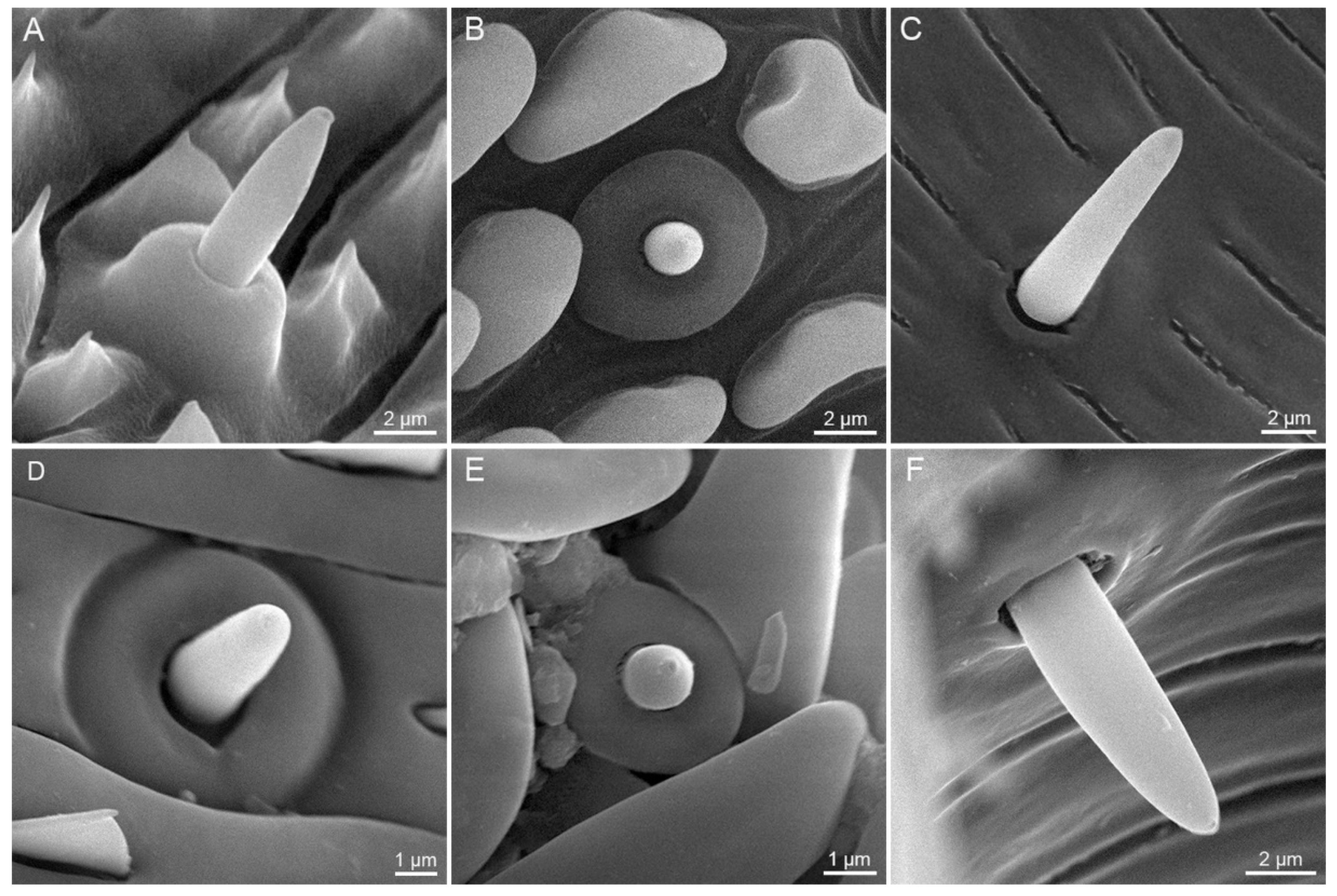
Sensilla basiconica are distributed on both the external galeal surface and the food canal along the whole proboscis. Each sensillum basiconicum consists of a short smooth cone with a sensory pore at the blunt tip, protruding from a round socket (Figure 7).
External sensilla basiconica are irregularly distributed on the galeal surface, and become shorter at the distal region of the proboscis (Figure 7A,B,D,E). S. trifolii bears significantly longer and wider external sensilla basiconica on Zone 1 and 2 than P. scutosa (Table 2). Males of S. trifolii have significantly shorter and wider external sensilla basiconica on Zone 1 than females (Table 3). No significant differences are found in the length or width of external sensilla basiconica between sexes of P. scutosa (Table 4).
Internal sensilla basiconica (Figure 7C,F) are regularly spaced in a row in the food canal. The internal sensilla basiconica of S. trifolii are noticeably longer and wider than those of P. scutosa (Table 2). Males of S. trifolii have significantly wider internal sensilla basiconica than females (Table 3). No significant differences are observed in the length and width between sexes of P. scutosa (Table 4).

3.2.3. Sensilla Styloconica

Sensilla styloconica of S. trifolii and P. scutosa are distributed exclusively on Zone 2 and 3 (Figure 8). Each sensillum styloconicum is composed of a uniporous sensory cone inserted into an elongated stylus, bearing 5–6 longitudinal ribs and protruding from a lotus-shaped base (Figure 8C–F). Sensilla styloconica are progressively shortened toward the tip of the proboscis, generating a brush-shaped appearance on Zone 2 and 3. S. trifolii has significantly longer and wider sensilla styloconica than P. scutosa (Table 2). Females of S. trifolii and P. scutosa have markedly wider sensilla styloconica than males (Table 3 and Table 4).

4. Discussion

This study made a detailed morphological comparison of proboscis and the associated sensilla between the clover cutworm, Scotogramma trifolii, and the spotted clover moth, Protoschinia scutosa. The two distant species, S. trifolii of Noctuinae and P. scutosa of Heliothinae, possess similar proboscis in morphology and structure, both including three sensillum types and three zones (Zone 1–3). The proboscis structure is relatively conservative in Noctuidae, and sensilla morphology may not be useful for comparative studies of nearby species. In Noctuinae, Mythimna separata [12] and Athetis lepigone [20] were reported to lack Zone 3 at the proboscis tip, while S. trifolii possesses Zone 3 (Figure 3D), suggesting a somewhat complex pattern in proboscis morphology. By contrast, both P. scutosa and Helicoverpa armigera [12] bear Zone 3 at the proboscis tip, which might suggest that Zone 3 is relatively conservative in Heliothinae. Our study indicates that S. trifolii of Noctuinae and P. scutosa of Heliothinae exhibit significant differences in the dimension of each zone, as well as in the sensilla size. These morphological results probably reinforce the previous conclusion that the proboscis and associated sensilla could provide potential values in the systematic and phylogenetic analyses at the subfamily level within Noctuidae.
The proboscises of S. trifolii and P. scutosa are tightly coiled in the resting position, and the cuticular processes might help maintain the resting position [36]. The proboscis extends to a relatively straight shape when feeding, producing converse effects on the antero-dorsal and postero-ventral surfaces [37]. Two groups of maxillary muscles, galeal and stipital muscles, are used to control the proboscis movement, as reported in other lepidopterans [38]. The proboscis uncoiling is caused by the increase of hemolymph pressure of stipites forcing hemolymph into galeae [39]. By contrast, proboscis recoiling is supported by the elasticity of the galeal cuticle [1,40] and the contraction of the oblique longitudinal intragaleal muscles [41]. The proboscis coiling starts at the tip and progresses toward the base. The diameter of the spiral widens due to its elastic properties until the proboscis props itself against the ventral side of the head [36]. This elastic effect combined with the tightly linking cuticular processes may hold the resting position of the proboscis. Coiling and bending not only help package and protect the proboscis, but also provide additional means to optimize fluid intake [42].
The lepidopteran proboscis is functionally divided into a hydrophobic and a hydrophilic region [35]. Zone 1 with a closed interlocking structure is considered to be a hydrophobic non-drinking region, whereas Zones 2 and 3 are regarded as hydrophilic drinking regions [22]. The hydrophobic region bears overlapping dorsal legulae and small interlegular spaces with a large proportion of the circumference covered with hydrophobic microbumps [43]. The hydrophilic regions of S. trifolii and P. scutosa account for 5–20% of the total length of the proboscis [44,45], and bear slits at the tip which actively lead fluid into the food canal [46,47]. Liquid absorption was originally explained by the drinking-straw model of the proboscis [48]. However, recent studies have revealed that Lepidoptera pull fluid from the porous surface into the food canal by capillary action via the interlegular spaces and the spaces between the hydrophilic sensilla styloconica and the pressure gradient created by the sucking sump [45,49]. Fluid uptake with the proboscis is mainly comprised of four steps: wetting, dewetting, absorbing, and pumping [50,51]. Many physical determinants represent the fundamental architecture of the proboscis affecting fluid uptake [52]. For example, the absorption efficiency is affected by increased resistance from tapering of the food canal in the drinking region and the viscous resistance of the membranes spreading along the food canal [46,49]. S. trifolii possess significantly longer Zones 2 and 3 than P. scutosa, probably suggesting a better ability of fluid absorption of S. trifolii.
Three sensillum types on the proboscis of S. trifolii and P. scutosa play different roles in feeding behavior. The aporous sensilla chaetica are considered to be mechanosensitive [16,53,54], probably involved in detecting the depth of proboscis insertion during flower probing and monitoring the coiling status in the resting position [36]. The external sensilla basiconica are uniporous and act as chemoreceptors (taste and gustatory receptors) [16,53,55]. The internal sensilla basiconica on the food canal could provide information on the passage from nectar to the pharynx, as reported in butterfly species [16,56]. The sensilla styloconica of both species are located exclusively at the proboscis tip in a dense brush-like configuration, similar to those of Hypsoropha hormos [57], Choristoneura fumifernana [55], and Laspeyresia pomonella [58]. In most lepidopterans, each sensillum styloconicum bears a single pore at the apex of the cone, and is inferred to be derived from sensillum basiconicum [16]. The uniporous sensilla styloconica function as contact chemo-mechanoreceptors to explore the corolla entrance of the flower and taste nectar [59,60]. Each sensillum styloconicum is reported to be innervated by dendrites from receptor cells [61], and sensitive to sugars, nicotine, and other substances [62,63].
The noctuid moths are mostly flower-visiting pollinators, and use the proboscis tip to pollinate [64,65,66]. These two species, S. trifolii and P. scutosa, are most likely to be flower-visiting noctuids, and characterized by elongated sensilla styloconica and modified dorsal legulae on the proboscis, as reported in previous studies [15,19,20,67,68]. The conclusion is also reinforced by the presence of floral pollen at the proboscis tip of P. scutosa (Figure 2). The morphological adaptability of the proboscis is mainly manifested in its length and the tip region with specially shaped sensilla and cuticular structures [14,23,69,70]. The proboscis morphology of flower visitors generally varies according to the diverse floral structures [22]. The longer and more finely pointed the proboscis, the more readily accessible is the corolla for feeding and nutrient acquisition [71]. Compared with S. trifolii, P. scutosa possess a long proboscis with a narrow tip, which might suggest that P. scutosa could better stretch into a long and slender flower tube than S. trifolii. In the potential flower visiting noctuids, Zone 3 is present on the proboscis tip of S. trifolii and P. scutosa, and absent on the proboscis tip of M. separata and A. lepigone [12,20,72]. The presence of Zone 3 might have adaptive value in facilitating the proboscis to enter narrow floral corollas, as described in Lehnert et al. [22]. Further studies may be needed to explore the function of Zone 3 in the evolution of adult feeding habits of noctuid species.

5. Conclusions

This study shows that the proboscises of S. trifolii and P. scutosa are structurally similar, both composed of two tightly linked maxillary galeae that enclose the central food canal by dorsal and ventral legulae. The proboscis was structurally delineated into three zones (Zone 1–3), and possesses three types of sensilla, sensilla chaetica, basiconica, and styloconica. However, S. trifolii and P. scutosa exhibit significant differences in the length of the proboscis and each zone, as well as in the dimension of each sensillum type. The presence of floral pollen, in combination with the characteristics of the distal proboscis indicates that S. trifolii and P. scutosa are very likely flower visitors. These findings provide a morphological basis to better understand the feeding mechanisms of both species.

Author Contributions

Conceptualization, G.-L.H. and J.-Q.L.; methodology, C.-M.Z.; software, C.-M.Z.; validation, G.-L.H.; formal analysis, C.-M.Z. and Y.N.; investigation, C.-M.Z.; resources, G.-L.H. and J.-Q.L.; data curation, C.-M.Z. and Y.N. writing—original draft preparation, C.-M.Z.; writing—review and editing, C.-M.Z., G.-L.H. and J.-Q.L.; visualization, C.-M.Z. and G.-L.H.; supervision, J.-Q.L.; project administration, G.-L.H. and J.-Q.L.; funding acquisition, G.-L.H. All authors wrote and edited the paper. All authors have read and agreed to the published version of the manuscript.

Funding

China Postdoctoral Science Foundation (Grant no. 2019M662540); Key Research Projects of Henan Higher Education Institutions, Grant No. 21A180026.

Institutional Review Board Statement

Not applicable.

Informed Consent Statement

Informed consent was obtained from all subjects involved in the study.

Data Availability Statement

The data presented in this study are available on request from the corresponding author.

Acknowledgments

We are indebted to Yan-Yan Zhou and Dan Long for specimen collection. We thank Fu-Zhen Guo, Xiao-Hua He, and Le-Le He for technical assistance in scanning electron microscopy. This research was supported by the China Postdoctoral Science Foundation (Grant no. 2019M662540) and the Key Research Projects of Henan Higher Education Institutions (Grant No. 21A180026).

Conflicts of Interest

The authors report no conflict of interest.

References

  1. Krenn, H.W. Feeding mechanisms of adult Lepidoptera: Structure, function, and evolution of the mouthparts. Annu. Rev. Entomol. 2010, 55, 307–327. [Google Scholar] [CrossRef] [Green Version]
  2. Blanke, A.; Ruhr, P.T.; Mokso, R.; Villanueva, P.; Wilde, F.; Stampanoni, M.; Uesugi, K.; Machida, R.; Misof, B. Structural mouthpart interaction evolved already in the earliest lineages of insects. Proc. Biol. Sci. 2015, 282, 20151033. [Google Scholar] [CrossRef] [Green Version]
  3. Krenn, H.W. Form and function of insect mouthparts. In Insect Mouthparts: Form, Function, Development and Performance; Krenn, H.W., Ed.; Springer Nature: Gewerbestrasse, Cham, Switzerland, 2019; pp. 9–46. [Google Scholar]
  4. Krenn, H.W.; Fournel, J.; Bauder, J.A.; Hugel, S. Mouthparts and nectar feeding of the flower visiting cricket Glomeremus orchidophilus (Gryllacrididae). Arthropod Struct. Dev. 2016, 45, 221–229. [Google Scholar] [CrossRef] [PubMed]
  5. Betz, O.; Thayer, M.K.; Newton, A.F. Comparative morphology and evolutionary pathways of the mouthparts in spore–feeding Staphylinoidea (Coleoptera). Acta Zool. 2003, 84, 179–238. [Google Scholar] [CrossRef]
  6. Lei, G.; Fu, Y.; Wu, W. Fine structure of mouthparts and forelegs of Euplatypus parallelus (Coleoptera: Curculionidae) with emphasis on the behavior of gallery excavation. Micron 2020, 130, 102815. [Google Scholar] [CrossRef]
  7. Moon, M.J.; Park, J.G.; Kim, K.H. Fine structure of the mouthparts in the ambrosia beetle Platypus koryoensis (Coleoptera: Curculionidae: Platypodinae). Anim. Cells Syst. 2008, 12, 101–108. [Google Scholar] [CrossRef]
  8. Krenn, H.W.; Aspock, H. Form, function and evolution of the mouthparts of blood-feeding Arthropoda. Arthropod Struct. Dev. 2012, 41, 101–118. [Google Scholar] [CrossRef]
  9. Wang, T.; Pan, L.; Zhang, Y.; Dai, W. Morphology of the mouthparts of the spittlebug Philagra albinotata Uhler (Hemiptera: Cercopoidea: Aphrophoridae). Arthropod Struct. Dev. 2015, 44, 121–130. [Google Scholar] [CrossRef]
  10. Wang, Y.; Zhang, J.; Wang, W.; Brozek, J.; Dai, W. Unique fine morphology of mouthparts in Haematoloecha nigrorufa (Stal) (Hemiptera: Reduviidae) adapted to millipede feeding. Insects 2020, 11, 386. [Google Scholar] [CrossRef] [PubMed]
  11. Krenn, H.W.; Mauss, V.; Plant, J. Evolution of the suctorial proboscis in pollen wasps (Masarinae, Vespidae). Arthropod Struct. Dev. 2002, 31, 103–120. [Google Scholar] [CrossRef]
  12. Chen, Q.X.; Li, W.L.; Chen, Y.W.; Chen, J.; Song, Y.Q. Morphological comparison of proboscides and associated sensilla of Helicoverpa armigera and Mythimna separate (Lepidoptera: Noctuidae). Arthropod Struct. Dev. 2019, 49, 119–127. [Google Scholar] [CrossRef] [PubMed]
  13. Krenn, H.W.; Kristensen, N.P. Early evolution of the proboscis of Lepidoptera (Insecta): External morphology of the galea in basal glossatan moth lineages, with remarks on the origin of the pilifers. Zool. Anz. 2000, 239, 179–196. [Google Scholar]
  14. Bauder, J.A.; Handschuh, S.; Metscher, B.D.; Krenn, H.W. Functional morphology of the feeding apparatus and evolution of proboscis length in metalmark butterflies (Lepidoptera: Riodinidae). Biol. J. Linn. Soc. 2013, 110, 291–304. [Google Scholar] [CrossRef] [Green Version]
  15. Zenker, M.M.; Penz, C.; Paris, M.; Specht, A. Proboscis morphology and its relationship to feeding habits in noctuid moths. J. Insect Sci. 2011, 11, 1–10. [Google Scholar] [CrossRef]
  16. Faucheux, M.J. Sensillum types on the proboscis of the Lepidoptera: A review. Ann. Soc. Entomol. Fr. 2013, 49, 73–90. [Google Scholar] [CrossRef]
  17. Krenn, H.W.; Plant, J.D.; Szucsich, N.U. Mouthparts of flower-visiting insects. Arthropod Struct. Dev. 2005, 34, 1–40. [Google Scholar] [CrossRef]
  18. Xue, S.; Hu, Y.Q.; Hua, B.Z. Morphological comparison of proboscis sensilla between Plusiinae and Noctuinae (Lepidoptera: Noctuidae). Zool. Anz. 2016, 263, 75–83. [Google Scholar] [CrossRef]
  19. Xue, S.; Hua, B.Z. Proboscis sensilla of the black cutworm Agrotis ypsilon (Rottemberg) (Lepidoptera: Noctuidae). J. Asia–Pac. Entomol. 2014, 17, 295–301. [Google Scholar] [CrossRef]
  20. Hu, G.L.; Zhang, C.M.; Wang, Z.Q.; Chen, Q.X.; Lu, J.Q. Sensilla of the antenna and proboscis of Athetis lepigone (Möschler) (Lepidoptera: Noctuidae). J. Morphol. 2021, 282, 733–745. [Google Scholar] [CrossRef]
  21. Krenn, H.W.; Zulka, K.P.; Gatschnegg, T. Proboscis morphology and food preferences in nymphalid butterflies (Lepidoptera: Nymphalidae). J. Zool. 2001, 254, 17–26. [Google Scholar] [CrossRef]
  22. Lehnert, M.S.; Beard, C.E.; Gerard, P.D.; Kornev, K.G.; Adler, P.H. Structure of the Lepidopteran proboscis in relation to feeding guild. J. Morphol. 2016, 277, 167–182. [Google Scholar] [CrossRef]
  23. Molleman, F.; Krenn, H.W.; Van Alphen, M.E.; Brakefield, P.M.; Devries, P.J.; Zwaan, B.J. Food intake of fruit-feeding butterflies: Evidence for adaptive variation in proboscis morphology. Biol. J. Linn. Soc. 2005, 86, 333–343. [Google Scholar] [CrossRef]
  24. Kramer, V.R.; Reiter, K.E.; Lehnert, M.S. Proboscis morphology suggests reduced feeding abilities of hybrid Limenitis butterflies (Lepidoptera: Nymphalidae). Biol. J. Linn. Soc. 2018, 125, 535–546. [Google Scholar] [CrossRef] [Green Version]
  25. Mitchell, A.; Mitter, C.; Regier, J.C. Systematics and evolution of the cutworm moths (Lepidoptera: Noctuidae): Evidence from two protein-coding nuclear genes. Syst. Entomol. 2005, 31, 21–46. [Google Scholar] [CrossRef]
  26. Nieukerken, E.J.; Kaila, L.; Kitching, I.J.; Kristensen, N.P.; Lees, D.C.; Minet, J.; Mitter, C.; Mutanen, M.; Regier, J.C.; Simonsen, T.J.; et al. Order Lepidoptera Linnaeus, 1758. In: Zhang, Z.-Q. (Ed.) Animal biodiversity: An outline of higher-level classification and survey of taxonomic richness. Zootaxa 2011, 3148, 212–221. [Google Scholar] [CrossRef]
  27. Federici, B.A. A new type of insect pathogen in larvae of the clover cutworm, Scotogramma trifolii. J. Invertebr. Pathol. 1982, 40, 41–54. [Google Scholar] [CrossRef]
  28. Zhao, S.; Fu, X.; Guo, J.; Zhou, Y.; Wyckhuys, K.A.G.; Wu, K. Seasonal patterns of Protoschinia scutosa (Lepidoptera: Noctuidae) migration across China’s Bohai strait. Environ. Entomol. 2018, 47, 927–934. [Google Scholar] [CrossRef]
  29. Zhang, Y.H.; Chen, L.; Cheng, D.F.; Zhang, Y.J.; Jiang, Y.Y.; Jiang, J.W. Radar observation and population analysis on the migration of the clover cutworm, Scotogramma trifolii Rottemberg (Lepidoptera: Pyralidae). Acta Entomol. Sin. 2007, 50, 494–500. [Google Scholar]
  30. Cao, K.L.; Wang, S.Y.; Yu, B.J.; Liu, C.C.; Jashenko, R.; Ji, R. Entomological radar observation of migratory insects in China–Kazakhstan border areas. Plant Prot. 2020, 46, 30–37. [Google Scholar]
  31. Sun, Z.H.; Zhao, S.Z. Occurrence of clover cutworm and its control. J. Neimenggu Sci. Technol. 1995, 2, 23–24. [Google Scholar]
  32. Yu, J.N.; Bao, Y.Q. The occurrence of clover cutworm in cotton region of Xinjiang. Xinjiang Agric. Sci. 1996, 1, 34. [Google Scholar]
  33. Zhao, Z.J.; Chen, E.X.; Zhang, Y. Studies on the biological characteristic of Scotogramma trifolii (Rott) and its control. Zhongguo Tiancai 1992, 25–28. [Google Scholar]
  34. Li, Q.R.; Li, F.G.; Wei, Y.H.; Zhu, H.X.; Lai, Y.P.; Hou, L.; Guo, Q.Y. Checklist of insect pests, their enemies and description of five species of pests in Chenopodium quinoa Willd. in arid area of Qinghai Plateau. Plant Prot. 2019, 45, 190–198. [Google Scholar]
  35. Lehnert, M.S.; Monaenkova, D.; Andrukh, T.; Beard, C.E.; Adler, P.H.; Kornev, K.G. Hydrophobic-hydrophilic dichotomy of the butterfly proboscis. J. R. Soc. Interface 2013, 10, 20130336. [Google Scholar] [CrossRef]
  36. Krenn, H.W. Functional morphology and movements of the proboscis of Lepidoptera (Insecta). Zoomorphology 1990, 110, 105–114. [Google Scholar] [CrossRef]
  37. Bauder, J.A.S.; Karolyi, F. Superlong proboscises as co-adaptations to flowers. In Insect Mouthparts: Form, Function, Development and Performance; Krenn, H.W., Ed.; Springer Nature: Gewerbestrasse, Cham, Switzerland, 2019; pp. 479–527. [Google Scholar]
  38. Krenn, H.W.; Kristensen, N.P. Evolution of proboscis musculature in Lepidoptera. Eur. J. Entomol. 2004, 101, 565–575. [Google Scholar] [CrossRef] [Green Version]
  39. Wannenmacher, G.; Wasserthal, L.T. Contribution of the maxillary muscles to proboscis movement in hawkmoths (Lepidoptera: Sphingidae)-an electrophysiological study. J. Insect Physiol. 2003, 49, 765–776. [Google Scholar] [CrossRef]
  40. Hepburn, H.R. Proboscis extension and recoil in Lepidoptera. J. Insect Physiol. 1971, 17, 637–656. [Google Scholar] [CrossRef]
  41. Krenn, H.W. Proboscis musculature in the butterfly Vanessa cardui (Nymphalidae, Lepidoptera): Settling the proboscis recoiling controversy. Acta Zool. (Stockh.) 2001, 81, 259–266. [Google Scholar] [CrossRef]
  42. Zhang, C.; Beard, C.E.; Adler, P.H.; Kornev, K.G. Effect of curvature on wetting and dewetting of proboscises of butterflies and moths. R. Soc. Open Sci. 2018, 5, 171241. [Google Scholar] [CrossRef] [PubMed] [Green Version]
  43. Lehnert, M.S.; Wei, Q.H. Hierarchical microstructures and functions of the Lepidopteran proboscis cuticle. In Insect Mouthparts: Form, Function, Development and Performance; Krenn, H.W., Ed.; Springer Nature: Gewerbestrasse, Cham, Switzerland, 2019; pp. 315–334. [Google Scholar]
  44. Wang, N.; Wei, J.S.; Dang, L.; Wang, J.B.; Hua, B.Z. Ultramorphology of sensilla on the proboscis in three noctuid moths (Lepidoptera:Noctuidae). Acta Entomol. Sin. 2012, 55, 877–884. [Google Scholar] [CrossRef]
  45. Lee, S.C.; Kim, J.H.; Lee, S.J. Adhesion and suction functions of the tip region of a nectar-drinking butterfly proboscis. J. Bionic Eng. 2017, 14, 600–606. [Google Scholar] [CrossRef]
  46. Tsai, C.C.; Monaenkova, D.; Beard, C.E.; Adler, P.H.; Kornev, K.G. Paradox of the drinking-straw model of the butterfly proboscis. J. Exp. Biol. 2014, 217, 2130–2138. [Google Scholar] [CrossRef] [PubMed] [Green Version]
  47. Kwauk, K.J.; Hasegawa, D.K.; Lehnert, M.S.; Beard, C.E.; Gerard, P.D.; Kornev, K.G.; Adler, P.H. Drinking with an unsealed tube: Fluid uptake along the butterfly proboscis. Ann. Entomol. Soc. Am. 2014, 107, 886–892. [Google Scholar] [CrossRef]
  48. Kingsolver, J.G.; Daniel, T.L. Mechanics of food handling by fluid-feeding insects. In Regulatory Mechanisms in Insect Feeding; Chapman, R.F., Ed.; Springer: Boston, MA, USA, 1995; pp. 32–73. [Google Scholar]
  49. Kornev, K.G.; Salamatin, A.A.; Adler, P.H.; Beard, C.E. Structural and physical determinants of the proboscis-sucking pump complex in the evolution of fluid-feeding insects. Sci. Rep. 2017, 7, 6582. [Google Scholar] [CrossRef] [Green Version]
  50. Salamatin, A.A.; Adler, P.H.; Kornev, K.G. Lepidopteran mouthpart architecture suggests a new mechanism of fluid uptake by insects with long proboscises. J. Theor. Biol. 2021, 510, 110525. [Google Scholar] [CrossRef]
  51. Monaenkova, D.; Lehnert, M.S.; Andrukh, T.; Beard, C.E.; Rubin, B.; Tokarev, A.; Lee, W.K.; Adler, P.H.; Kornev, K.G. Butterfly proboscis: Combining a drinking straw with a nanosponge facilitated diversification of feeding habits. J. R. Soc. Interface 2012, 9, 720–726. [Google Scholar] [CrossRef] [Green Version]
  52. Kornev, K.G.; Adler, P.H. Physical determinants of fluid-feeding in insects. In Insect Mouthparts: Form, Function, Development and Performance; Krenn, H.W., Ed.; Springer Nature: Gewerbestrasse, Cham, Switzerland, 2019; pp. 263–314. [Google Scholar]
  53. Krenn, H.W. Proboscis sensilla in Vanessa cardui (Nymphalidae, Lepidoptera): Functional morphology and significance in flower–probing. Zoomorphology 1998, 118, 23–30. [Google Scholar] [CrossRef]
  54. Song, Y.Q.; Sun, H.Z.; Wu, J.X. Ultrastructural characteristics of the proboscis and the labial palp pit organ in the oriental fruit moth, Grapholita Molesta. Bull. Insectol. 2016, 69, 59–66. [Google Scholar]
  55. Walters, B.D.; Albert, P.J.; Zacharuk, R.Y. Morphology and ultrastructure of sensilla on the proboscis of the adult spruce budworm, Choristoneura fumiferana (Clem.) (Lepidoptera: Tortricidae). Can. J. Zool. 1998, 76, 466–479. [Google Scholar] [CrossRef]
  56. Inoue, T.A.; Asaoka, K.; Seta, K.; Imaeda, D.; Ozaki, M. Sugar receptor response of the food-canal taste sensilla in a nectar-feeding swallowtail butterfly, Papilio xuthus. Sci. Nat. 2009, 96, 355–363. [Google Scholar] [CrossRef] [Green Version]
  57. Zaspel, J.M.; Weller, S.J.; Branham, M.A. A comparative survey of proboscis morphology and associated structures in fruit-piercing, tear-feeding, and blood-feeding moths in Calpinae (Lepidoptera: Erebidae). Zoomorphology 2011, 130, 203–225. [Google Scholar] [CrossRef]
  58. Goldware, M.A.; Barnes, M.M. Mouthparts of the adult codling moth, Laspeyresia pomonella (Lepidoptera: Olethreutidae). Ann. Entomol. Soc. Am. 1973, 66, 349–351. [Google Scholar] [CrossRef]
  59. Städler, E.; Seabrook, W.D. Chemoreceptors on the proboscis of the female eastern spruce budworm: Electrophysiological study. Entomol. Exp. Appl. 1975, 18, 153–160. [Google Scholar] [CrossRef]
  60. Jorgensen, K.; Kvello, P.; Almaas, T.J.; Mustaparta, H. Two closely located areas in the suboesophageal ganglion and the tritocerebrum receive projections of gustatory receptor neurons located on the antennae and the proboscis in the moth Heliothis virescens. J. Comp. Neurol. 2006, 496, 121–134. [Google Scholar] [CrossRef] [PubMed]
  61. Nagnan-Le Meillour, P.; Cain, A.H.; Jacquin-Joly, E.; Francois, M.C.; Ramachandran, S.; Maida, R.; Steinbrecht, R.A. Chemosensory proteins from the proboscis of Mamestra brassicae. Chem. Senses 2000, 25, 541–553. [Google Scholar] [CrossRef] [PubMed] [Green Version]
  62. Blaney, W.M.; Simmonds, M.S.J. Food selection in adults and larvae of three species of Lepidoptera: A behavioural and electrophysiological study. Entomol. Exp. Appl. 1988, 49, 111–121. [Google Scholar] [CrossRef]
  63. Shields, V.D. Fine structure of the galeal styloconic sensilla of larval Lymantria dispar (Lepidoptera: Lymantriidae). Ann. Entomol. Soc. Am. 2009, 102, 1116–1125. [Google Scholar] [CrossRef] [PubMed] [Green Version]
  64. Sakagami, K.; Matsubara, S.; Sugiura, S. Nocturnal moths as potential pollinators of Marsdenia formosana (Apocynaceae: Asclepiadoideae). Taiwania 2019, 64, 195–197. [Google Scholar] [CrossRef]
  65. Guo, P.; Wang, G.P.; Jin, L.J.; Fan, X.Q.; He, H.L.; Zhou, P.W.; Guo, X.R.; Li, W.Z.; Yuan, G.H. Identification of summer nectar plants contributing to outbreaks of Mythimna separata (Walker) (Lepidoptera: Noctuidae) in North China. J. Integr. Agric. 2018, 17, 1516–1526. [Google Scholar] [CrossRef] [Green Version]
  66. Sakagami, K.; Sugiura, S. Noctuid moths as pollinators of Habenaria sagittifera (Orchidaceae): Floral adaptations for the transfer of pollinaria on the thoraxes of moths. Sci. Nat. 2019, 106, 58. [Google Scholar] [CrossRef]
  67. Yu, J.N.; Zhou, X.H.; Ma, Y.P.; Bao, Y.Q. A study of Scotogramma trifolii and its control. J. Xinjiang Agric. Univ. 1997, 20, 70–73. [Google Scholar]
  68. Büttiker, W.; Krenn, H.W.; Putterill, J.F. The proboscis of eye-frequenting and piercing Lepidoptera (Insecta). Zoomorphology 1996, 116, 77–83. [Google Scholar] [CrossRef]
  69. Hilgartner, R.; Raoilison, M.; Büttiker, W.; Lees, D.C.; Krenn, H.W. Malagasy birds as hosts for eye-frequenting moths. Biol. Lett. 2007, 3, 117–120. [Google Scholar] [CrossRef] [Green Version]
  70. Knopp, M.C.N.; Krenn, H.W. Efficiency of fruit juice feeding in Morpho peleides (Nymphalidae, Lepidoptera). J. Insect Behav. 2003, 16, 67–77. [Google Scholar] [CrossRef]
  71. Danaher, M.W.; Ward, C.; Zettler, L.W.; Covell, C.V. Pollinia removal and suspected pollination of the endangered ghost orchid, Dendrophylax lindenii (Orchidaceae) by various hawk moths (Lepidoptera: Sphingidae): Another mystery dispelled. Fla. Entomol. 2020, 102, 671. [Google Scholar] [CrossRef]
  72. Wang, Y.; Li, L.; Dai, W. Fine morphology of the mouthparts in Cheilocapsus nigrescens (Hemiptera: Heteroptera: Miridae) reflects adaptation for phytophagous habits. Insects 2019, 10, 143. [Google Scholar] [CrossRef] [PubMed] [Green Version]
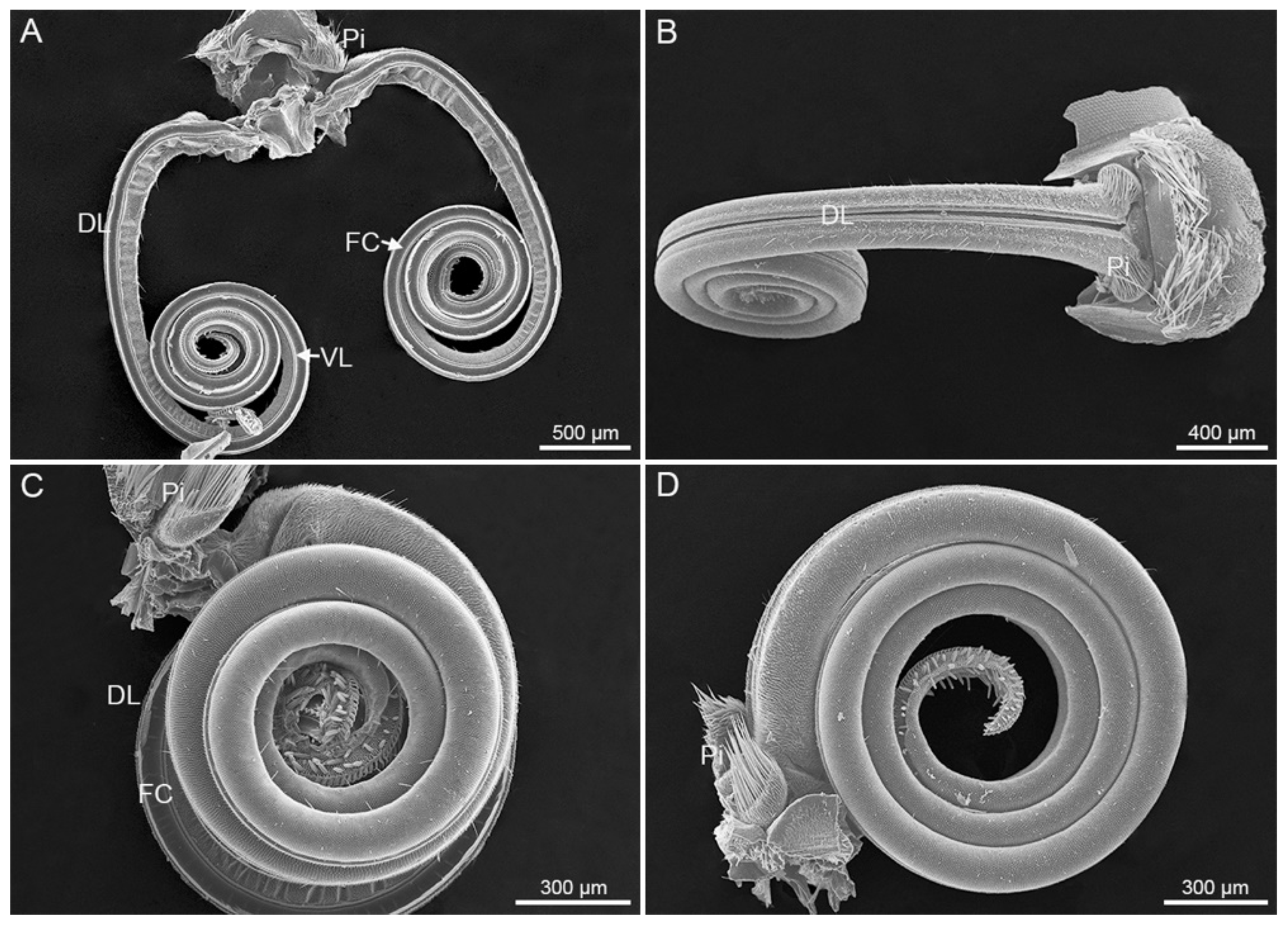
Figure 1. The proboscis morphology. (A) General interior view of Scotogramma trifolii; (B) dorsal view of Protoschinia scutosa; (C) lateral view of S. trifolii; (D) lateral view of P. scutosa. DL, dorsal legulae; FC, food canal; Pi, pilifer; VL, ventral legulae.
Insects 12 00992 g001
Figure 2. The proboscis of Protoschinia scutosa. (A) Lateral view; (B) enlarged view of the dotted rectangle in (A); (C) pollen grain on the proboscis tip. DL, dorsal legulae; Pi, pilifer; SC, sensilla chaetica; SS, sensilla styloconica.
Insects 12 00992 g002
Figure 3. The division of proboscis. (A) External view of the boundary between Zones 1 and 2 of S. trifolii; (B) external view of the boundary between Zones 1 and 2 of P. scutosa; (C) inner view of the boundary between Zones 1 and 2 of S. trifolii (indicated by white dashed line); (D) inner view of the boundary between Zones 2 and 3 of S. trifolii (indicated by white arrow); (E) inner view of the boundary between Zones 2 and 3 of P. scutosa (indicated by white arrow). DL, dorsal legulae; ISB, internal sensilla basiconica; SS, sensilla styloconica; VL, ventral legulae.
Insects 12 00992 g003
Figure 4. Zone 1. (A) Dorsal view of S. trifolii; (B) internal view of S. trifolii; (C,E) internal view of P. scutosa; (D) dorsal view of P. scutosa. DL, dorsal legulae; ESB, external sensilla basiconica; FC, food canal; ISB, internal sensilla basiconica; SC, sensilla chaetica; VL, ventral legulae.
Insects 12 00992 g004
Figure 5. Zones 2 and 3. (A) Internal view of Zone 2 of S. trifolii; (B) internal view of Zone 2 of P. scutosa; (C) external view of Zone 2 of S. trifolii; (D) external view of Zone 2 of P. scutosa; (E) internal view of Zones 2 and 3 of S. trifolii; (F) internal view of Zone 3 of S. trifolii, with the white arrow indicating the boundary between Zones 2 and 3; (G) lateral view of Zone 3 of P. scutosa, with the white arrow indicating the boundary between Zones 2 and 3. DL, dorsal legulae; ESB, external sensilla basiconica; FC, food canal; ISB, internal sensilla basiconica; SC, sensilla chaetica; SS, sensilla styloconica; VL, ventral legulae.
Insects 12 00992 g005
Figure 6. Sensilla chaetica. (A,B,D) Sensillum chaeticum on Zone 1 of S. trifolii; (C,E,F) sensillum chaeticum on Zone 1 of P. scutosa; (G) sensillum chaeticum on Zone 2 of S. trifolii; (H) sensillum chaeticum on Zone 2 of P. scutosa.
Insects 12 00992 g006
Figure 7. Sensilla basiconica. (A) External sensillum basiconicum on Zone 1 of S. trifolii; (B) external sensillum basiconicum on Zone 2 of S. trifolii; (C) internal sensillum basiconicum on food canal of S. trifolii; (D) external sensillum basiconicum on Zone 1 of P. scutosa; (E) external sensillum basiconicum on Zone 2 of P. scutosa showing a sensory pore at the blunt tip; (F) Internal sensillum basiconicum on food canal of P. scutosa.
Insects 12 00992 g007
Figure 8. Sensilla styloconica. (A) Sensilla styloconica on Zones 2 and 3 of S. trifolii; (B) sensilla styloconica on Zones 2 and 3 of P. scutosa; (C,D) sensillum styloconicum on Zone 2 of S. trifolii; (E) sensillum styloconicum on Zone 2 of P. scutosa. DL, dorsal legulae; ESB, external sensilla basiconica; SC, sensilla chaetica; SS, sensilla styloconica.
Insects 12 00992 g008
Table 1. Length of proboscis and width of food canal of Scotogramma trifolii and Protoschinia scutosa.
TypeLength/Width (μm)t-Test
S. trifoliiP. scutosa
Proboscis7478.07 ± 159.76 (14)7858.15 ± 157.38 (14)NS
Food canal61.58 ± 0.92 (28)59.42 ± 0.66 (28)*
Zone 16426.14 ± 165.60 (14)7157.51 ± 156.61 (14)*
Zone 21049.68 ± 36.54 (14)816.44 ± 25.68 (14)*
Zone 335.03 ± 1.49 (8)39.03 ± 0.65 (14)*
Note: Data are presented as Mean ± SE (n); n, sample size; NS, nonsignificant differences; * indicates p < 0.05 in the independent samples t-test.
Table 2. Length and basal width of each sensillum type of Scotogramma trifolii and Protoschinia scutosa.
TypeLength (μm)t-TestBasal Width (μm)t-Test
S. trifoliiP. scutosaS. trifoliiP. scutosa
SC46.41 ± 1.93 (140)60.20 ± 2.13 (140)*3.23 ± 0.06 (140)3.11 ± 0.06 (140)NS
SST37.28 ± 0.52 (280)30.90 ± 0.48 (280)*8.26 ± 0.09 (280)7.50 ± 0.09 (280)*
ESB (Zone 1)9.67 ± 0.28 (140)6.81 ± 0.21 (140)*2.37 ± 0.04 (140)2.15 ± 0.05 (140)*
ESB (Zone 2)6.67 ± 0.43 (28)4.41 ± 0.30 (28)*2.14 ± 0.07 (28)1.87 ± 0.08 (28)*
ISB8.01 ± 0.30 (50)6.82 ± 0.29 (50)*2.46 ± 0.05 (50)2.49 ± 0.05 (50)NS
Note: Data are presented as Mean ± SE (n); n, sample size; NS, nonsignificant differences; * indicates p < 0.05 in the independent samples t-test. Abbreviations: SC, sensilla chaetica; SST, sensilla styloconica; ESB, external sensilla basiconica; ISB, internal sensilla basiconica.
Table 3. Length/Width of proboscis and and each sensillum type of Scotogramma trifolii males and females.
TypesLength (μm)t-TestBasal Width (μm)t-Test
MaleFemaleMaleFemale
Proboscis7583.81 ± 640.84 (7)7372.33 ± 580.87 (7)NS
Zone 16570.53 ± 253.31 (7)6281.75 ± 218.44 (7)NS
Zone 21008.78 ± 46.84 (7)1090.58 ± 55.07 (7)NS
Zone 333.67 ± 2.01 (4)36.39 ± 2.24 (4)NS
SC44.24 ± 3.33 (70)48.58 ± 2.82 (70)NS3.44 ± 0.09 (70)3.03 ± 0.06 (70)*
SST36.43 ± 0.63 (140)38.13 ± 0.82 (140)NS9.10 ± 0.13 (140)7.43 ± 0.08 (140)*
ESB (Zone 1)8.99 ± 0.24 (70)10.36 ± 0.49 (70)*2.50 ± 0.06 (70)2.24 ± 0.05 (70)*
ESB (Zone 2)6.39 ± 0.39 (14)6.94 ± 0.78 (14)NS2.27 ± 0.08 (14)2.01 ± 0.10 (14)NS
ISB7.54 ± 0.46 (25)8.49 ± 0.36 (25)NS2.64 ± 0.07 (25)2.28 ± 0.06 (25)*
Note: Data of proboscis sensilla are presented as Mean ± SE (n); n, sample size; NS, nonsignificant differences; * indicates p < 0.05 in the independent samples t-test; –, present but not counted. Abbreviations: SC, sensilla chaetica; SST, sensilla styloconica; ESB, external sensilla basiconica; ISB, internal sensilla basiconica.
Table 4. Length/Width of proboscis and each sensillum type of Protoschinia scutosa males and females.
TypesLength (μm)t-TestBasal Width (μm)t-Test
MaleFemaleMaleFemale
Proboscis8065.79 ± 195.46 (7)7650.50 ± 234.00 (7)NS
Zone 17362.79 ± 186.98 (7)6952.23 ± 239.31 (7)NS
Zone 2865.20 ± 39.11 (7)767.67 ± 23.16 (7)NS
Zone 338.83 ± 0.64 (7)39.24 ± 1.18 (7)NS
SC59.02 ± 3.19 (70)61.38 ± 2.83 (70)NS3.27 ± 0.09 (70)2.94 ± 0.09 (70)*
SST31.16 ± 0.65 (140)30.64 ± 0.72 (140)NS7.82 ± 0.13 (140)7.18 ± 0.13 (140)*
ESB (Zone 1)6.54 ± 0.27 (70)7.16 ± 0.31 (70)NS2.22 ± 0.07 (70)2.10 ± 0.07 (70)NS
ESB (Zone 2)4.28 ± 0.40 (14)4.54 ± 0.46 (14)NS1.89 ± 0.1 (14)1.86 ± 0.09 (14)NS
ISB6.80 ± 0.45 (25)6.85 ± 0.39 (25)NS2.48 ± 0.09 (25)2.49 ± 0.07 (25)NS
Note: Data of proboscis sensilla are presented as Mean ± SE (n); n, sample size; NS, nonsignificant differences; * indicates p < 0.05 in the independent samples t-test; –, present but not counted. Abbreviations: SC, sensilla chaetica; SST, sensilla styloconica; ESB, external sensilla basiconica; ISB, internal sensilla basiconica.
Publisher’s Note: MDPI stays neutral with regard to jurisdictional claims in published maps and institutional affiliations.