
Validation of an Effective Protocol for Culicoides Latreille (Diptera: Ceratopogonidae) Detection Using eDNA Metabarcoding


 and
and
Abstract
:Simple Summary
Abstract
1. Introduction
2. Materials and Methods
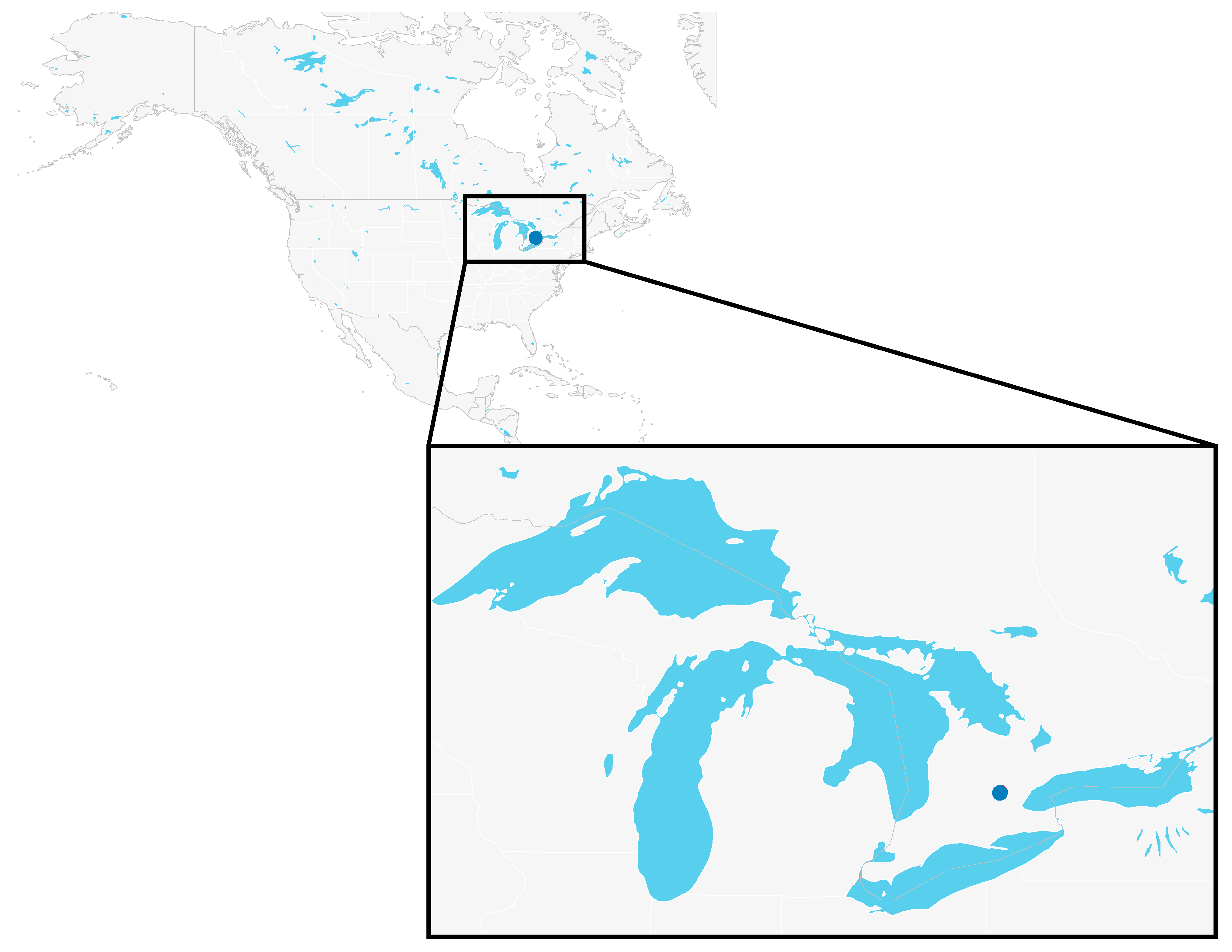
2.1. Sampling
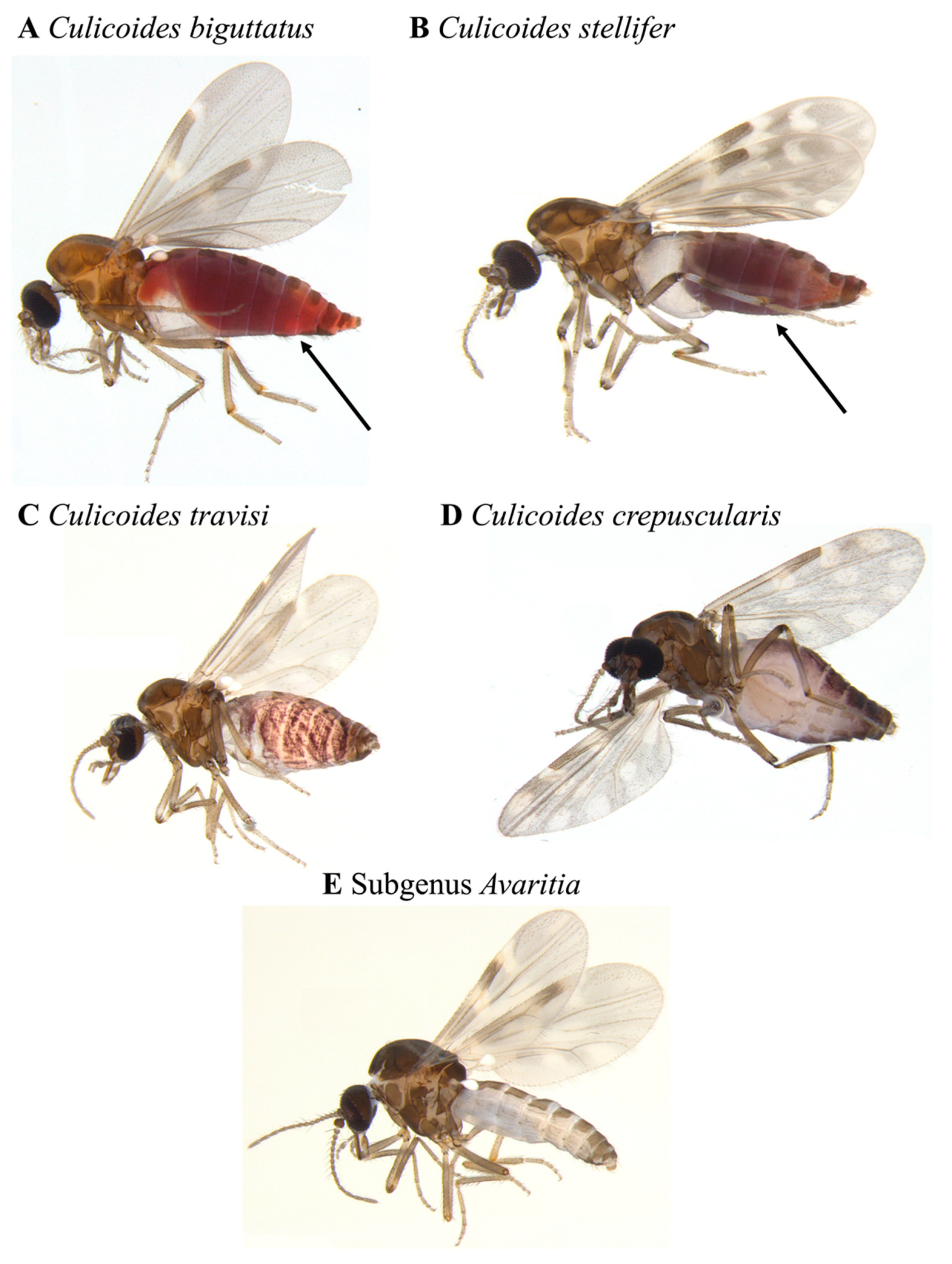
2.2. Morphological Identification
2.3. Salt Trap Solution Filtration
2.4. DNA Extraction
2.5. Library Preparation
2.6. High-Throughput Sequencing
2.7. Data Analysis
3. Results
3.1. Morphological Identification
3.2. Molecular Identification
4. Discussion
5. Conclusions
Supplementary Materials
Author Contributions
Funding
Institutional Review Board Statement
Data Availability Statement
Acknowledgments
Conflicts of Interest
References
- Makiola, A.; Compson, Z.G.; Baird, D.J.; Barnes, M.A.; Boerlijst, S.P.; Bouchez, A.; Brennan, G.; Bush, A.; Canard, E.; Cordier, T.; et al. Key Questions for Next-Generation Biomonitoring. Front. Environ. Sci. 2020, 7, 197. [Google Scholar] [CrossRef] [Green Version]
- Piper, A.M.; Batovska, J.; Cogan, N.O.I.; Weiss, J.; Cunningham, J.P.; Rodoni, B.C.; Blacket, M.J. Prospects and Challenges of Implementing DNA Metabarcoding for High-Throughput Insect Surveillance. Gigascience 2019, 8, giz092. [Google Scholar] [CrossRef] [PubMed]
- Taberlet, P.; Coissac, E.; Pompanon, F.; Brochmann, C.; Willerslev, E. Towards Next-Generation Biodiversity Assessment Using DNA Metabarcoding. Mol. Ecol. 2012, 21, 2045–2050. [Google Scholar] [CrossRef] [PubMed]
- Creer, S.; Deiner, K.; Frey, S.; Porazinska, D.; Taberlet, P.; Thomas, W.K.; Potter, C.; Bik, H.M. The Ecologist’s Field Guide to Sequence-Based Identification of Biodiversity. Methods Ecol. Evol. 2016, 7, 1008–1018. [Google Scholar] [CrossRef]
- Ruppert, K.M.; Kline, R.J.; Rahman, M.S. Past, Present, and Future Perspectives of Environmental DNA (EDNA) Metabarcoding: A Systematic Review in Methods, Monitoring, and Applications of Global EDNA. Glob. Ecol. Conserv. 2019, 17, e00547. [Google Scholar] [CrossRef]
- Stat, M.; John, J.; DiBattista, J.D.; Newman, S.J.; Bunce, M.; Harvey, E.S. Combined Use of EDNA Metabarcoding and Video Surveillance for the Assessment of Fish Biodiversity. Conserv. Biol. 2019, 33, 196–205. [Google Scholar] [CrossRef] [Green Version]
- Borkent, A.; Dominiak, P. Catalog of the Biting Midges of the World (Diptera: Ceratopogonidae). Zootaxa 2020, 4787, 1–377. [Google Scholar] [CrossRef] [PubMed]
- Mands, V.; Kline, D.L.; Blackwell, A. Culicoides Midge Trap Enhancement with Animal Odour Baits in Scotland. Med. Vet. Entomol. 2004, 18, 336–342. [Google Scholar] [CrossRef]
- Downes, J.A.; Wirth, W.W. Ceratopogonidae. In Manual of Nearctic Diptera; Biosystematics Research Institute: Ottawa, ON, Canada, 1981; Volume 1, pp. 93–421. [Google Scholar]
- Borkent, A.; William, L.; Grogan, J. Catalog of the New World Biting Midges North of Mexico (Diptera: Ceratopogonidae). Zootaxa 2009, 2273, 1–48. [Google Scholar] [CrossRef]
- Harrup, L.E.; Bellis, G.A.; Balenghien, T.; Garros, C. Culicoides Latreille (Diptera: Ceratopogonidae) Taxonomy: Current Challenges and Future Directions. Infect. Genet. Evol. 2015, 30, 249–266. [Google Scholar] [CrossRef] [Green Version]
- Borkent, A. The biting midges, the Ceratopogonidae (Diptera). In Biology of Diseases Vectors; Elsevier: Amsterdam, The Netherlands, 2004; pp. 113–126. [Google Scholar]
- Jewiss-Gaines, A.; Barelli, L.; Hunter, F.F. First Records of Culicoides Sonorensis (Diptera: Ceratopogonidae), a Known Vector of Bluetongue Virus, in Southern Ontario. J. Med. Entomol. 2017, 54, 757–762. [Google Scholar] [CrossRef] [PubMed]
- Viennet, E.; Garros, C.; Lancelot, R.; Allène, X.; Gardès, L.; Rakotoarivony, I.; Crochet, D.; Delécolle, J.-C.; Moulia, C.; Baldet, T.; et al. Assessment of Vector/Host Contact: Comparison of Animal-Baited Traps and UV-Light/Suction Trap for Collecting Culicoides Biting Midges (Diptera: Ceratopogonidae), Vectors of Orbiviruses. Parasites Vectors 2011, 4, 119. [Google Scholar] [CrossRef] [PubMed] [Green Version]
- Desvars, A.; Grimaud, Y.; Guis, H.; Esnault, O.; Allène, X.; Gardès, L.; Balenghien, T.; Baldet, T.; Delécolle, J.C.; Garros, C. First Overview of the Culicoides Latreille (Diptera: Ceratopogonidae) Livestock Associated Species of Reunion Island, Indian Ocean. Acta Trop. 2015, 142, 5–19. [Google Scholar] [CrossRef] [PubMed]
- Milián-García, Y.; Young, R.G.; Madden, M.; Bullas-Appleton, E.; Hanner, R.H. Optimization and Validation of a Cost-Effective Protocol for Biosurveillance of Invasive Alien Species. Ecol. Evol. 2020. [Google Scholar] [CrossRef]
- Young, R.G.; Milián-García, Y.; Yu, J.; Bullas-Appleton, E.; Hanner, R.H. Biosurveillance for Invasive Insect Pest Species Using an Environmental DNA Metabarcoding Approach and a High Salt Trap Collection Fluid. Ecol. Evol. 2020. [Google Scholar] [CrossRef]
- Ratnasingham, S.; Hebert, P.D.N. BOLD: The Barcode of Life Data System (Http://Www.Barcodinglife.Org). Mol. Ecol. Notes 2007, 7, 355–364. [Google Scholar] [CrossRef] [PubMed] [Green Version]
- Jamnback, H.; Wirth, W.W. The Species of Culicoides Related to Obsoletus in Eastern North America (Diptera: Ceratopogonidae). Ann. Entomol. Soc. Am. 1963, 56, 185–198. [Google Scholar] [CrossRef]
- Foote, R.H.; Pratt, H.D. The Culicoides of the Eastern United States (Diptera, Heleidae), a Review; Public Health Monograph: Washington, DC, USA, 1954; Volume 18, pp. 1–53.
- Hoffman, W.A. A Review of the Species of Culicoides of North and Central America and the West Indies. Am. J. Hyg. 1925, 5, 274–301. [Google Scholar] [CrossRef]
- Wirth, W.W.; Dyce, A.L.; Peterson, B.V.; Roper, I. An Atlas of Wing Photographs, with a Summary of the Numerical Characters of the Nearctic Species of Culicoides (Diptera: Ceratopogonidae). Contrib. Am. Entomol. Inst. 1984, 22, 46. [Google Scholar]
- Root, F.M.; Hoffman, W.A. The North American Species of Culicoides. Am. J. Epidemiol. 1937, 25, 150–176. [Google Scholar] [CrossRef]
- Dempster, E.l.; Pryor, K.v.; Francis, D.; Young, J.e.; Rogers, H.j. Rapid DNA Extraction from Ferns for PCR–Based Analyses. BioTechniques 1999, 27, 66–68. [Google Scholar] [CrossRef] [PubMed] [Green Version]
- Coyne, K.J.; Handy, S.M.; Demir, E.; Whereat, E.B.; Hutchins, D.A.; Portune, K.J.; Doblin, M.A.; Cary, S.C. Improved Quantitative Real-Time PCR Assays for Enumeration of Harmful Algal Species in Field Samples Using an Exogenous DNA Reference Standard. Limnol. Oceanogr. Methods 2005, 3, 381–391. [Google Scholar] [CrossRef] [Green Version]
- Barnes, M.A.; Turner, C.R.; Jerde, C.L.; Renshaw, M.A.; Chadderton, W.L.; Lodge, D.M. Environmental Conditions Influence EDNA Persistence in Aquatic Systems. Environ. Sci. Technol. 2014, 48, 1819–1827. [Google Scholar] [CrossRef] [PubMed]
- Turner, C.R.; Barnes, M.A.; Xu, C.C.Y.; Jones, S.E.; Jerde, C.L.; Lodge, D.M. Particle Size Distribution and Optimal Capture of Aqueous Macrobial EDNA. Methods Ecol. Evol. 2014, 5, 676–684. [Google Scholar] [CrossRef] [Green Version]
- Renshaw, M.A.; Olds, B.P.; Jerde, C.L.; McVeigh, M.M.; Lodge, D.M. The Room Temperature Preservation of Filtered Environmental DNA Samples and Assimilation into a Phenol–Chloroform–Isoamyl Alcohol DNA Extraction. Mol. Ecol. Resour. 2015, 15, 168–176. [Google Scholar] [CrossRef] [PubMed] [Green Version]
- Dougherty, M.M.; Larson, E.R.; Renshaw, M.A.; Gantz, C.A.; Egan, S.P.; Erickson, D.M.; Lodge, D.M. Environmental DNA (EDNA) Detects the Invasive Rusty Crayfish Orconectes Rusticus at Low Abundances. J. Appl. Ecol. 2016, 53, 722–732. [Google Scholar] [CrossRef] [PubMed] [Green Version]
- 16S Metagenomic Sequencing Library Preparation. Available online: https://support.illumina.com/downloads/16s_metagenomic_sequencing_library_preparation.html (accessed on 3 February 2020).
- Braukmann, T.W.A.; Ivanova, N.V.; Prosser, S.W.J.; Elbrecht, V.; Steinke, D.; Ratnasingham, S.; de Waard, J.R.; Sones, J.E.; Zakharov, E.V.; Hebert, P.D.N. Metabarcoding a Diverse Arthropod Mock Community. Mol. Ecol. Resour. 2019, 19, 711–727. [Google Scholar] [CrossRef] [Green Version]
- Ratnasingham, S.; Hebert, P.D.N. A DNA-Based Registry for All Animal Species: The Barcode Index Number (BIN) System. PLoS ONE 2013, 8, e66213. [Google Scholar] [CrossRef] [Green Version]
- Martins, F.M.S.; Porto, M.; Feio, M.J.; Egeter, B.; Bonin, A.; Serra, S.R.Q.; Taberlet, P.; Beja, P. Modelling Technical and Biological Biases in Macroinvertebrate Community Assessment from Bulk Preservative Using Multiple Metabarcoding Markers. Mol. Ecol. 2020. [Google Scholar] [CrossRef]
- Beck, E.C. Two New Species of Culicoides from Florida (Diptera: Heleidae). Fla. Entomol. 1957, 40, 103–105. [Google Scholar] [CrossRef]
- Guirgis, S.S. New Records of Culicoides (Diptera: Ceratopogonidae) for New York State and Suffolk County, Long Island, NY. Mosq. News (USA) 1984, 44, 400–403. [Google Scholar]
- Borkent, A. The Subgeneric Classification of Species of Culicoides—Thoughts and a Warning. Available online: http://wwx.inhs.illinois.edu/files/5014/6532/8290/CulicoidesSubgenera.pdf (accessed on 28 March 2021).
- Tomazatos, A.; Jöst, H.; Schulze, J.; Spînu, M.; Schmidt-Chanasit, J.; Cadar, D.; Lühken, R. Blood-Meal Analysis of Culicoides (Diptera: Ceratopogonidae) Reveals a Broad Host Range and New Species Records for Romania. Parasites Vectors 2020, 13, 79. [Google Scholar] [CrossRef] [PubMed]


| Taxa | Arboretum | Dairy Barn |
|---|---|---|
| Culicoides biguttatus | 3 | 13 |
| Culicoides crepuscularis | - | 1 |
| Culicoides stellifer | - | 13 |
| Culicoides travisi | 1 | 1 |
| Subgenus Avaritia | 3 | 7 |
| Run Name | Reads | BINs | # Sequences | OTU Count | Filtered % | Dereplicated % |
|---|---|---|---|---|---|---|
| FAP3ST-ARB-04-COI | 320,117 | 313 | 284,362 | 61 | 0.26 | 67.67 |
| FAP3ST-DAB-03-COI | 291,847 | 171 | 239,954 | 48 | 0.13 | 65.1 |
| Arboretum (mBRAVE) | ||||||||
|---|---|---|---|---|---|---|---|---|
| BIN/Taxon ID | Order | Family | Genus | Species | Seq | MS (%) | MO (bp) | Length |
| BOLD:ACC8633 | Diptera | Ceratopogonidae | Culicoides | Culicoides mulrennani | 3 | 98.06 | 395 | 395 |
| Dairy Barn (mBRAVE) | ||||||||
| BOLD:AAG6468 | Diptera | Ceratopogonidae | Culicoides | Culicoides biguttatus | 103 | 99.45 | 397.6 | 397.47 |
| BOLD:ABA0803 | Diptera | Ceratopogonidae | Culicoides | Culicoides stellifer | 4 | 97.64 | 402.5 | 402.5 |
| BOLD:AAO7718 | Diptera | Ceratopogonidae | Culicoides | Culicoides obsoletus | 69 | 99.06 | 391.25 | 399.09 |
| Dairy Barn (Geneious) | ||||||||
| Accession | Order | Family | Genus | Species | Seq | IS (%) | PI (%) | Length |
| HQ583003, JF879904 | Diptera | Ceratopogonidae | Culicoides | Culicoides biguttatus | 10 | 94.92 | 94.96 | 340.2 |
| KR659902 | Diptera | Ceratopogonidae | Culicoides | Culicoides stellifer | 2 | 98 | 98 | 350 |
| MW642460 | Diptera | Ceratopogonidae | Culicoides | Culicoides obsoletus | 2 | 100 | 100 | 351 |
Publisher’s Note: MDPI stays neutral with regard to jurisdictional claims in published maps and institutional affiliations. |
© 2021 by the authors. Licensee MDPI, Basel, Switzerland. This article is an open access article distributed under the terms and conditions of the Creative Commons Attribution (CC BY) license (https://creativecommons.org/licenses/by/4.0/).
Share and Cite
Milián-García, Y.; Janke, L.A.A.; Young, R.G.; Ambagala, A.; Hanner, R.H. Validation of an Effective Protocol for Culicoides Latreille (Diptera: Ceratopogonidae) Detection Using eDNA Metabarcoding. Insects 2021, 12, 401. https://doi.org/10.3390/insects12050401
Milián-García Y, Janke LAA, Young RG, Ambagala A, Hanner RH. Validation of an Effective Protocol for Culicoides Latreille (Diptera: Ceratopogonidae) Detection Using eDNA Metabarcoding. Insects. 2021; 12(5):401. https://doi.org/10.3390/insects12050401
Chicago/Turabian StyleMilián-García, Yoamel, Lauren A. A. Janke, Robert G. Young, Aruna Ambagala, and Robert H. Hanner. 2021. "Validation of an Effective Protocol for Culicoides Latreille (Diptera: Ceratopogonidae) Detection Using eDNA Metabarcoding" Insects 12, no. 5: 401. https://doi.org/10.3390/insects12050401
APA StyleMilián-García, Y., Janke, L. A. A., Young, R. G., Ambagala, A., & Hanner, R. H. (2021). Validation of an Effective Protocol for Culicoides Latreille (Diptera: Ceratopogonidae) Detection Using eDNA Metabarcoding. Insects, 12(5), 401. https://doi.org/10.3390/insects12050401