Article

Expression and Role of Vitellogenin Genes in Ovarian Development of Zeugodacus cucurbitae

1
Chongqing Key Laboratory of Entomology and Pest Control Engineering, College of Plant Protection, Southwest University, Chongqing 400715, China
2
Academy of Agricultural Sciences, Southwest University, Chongqing 400715, China
*
Author to whom correspondence should be addressed.
Insects 2022, 13(5), 452; https://doi.org/10.3390/insects13050452
Submission received: 20 March 2022 / Revised: 3 May 2022 / Accepted: 9 May 2022 / Published: 11 May 2022
(This article belongs to the Section Insect Molecular Biology and Genomics)

Abstract

:

Simple Summary

Vitellogenin (Vg) is the precursor of the yolk protein gene, which is crucial for insect reproduction. In this study, we identified four Vg genes in Zeugodacus cucurbitae (Coquillett). Their molecular characteristics and expression patterns were analyzed. The four genes are mainly expressed in the fat body tissue of adult female melon flies, and their expression is regulated by the juvenile hormone and ecdysone. Nutritional stress significantly down-regulated their expression, indicating that nutrition-dependent vitellogenic development occurs during ovarian development. RNAi-mediated inhibition of the expression of the four genes resulted in significantly delayed ovarian development in Z. cucurbitae. The results indicate that the four genes play an important role in the development of ovaries in Z. cucurbitae.

Abstract

Vitellogenin (Vg) genes encode the major egg yolk protein precursor in arthropods. In this study, four Vgs were identified in Zeugodacus cucurbitae (Coquillett). Sequence analysis showed that four ZcVgs had the conserved Vg domain. Phylogenetic analysis indicated that four ZcVgs were homologous to the Vgs of Tephritidae insects. The temporal and spatial expression patterns of ZcVgs were analyzed by quantitative real-time polymerase chain reaction (RT-qPCR), and the four ZcVgs showed high expression levels in female adults, especially in the fat body. The expression of ZcVg1 and ZcVg3 was down-regulated by a low dosage (0.5 μg) of 20-hydroxyecdysone (20E), and ZcVg2, ZcVg3, and ZcVg4 were up-regulated by a high dosage (1.0 and 2.0 μg) of 20E. The expression of ZcVg1 and ZcVg2 was up-regulated by 5 μg of juvenile hormone (JH), while all of the ZcVgs were down-regulated by a low and high dosage of JH. Expression of ZcVgs was down-regulated after 24 h of starvation and recovered to normal after nutritional supplementation. After micro-injection of the gene-specific double-stranded RNA, the ZcVgs’ expression was significantly suppressed, and ovarian development was delayed in Z. cucurbitae females. The results indicate that RNA interference of reproduction-related genes is a potential pest control method that works by manipulating female fertility.

1. Introduction

Vitellogenin (Vg) genes encode the major egg yolk protein precursor in arthropods. In insects, Vg is mainly synthesized in the fat body in a tissue-, sexual-, and stage-specific manner [1]. It is secreted into the hemolymph, transported to the ovary, and absorbed by the receptor in oocytes [2]. The insect Vg protein was first discovered in the cecropia moth, Hyalophora cecropia, as a female-specific protein precursor for vitellin or yolk protein [1]. The Vg protein is a large glycolipophosphoprotein. The molecular weights vary from 150 to 200 kDa for large subunits and 40 to 65 kDa for small subunits belonging to the large lipid transfer protein (LLTP) superfamily [3]. Vg protein sequences are evolutionarily conserved across insect orders except for Diptera [2]. The identification of insect Vgs has revealed that they share similar structural motifs, including an N-terminal lipid-binding domain (LPD_N) and a von Willebrand factor type D similar domain (VWD) [2]. As in most insects, the consensus cleavage site for the primary Vg gene product is conserved near the N-terminus and is flanked by polyserine domains [4]. The most striking characteristic of insect Vgs is the existence of polyserine tracts, which serve as good phosphorylation sites [4]. However, the function of Vg phosphorylation remains elusive. Glycosylation of Vg has an essential role in folding and subunit assembly to achieve the mature protein in the hemolymph and ovary [5]. Vg has been studied extensively in a wide group of vertebrates and invertebrates, including insects. The number of Vgs varies greatly in different species [4]. For example, three Vgs have been found in Drosophila melanogaster [6], Aedes aegypti [7], and Plautia stali [8], and two Vgs have been found in Riptortus clavatus [4] and Periplaneta americana [9].
Generally, Vgs in insects are categorized into four types [1]. In the first, Vg is synthesized in the fat body in a sex-biased manner and absorbed by the developing oocytes in the ovary, as in the Vgs in Perillus bioculatus [10]. In the second, Vg is produced in both the fat body and ovarian follicle cells in females and is included in the development of oocytes, as in the Vg in Musca domestica [1]. In the third, Vg is synthesized in the ovarian follicle cells and included in the development of oocytes, as in the Vg in Bombyx mori [4]. Finally, in the fourth, Vg is produced in a non-sex-biased manner in the fat body, secreted into the hemolymph, and absorbed in the development of oocytes, as in a 30 kD protein in B. mori [11]. However, Vg expression is not female-specific and can be expressed in smaller amounts in the males of some species, such as Apis mellifera [1].
In insects, Vg synthesis is regulated by multiple signals. The regulation of Vg in insects is directed by hormones at the transcriptional level [2]. The mechanisms of hormone-based regulation of Vg gene transcription in insects are generally classified into three categories [1]. In the first, Vg gene transcription is only regulated by the juvenile hormone (JH), such as in Heliothis virescens [12] and Maruca vitrata [13]. In addition to JH, insects with the second mechanism also need the ecdysone hormone to co-regulate Vg gene transcription, such as in M. domestica [14]. Insects with the third type of mechanism require JH, ecdysone, and other hormones to co-regulate Vg gene transcription, such as in A. aegypti [15]. Nevertheless, JH is not responsible for Vg synthesis in social insects, and it has a primary role in regulating the age-based division of labor among workers, such as in Pogonomyrmex californicus [16]. In addition to these hormones, Vg synthesis is also affected by nutritional signals [2]. The starvation of female beetles resulted in a block in Vg synthesis but not in the progression of primary oocyte development to the resting stage in Tribolium castaneum [17]. Vg is essential for the maturation of adult eggs and the development of embryos in insects [2]. RNA interference (RNAi)-mediated suppression of the Cadra cautella Vg gene curtails oogenesis in the almond moth, C. cautella [18]. The silencing of the Vg1 or Vg2 genes inhibits oviposition in the Chagas disease vector Triatoma infestans [19]. The down-regulation of ClVg and ClVg-like expression in Cimex lectularius leads to the atrophy of ovarian tissue and reduced oviposition [20]. In Lepidoptera, the silencing of the Vg gene in Corcyra cephalonica caused severely abnormal ovaries [21]. In addition, the Vg in A. mellifera has multiple functions, including labor differentiation, reproductive competition, and climate adaptation [22].
The melon fly, Zeugodacus cucurbitae (Coquillett) [23], is one of the most troublesome agricultural pests [24]. It is widely distributed, including in Africa, temperate Asia, and many Pacific islands [25]. The genome of Z. cucurbitae has been sequenced and released, providing sequence information for gene annotation and functional research [26]. In this study, four Vgs were identified in Z. cucurbitae from the genome. The expression levels of ZcVgs in multiple development stages and tissues were investigated by quantitative real-time polymerase chain reaction (RT-qPCR). The effects of JH, 20-hydroxyecdysone (20E), and starvation on the expression of ZcVgs were also evaluated. The expected roles of these ZcVgs were also explored using RNAi, revealing their potential as pest control targets.

2. Materials and Methods

2.1. Insect Rearing

The insect stock was collected from Haikou, Hainan Province, China, in 2016. The adults were reared in an environmentally controlled insectary, with a temperature of 26–27 °C, a relative humidity (RH) of 65–75%, and a photoperiod of 14:10 h (light: dark). As prescribed in previously published work, Z. cucurbitae larvae were reared on an artificial diet consisting of bitter gourd, yeast powder, corn, wheat flour, and sucrose; adults were fed a mixture of sucrose, yeast powder, and honey [27].

2.2. RNA Isolation and cDNA Synthesis

Same-aged females were cultured under the same conditions as the stock flies. Five-day-old virgin female adults were collected. The total RNA was isolated using TRIzol reagent (Invitrogen, Carlsbad, CA, USA) according to the manufacturer’s instructions. The RNA sample’s concentration was determined by a NanoDrop One spectrophotometer (Thermo Scientific, Madison, WI, USA), that is, the purity of RNA samples (OD260/280 and OD260/230). The genomic DNA contamination of 1 μg of total RNA was removed using an RQ1 RNase-Free DNase kit (Promega, Madison, WI, USA). The first-strand cDNA was synthesized using the PrimeScript®RT reagent Kit (TaKaRa, Dalian, China) and stored at −20 °C until use.

2.3. Molecular Cloning and Sequence Analysis

The homologous Vgs from Bactrocera dorsalis (AF368053.1, AF368054.1, and ARV91014.1) were used as the query sequences to find the Vgs in Z. cucurbitae with a BLAST search against the genome in the National Center for Biotechnology Information (NCBI) database. Four fragment candidates were downloaded for the full-length confirmation by PCR. The accession number for the melon fly genome was ASM80634v1. The gene-specific primers were designed by Primer Premier 5.0 (PREMIER Biosoft International, Palo Alto, CA, USA) (Table 1). The PCR amplification was run on a Bio-Rad PCR machine (Bio-Rad, Singapore). PCR was conducted with 25 μL of a mixture consisting of 12.5 μL of 2× Taq PCR Master Mix (Biomed, Beijing, China), 9.5 μL of RNase-free water, 1.0 μL of the cDNA template (described above), and 1.0 μL each of forward and reverse primers (10 μM). PCR amplification was performed using the following procedure: initial denaturation at 95 °C for 3 min, followed by 35 cycles at 95 °C for 30 s, 55 °C for 30 s, 72 °C for 1 min, and a final extension at 72 °C for 10 min. The product was detected by 1% agarose gel electrophoresis. A QIAquick PCR Purification Kit (QIAGEN, Dusseldorf, Germany) was used to purify the positive products. The target product was connected to a pGEM-T by pGEM®-T Easy Vector (Promega, Madison, WI, USA) and transformed into E. coli Trans 5α (TSINGKE, Beijing, China). Finally, positive clones were obtained through blue-white spot screening and then sequenced.
The conserved domains of ZcVgs were analyzed by the SMART online website (http://smart.embl-heidelberg.de, accessed on 16 August 2021) [28]. For the alignment and phylogenetic tree construction, the homologs of Vg in other insects were derived from the NCBI database by BLASTp. Multiple sequence alignment was performed using Clustal Omega and visualized with Jalview 2.10 software [29]. The unrooted phylogenetic tree was constructed by the neighbor-joining method with 1000 bootstrap replicates using MEGA7 software [30].

2.4. Spatio-Temporal Expression Revealed by RT-qPCR

Virgin female adults were reared separately and collected to analyze the expression pattern of ZcVgs on each day from 0 to 9 d, with four biological replicates. Total RNA and cDNA preparation was performed as described above. Ribosomal protein subunit 3 (Rps3) and ribosomal protein L13 (Rpl13) were selected as the internal reference genes to explore the expression patterns of ZcVgs at different developmental stages [31]. The tissue samples from the midgut, fat body, Malpighian tubules, and ovary were dissected from 5 d old virgin female adults to determine the expression pattern of ZcVgs. Four biological replicates were prepared, and each sample was dissected from 20 individuals. α-Tubulin (α-Tub) and β-tubulin 1 (β-Tub1) were selected as the internal reference genes to analyze the expression in various tissues of female adults [31]. The primers for the RT-qPCR were designed by Primer Premier 5.0 (Table 1), followed by the amplification efficiency determination. After the primers were synthesized, the standard curve was calculated to evaluate the amplification efficiency. Efficiencies in the range of 90–110% were acceptable. The qPCR reaction was run on a CFX384 Optics Module (Bio-Rad, Singapore) using a NovoStart SYBR qPCR SuperMix Plus Kit (Novoprotein, Shanghai, China). RT-qPCR was conducted with 10 μL of a mixture consisting of 5 μL of 2× qPCR mixture, 3.5 μL of RNase-free water, 0.5 μL of cDNA, and 0.5 μL each of forward and reverse primers (10 μM). The reaction program was 95 °C for 2 min, followed by 40 cycles at 95 °C for 30 s, 60 °C for 30 s, and a final at 60 °C for 1 min, 95 °C for 10 min. Melting curve analysis at 60–95 °C was performed to ensure the specificity of each primer. The gene expression was analyzed using qBase plus software [32].

2.5. Hormone-Induced Expression

First, 20E (TCI, Shanghai, China) was diluted in ethanol (Chuandong, Chongqing, China) to produce a stock solution with a concentration of 20 mg/mL. Then, the stock solution was diluted in ethanol to 0.5, 1.0, and 2.0 μg/μL as the work solutions. More than 300 female flies that had emerged on the same day were collected. After that, 1 μL of each solution was dripped onto the pronotum of each female adult on day 4, that is, 0.5, 1.0, and 2.0 μg/fly [33]. After treatment, all females were reared and collected for total RNA isolation after 12, 24, and 48 h. Females with an equal amount of ethanol dripped onto them served as the control group.
The JH analog Methoprene (Sigma, St. Louis, MO, USA) was dissolved in acetone (Chuandong, Chongqing, China) to produce a stock solution with a concentration of 20 mg/mL. The stock solution was then diluted in acetone to 2.5, 5.0, and 10.0 μg/μL. Then, 1 μL of each solution was dripped onto the pronotum of each female adult on day 4, that is, 2.5, 5.0, and 10.0 μg/fly. After treatment, all females were reared and collected for total RNA isolation after 12, 24 and 48 h. The females treated with an equal amount of acetone served as the control group. The Rps3 and actin 3 (Act3) were preliminarily evaluated as the internal reference genes to explore ZcVgs’ expression induced by 20E; Rps3 and α-Tub were preliminarily evaluated as the internal reference genes to explore the expression of ZcVgs induced by Methoprene. The expression of ZcVgs was detected by RT-qPCR.

2.6. Nutritional Stress

A total of 150 4 d old virgin females were reared and divided into three groups. Fifty females in group I were normally raised with sufficient water and nutrition. The females in group II were provided water only. After 12, 24 and 48 h, the females in groups I and II were collected for total RNA isolation. The females in group III were starved for 24 h and then provided with an adult diet for supplementary nutrition for another 24 h. The females in groups III were collected for total RNA isolation. Rps3 and RPl13 were selected as the internal reference genes to explore ZcVgs’ expression patterns under nutritional stress. The expression of ZcVgs was detected by RT-qPCR.

2.7. RNA Interference and Functional Analysis

Gene-specific primers of four ZcVgs were designed for dsRNA synthesis using the Primer Premier 5.0. After amplifying the target fragments of 577, 533, 489, and 571 bp in ORFs, the dsRNAs were synthesized and purified using a Transcript Aid T7 High Yield Transcription Kit (Thermo Scientific, Vilnius, Lithuania) according to the manufacturer’s instructions. The concentration of dsRNA was tested with a NanoDrop One spectrophotometer. The integrity of the dsRNA was detected by 1% agarose gel electrophoresis. More than 500 female flies that had emerged on the same day were collected. Based on the spatio-temporal expression pattern of melon fly ZcVgs, the 3 d old Z. cucurbitae females were injected with 2 μg (50 nL) of dsRNA into the last ventral segment using a micromanipulator M3301R (World Precision Instruments, Sarasota, FL, USA). The females who delivered dsGFP fragment (378 bp) were set as the control group. Four biological replicates were conducted. Total RNA and cDNA preparation was performed as described above. Rps3 and α-Tub were selected as reference genes to normalize the expression of ZcVgs on day 5 after RNAi [31]. Ovaries were dissected to determine the size on day 5. Thirty samples were dissected in each treatment, and the ovarian area (mm2) was measured using a Digital MicroscopeVHX-5000 (KEYENCE, Itasca, IL, USA).

2.8. Statistical Analysis

Gene expression was analyzed using the qBase plus software [32]. The significant differences among spatio-temporal expression and gene expression induced by the hormones were analyzed by the one-way analysis of variance with the least significant difference method (p < 0.05). The gene-silencing efficiency and phenotype (ovarian size) were analyzed by an independent sample t-test (p < 0.05). All of the analyses were performed by SPSS Statistics 22.0 (IBM, Chicago, IL, USA).

3. Results

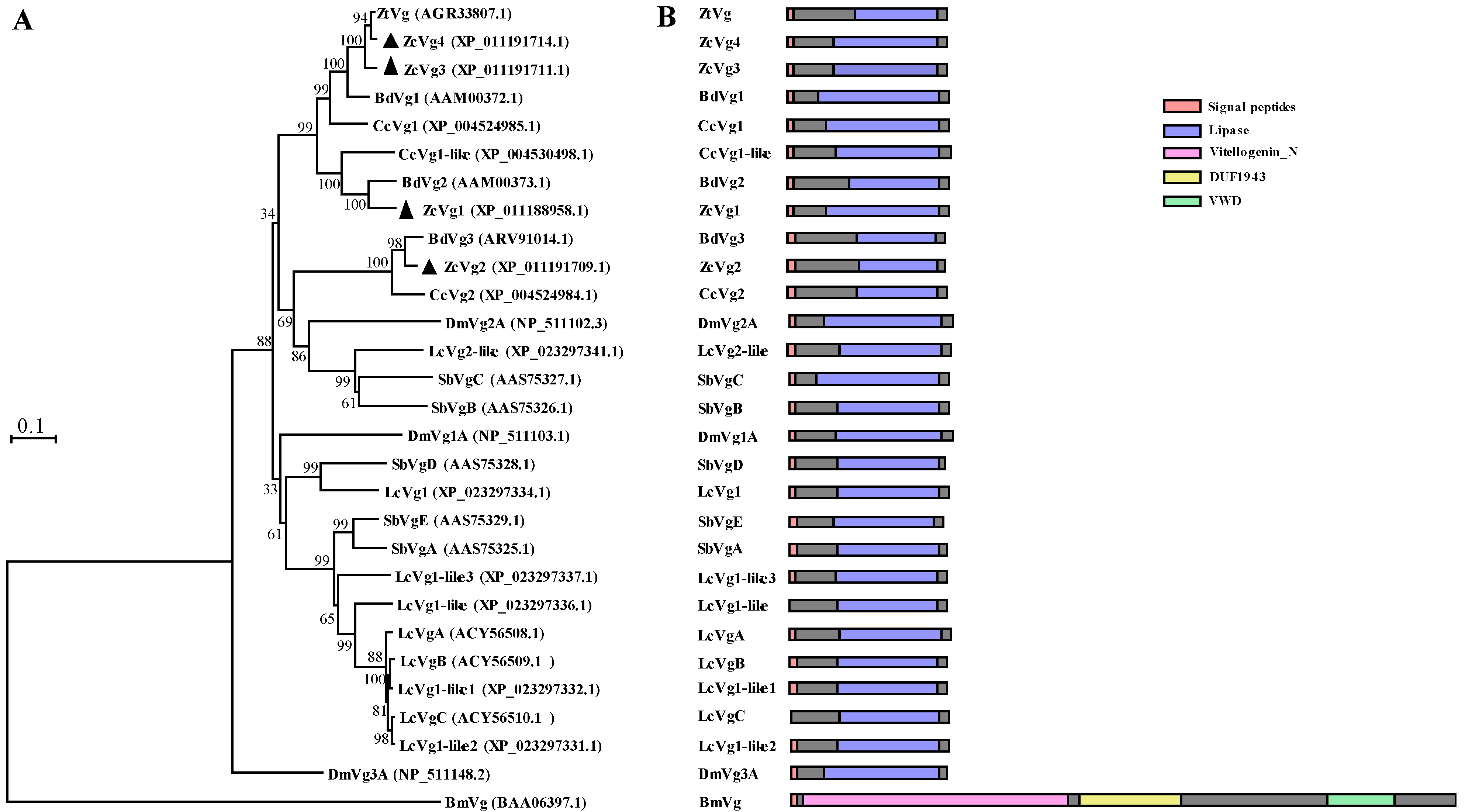
3.1. Sequence Analysis and Phylogenetic Analysis

The full-length sequence confirmation showed that ZcVg1, ZcVg2, ZcVg3, and ZcVg4 had open reading frames (ORFs) of 1308, 1275, 1290, and 1296 bp, respectively, encoding 435, 424, 429, and 427 amino acids, respectively. The sequence similarity of these four ZcVgs ranged from 53.90% to 92.81%. The sequence similarity of the four ZcVgs compared to other Dipteran Vgs ranged from 47.30% to 97.45%, while the sequence similarity of Vgs between Dipteran and B. mori Vgs ranged from 2.02% to 2.97% (Table S1). Sequences of the four genes (ZcVg1, ZcVg2, ZcVg3, and ZcVg4) were deposited in the NCBI GenBank database with the accession numbers OL546555, OL546556, OL546557, and OL546558, respectively. Sequence analysis revealed that all four ZcVgs possessed conserved domains with the LPD_N. They had putative cleavage sites (R/KXXR/K) and signal peptides. In addition, serine residues (the potential phosphorylation sites) and asparagines (NELV, NGPA, NVIE, and NERN–the glycosylation sites) were identified (Figure 1). The conserved lipase domains of ZcVg1, ZcVg2, ZcVg3, and ZcVg4 existed in 107–407, 91–399, 96–403, and 101–405 amino acids, respectively. The phylogenetic tree showed that ZcVg3 and ZcVg4 are most closely related to the Vgs of Z. tau, but ZcVg1 and ZcVg2 are most closely related to the Vgs of B. dorsalis (Figure 2).

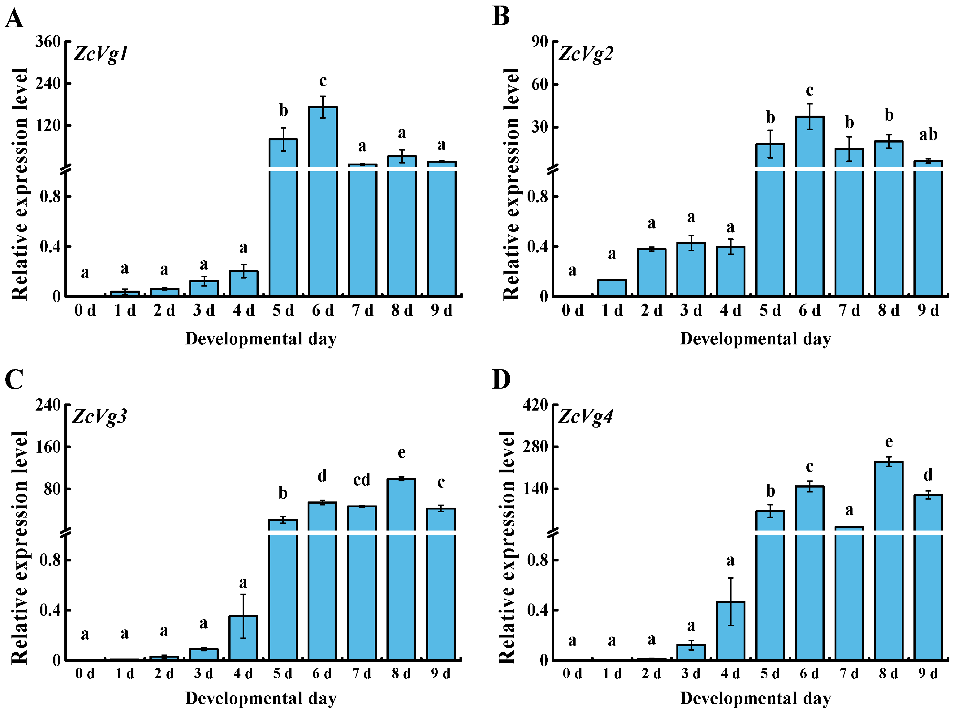
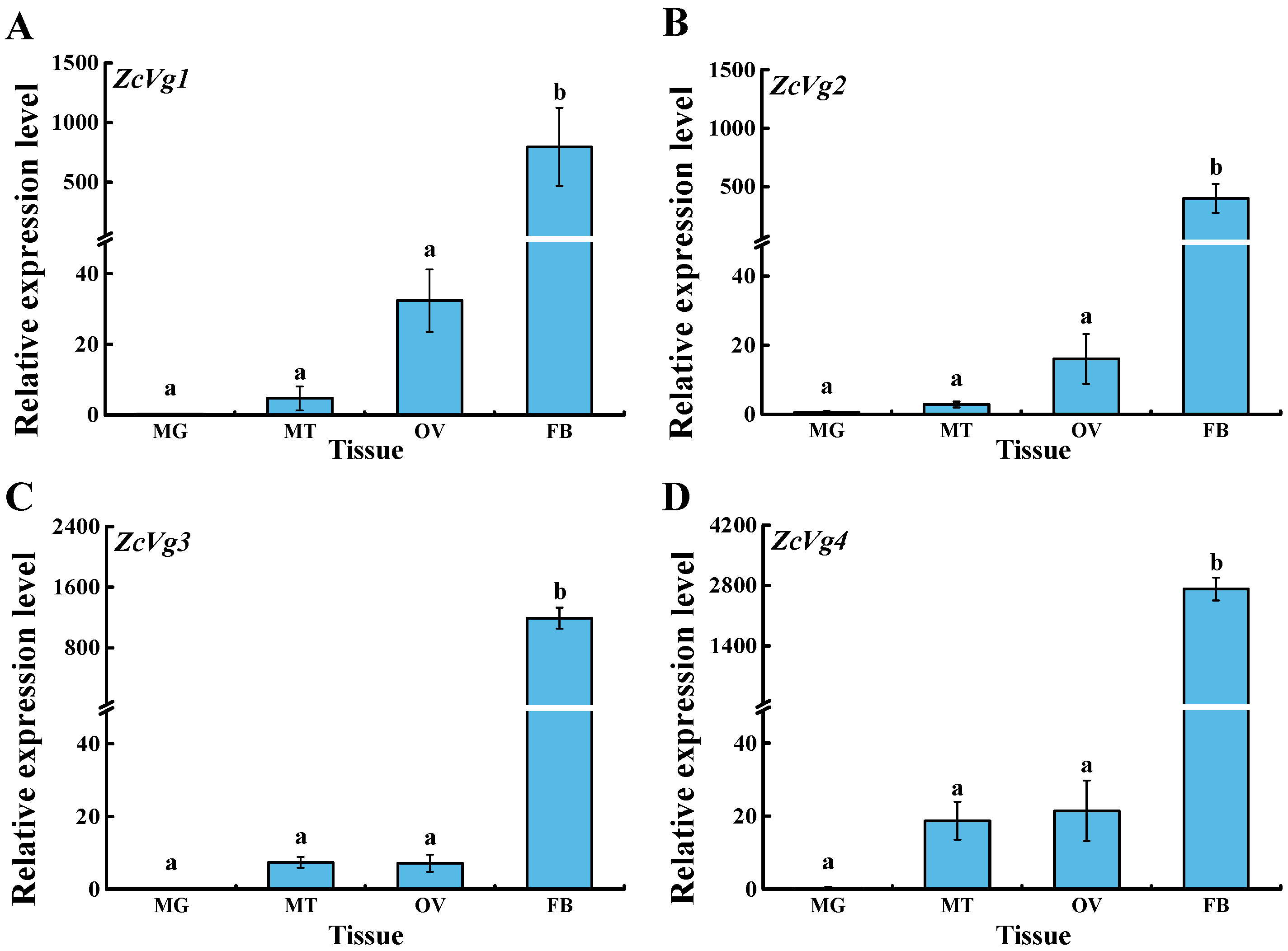
3.2. Spatio-Temporal Expression of ZcVgs

The expression patterns of ZcVgs in different developmental stages showed that all of the genes were highly expressed from 5 to 9 d (Figure 3). In particular, ZcVg1 and ZcVg2 were significantly highly expressed in females from 5 to 9 d, with the highest expression at 6 d. ZcVg3 and ZcVg4 were significantly highly expressed in females from 5 to 9 d, with the highest expression at 8 d.
The expression patterns of ZcVgs in different tissues of female adults were analyzed. The results showed that the expression levels of ZcVg1, ZcVg2, ZcVg3, and ZcVg4 in the fat body were significantly higher than those in other tissues (Figure 4). Therefore, the elevated transcript levels in the fat body are consistent with Vg biosynthesis in this tissue. In addition, these genes are also expressed in the ovary but with low expression levels.

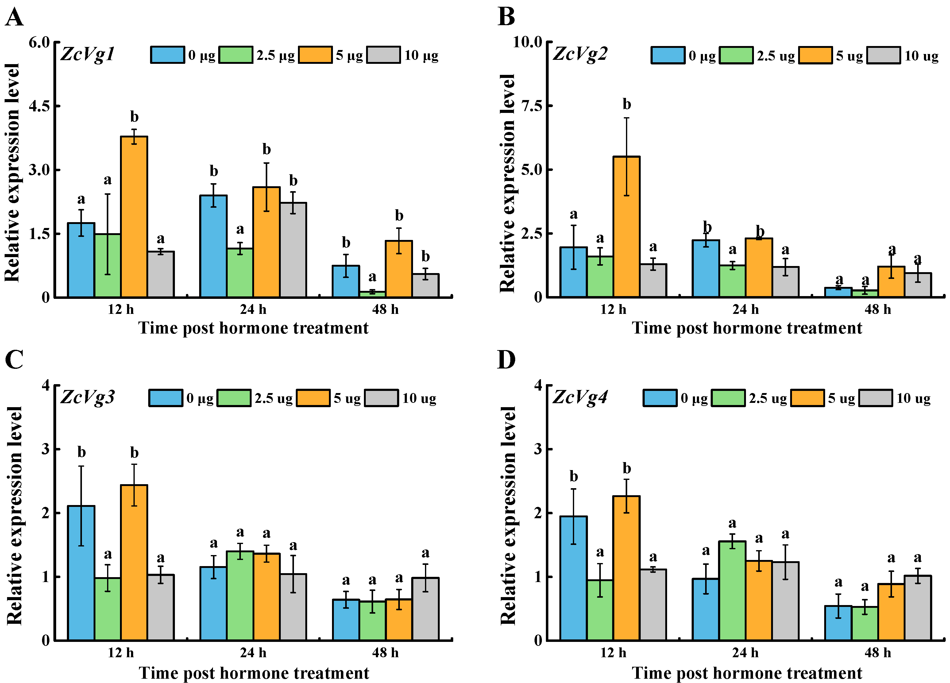
3.3. Effects of Hormone Induction on ZcVgs’ Expression

Four ZcVgs were induced by the exogenous hormone 20E with a different pattern (Figure 5). After 12 h, the expression of ZcVg2, ZcVg3, and ZcVg4 in the 1 μg treatment group was up-regulated, and the expression of ZcVg4 in the 2 μg treatment group was up-regulated. After 24 h, the expression of ZcVg3 in the 0.5 μg treatment group was down-regulated. After 48 h, the expression of ZcVg1 in the 0.5 μg treatment group was down-regulated.
Four ZcVgs were induced by the exogenous JH analogue Methoprene (Figure 6). After 12 h, the expression of ZcVg1 and ZcVg2 in the 5 μg treatment group was up-regulated, and the expression of ZcVg3 and ZcVg4 in the 2.5 μg and 10 μg treatment groups was down-regulated. After 24 h, the expression of ZcVg1 and ZcVg2 in the 2.5 μg treatment group was down-regulated, and the expression of ZcVg2 in the 10 μg treatment group was down-regulated. After 48 h, the expression of ZcVg1 in the 2.5 μg treatment group was down-regulated.

3.4. Effects of Starvation on ZcVgs’ Expression

The results showed that the expression levels of four ZcVgs were down-regulated after 12, 24, and 48 h of starvation (Figure 7). After 12 h of starvation, the expression of ZcVg1 was relatively less affected. Feeding after starvation can significantly induce Vg gene expression and return it to a normal level, or even a higher level than normal, such as in ZcVg3.

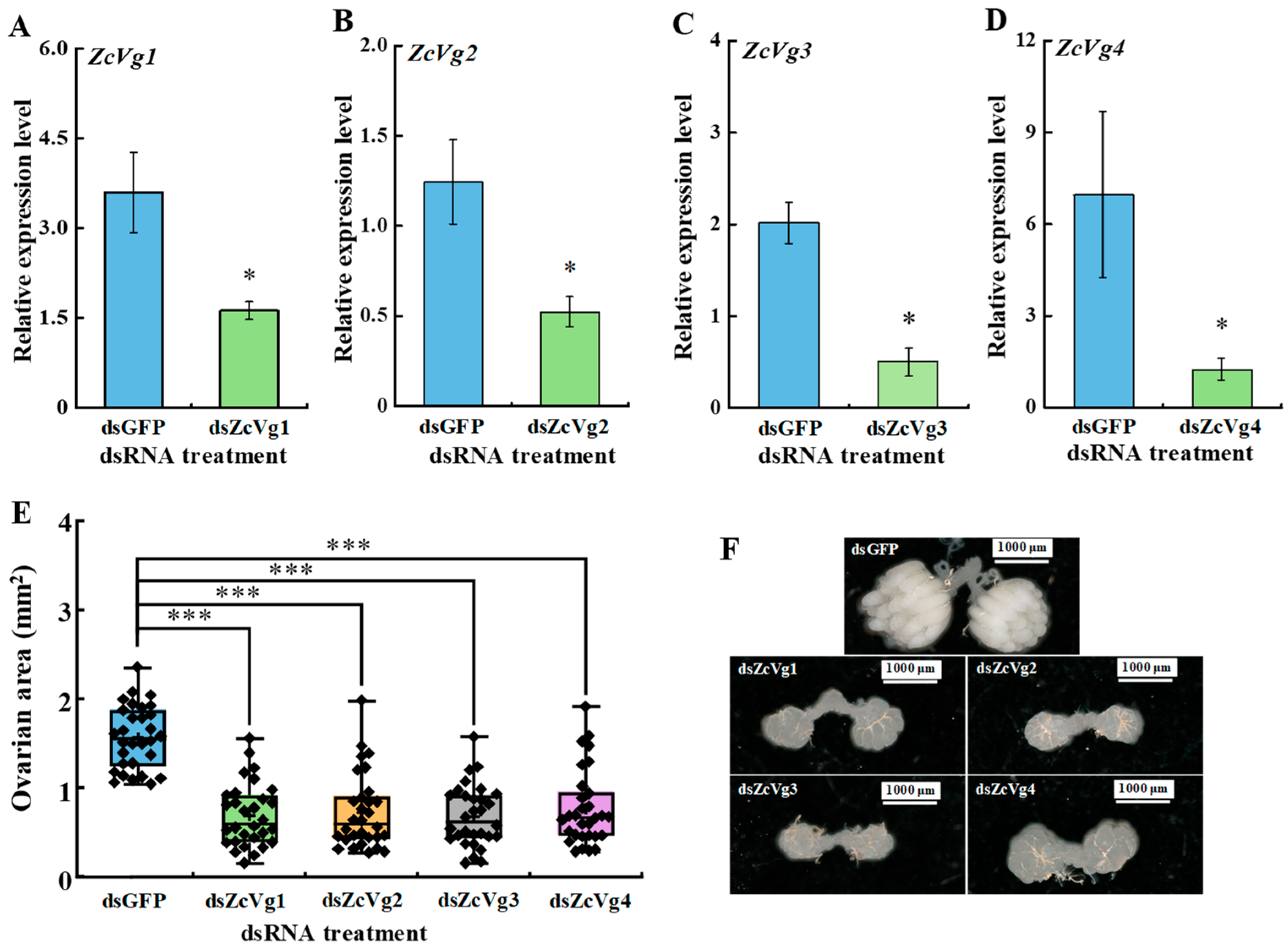
3.5. Effects of RNAi-Mediated Knockdown on Ovarian Development

The results of RNAi showed that ZcVg1, ZcVg2, ZcVg3, and ZcVg4 were significantly down-regulated by 54.9%, 58.0%, 75.1%, and 82.1%, respectively (Figure 8A–D). The ovarian area of the dsVgs treatment group was significantly smaller than that of the dsGFP control group, and ovarian development was significantly delayed compared to the control group (Figure 8E,F).

4. Discussion

The Vg hemolymph proteins are the precursors of yolk protein in the egg. The conserved Vgs have been studied extensively in various animals, both vertebrates and invertebrates, including insects [4]. In this study, we identified four Vgs in Z. cucurbitae based on the genome: ZcVg1, ZcVg2, ZcVg3, and ZcVg4. Multiple Vgs work together to ensure effective Vg production required for ovarian development. Variations in the Vg gene number in insects may reflect evolutionary selection and strategies to adapt to the environment [2]. Sequence analysis of Vgs revealed that the four ZcVgs possess conserved domains in Vgs and contain putative cleavage sites (R/KXXR/K), serine residues, and putative asparagine-linked glycosylation sites. These polyserine bundles are considered to be substrates for kinases. The negatively charged phosphoserine may affect protein solubility or chelate essential metal ions required for yolk production [34]. N-glycans may have a significant role in keeping this large, hydrophobic protein in the hemolymph to improve its transport to the ovary [5]. Vg, composed of the LPD_N for lipid binding, DUF1943, and vWFD, is highly conserved in most insect species except for aphids [35]. In addition, the length of the primary protein structure of the four ZcVgs is much smaller than that of most other order insects [36]. These much smaller Vgs are associated with the mammalian triglyceride lipase gene family [4]. Phylogenetic analysis indicated that ZcVgs are most closely related to the Vgs of other Tephritidae, such as B. dorsalis and Z. tau.
In this study, ZcVg1, ZcVg2, ZcVg3, and ZcVg4 showed similar expression patterns, with the highest expression in the fat body. This expression pattern was similar to that of B. dorsalis, and Vg was synthesized in the fat body [37]. The VgR takes Vg up from the hemolymph and transports it into developing oocytes [4]. Therefore, based on the expression pattern of Vgs in Z. cucurbitae, the specific expression pattern of Vg indicates its function in synthesizing nutrition in the fat body. In contrast, low expression of Vg was observed in the male fat body and testis of B. dorsalis [37]. Analysis of the expression levels of ZcVgs in 0 to 9 d old female adults in Z. cucurbitae revealed that the mRNA level of ZcVgs was very high in 5 to 9 d old adults during the vitellogenic stage. Therefore, the expression of ZcVg1, ZcVg2, ZcVg3, and ZcVg4 is correlated with the ovarian development of Z. cucurbitae. There are similar expression patterns in other insects, such as Bemisia tabaci [38] and Actias selene [39]. However, the up-regulation of Vg expression in some insects is observed earlier, with up-regulation beginning during the pupal stage. For instance, Vg expression is first detected during the late pupal stage in Spodoptera litura [40].
The expression of Vgs in insects is regulated by the ecdysone and JH, while the transcriptional mechanisms are different [1,41]. For instance, the transcription of the Vg gene in Heliothis virescens is only regulated by JH [1], while in T. castaneum, both JH and 20E are required for Vg gene expression [42]. After 20E induction, the expression of ZcVg1 and ZcVg3 was down-regulated by a low dosage, and the expression of ZcVg2, ZcVg3, and ZcVg4 was up-regulated by a high dosage. Only the expression of ZcVg1 and ZcVg2 was up-regulated by 5 μg of JH, while all of the ZcVgs genes were down-regulated by a low and high dosage of JH. The following inferences are possible. The pathways activated by exogenous 20E after induction of a low dose and high dose were different in regulating Vg synthesis. A low dose and high dose of exogenous JH induced the same activation of the Vg synthesis pathway, but 5 μg of exogenous JH inducing the activation of the Vg synthesis pathway differs with a low dose or high dose. The pathway activated by low-dose exogenous 20E to regulate Vg synthesis has the same function as the pathway activated by low-dose and high-dose exogenous JH to promote Vg expression. The activation of the Vg synthesis pathway induced by exogenous 20E at a high dose was the same as that induced by exogenous JH at 5 μg, which inhibited Vg expression. The results of this study indicate that ZcVgs are regulated by the ecdysis hormone and JH, and the regulatory system is relatively complex. However, in the female American cockroach (Periplaneta americana), the Vg gene is activated by JH III and repressed by 20E via cis-regulatory elements in a dose-dependent manner [43]. JH III at doses of 0.1, 1, and 10 µg induced Vg synthesis in a dose-dependent manner, with doses as low as 0.1 µg being sufficient to significantly induce Vg synthesis in Blattella germanica [44]. The transcription of Vg1 and Vg2 in P. americana is induced by JH and inhibited by 20E in a dose-dependent manner [1], while both JH and 20E induce the Vg expression in T. castaneum [42]. Interestingly, in Lymantria dispar, JH induced or even down-regulated the expression of the Vg gene [1]. Both 20E and JH are extremely important for ovarian development by regulating the Vg expression. In Nilaparvata lugens, gene silencing using RNAi demonstrates the importance of JH signaling in ovarian development [45].
Nutrients play a pivotal role in insect vitellogenesis [2]. In this study, during ovarian development, the expression of ZcVgs was down-regulated after 24 h of starvation in melon flies, and the expression of ZcVgs could return to normal levels after nutritional supplementation. These results are similar to the effect of nutrition on the expression of Vg in T. castaneum, in which the starvation of female T. castaneum caused Vg synthesis to be blocked but did not cause oocyte development to enter the quiescent phase. Vg synthesis continued and oocyte development shifted from the quiescent phase to the mature phase after nutritional supplementation [17].
Gene silencing using RNAi is an effective method to explore gene function [46], and its use has proven successful in Diptera [47]. In this study, gene-specific dsRNA suppressed the ZcVgs’ transcription and delayed ovarian development. These results indicate that ZcVgs play an important role in the development of the ovary in Z. cucurbitae. Interference with the expression of these four genes had a negative effect on ovarian development. This suggests that the expression of ZcVg1, ZcVg2, ZcVg3, and ZcVg4 is required for ovarian development. It also demonstrates that multiple Vgs work together to ensure effective production of the Vg required for ovarian development. In addition, RNAi-mediated inhibition of ZcVgs showed an off-target effect as the conserved domain in sequences. It was speculated that the expression of one ZcVg was affected by the other ZcVgs. This off-target silencing of Vgs was also found in Haemaphysalis longicornis; after silencing each Vg, the expression of other Vgs was also significantly reduced, and the body weight and egg weight of ticks were significantly reduced [48]. In any case, the role of ZcVgs in ovarian development was validated by the RNAi work, showing potential targets for pest control. Since these four Vg sequences have a high similarity, this conserved sequence may be an excellent fragment target for pest control based on RNA interference. Previous studies have shown that the successful reproduction of insects depends on the production and deposition of Vg and the uptake of Vgs by VgR in developing oocytes [49]. The silencing of Vgs also leads to decreased reproduction in other insects and abnormal ovarian development. For example, suppressing C. lectularius Vg caused ovarian tissue atrophy and reduced egg production [20].

5. Conclusions

In this study, four ZcVgs were identified and cloned. Their molecular characteristics and expression patterns were analyzed. ZcVgs are mainly expressed in the fat body tissue of adult female melon flies. In addition, the expression of ZcVgs is regulated by JH and 20E in a dose-dependent manner. Nutritional stress significantly down-regulated the ZcVgs’ expression, indicating that nutrition-dependent vitellogenic development occurs during ovarian development. RNAi-mediated inhibition of the ZcVgs’ expression resulted in significantly delayed ovarian development. These results indicate that ZcVgs play an important role in the ovarian development of Z. cucurbitae. The results demonstrate that Vgs are potential candidates for a pest control method that works by suppressing their expression using RNAi to manipulate ovarian development.

Supplementary Materials

The following supporting information can be downloaded at: https://www.mdpi.com/article/10.3390/insects13050452/s1, Table S1. Identities of amino acid sequences of ZcVgs compared to other insect Vgs.

Author Contributions

Conceptualization, D.W. and J.-J.W.; methodology, D.C. and H.-L.H.; resources, D.C. and H.-L.H.; data analysis, D.C. and W.-J.L.; writing—original draft preparation, D.C. and W.-J.L.; writing—review and editing, D.W. and J.-J.W.; funding acquisition, D.W. and J.-J.W. All authors have read and agreed to the published version of the manuscript.

Funding

This study was supported in part by the National Key Research and Development Program (2021YFC2600100), the Chongqing Research Program of Basic Research and Frontier Technology (CSTC2016jcyjA0019), and the 111 Project (B18044).

Institutional Review Board Statement

Not applicable.

Data Availability Statement

Data is contained within the article and Supplementary Material.

Conflicts of Interest

The authors declare that they do not have any conflict of interest.

References

  1. Tufail, M.; Nagaba, Y.; Elgendy, A.M.; Takeda, M. Regulation of vitellogenin genes in insects. Entomol. Sci. 2014, 17, 269–282. [Google Scholar] [CrossRef]
  2. Wu, Z.; Yang, L.; He, Q.; Zhou, S. Regulatory mechanisms of vitellogenesis in insects. Front. Cell Dev. Biol. 2021, 8, 1863. [Google Scholar] [CrossRef] [PubMed]
  3. Lee, K.Y.; Yoon, H.J.; Jin, B.R. Osmia cornifrons vitellogenin: cDNA cloning, structural analysis and developmental expression. Entomol. Res. 2015, 45, 94–101. [Google Scholar] [CrossRef]
  4. Tufail, M.; Takeda, M. Molecular characteristics of insect vitellogenins. J. Insect Physiol. 2008, 54, 1447–1458. [Google Scholar] [CrossRef]
  5. Khalaila, I.; Peter-Katalinic, J.; Tsang, C.; Radcliffe, C.M.; Aflalo, E.D.; Harvey, D.J.; Dwek, R.A.; Rudd, P.M.; Sagi, A. Structural characterization of the N-glycan moiety and site of glycosylation in vitellogenin from the decapod crustacean Cherax quadricarinatus. Glycobiology 2004, 14, 767–774. [Google Scholar] [CrossRef] [Green Version]
  6. Tarone, A.M.; McIntyre, L.M.; Harshman, L.G.; Nuzhdin, S.V. Genetic variation in the Yolk protein expression network of Drosophila melanogaster: Sex-biased negative correlations with longevity. Heredity 2012, 109, 226–234. [Google Scholar] [CrossRef] [Green Version]
  7. Isoe, J.; Hagedorn, H.H. Mosquito vitellogenin genes: Comparative sequence analysis, gene duplication, and the role of rare synonymous codon usage in regulating expression. J. Insect Sci. 2007, 7, 1–49. [Google Scholar] [CrossRef]
  8. Lee, J.M.; Hatakeyama, M.; Oishi, K. A simple and rapid method for cloning insect vitellogenin cDNAs. Insect Biochem. Mol. Biol. 2000, 30, 189–194. [Google Scholar] [CrossRef]
  9. Tufail, M.; Hatakeyama, M.; Takeda, M. Molecular evidence for two vitellogenin genes and processing of vitellogenins in the American cockroach, Periplaneta americana. Arch. Insect Biochem. Physiol. 2001, 48, 72–80. [Google Scholar] [CrossRef]
  10. Adams, T.S.; Filipi, P.A.; Yi, S.X. Effect of age, diet, diapause and juvenile hormone on oogenesis and the of amount of vitellogenin and vitellin in the twospotted stink bug, Perillus bioculatus (Heteroptera: Pentatomidae). J. Insect Physiol. 2002, 48, 477–486. [Google Scholar] [CrossRef]
  11. Shi, X.F.; Li, Y.N.; Yi, Y.Z.; Xiao, X.G.; Zhang, Z.F. Identification and characterization of 30 k protein genes found in Bombyx mori (Lepidoptera: Bombycidae) transcriptome. J. Insect Sci. 2015, 15, 47–60. [Google Scholar] [CrossRef] [PubMed] [Green Version]
  12. Rachinsky, A.; Srinivasan, A.; Ramaswamy, S.B. Regulation of juvenile hormone biosynthesis in Heliothis virescens by Manduca sexta allatotropin. Arch. Insect Biochem. Physiol. 2003, 54, 121–133. [Google Scholar] [CrossRef] [PubMed]
  13. Al Baki, M.A.; Lee, D.W.; Jung, J.K.; Kim, Y. Insulin signaling mediates previtellogenic development and enhances juvenile hormone-mediated vitellogenesis in a lepidopteran insect, Maruca vitrata. BMC Dev. Biol. 2019, 19, 14. [Google Scholar] [CrossRef] [PubMed] [Green Version]
  14. Adams, T.S.; Filipi, P.A. Interaction between juvenile-hormone, 20-hydroxyecdysone, the corpus cardiacum allatum complex, and the ovaries in regulating vitellogenin levels in the housefly, Musca domestica. J. Insect Physiol. 1988, 34, 11–19. [Google Scholar] [CrossRef]
  15. Hansen, I.A.; Attardo, G.M.; Rodriguez, S.D.; Drake, L.L. Four-way regulation of mosquito yolk protein precursor genes by juvenile hormone-, ecdysone-, nutrient-, and insulin-like peptide signaling pathways. Front. Physiol. 2014, 5, 103. [Google Scholar] [CrossRef] [PubMed] [Green Version]
  16. Trumbo, S.T. Juvenile hormone and parental care in subsocial insects: Implications for the role of juvenile hormone in the evolution of sociality. Curr. Opin. Insect Sci. 2018, 28, 13–18. [Google Scholar] [CrossRef] [PubMed]
  17. Parthasarathy, R.; Palli, S.R. Molecular analysis of nutritional and hormonal regulation of female reproduction in the red flour beetle, Tribolium castaneum. Insect Biochem. Mol. Biol. 2011, 41, 294–305. [Google Scholar] [CrossRef] [Green Version]
  18. Husain, M.; Rasool, K.G.; Tufail, M.; Alwaneen, W.S.; Aldawood, A.S. RNAi-mediated silencing of vitellogenin gene curtails oogenesis in the almond moth Cadra cautella. PLoS ONE 2021, 16, e0245928. [Google Scholar] [CrossRef]
  19. Blariza, M.J.; Grosso, C.G.; Garcia, B.A. Silencing of Two Vitellogenin Genes Inhibits Oviposition in the Chagas Disease Vector Triatoma infestans (Hemiptera: Reduviidae). Am. J. Trop. Med. Hyg. 2017, 97, 477–480. [Google Scholar] [CrossRef] [Green Version]
  20. Moriyama, M.; Hosokawa, T.; Tanahashi, M.; Nikoh, N.; Fukatsu, T. Suppression of bedbug’s reproduction by RNA interference of vitellogenin. PLoS ONE 2016, 11, e0153984. [Google Scholar] [CrossRef] [Green Version]
  21. Veerana, M.; Kubera, A.; Ngernsiri, L. Analysis of the vitellogenin gene of rice moth, Corcyra cephalonica stainton. Arch. Insect Biochem. Physiol. 2014, 87, 126–147. [Google Scholar] [CrossRef] [PubMed]
  22. Yan, Y.; Peng, L.; Wan, F.H. Pleiotropic functions of insect vitellogenin: With honey bee Apis mellifera as an example. Acta Entomol. Sin. 2010, 53, 335–348. [Google Scholar]
  23. De Meyer, M.; Delatte, H.; Mwatawala, M.; Quilici, S.; Vayssieres, J.F.; Virgilio, M. A review of the current knowledge on Zeugodacus cucurbitae (Coquillett) (Diptera, Tephritidae) in Africa, with a list of species included in Zeugodacus. Zookeys 2015, 540, 539–557. [Google Scholar] [CrossRef] [PubMed] [Green Version]
  24. Li, W.J.; Song, Y.J.; Han, H.L.; Xu, H.Q.; Wei, D.; Smagghe, G.; Wang, J.J. Genome-wide analysis of long non-coding RNAs in adult tissues of the melon fly, Zeugodacus cucurbitae (Coquillett). BMC Genom. 2020, 21, 600. [Google Scholar] [CrossRef]
  25. McQuate, G.T.; Teruya, T. Melon fly, Bactrocera cucurbitae (Diptera: Tephritidae), infestation in host fruits in the Southwestern islands of Japan before the initiation of island-wide population suppression, as recorded in publications of Japanese public institutions. Int. J. Trop. Insect Sci. 2015, 7, 27–37. [Google Scholar] [CrossRef] [Green Version]
  26. Sim, S.B.; Geib, S.M. A chromosome-scale assembly of the Bactrocera cucurbitae genome provides insight to the genetic basis of white pupae. G3-Genes Genomes Genet. 2017, 7, 1927–1940. [Google Scholar] [CrossRef] [Green Version]
  27. Wei, D.; Xu, H.Q.; Chen, D.; Zhang, S.Y.; Li, W.J.; Smagghe, G.; Wang, J.J. Genome-wide gene expression profiling of the melon fly, Zeugodacus cucurbitae, during thirteen life stages. Sci. Data 2020, 7, 45. [Google Scholar] [CrossRef] [Green Version]
  28. Letunic, I.; Doerks, T.; Bork, P. SMART: Recent updates, new developments and status in 2015. Nucleic Acids Res. 2015, 43, D257–D260. [Google Scholar] [CrossRef]
  29. Procter, J.B.; Carstairs, G.M.; Soares, B.; Mourao, K.; Ofoegbu, T.C.; Barton, D.; Lui, L.; Menard, A.; Sherstnev, N.; Roldan-Martinez, D.; et al. Alignment of biological sequences with jalview. Methods Mol. Biol. 2021, 2231, 203–224. [Google Scholar]
  30. Kumar, S.; Stecher, G.; Tamura, K. MEGA7: Molecular evolutionary genetics analysis version 7.0 for bigger datasets. Mol. Biol. Evol. 2016, 33, 1870–1874. [Google Scholar] [CrossRef] [Green Version]
  31. Li, W.J.; Song, Y.J.; Xu, H.Q.; Wei, D.; Wang, J.J. Vitelline membrane protein gene ZcVMP26Ab and its role in preventing water loss in Zeugodacus cucurbitae (Coquillett) embryos. Entomol. Gen. 2021, 41, 279–288. [Google Scholar] [CrossRef]
  32. Hellemans, J.; Mortier, G.; De Paepe, A.; Speleman, F.; Vandesompele, J. QBase relative quantification framework and software for management and automated analysis of real-time quantitative PCR data. Genome Biol. 2007, 8, R19. [Google Scholar] [CrossRef] [PubMed] [Green Version]
  33. Pereira, R.; Sivinski, J.; Teal, P.E. Influence of methoprene and dietary protein on male Anastrepha suspensa (Diptera: Tephritidae) mating aggregations. J. Insect Physiol. 2009, 55, 328–335. [Google Scholar] [CrossRef] [PubMed]
  34. Smolenaars, M.M.W.; Madsen, O.; Rodenburg, K.W.; Van der Horst, D.J. Molecular diversity and evolution of the large lipid transfer protein superfamily. J. Lipid Res. 2007, 48, 489–502. [Google Scholar] [CrossRef] [PubMed] [Green Version]
  35. Morandin, C.; Havukainen, H.; Kulmuni, J.; Dhaygude, K.; Trontti, K.; Helantera, H. Not only for egg yolk-functional and evolutionary insights from expression, selection, and structural analyses of formica ant vitellogenins. Mol. Biol. Evol. 2014, 31, 2181–2193. [Google Scholar] [CrossRef] [Green Version]
  36. Shang, F.; Niu, J.Z.; Ding, B.Y.; Zhang, Q.; Ye, C.; Zhang, W.; Smagghe, G.; Wang, J.J. Vitellogenin and its receptor play essential roles in the development and reproduction of the brown citrus aphid, Aphis (Toxoptera) citricidus. Insect Mol. Biol. 2018, 27, 221–233. [Google Scholar] [CrossRef]
  37. Wei, D.; Jia, H.T.; Zhang, M.Y.; Li, R.; Smagghe, G.; Wang, J.J. Comparative analysis of differential gene expression profiling of sex-bias fat body of Bactrocera dorsalis (Diptera: Tephritidae) identifying a new vitellogenin gene. Ann. Entomol. Soc. Am. 2018, 111, 43–54. [Google Scholar] [CrossRef]
  38. Upadhyay, S.K.; Singh, H.; Dixit, S.; Mendu, V.; Verma, P.C. Molecular characterization of vitellogenin and vitellogenin receptor of Bemisia tabaci. PLoS ONE 2016, 11, e0155306. [Google Scholar] [CrossRef] [Green Version]
  39. Qian, C.; Liu, C.; Zhu, B.; Meng, Y.; Wei, G. Identification and expression analysis of vitellogenin from silk-producing insect, Actias selene Hubner. Afr. J. Biotechnol. 2011, 10, 999–1010. [Google Scholar]
  40. Shu, Y.; Zhou, J.; Tang, W.; Lu, K.; Zhou, Q.; Zhang, G. Molecular characterization and expression pattern of Spodoptera litura (Lepidoptera: Noctuidae) vitellogenin, and its response to lead stress. J. Insect Physiol. 2009, 55, 608–616. [Google Scholar] [CrossRef]
  41. Swevers, L. An update on ecdysone signaling during insect oogenesis. Curr. Opin. Insect Sci. 2019, 31, 8–13. [Google Scholar] [CrossRef] [PubMed]
  42. Parthasarathy, R.; Sun, Z.; Bai, H.; Palli, S.R. Juvenile hormone regulation of vitellogenin synthesis in the red flour beetle, Tribolium castaneum. Insect Biochem. Mol. Biol. 2010, 40, 405–414. [Google Scholar] [CrossRef] [PubMed] [Green Version]
  43. Elgendy, A.M.; Tufail, M.; Mohamed, A.A.; Takeda, M. A putative direct repeat element plays a dual role in the induction and repression of insect vitellogenin-1 gene expression. Comp. Biochem. Physiol. Part B Biochem. Mol. Biol. 2019, 234, 1–8. [Google Scholar] [CrossRef] [PubMed]
  44. Comas, D.; Piulachs, M.D.; Belles, X. Fast induction of vitellogenin gene expression by juvenile hormone III in the cockroach Blattella germanica (L.) (Dictyoptera, Blattellidae). Insect Biochem. Mol. Biol. 1999, 29, 821–827. [Google Scholar] [CrossRef]
  45. Li, Y.; Gao, H.; Zhang, Y.; Lin, X. Role of the transcription factor Taiman in moulting and ovarian development of Nilaparvata lugens. Entomol. Gen. 2021, 41, 169–177. [Google Scholar] [CrossRef]
  46. Hannon, G.J. RNA interference. Nature 2002, 418, 244–251. [Google Scholar] [CrossRef]
  47. Maktura, G.C.; Paranhos, B.J.; Marques-Souza, H. RNAi in fruit flies (Diptera: Tephritidae): Successes and challenges. J. Appl. Entomol. 2021, 145, 740–756. [Google Scholar] [CrossRef]
  48. Boldbaatar, D.; Umemiya-Shirafuji, R.; Liao, M.; Tanaka, T.; Xuan, X.; Fujisaki, K. Multiple vitellogenins from the Haemaphysalis longicornis tick are crucial for ovarian development. J. Insect Physiol. 2010, 56, 1587–1598. [Google Scholar] [CrossRef]
  49. Sappington, T.W.; Raikhel, A.S. Molecular characteristics of insect vitellogenins and vitellogenin receptors. Insect Biochem. Mol. Biol. 1998, 28, 277–300. [Google Scholar] [CrossRef]
Figure 1. Sequence alignment comparison and analysis of ZcVgs and other insect Vgs. Multiple sequence alignment was generated using the amino acid sequences of Vgs from Drosophila melanogaster (Dm), Bactrocera dorsalis (Bd), and Zeugodacus cucurbitae (Zc). Putative cleavage sites (R/KXXR/K) are marked by a black dashed line box. Serine residues are marked by a black triangle under the alignment. Putative asparagine-linked glycosylation sites are marked by a black line box. The signal peptide is underlined by a black line. The height of the yellow/brown bars below the alignments indicates the degree of similarity of the amino acid above.
Insects 13 00452 g001
Figure 2. Phylogenetic analysis (A) and schematic comparison of protein structures (B) of Vgs in insects. The unrooted phylogenetic tree was generated with MEGA7 using the amino acid sequences of Vgs from Z. cucurbitae (Zc), Drosophila melanogaster (Dm), Ceratitis capitata (Cc), Zeugodacus tau (Zt), Bactrocera dorsalis (Bd), Sarcophaga bullata (Sb), Lucilia cuprina (Lc), and Bombyx mori (Bm). The black triangle indicates the ZcVgs. The accession number of each sequence is listed at the end. Signal peptide, lipase domain, vitellogenin_N domain, DUF1943 domain, and VWD domain are marked by pink, blue, purple, yellow, and green rectangles, respectively.
Insects 13 00452 g002
Figure 3. Expression patterns of ZcVgs in the development from eclosion to the sexual maturity of Z. cucurbitae. (AD) Gene expression of ZcVg1, ZcVg2, ZcVg3 and ZcVg4, respectively. The different letters on the bars represent the significant differences analyzed by SPSS 22.0 (p < 0.05, LSD in ANOVA).
Insects 13 00452 g003
Figure 4. Expression patterns of ZcVgs in different tissues of 5 d old virgin female adults of Z. cucurbitae. (AD) Gene expression of ZcVg1, ZcVg2, ZcVg3 and ZcVg4, respectively. FB, MG, MT, and OV represent the fat body, midgut, Malpighian tubule, and ovary, respectively. The different letters on the bars represent significant differences (p < 0.05, LSD in ANOVA).
Insects 13 00452 g004
Figure 5. Effects of 20E on the expression of ZcVgs in Z. cucurbitae. (AD) Gene expression of ZcVg1, ZcVg2, ZcVg3 and ZcVg4, respectively. The different letters on the bars represent significant differences in the dose-dependent treatments at the same time-point (p < 0.05, LSD in ANOVA).
Insects 13 00452 g005
Figure 6. Effects of JH on the expression of ZcVgs in Z. cucurbitae. (AD) Gene expression of ZcVg1, ZcVg2, ZcVg3 and ZcVg4, respectively. The different letters on the bars represent significant differences in the dose-dependent treatments at the same time-point (p < 0.05, LSD in ANOVA).
Insects 13 00452 g006
Figure 7. Effects of nutritional stress on the expression of ZcVgs in Z. cucurbitae. (AD) Gene expression of ZcVg1, ZcVg2, ZcVg3 and ZcVg4, respectively. The asterisks on the bars indicate significant differences analyzed by an independent sample t-test (* p < 0.05).
Insects 13 00452 g007
Figure 8. RNAi-mediated silencing affects the ovarian development of Z. cucurbitae. (AD) RNAi efficiencies of four ZcVgs. (E) The ovarian size after RNAi treatments (n = 30). (F) Examples of the ovaries dissected in each treatment. A scale bar is provided in the upper right corner. The asterisks on the bars indicate significant differences between treatments and controls analyzed by an independent sample t-test (* p < 0.05, *** p < 0.001).
Insects 13 00452 g008
Table 1. Primers used in this study.
GeneUpstream Primer (5′-3′)Downstream Primer (5′-3′)Purpose
ZcVg1ATCTAGAGCACAATATGAATATACTGAGGACACATTAGTATTGGCTGTORF cloning
ZcVg2CCATGAATCCTTTAAGCATTTAACCGTAGTTTAGTTGTTCGA
ZcVg3GATCATGAATTCACTGAAGATTTTGTTAGCATATTAATCCTGGTTT
ZcVg4CATGAATCCGCTGAAGATTGTTAGCATATCAATCCTGGT
ZcVg1GCCAAATGATCGGCAAGACCGAAGATCTTTGCGGGGTCCART-qPCR
ZcVg2TCTAGCCGTTCAAGCAGTCAGTGTTGCCGGTTTGACG
ZcVg3GCTCCACACTCACCAACATGCGAACATCTTGGCAGGGT
ZcVg4CACTCACCAACATGAAGCGTTTAGCAGGGTCCAAAGC
Act3GAAACCTTCAACACACCCGCCGGCCAAATCCAAACGAAGG
Rps3TAAGTTGACCGGAGGTTTGGTGGATCACCAGAGTGGATCA
Rpl13GTTGTGCGTTGCGAGGAATTGCTTGTCGTATGGTGGTGGA
α-TubCGCATTCATGGTTGATAACGGGGCACCAAGTTAGTCTGGA
β-Tub1GAATTGATGCGACTGGTGCCCTGAATCCATGGTGCCAGGT
dsZcVg1taatacgactcactatagggTCATCTTTCCAAAATCGATtaatacgactcactatagggACCAGTCCAGCGTTTGTAdsRNA synthesis
dsZcVg2taatacgactcactatagggCAGAAGCAACTCAATACGTGtaatacgactcactatagggCCAAATCAATGACCACTAAA
dsZcVg3taatacgactcactatagggACATGTCAATGGAAGAAGCtaatacgactcactatagggCAACATCGATAAGAGCTAAT
dsZcVg4taatacgactcactatagggATGTCAGTGGAAGAGGCCtaatacgactcactatagggCAACAATGTGGATGATCTC
dsGFPtaatacgactcactatagggTGAGCAAGGGCGAGGAGCTGtaatacgactcactatagggTCGATGCGGTTCACCAG
Note: T7 promoter sequence: taatacgactcactataggg.
Publisher’s Note: MDPI stays neutral with regard to jurisdictional claims in published maps and institutional affiliations.

