Article

Species Identification of the Major Japanese Encephalitis Vectors within the Culex vishnui Subgroup (Diptera: Culicidae) in Thailand Using Geometric Morphometrics and DNA Barcoding

by
Tawee Saiwichai
1,
Sedthapong Laojun
1,
Tanawat Chaiphongpachara
2 and
Suchada Sumruayphol
3,*
1
Department of Parasitology and Entomology, Faculty of Public Health, Mahidol University, Bangkok 10400, Thailand
2
Department of Public Health and Health Promotion, College of Allied Health Science, Suan Sunandha Rajabhat University, Samut Songkhram 75000, Thailand
3
Department of Medical Entomology, Faculty of Tropical Medicine, Mahidol University, Bangkok 10400, Thailand
*
Author to whom correspondence should be addressed.
Insects 2023, 14(2), 131; https://doi.org/10.3390/insects14020131
Submission received: 16 December 2022 / Revised: 23 January 2023 / Accepted: 24 January 2023 / Published: 26 January 2023
(This article belongs to the Section Medical and Livestock Entomology)

Abstract

:

Simple Summary

Culex pseudovishnui, Cx. tritaeniorhynchus, and Cx. vishnui are members of the Cx. vishnui subgroup and are vectors of Japanese encephalitis (JE) in Thailand. However, their extremely similar morphologies make identification difficult. Thus, geometric morphometrics (GM) and DNA barcoding were applied to identify Cx. pseudovishnui, Cx. tritaeniorhynchus, and Cx. vishnui in Thailand. Our results of cross-validation reclassification demonstrated that GM based on wing shape was relatively high potential for distinguishing three Culex species. Meanwhile, DNA barcoding was highly effective at identifying all three species based on the DNA barcode gap.

Abstract

Japanese encephalitis (JE) is a viral infection of the brain caused by the Japanese encephalitis virus, which spreads globally, particularly in 24 countries of Southeast Asia and the Western Pacific region. In Thailand, the primary vectors of JE are Cx. pseudovishnui, Cx. tritaeniorhynchus, and Cx. vishnui of the Cx. vishnui subgroup. The morphologies of three mosquito species are extremely similar, making identification challenging. Thus, geometric morphometrics (GM) and DNA barcoding were applied for species identification. The results of cross-validation reclassification revealed that the GM technique based on wing shape analysis had relatively high potential for distinguishing Cx. pseudovishnui, Cx. tritaeniorhynchus, and Cx. vishnui (total performance = 88.34% of correctly assigned individuals). While the DNA barcoding yielded excellent results in identifying these Culex species based on the DNA barcode gap (average intraspecific genetic distance = 0.78% ± 0.39% and average interspecific genetic distance = 6.14% ± 0.79%). However, in the absence of the required facilities for DNA barcoding, GM techniques can be employed in conjunction with morphological methods to enhance the reliability of species identification. Based on the results of this study, our approach can help guide efforts to identify members of the Cx. vishnui subgroup, which will be useful for the effective vector control of JE in Thailand.

1. Introduction

Japanese encephalitis (JE) is a brain infection caused by the Japanese encephalitis virus (JEV), which spreads globally, particularly in 24 Southeast Asian and Western Pacific countries [1]. JE has a 30% fatality rate, and its survivors often suffer from aphasia [2]. According to the World Health Organization (WHO), more than 3 billion people are at risk of JEV infection. In 2017, 2018, and 2019, the WHO recorded 4668, 4402, and 3709 global cases of JE, respectively [3]. Currently, there is no effective medication for JE, although at-risk populations can be protected by JE vaccination. Vaccine accessibility has become a significant public health issue, because numerous rural populations in many countries, including Thailand, still lack access to immunization [2]. According to data from the Bureau of Epidemiology, Thailand reported 14, 18, and 8 cases of JE in 2017, 2018, and 2019, respectively [4]. Patient morbidity of JE is not considered very high; however, the morbidity rates of mosquito-borne diseases in several Thai provinces are increasing [5]. Consequently, surveillance of JE is essential.
Mosquitoes pose a significant threat to global public health, particularly in tropical and subtropical regions, as they are the main vectors for transmitting deadly diseases to humans [6,7]. Most JE is spread by specific Culex species [8]. Of the 769 species of Culex mosquitoes worldwide [9], 82 are found in Thailand [10], where Cx. vishnui subgroup mosquitoes, comprising Cx. pseudovishnui, Cx. tritaeniorhynchus, and Cx. vishnui, are considered the principal JE vectors [10,11]. The habitats and breeding sites of these three Culex species comprise wells, marshy depressions, ditches, pits, sand pools, ground pools, stream pools, rice fields, footprints, wheel tracks, rock pools, and holes in natural containers [10]. In areas where mosquito-borne diseases are endemic, successful disease control requires knowledge of the vector species [12].
According to previous studies, species members of the Cx. vishnui subgroup exhibited distinct behaviors associated with disease outbreaks. For example, a survey of host selection or preference of mosquito species in Kandal province, Cambodia revealed that Cx. vishnui preferred humans more than animals, whereas Cx. tritaeniorhynchus preferred animals more than humans [13]. However, the three mosquito species that transmit JEV have very similar characteristics, making species identification problematic [10]. Normally, Cx. tritaeniorhynchus can be distinguished from Cx. vishnui and Cx. pseudovishnui based on a pale band on the proboscis. The proboscis of Cx. tritaeniorhynchus appears as a pale ring with a proximal extension on the ventral surface, which is absent in the Cx. vishnui and Cx. pseudovishnui proboscis [10]. Meanwhile, Cx. pseudovishnui can be distinguished from Cx. vishnui by a pale stripe on the anterior surface of the hindfemur of Cx. vishnui, which does not distinctly contrast from the dark-scaled area, whereas this pale stripe contrasts well with the dark-scaled area in Cx. pseudovishnui [10]. These important characteristics of the three Culex species are fragile and easily damaged, especially during the act of collecting the adult mosquitoes in the field using mosquito traps, making it challenging to identify species morphologically [14].
Successfully controlling these three Culex populations can be difficult due to their incorrect identification. There are several effective methods for controlling mosquito vectors; however, selection of the appropriate method relies on an accurate identification of the mosquito species, because most methods rely on species-specific characteristics and behaviors, such as larval habitat and feeding and resting behaviors [12,15]. The standard method for species identification of the Cx. vishnui subgroup relies on morphological traits that are unique to each species. However, this method is limited by the need for complete morphological specimens and practitioner expertise [10]. Currently, effective alternative methods for species identification include geometric morphometrics and DNA barcoding; both may support the standard method to accurately identify mosquito samples that are morphologically similar [16,17,18,19,20,21,22].
Geometric morphometrics (GM) is a modern alternative technique for identifying mosquito species. It is gaining popularity because it is quick, inexpensive, and does not require advanced laboratory equipment [23]. GM is based on the analysis and comparison of structural variations, including the shape and size of organisms [23,24,25]. The landmark-based GM approach is the most popular, and it is based on the analysis of the size and shape of wing based on the positions of anatomical landmark coordinates (also called true landmarks) and the distance between landmarks that contains both size and shape data [23,24]. The results of previous research carried out in Thailand demonstrated that the GM technique is an effective tool for identifying various mosquito species [15,17,20,26,27,28].
DNA barcoding refers to one of the molecular techniques that uses a short section of DNA from a specific gene in eukaryotic organisms for rapid species identification, especially among members of the kingdom Animalia [29]. The mitochondrial cytochrome c oxidase subunit I (COI) gene is frequently used as the marker of choice for DNA barcoding [30] and metabarcoding approach [31]. Currently, DNA barcodes are being widely used to identify several animal species, particularly insects in the order Diptera. Moreover, this technique has been very successful in identifying mosquitoes in Thailand [21,28].
Here, we applied DNA barcoding and the landmark-based GM approach to identify Cx. pseudovishnui, Cx. tritaeniorhynchus, and Cx. vishnui. Based on the results of this study, our approach can help guide efforts to identify members of the Cx. vishnui subgroup, which will be useful for the effective vector control of JE in Thailand.

2. Materials and Methods

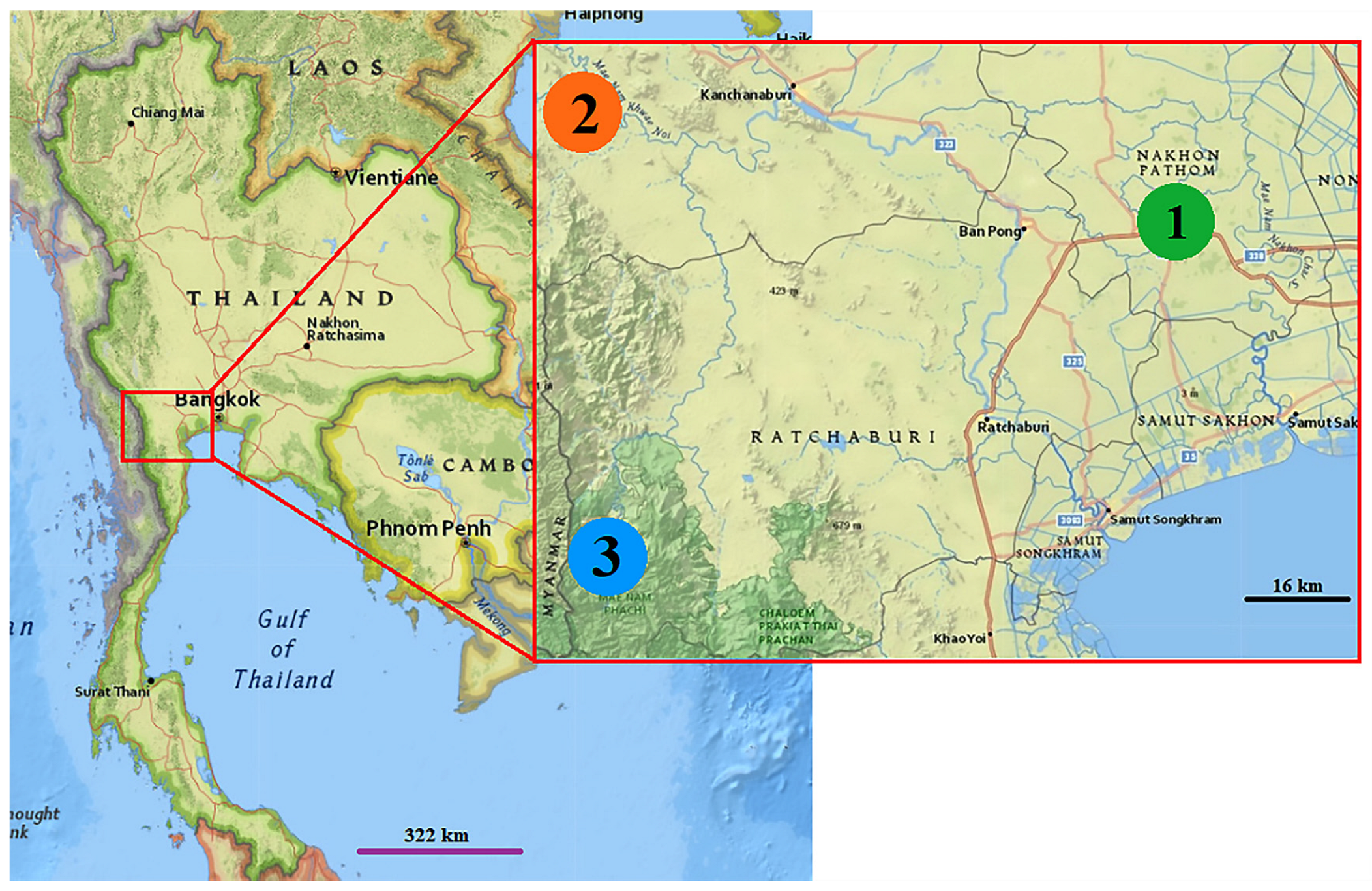
2.1. Mosquito Collections

Adult Cx. pseudovishnui, Cx. tritaeniorhynchus, and Cx. vishnui were collected from three provinces in Thailand: Nakhon Pathom (13°53′29.6″ N, 100°00′08.8″ E), Kanchanaburi (14°07′06.2″ N, 99°01′16.2″ E), and Ratchaburi (13°21′35.1″ N, 99°15′04.5″ E) (Figure 1). Ten BG-Pro CDC-style traps (Biogents, Regensburg, Germany) used in with dry ice and BG-lure (Biogents, Regensberg, Germany) were used in trapping adult mosquito for ten consecutive nights per site during July and September 2021 between 6 p.m. (start time) and 6 a.m. (end time). Ten traps were hung at heights of 1.5 and 5 m around cottages near rice fields, which are the breeding sites for mosquitoes in the Cx. vishnui subgroup. In the morning (6 a.m. (end time)), the field collecting bag was taken out of the trap and cooled for approximately 30 min in an ice box containing 1 kg of dry ice to kill all mosquitoes.
This entomological study was approved by the Institute of Animal Care and Use Committee (MU-IACUC) of the Faculty of Tropical Medicine, Mahidol University, Thailand (reference no. 008-2021E).

2.2. Species Identification Based on the Morphology

Mosquitoes were transported from the field to the laboratory of the College of Allied Health Sciences, Suan Sunandha Rajabhat University, Samut Songkhram Campus for species identification based on morphological characters. All samples were stored at −20 °C in the laboratory’s freezer pending species identification. Adult female Culex mosquitoes were examined under a stereomicroscope (Nikon SMZ 745; Nikon Corp., Tokyo, Japan) and identified using illustrated keys to the medically important mosquitoes of Thailand [10]. Only complete Culex specimens without damaged unique portions and morphologically identifiable were used for analyses. While morphologically ambiguous specimens were excluded from this experiment.

2.3. Sample Preparation for GM and DNA Barcoding

The right wings of Cx. pseudovishnui, Cx. tritaeniorhynchus, and Cx. vishnui were used for the GM analysis, whereas their legs were used for DNA barcoding. The wings of mosquitoes were dissected from the thorax, placed on a microscope slide, and then mounted in Hoyer’s mounting medium solution. The plume scales were removed using bristle needles, and the specimen was covered with a cover slip. Images of all wing slides were captured using a (Nikon SMZ 800N) digital camera coupled to a stereomicroscope at 40× magnification using the NIS-Elements application. Additionally, a 1 mm scale bar was placed in the upper left corner of each wing image.
The legs of 10 mosquitoes per species were randomly collected from samples that were used for the GM analysis. The legs were placed in a 1.5 mL tube (one individual per one tube) pending DNA extraction.

2.4. Geometric Morphometrics

In this study, we used XYOM (freely available at https://xyom.io/ (accessed on 4 March 2022)) for GM analysis [32]. Eighteen anatomical landmarks on the wing veins were digitized (Figure 2) according to criteria used in previous studies [17,18,33].
After that, epeatability was computed to ensure the precision of landmark digitization. Two sets of wing images were used to find the value of shape repeatability using Procrustes analysis [34]. Then, the relationship between the wing size and wing shape of all samples was determined by allometric examination, which involves the calculation of the linear determination coefficient after regressing principal components of wing shape on wing size.
The wing size is expressed as the centroid size (CS), which is the result of the square root of the sum of the squared distances from the centroid to the 18 wing landmarks [35]. The mean wing sizes among the three Culex species were evaluated by a nonparametric ANOVA with 1000 permutations. Statistical significance was set at p < 0.05.
The variables for wing shape were calculated by principal component analysis of the partial warp after the generalized Procrustes analysis [36,37]. After that, discriminant analysis and Mahalanobis distance were computed using the final shape variables. The Mahalanobis distance refers to the statistical distance between the wing shapes of the three Culex species. Wing shape difference based on Mahalanobis distances between Culex species were compared using a nonparametric test (1000 permutations) with Bonferroni correction at p-values < 0.05.
The accuracy of species identification based on wing size was validated by maximum likelihood-based classification [38], whereas the accuracy of identification based on wing shape was validated by Mahalanobis-based classification [39]. For cross-validation reclassification, each individual is iteratively excluded from the total sample and assigned to the closest group to see how resampling the dataset affects the assignment estimation. A hierarchical clustering tree based on the Mahalanobis distances was constructed to examine the relationship between Culex species based on wing shape.

2.5. DNA Barcoding

Thirty Culex samples composed of 10 individuals each of Cx. pseudovishnui, Cx. tritaeniorhynchus, and Cx. vishnu were used for DNA barcoding analysis. The DNA from at least 2 legs per individual of all 30 samples were extracted using the FavorPrep™ tissue genomic DNA extraction mini kit (Favorgen Biotech, Ping-Tung, Taiwan) following the manufacturer’s instructions. PCR was used to amplify a fragment of the COI gene using forward (5′GGATTTGGAAATTGATTAGTTCCTT3′) and reverse (5′AAAAATTTTAA TTCCAGTTGGAACAGC3′) barcoding primers [40]. Each PCR reaction consisted of a 25-µL solution containing 4 µL of DNA, 1.5 unit of Platinum Taq DNA polymerase (Invitrogen), 0.2 μM of each primer, 0.2 mM of dNTPs, and 1x reaction buffer, 1.5 mM MgCl2, 5% dimethyl sulfoxide, and distilled water up to 25 μL. The PCR cycles consisted of 95 °C for 5 min, followed by 5 cycles at 94 °C for 40 s, 45 °C for 60 s, and 72 °C; 35 cycles at 94 °C for 40 s, 54 °C for 60 s, and 72 °C for 1 min; and a final extension at 72 °C for 10 min.
The PCR products were visualized by electrophoresis in 1.5% agarose gels with Midori Green Advance DNA staining solution (Nippon Gene, Tokyo, Japan). The Gel Doc™ image Quant LAS 500 (GE Healthcare Bio-Sciences AB, Uppsala, Sweden) was used to examine and photograph DNA bands on agarose gels to evaluate the quality of PCR products. PCR products of sufficient quality were then submitted to SolGent Co., Ltd. (Daejeon, Republic of Korea) for purification and sequencing.
After Sanger sequencing, chromatogram trace files of COI sequences were manually edited, then ambiguous sites were removed, and consensus sequences were created by BioEdit software. To confirm the identities our COI sequences, they were compared to sequences published in GenBank (https://blast.ncbi.nlm.nih.gov/, accessed on 11 April 2022) and BOLD Systems (https://www.boldsystems.org/index.php/, accessed on 11 April 2022). DNA sequences were aligned using the Clustal W tool [41] within Molecular Evolutionary Genetics Analysis (MEGA) version X [42]. Intra- and interspecific pairwise distances based on the Kimura two-parameter (K2P) model for the COI barcodes of Cx. pseudovishnui, Cx. tritaeniorhynchus, and Cx. vishnui were calculated using MEGA X. Furthermore, the nucleotide sequences were used to build phylogenetic trees based on the neighbor-joining (NJ) method with bootstrapping (1000 replications) by MEGA X to determine the phylogenetic relationships among Cx. pseudovishnui, Cx. tritaeniorhynchus, and Cx. vishnui. Culex quinquefasciatus (OL743030) was included in the phylogenetic tree as an outgroup. The FigTree v.1.4.3 (available at http://tree.bio.ed.ac.uk/software/Figtree/, accessed on 2 May 2022) was used to decorate, modify and create a phylogenetic tree.
For molecular species delimitation, the online assemble species by automatic partitioning (ASAP) approach based on the simple distance (p-distances) was used (https://bioinfo.mnhn.fr/abi/public/asap/, accessed on 10 May 2022). Our COI sequences were submitted to the GenBank database under accession numbers OK147859–OK147868 (for Cx. pseudovishnui), OK147839–OK147848 (for Cx. tritaeniorhynchus), and OK147849–OK147858 (for Cx. vishnui).

3. Results

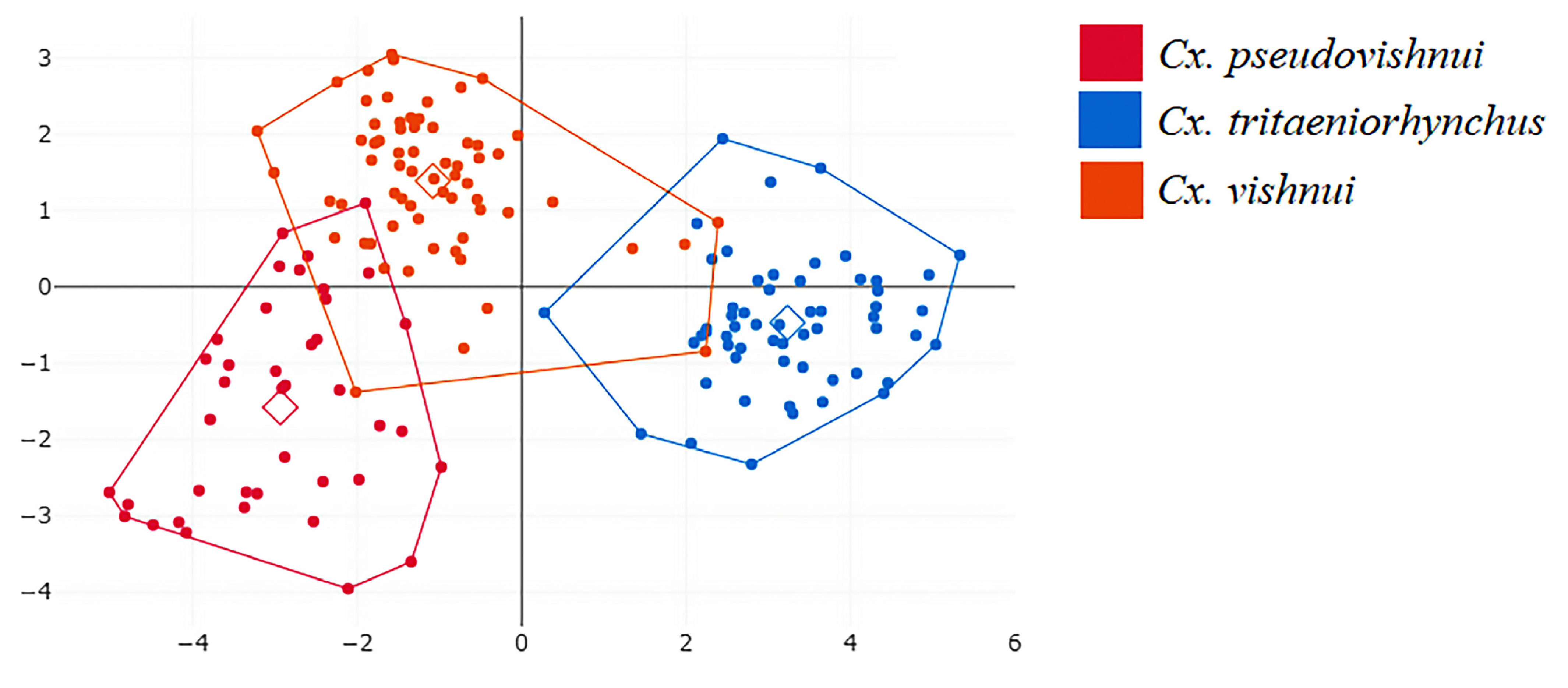
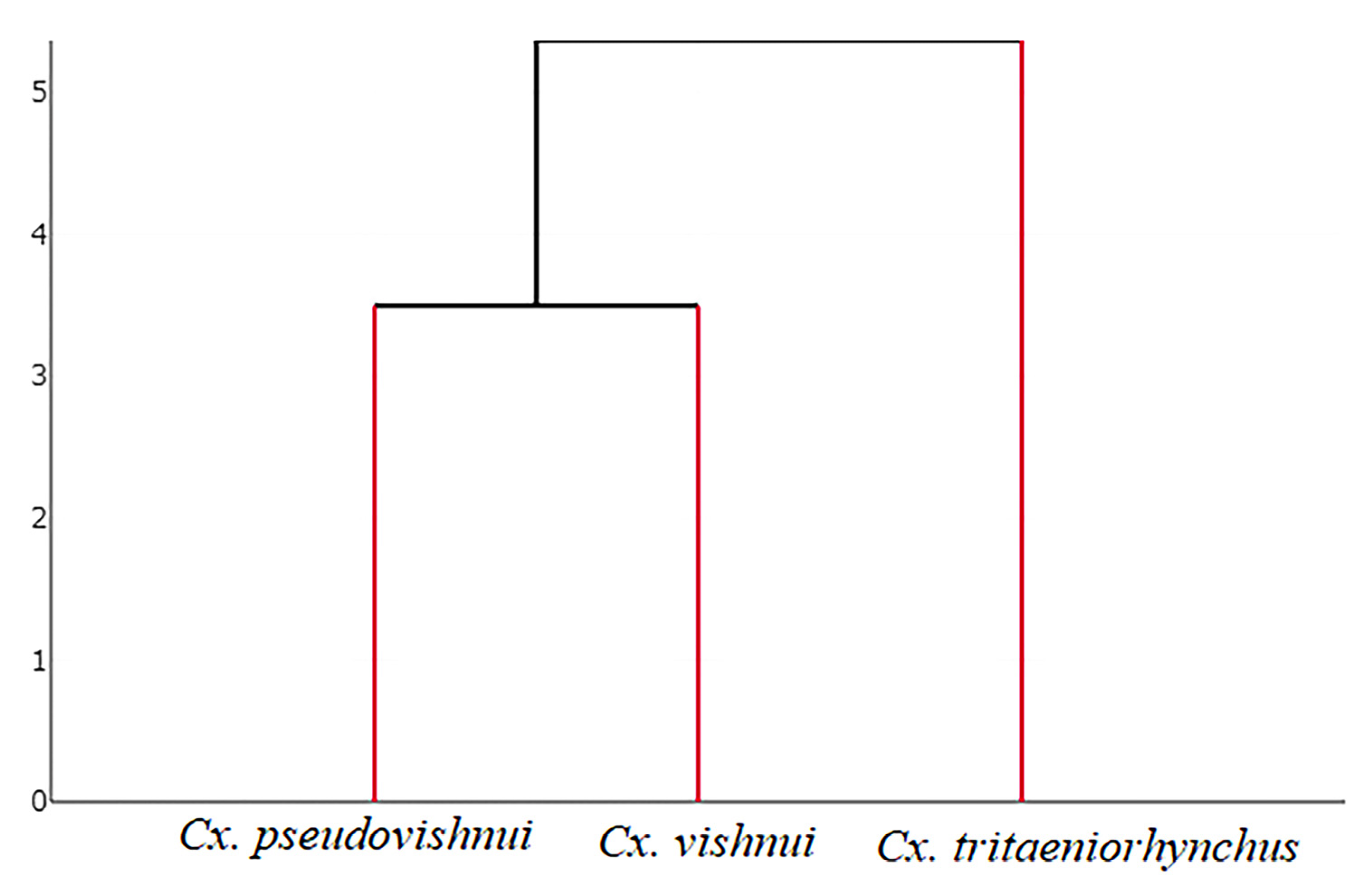
3.1. Geometric Morphometrics for Species Identification

A total of 163 wings of the Cx. vishnui subgroup were analyzed by GM, including 40, 58, and 65 wings of Cx. pseudovishnui, Cx. tritaeniorhynchus, and Cx. vishnui, respectively. The repeatability value of the wing image set was 95%, while the measurement error was 5%, indicating that the quality of our landmark digitization was high.
The relationship between size and shape (allometry) was estimated using the scatter diagram in Figure 3, which indicates a linear relationship between the two variables. The slope of the scatter diagram indicates a negative linear relationship between size and shape, with an r2 value of 11.60 (p < 0.05).
Analysis of wing size variation based on wing CS calculations of Cx. pseudovishnui, Cx. tritaeniorhynchus, and Cx. vishnui is shown in Figure 4. According to wing CS analysis, Cx. vishnui had the largest wings (CS = 2.58 mm), followed by Cx. pseudovishnui (CS = 2.45 mm) and Cx. tritaeniorhynchus (CS = 2.23 mm). Results of statistical analysis confirmed the differences in wing size of the three Culex species. (p < 0.05, Table 1).
Establishing the superposition of the landmark sites on the wings of the Cx. vishnui subgroup after alignment, as depicted in Figure 5, reveals the various different characteristics of wing shape between Culex species.
Figure 6 shows a factor map based on discriminant analysis of shape variables, and it shows that wing shape varies between Cx. pseudovishnui, Cx. tritaeniorhynchus, and Cx. vishnui, i.e., although the group space of three Culex species have overlapping distributions, the wing shapes differ significantly based on the pairwise Mahalanobis distances (1000 runs, p < 0.05, Table 2). The hierarchical clustering tree based on the wing shapes of the three Cx. vishnui subgroup species is shown in Figure 7.
Analysis of wing size based on centroid size reclassification of the Cx. vishnui subgroup shows a lower total percentage score (58.28%) compared to the cross-validated reclassification based on wing shape obtained from Mahalanobis distance values (88.34%, Table 3).
According to the validated reclassification based on wing shape, Cx. tritaeniorhynchus had the highest percentage of correctly classified individual (98.28%), followed by Cx. vishnui (87.69%), and Cx. pseudovishnui (75%).

3.2. Species Identification by DNA Barcoding

DNA barcoding was used in parallel with the GM approach for the identification of Cx. pseudovishnui, Cx. tritaeniorhynchus, and Cx. vishnui. The length of the mitochondrial COI sequences analyzed was 735 bp. The average ATGC composition of 30 Culex sequences were 30.3%, 37.7%, 16.1%, and 15.8%, respectively. Sequence comparisons to the GenBank and Bold databases indicated that the identities of our mosquito species based on morphology were correct (>98% sequence similarity).
The average intraspecific genetic distance among the Culex species based on K2P distance calculations was 0.78% ± 0.39% (average ± SD). The highest average intraspecific divergence was found 0.90% ± 0.40% in Cx. triteaniorhynchus, followed by Cx. pseudovishnui at 0.81% ± 0.31% and Cx. vishnui at 0.67% ± 0.41% (Table 4). Meanwhile, the average interspecific genetic distance was 6.14% ± 0.79%. The highest average interspecific divergence was between Cx. triteaniorhynchus and Cx. pseudovishnui (6.87% ± 0.30%), followed by Cx. triteaniorhynchus and Cx. vishnui (5.40% ± 0.27%) and Cx. vishnui and Cx. pseudovishnui (4.71% ± 0.43%).
An NJ tree based on COI sequences revealed that Cx. vishnui and Cx. pseudovishnui formed a well-supported clade that was sister to Cx. tritaeniorhynchus (bootstrap support > 93%, Figure 8). The ASAP procedure indicated concordant results with morphological taxonomy (the “best” ASAP score = 1.00 and a threshold distance = 0.025).

4. Discussion

The use of morphology to identify mosquito species is the most commonly used method of identification. However, this method is prone to error, especially when identifying mosquitoes belonging to a species complex, as well as isomorphic species, cryptic species, and samples damaged during collection [12]. Therefore, several more advanced and accurate modern techniques have been used to identify mosquitoes, and these have greatly increased our knowledge and understanding of mosquito vectors [16,17,18,19,20,21,22]. Culex pseudovishnui, Cx. tritaeniorhynchus, and Cx. vishnui are the major vectors of JE and are common in many Asian countries [43,44]. However, the morphology of each of these Culex species is slightly different, and specimens tend to be easily damaged [10]. This study is the first to use the landmark-based GM approach to distinguish between Cx. pseudovishnui, Cx. tritaeniorhynchus, and Cx. vishnui. Furthermore, we applied DNA barcoding in parallel with the GM approach to identify these three Culex species.
Only undamaged samples are used in the GM analysis to reduce sources of error. The result of the allometric examination based on the linear determination coefficient shows that wing size and wing shape of our Culex samples are negatively correlated. Similar observations have been reported for several mosquito species, such as Anopheles dissidens, An. wejchoochotei, and An. saeungae in Thailand [18]. Although the relationship between wing size and shape impacts sexual shape dimorphism, it has no effect on species identification based on landmark-based GM analysis [23].
The results of GM analysis showed that the wings of all three Culex species differ significantly in size. Nevertheless, the maximum likelihood-based validated classification based on wing size yielded poor results in distinguishing between the three Culex species (total performance = 58.28%). This result is consistent with the findings of previous research, indicating that wing size is unsuitable for the identification of mosquito species [17,18,27,28]. The wing size is more sensitive to the environment, particularly the influence of temperature, humidity, and food availability in breeding sites, during the development of the immature stage of mosquito [28]. In contrast, wing shape analyses yielded better outcomes than wing size.
The validated classification based on wing shape (total performance = 88.34% of correctly assigned individuals) clearly outperformed wing size analyses. This is consistent with previous studies that applied the GM approach in identifying mosquito vectors and found that wing shape is more effective criterion compared to wing size [27,28]. Several studies have demonstrated that the GM technique is effective for the identification of mosquito species in Thailand, including Aedes species (Ae. aegypti, Ae. albopictus, and Ae. scutellaris [27,45]); Mansonia species (Ma. dives and Ma. bonneae [28]); Culex species (Cx. visnui, Cx. sitiens, and Cx. whitmorei [14]); and Anopheles species (An. dirus, An. baimaii [17], An. maculatus, An. sawadwongporni, and An. pseudowillmori [18]). Nevertheless, a performance level of 88.34%, as we found, is not considered perfect but still has a relatively high potential. The results of discriminant analysis indicate that Cx. pseudovishnui, Cx. tritaeniorhynchus, and Cx. vishnui share very similar wing characteristics that overlap among species. If species identification is limited by financial constraints, then the GM technique based on wing shape can be used in conjunction with the morphological method, which is better than visual inspection alone [28].
DNA barcoding is a highly effective molecular technique for the identification of mosquito species that are morphologically similar or indistinguishable [21,22,46,47]. Our genetic analysis and the ASAP method suggested that DNA barcoding based on COI sequences can be used to distinguish between Cx. pseudovishnui, Cx. tritaeniorhynchus, and Cx. vishnui and the results were consistent with those of morphological identification. Analysis of the genetic distance between species shows that the species are separated by a DNA barcoding gap, which is the difference between the greatest intraspecific distance and the smallest interspecific distance [21,22,46]. Our results of DNA barcoding are consistent with those of a previous study in Sri Lanka that used DNA barcoding to successfully identify members in the Cx. vishnui subgroup [30]. In addition, the NJ tree based on COI sequences is consistent with the hierarchical clustering tree based on wing shape (GM) analysis. Both trees indicate that the Cx. tritaeniorhynchus clade is distinct from the other two clades, while Cx. vishnui and Cx. pseudovishnui are more closely related to each other. A previous study that applied GM and genetic techniques to compare variability between male and female Ae. aegypti populations in the Philippines found that phenotypic and genetic patterns in male Ae. aegypti are highly congruent [48], suggesting that wing shape is genetically influenced.

5. Conclusions

The cross-validation reclassification revealed that the GM technique had relatively high potential for distinguishing Cx. pseudovishnui, Cx. tritaeniorhynchus, and Cx. vishnui based on wing shape analysis (88.34% of correctly assigned individuals). While the DNA barcoding yielded excellent results in identifying these Culex species based on the DNA barcode gap. Therefore, we suggest that DNA barcoding based on the COI gene is an excellent method for identifying Cx. pseudovishnui, Cx. tritaeniorhynchus, and Cx. vishnui, which are difficult to identify morphologically. However, in the absence of the required facilities for DNA barcoding, GM techniques can be employed in conjunction with morphological methods to enhance the reliability of species identification. The GM technique offers an inexpensive, and rapid analysis of species identification (approximately 5–10 min). Currently, there are several software freely available for studying geometric morphometrics. In addition, it can be implemented using basic equipment found in most entomology laboratories. However, the wing slide preparation is a rather time-consuming process (approximately 10 wings per hour). Our results will help guide efforts to identify members of the Cx. vishnui subgroup, which is a requirement for the effective control of the vector of JE in Thailand.

Author Contributions

Conceptualization, T.S., T.C. and S.S.; methodology, T.S., S.L., T.C. and S.S.; validation, T.S., S.L., T.C. and S.S.; investigation, S.L. and T.C.; data curation, T.S., S.L., T.C. and S.S.; writing—original draft preparation, T.S., S.L., T.C. and S.S.; writing—review and editing, T.S., T.C. and S.S.; project administration, T.S., T.C. and S.S. All authors have read and agreed to the published version of the manuscript.

Funding

This study was financially supported by the Faculty of Tropical Medicine, Mahidol University.

Institutional Review Board Statement

This entomological study has been approved by the Institute Animal Care and Use Committee (MU-IACUC) of the Faculty of Tropical Medicine, Mahidol University, Thailand (reference no. 008-2021E).

Informed Consent Statement

Not applicable.

Data Availability Statement

Our COI sequences were submitted in the GenBank database with the following accession numbers: Cx. pseudovishnui: OK147859–OK147868; Cx. tritaeniorhynchus: OK147839–OK147848; Cx. vishnui: OK147849–OK147858.

Acknowledgments

We thank the Mahidol University and the Suan Sunandha Rajabhat University, Thailand for supporting laboratory facilities and equipment.

Conflicts of Interest

The authors declare no conflict of interest.

References

  1. World Health Organization. Japanese Encephalitis 2019. Available online: https://www.who.int/news-room/fact-sheets/detail/japanese-encephalitis (accessed on 10 July 2022).
  2. Amicizia, D.; Zangrillo, F.; Lai, P.L.; Iovine, M.; Panatto, D. Overview of Japanese Encephalitis Disease and Its Prevention. Focus on IC51 Vaccine (IXIARO®). J. Prev. Med. Hyg. 2018, 59, E99–E107. [Google Scholar] [PubMed]
  3. World Health Organization. Japanese Encephalitis Reported Cases by WHO Region 2020. Available online: https://apps.who.int/gho/data/view.main.1520_42lang=en (accessed on 20 December 2022).
  4. Ministry of Public Health, Thailand. Annual Report. Bureau of Epidemiology: MOPH; Japanese B encephalitis. 2017–2019. Available online: http://www.boe.moph.go.th/boedb/surdata/index.php (accessed on 10 July 2022).
  5. Ministry of Public Health, Thailand. Epidemiological Report. Available online: http://www.boe.moph.go.th/boedb/surdata/index.php (accessed on 10 July 2022).
  6. World Health Organization. Mosquito Born Diseases. Available online: http://www.who.int/neglected_diseases/vector_ecology/mosquito-borne-diseases/en/ (accessed on 15 July 2022).
  7. Chaiphongpachara, T.; Laojun, S. Effectiveness of Ultraviolet (UV) Insect Light Traps for Mosquitoes Control in Coastal Areas of Samut Songkhram Province, Thailand. J. Anim. Behav. Biometeorol. 2019, 7, 25–30. [Google Scholar]
  8. Lee, H.; Halverson, S.; Ezinwa, N. Mosquito-Borne Diseases. Prim. Care-Clin. Off. Pract. 2018, 45, 393–407. [Google Scholar] [CrossRef] [PubMed]
  9. Harbach, R.E. Mosquito Taxonomic Inventory. Available online: https://mosquito-taxonomic-inventory.myspecies.info/simpletaxonomy/term/6192 (accessed on 1 October 2022).
  10. Rattanarithikul, R.; Harbach, R.E.; Harrison, B.A.; Panthusiri, P.; Jones, J.W.; Coleman, R.E. Illustrated Keys to the Mosquitoes of Thailand. II. Genera Culex and Lutzia. Southeast Asian J. Trop. Med. Public Health 2005, 36, 1–97. [Google Scholar] [PubMed]
  11. Gingrich, J.B.; Nisalak, A.; Latendresse, J.R.; Sattabongkot, J.; Hoke, C.H.; Pomsdhit, J.; Chantalakana, C.; Satayaphanta, C.; Uechiewcharnkit, K.; Innis, B.L. Japanese Encephalitis Virus in Bangkok: Factors Influencing Vector Infections in Three Suburban Communities. J. Med. Entomol. 1992, 29, 436–444. [Google Scholar] [CrossRef]
  12. Sumruayphol, S.; Chaiphongpachara, T.; Samung, Y.; Ruangsittichai, J.; Cui, L.; Zhong, D.; Sattabongkot, J.; Sriwichai, P. Seasonal Dynamics and Molecular Differentiation of Three Natural Anopheles Species (Diptera: Culicidae) of the Maculatus Group (Neocellia Series) in Malaria Hotspot Villages of Thailand. Parasit. Vectors 2020, 13, 574. [Google Scholar] [CrossRef]
  13. Kshitiz, S.; Sebastien, B.; Veronique, C.; Chalalai, R. Host Preference of Culex Mosquitoes in an Area with High Transmission Rate of Japanese Encephalitis of Kandal Province, Cambodia. J. Kasetsart Vet. 2008, 28, 126–137. [Google Scholar]
  14. Chaiphongpachara, T.; Laojun, S. Effectiveness of Landmark- and Semi-Landmark-Based Geometric Morphometric to Identify Four Species of Culex Mosquitoes in Thailand. J. Adv. Vet. Anim. Res. 2019, 6, 278–283. [Google Scholar] [CrossRef]
  15. Chaiphongpachara, T.; Sriwichai, P.; Samung, Y.; Ruangsittichai, J. Geometric Morphometrics Approach towards Discrimination of Three Member Species of Maculatus Group in Thailand. Acta Trop. 2019, 192, 66–74. [Google Scholar] [CrossRef]
  16. Champakaew, D.; Junkum, A.; Sontigun, N.; Sanit, S.; Limsopatham, K.; Saeung, A.; Somboon, P.; Pitasawat, B. Geometric Morphometric Wing Analysis as a Tool to Discriminate Female Mosquitoes from Different Suburban Areas of Chiang Mai Province, Thailand. PLoS ONE 2021, 16, e0260333. [Google Scholar] [CrossRef]
  17. Chaiphongpachara, T.; Changbunjong, T.; Sumruayphol, S.; Laojun, S.; Suwandittakul, N.; Kuntawong, K. Geometric Morphometrics versus DNA Barcoding for the Identification of Malaria Vectors Anopheles dirus and An. baimaii in the Thai-Cambodia Border. Sci. Rep. 2022, 12, 13236. [Google Scholar] [CrossRef]
  18. Chaiphongpachara, T.; Changbunjong, T.; Laojun, S. Geometric Morphometric and Molecular Techniques for Discriminating among Three Cryptic Species of the Anopheles barbirostris Complex (Diptera: Culicidae) in Thailand. Heliyon 2022, 8, e11261. [Google Scholar] [CrossRef]
  19. Oliveira-Christe, R.; Marrelli, M.T. Using Geometric Morphometric Analysis of Wings to Identify Mosquito Species from the Subgenus Microculex (Diptera: Culicidae). J. Vector Ecol. 2021, 46, 221–225. [Google Scholar] [CrossRef]
  20. Chatpiyaphat, K.; Sumruayphol, S.; Dujardin, J.P.; Samung, Y.; Phayakkaphon, A.; Cui, L.; Ruangsittichai, J.; Sungvornyothin, S.; Sattabongkot, J.; Sriwichai, P. Geometric Morphometrics to Distinguish the Cryptic Species Anopheles minimus and An. harrisoni in Malaria Hot Spot Villages, Western Thailand. Med. Vet. Entomol. 2021, 35, 293–301. [Google Scholar]
  21. Chaiphongpachara, T.; Changbunjong, T.; Laojun, S.; Nutepsu, T.; Suwandittakul, N.; Kuntawong, K.; Sumruayphol, S.; Ruangsittichai, J. Mitochondrial DNA Barcoding of Mosquito Species (Diptera: Culicidae) in Thailand. PLoS ONE 2022, 17, e0275090. [Google Scholar] [CrossRef]
  22. Ortega-Morales, A.I.; Hernandez-Triana, L.M.; Chan-Chable, R.J.; Garza-Hernandez, J.A.; Gonzalez-Alvarez, V.H.; Ruiz-Arrondo, I.; Nikolova, N.I.; Martinez-Arce, A.; Fooks, A.R.; Rodriguez-Perez, M.A. Dna Barcoding of Mosquitoes from the Pantanos de Centla Biosphere Reserve, Southeastern Mexico. J. Am. Mosq. Control Assoc. 2021, 37, 198–207. [Google Scholar] [CrossRef]
  23. Lorenz, C.; Almeida, F.; Almeida-Lopes, F.; Louise, C.; Pereira, S.N.; Petersen, V.; Vidal, P.O.; Virginio, F.; Suesdek, L. Geometric Morphometrics in Mosquitoes: What Has Been Measured? Infect. Genet. Evol. 2017, 54, 205–215. [Google Scholar] [CrossRef]
  24. Dujardin, J.P. Morphometrics Applied to Medical Entomology. Infect. Genet. Evol. 2008, 8, 875–890. [Google Scholar] [CrossRef]
  25. Suwandittakul, N.; Mungthin, M.; Kuntawong, K.; Laojun, S.; Pimsuka, S.; Chaiphongpachara, T. A Novel Use of a Geometric Morphometric Technique to Distinguish Human Parasite Eggs of Twelve Different Species. Exp. Parasitol. 2022, 238, 108281. [Google Scholar] [CrossRef]
  26. Simões, R.F.; Wilke, A.B.B.; Chagas, C.R.F.; de Menezes, R.M.T.; Suesdek, L.; Multini, L.C.; Silva, F.S.; Grech, M.G.; Marrelli, M.T.; Kirchgatter, K. Wing Geometric Morphometrics as a Tool for the Identification of Culex Subgenus Mosquitoes of Culex (Diptera: Culicidae). Insects 2020, 11, 567. [Google Scholar] [CrossRef]
  27. Sumruayphol, S.; Apiwathnasorn, C.; Ruangsittichai, J.; Sriwichai, P.; Attrapadung, S.; Samung, Y.; Dujardin, J.P. DNA Barcoding and Wing Morphometrics to Distinguish Three Aedes Vectors in Thailand. Acta Trop. 2016, 159, 1–10. [Google Scholar] [CrossRef] [PubMed]
  28. Ruangsittichai, J.; Apiwathnasorn, C.; Dujardin, J.P. Interspecific and Sexual Shape Variation in the Filariasis Vectors Mansonia dives and Ma. bonneae. Infect. Genet. Evol. 2011, 11, 2089–2094. [Google Scholar] [CrossRef] [PubMed]
  29. Kress, W.J.; Erickson, D.L. DNA Barcodes: Genes, Genomics, and Bioinformatics. Proc. Natl. Acad. Sci. USA 2008, 105, 2761–2762. [Google Scholar] [CrossRef] [PubMed] [Green Version]
  30. Weeraratne, T.C.; Surendran, S.N.; Karunaratne, S.H.P.P. DNA Barcoding of Morphologically Characterized Mosquitoes Belonging to the Subfamily Culicinae from Sri Lanka. Parasit Vectors 2018, 11, 266. [Google Scholar] [CrossRef] [Green Version]
  31. Artur, T.; Anna, S.K.; James, J.B.; Neil, S.; Miroslawa, D. A New Method of Metabarcoding Microsporidia and Their Hosts Reveals High Levels of Microsporidian Infections in Mosquitoes (Culicidae). Mol. Ecol. Resour. 2020, 20, 1486–1504. [Google Scholar]
  32. Dujardin, S.; Dujardin, J.P. Geometric Morphometrics in the Cloud. Infect. Genet. Evol. 2019, 70, 189–196. [Google Scholar] [CrossRef]
  33. Demari-Silva, B.; Suesdek, L.; Sallum, M.A.M.; Marrelli, M.T. Wing Geometry of Culex Coronator (Diptera: Culicidae) from South and Southeast Brazil. Parasit. Vectors 2014, 7, 174. [Google Scholar] [CrossRef] [Green Version]
  34. Goodall, C.R. Procrustes Methods in the Statistical Analysis of Shape. J. R. Stat. Soc. Ser. B 1991, 53, 285–321. [Google Scholar] [CrossRef]
  35. Bookstein, F.L. Morphometric Tools for Landmark Data: Geometry and Biology; Cambridge University Press: Cambridge, UK, 1991. [Google Scholar]
  36. Rohlf, F.J. Rotational Fit (Procrustes) Methods. Proceedings of the Michigan Morphometrics Workshop; Rohlf, F.J., Bookstein, F.L., Eds.; University of Michigan, Museum of Zoology: Ann Arbor, MI, USA, 1990. [Google Scholar]
  37. Rohlf, F.J.; Slice, D.E. Extensions of de Procrustes Method for the Optimal Superimposition of Landmarks. Syst. Zool. 1990, 39, 40–59. [Google Scholar] [CrossRef] [Green Version]
  38. Dujardin, J.-P.; Dujardin, S.; Kaba, D.; Santillán-Guayasamín, S.; Villacís, A.G.; Piyaselakul, S.; Sumruayphol, S.; Samung, Y.; Morales Vargas, R. The Maximum Likelihood Identification Method Applied to Insect Morphometric Data. Zool. Syst. 2017, 42, 46–58. [Google Scholar]
  39. Dujardin, J.P.; Kaba, D.; Henry, A.B. The exchangeability of shape. BMC Res. Notes 2010, 3, 266. [Google Scholar] [CrossRef] [Green Version]
  40. Kumar, N.P.; Rajavel, A.R.; Natarajan, R.; Jambulingam, P. DNA Barcodes Can Distinguish Species of Indian Mosquitoes (Diptera: Culicidae). J. Med. Entomol. 2007, 44, 1–7. [Google Scholar] [CrossRef]
  41. Larkin, M.A.; Blackshields, G.; Brown, N.P.; Chenna, R.; Mcgettigan, P.A.; McWilliam, H.; Valentin, F.; Wallace, I.M.; Wilm, A.; Lopez, R.; et al. Clustal W and Clustal X Version 2.0. Bioinformatics 2007, 23, 2947–2948. [Google Scholar] [CrossRef] [Green Version]
  42. Kumar, S.; Stecher, G.; Li, M.; Knyaz, C.; Tamura, K. MEGA X: Molecular Evolutionary Genetics Analysis across Computing Platforms. Mol. Biol. Evol. 2018, 35, 1547–1549. [Google Scholar] [CrossRef]
  43. Kumari, R.; Kumar, K.; Rawat, A.; Singh, G.; Yadav, N.K.; Chauhan, L.S. First Indigenous Transmission of Japanese Encephalitis in Urban Areas of National Capital Territory of Delhi, India. Trop. Med. Int. Health 2013, 18, 743–749. [Google Scholar] [CrossRef]
  44. Faizah, A.N.; Kobayashi, D.; Isawa, H.; Amoa-Bosompem, M.; Murota, K.; Higa, Y.; Futami, K.; Shimada, S.; Kim, K.S.; Itokawa, K.; et al. Deciphering the Virome of Culex vishnui Subgroup Mosquitoes, the Major Vectors of Japanese Encephalitis, in Japan. Viruses 2020, 12, 264. [Google Scholar] [CrossRef] [Green Version]
  45. Chonephetsarath, S.; Raksakoon, C.; Sumruayphol, S.; Dujardin, J.P.; Potiwat, R. The Unequal Taxonomic Signal of Mosquito Wing Cells. Insects 2021, 12, 376. [Google Scholar] [CrossRef]
  46. Wang, G.; Li, C.; Guo, X.; Xing, D.; Dong, Y.; Wang, Z.; Zhang, Y.; Liu, M.; Zheng, Z.; Zhang, H.; et al. Identifying the Main Mosquito Species in China Based on DNA Barcoding. PLoS ONE 2012, 7, e47051. [Google Scholar] [CrossRef]
  47. Hernández-Triana, L.M.; Brugman, V.A.; Nikolova, N.I.; Ruiz-Arrondo, I.; Barrero, E.; Thorne, L.; de Marco, M.F.; Krüger, A.; Lumley, S.; Johnson, N.; et al. DNA Barcoding of British Mosquitoes (Diptera, Culicidae) to Support Species Identification, Discovery of Cryptic Genetic Diversity and Monitoring Invasive Species. Zookeys 2019, 832, 57–76. [Google Scholar] [CrossRef] [Green Version]
  48. Carvajal, T.M.; Amalin, D.M.; Watanabe, K. Wing Geometry and Genetic Analyses Reveal Contrasting Spatial Structures between Male and Female Aedes aegypti (L.) (Diptera: Culicidae) Populations in Metropolitan Manila, Philippines. Infect. Genet. Evol. 2021, 87, 104676. [Google Scholar] [CrossRef]
Figure 1. Study sites for mosquito sample collection in this study. This geographical map was taken from the USGS National Map Viewer (the public domain) at http://viewer.nationalmap.gov/viewer/ (accessed on 9 November 2022). Mosquito samples were collected from Nakhon Pathom (1, Green) in the central region, Kanchanaburi (2, Orange), and Ratchaburi (3, Blue) in the western region of Thailand.
Insects 14 00131 g001
Figure 2. Location of the 18 wing landmarks used in the geometric morphometric analyses.
Insects 14 00131 g002
Figure 3. Scatter diagram showing the relationship between wing size (the x-axis) and wing shape (the y-axis) using landmark-based GM analysis. The linear regression prediction result is represented by the orange dots. While blue dots indicate individual mosquitoes.
Insects 14 00131 g003
Figure 4. Boxplot of the wing centroid size of Cx. pseudovishnui, Cx. tritaeniorhynchus, and Cx. vishnui. Each box represents the wing size data of each Culex species, and the group median that separates the 25th and 75th quartiles is displayed in each box.
Insects 14 00131 g004
Figure 5. Rotated landmarks of aligned objects (A) and the mean landmark superposition (B) of Cx. pseudovishnui, Cx. tritaeniorhynchus, and Cx. vishnui wings.
Insects 14 00131 g005
Figure 6. Factor map generated by discriminant analysis of shape variables for Cx. pseudovishnui, Cx. tritaeniorhynchus, and Cx. vishnui. The wing shape variations of each Culex species are represented by each polygon, and each dot within the polygons represents an individual Culex specimen.
Insects 14 00131 g006
Figure 7. Hierarchical clustering tree based on wing shape among Cx. pseudovishnui, Cx. tritaeniorhynchus, and Cx. vishnui. The UPGMA algorithm is used to create a hierarchical clustering tree based on Mahalanobis distances between average group shapes.
Insects 14 00131 g007
Figure 8. Neighbor-joining tree showing the phylogenetic relationships between Cx. pseudovishnui, Cx. tritaeniorhynchus, and Cx. vishnui. The COI sequences generated in this study are black (n = 30), while those from GenBank are red (n = 6). The Cx. quinquefasciatus sequence from GenBank (OL743030) was used as an outgroup. Only bootstrap values ≥ 50% are shown. The result of species delimitation by the ASAP method is indicated by vertical red bars.
Insects 14 00131 g008
Table 1. Statistical differences in mean wing centroid size of Cx. pseudovishnui, Cx. tritaeniorhynchus, and Cx. vishnui.
SpeciesnMean (mm)Min–MaxSDSE
Cx. pseudovishnui402.45 a2.06–2.820.170.03
Cx. tritaeniorhynchus582.23 b1.98–2.480.140.02
Cx. vishnui652.58 c2.02–2.950.240.03
Different superscript letters after wing CS values indicate significant differences at p < 0.05.
Table 2. Statistical differences of Mahalanobis distance values based on wing shapes of Cx. Pseudovishnui, Cx. tritaeniorhynchus, and Cx. vishnui.
SpeciesCx. pseudovishnuiCx. tritaeniorhynchusCx. vishnui
Cx. pseudovishnui0.000
Cx. tritaeniorhynchus6.268 *0.000
Cx. vishnui3.498 *4.695 *0.000
The superscript asterisks after Mahalanobis distance values indicate significant differences between Culex species at p < 0.05.
Table 3. Cross-validation reclassification score (%) based on wing size (centroid size) and wing shape (Mahalanobis distance values) of the Cx. vishnui subgroup.
SpeciesCross-Validated Reclassification Score (%)
Wing Size
(Assigned/Observed)
Wing Shape
(Assigned/Observed)
Cx. pseudovishnui35.00% (14/40)75.00% (30/40)
Cx. tritaeniorhynchus75.86% (44/58)98.28% (57/58)
Cx. vishnui56.92% (37/65)87.69% (57/65)
Total performance58.28% (95/163)88.34% (144/163)
Table 4. Inter- and intraspecific genetic divergence between Cx. pseudovishnui, Cx. tritaeniorhynchus, and Cx. vishnui based on the Kimura two parameter model.
SpeciesAverage Percentage Genetic Divergence (Min–Max)
Cx. pseudovishnuiCx. tritaeniorhynchusCx. vishnui
Cx. pseudovishnui0.81%
(0.00–1.43)
Cx. tritaeniorhynchus6.87%
(6.33–7.89)
0.90%
(0.28–1.86)
Cx. vishnui4.71%
(3.91–5.90)
5.40%
(4.96–6.03)
0.67%
(0.00–1.57)
Disclaimer/Publisher’s Note: The statements, opinions and data contained in all publications are solely those of the individual author(s) and contributor(s) and not of MDPI and/or the editor(s). MDPI and/or the editor(s) disclaim responsibility for any injury to people or property resulting from any ideas, methods, instructions or products referred to in the content.

