Article

Ectoparasitism of the Flightless Drosophila melanogaster and D. hydei by the Mite Blattisocius mali (Acari: Blattisociidae)

by
Katarzyna Michalska
1,*,
Agnieszka Mrowińska
1 and
Marcin Studnicki
2
1
Section of Applied Entomology, Department of Plant Protection, Institute of Horticulture Sciences, Warsaw University of Life Sciences, Nowoursynowska 159, 02-776 Warsaw, Poland
2
Department of Biometry, Institute of Agriculture, Warsaw University of Life Sciences, Nowoursynowska 159, 02-776 Warsaw, Poland
*
Author to whom correspondence should be addressed.
Insects 2023, 14(2), 146; https://doi.org/10.3390/insects14020146
Submission received: 24 December 2022 / Revised: 19 January 2023 / Accepted: 28 January 2023 / Published: 31 January 2023
(This article belongs to the Section Insect Behavior and Pathology)

Abstract

:

Simple Summary

There are a number of reports on the dispersal of predatory mites on insects, although much less is known about their mutual interactions and possible relationships. While some mite species use insects only as means of transport, others also feed on them, significantly lowering the carriers’ fitness. Blattisocius mali is a promising predatory mite for pest control, and a thorough understanding of its dispersal pathways in crops could be crucial for successful plant protection. So far, it has been reported as transported by several species of drosophilids. Our research indicates an ectoparasitic relationship between this species and drosophilid fruit flies. We used the flightless D. melanogaster and D. hydei commercially raised as live pet food. Female mites not only attached to flies but also fed on them, and their presence resulted in an increase in fly mortality. Although both fly species used similar defense tactics, mites had more difficulties getting onto D. hydei. Whether the wild D. hydei and D. melanogaster can also transport B. mali will be shown by further laboratory and field studies.

Abstract

Predatory mites dispersing by means of insects are often ectoparasites and may use various tactics to get onto the host, counteract its defenses, and diminish its survival. Blattisocius mali is a promising biological control agent which has been reported as transported by several drosophilid species. Our goal was to determine the type of relationship between this mite and fruit flies. We used flightless females of Drosophila melanogaster and D. hydei, which were commercially raised as live pet food. The predatory females mostly attacked the tarsi of the flies and then preferentially moved to the cervix or close to coxa III, where they eventually drilled their chelicerae and started feeding. Although both fly species used similar defensive tactics, more B. mali females did not attack D. hydei or did so with a delay, and a higher percentage of mites fell off the D. hydei tarsi during the first hour of observation. After 24 h, we noted the increased mortality of flies exposed to the presence of mites. Our study indicates the ectoparasitic relationship of B. mali with drosophilids. However, further research is needed to confirm the transport of this mite on wild D. hydei and D. melanogaster, both in the laboratory and under natural conditions.

1. Introduction

Spreading using insects is a common mode of dispersal in mites [1,2,3,4,5]. Relationships between mites and the insects that carry them can take many forms, ranging from mutualism or commensalism to parasitism and predation [6,7]. As stated by Bartlow and Agosta [5], in the case of a phoresy one organism, the phoront transfers from one site to another with the aid of another organism, the dispersal host, and attaches to it, as a result of seeking or waiting for the host. As emphasized by other researchers, phoresy is temporary, and the phoront does not feed on the host or develop during transfer [1,4]. Predatory mites can also be transported by insects [4]. While some species are phoretic, such as e.g., Poecilochirus carabi G. Canestrini and R. Canestrini on the burying beetle Nicrophorus vespilloides Herbst [8] or Parasitellus fucorum (De Geer) on bumblebees Bombus sp. [9], others are ectoparasites that feed on insects during transport, such as Macrocheles muscaedomesticae (Scopoli) and M. subbadius (Berlese) [10,11]. Moreover, both phoronts and ectoparasites can also feed on eggs laid by the hosts and also in further developmental stages [8]. Despite many reports dealing with mite transfer by insects [5,12], there is a scarcity of detailed behavioral observations on the tactics employed by predatory mites to get onto the body of insects or the defense reactions of their carriers, which, apart from various ecological factors, could also determine the preference of predators towards the host species.
Over a dozen mite species are reported as associated with drosophilid flies, both in the wild and in laboratory cultures [13,14,15,16,17,18]. One of the predators on the list is Blattisocius mali (Oudemans). It is a promising biological control agent for acarid mites, eggs of the potato tuber moths, and nematodes [19,20,21,22,23,24,25]. However, there are still many gaps in our knowledge of its ecology and behavior, including the range of prey species and its dispersal methods.
Blattisocius mali belongs to Blattisociidae (Mesostigmata), a family of cosmopolitan predatory mites found both in the soil and in litter, on plants and stored products, and is frequently associated with rodents, insects, and birds [20]. Among species of Blattisocius Kegaan, B. keegani Fox, B. dendriticus (Berlese), B. patagorium Treat, and B. tarsalis (Berlese) have been reported as transported by noctuiid and pyralid moths [26,27,28,29]. Moreover, in B. patogorium and B. tarsalis, an ectoparasitic relationship with moths has been suggested [27,30]. Others, such as B. apis Basha and Yousef and B. trigonae Radhakrishnan and Ramaraju, were found on the body of honey bees, while B. capsicum Basha and Yousef was found in association with juveniles of psocopterous species inhabiting stored hot peppers [31,32].
So far, B. mali has been recorded only in fruit flies (Drosophilidae) from both the Drosophilinae and Steganinae subfamilies. In Finland, the mites (mostly deutonymphs and occasionally females) have been found on Drosophila littoralis Meigen, D. montana Stone, D. ezoana Takada and Okada, and D. lummei Hackman [15]. In the USA and Mexico, B. mali has been attached to D. hexastigma Patterson and Mainland [16]. Finally, in Hungary, Kerezsi et al. [17] found females of this mite on Steganinae fruit flies, Phortica semivirgo Máca. Blattisocius mali has also been reported as associated with drosophilids in laboratory cultures [15].
Phoretic mites attach themselves to insects in a variety of ways [1,4,5,33,34]. They can use chelicerae to grasp setae or folds of insect integument or attach to the host body by means of ambulaclar claws, while other mite phoronts possess special attachment devices, such as a pedicel formed by the secretion of anal glands in some Uropodina deutonymphs or the anal suckers of hypopus, which is a modified deutonymphal stage of some acarid mites. According to Lehtinen and Aspi [15], chelicerae tips of B. mali seemed to be inserted through the integument of fruit flies, which suggests that the association of this mite with drosophilids could be parasitic.
The aim of this study was to determine the type of relationship between B. mali and drosophilid flies. First, we examined the behavioral tactics the mite uses to get onto the body of the flies, where it preferentially attaches itself, and what the influence of the drosophilid species could be on this process. Secondly, we checked whether B. mali feeds on the fruit flies during transportation, and thirdly, whether the dispersal of the mite with the aid of the flies affects the survival of its carriers.

2. Materials and Methods

2.1. Insects and Mites

The stock population of B. mali and the mold mite Tyrophagus putrecentiae (Schrank) came from the laboratory mite cultures maintained in the Section of Applied Entomology, Department of Plant Protection at Warsaw University of Life Sciences (WULS). Blattisocius mali was identified by Dr. K. Michalska using the key by Karg [35] and subsequently by Prof. D. J. Gwiazdowicz. Blattisocius mali was mass-reared on various stages of T. putrescentiae in wheat bran on soaked foam platforms (22 cm × 15 cm × 2.5 cm), which were covered with foil and placed within broader vessels (50 cm × 70 cm × 20 cm) filled up with water. The cultures of B. mali were maintained in a climatic room, at 21–23 °C, with a photoperiod of 16/8 h (L/D). The population of T. putrescentiae was fed with instant yeasts and maintained in a desiccator (Chemland, Stargard, Poland) at 26 °C and 80–85% RH in darkness.
In this study, we used the flightless forms of two drosophilae species, D. melanogaster Meigen and D. hydei Sturtevant (Figure 1), both distributed commercially as live pet food [36]. The stock populations of these flies were obtained from the Amustela Zoological Centre, Warsaw, Poland. The mutation of various genes may lead to the flightlessness of drosophilids, from lack of wings or their deformation to dysfunction of the muscles controlling drosophilid flight [37,38,39,40,41]. The flightless fruit flies we used in this study had developed wings, but instead of flying, they were only hopping at a maximum height of several centimeters. We additionally identified them at the species level using DNA barcoding and the methods developed by Dabert et al. [42,43] and Mironov et al. [44]. Sequences generated in this study were deposited in GenBank (Supplementary File S1). The insects were reared on the standard fruit fly medium based on cornmeal, molasses, yeast, and propionic acid [45] in an incubator (Panasonic, Osaka, Japan) at 25 °C and a 12/12 (L/D) photoperiod.
We hypothesized that similar to wild drosophilids, flightless mutants can also transport B. mali on their bodies. Moreover, considering previous reports on the invasion of laboratory drosophilid cultures with B. mali [15], we hypothesized that this mite could feed on fruit flies and, as an ectoparasite, also suck up the body of adults. As the response of B. mali toward drosophilids could be innate, we tested naïve mites that had no previous contact with flies.

2.2. Experimental Set-Up

The tests were carried out within small observational chambers using pairs of randomly selected females of B. mali (n = 30) and females of D. melanogaster (n = 30) or D. hydei (n = 30). Apart from the treatment combination, a control was prepared in which female flies of D. melanogaster (n = 30) and D. hydei (n = 30) were kept singly, without mites. Observations were made at 5–50× magnification using a stereo microscope (Olympus. Tokyo, Japan) connected to a cooled light source. The observational chambers were constructed from colorless, transparent 200-μL Eppendorf pipette tips (FL Medical, Torreglia, Italy) that were 3 cm long. The chamber had a vent (ca. 0.5 mm in diam) at the narrower end, and at the broader end, it was plugged with a piece of cotton wrapped in gauze. First, single fruit fly females were introduced into the chamber, and then (after ca. 10 min when the flies became calm and the chamber became filled with their odor), the single 24-hour-starved females of B. mali were released into it. In contrast to fully satiated mites, the idiosoma of hungry mites was flat and brown. The mites were transferred with the aid of a fine brush, while the fruit flies were transferred using an aspirator (handmade). The mites were starved in isolation within a conical chamber of glass cages (4 cm × 3 cm × 0.3 cm) following the methods described by Robertson [46]. The conical chamber had two openings, an upper opening of 0.8 cm in diameter and the bottom one of 0.3 cm in diameter. The openings were sealed from above with a coverslip and at the bottom with permeable filter paper using warm paraffin. The cages containing the starving mites were put into a desiccator that was maintained in an incubator at 23 °C and 80–85% RH, with a 16/8 h (L/D) photoperiod.
The experiment was divided into two parts. In the first part, which lasted one hour, detailed ethological observations and time measurements of the B. mali attachments to D. melanogaster and D. hydei were conducted. We regarded the place of location of the mite’s mouthparts on the fly’s body as a site of the mite’s attachment. For the description of the flies’ thoracic morphology, we used the nomenclature from Fabian et al. [47]. The pairs of fruit fly or mite legs were numbered I, II, III, and IV. During the first hour, we also examined whether B. mali females fed on fruit flies and whether the flies defended themselves against them. The mite’s feeding on the fly was judged by the degree of filling of its idiosoma. We arbitrarily assumed three degrees of the mite’s idiosoma filling: flat, partially filled, and full. The times of particular mite behaviors were measured using a stopwatch.
In order to examine whether B. mali may remain attached to a fly and feed over a longer period of time and how this may affect the viability of the fly and the mite itself, observations were continued for the next 23 h. After the completion of 1-h observations, the position of the mite on the fly’s body, the degree of the filling of its idiosoma, and the viability of the flies and the mite were noted, and then the observational chambers with the flies and B. mali females, as well as the control flies, were placed in an incubator with a temperature of 23° C and a 16/8 h (L/D) photoperiod. Additionally, we kept the relative humidity in the incubator at 60–70% RH, which, as investigations by Shaffer et al. [48] and Kamal et al. [49] have shown, is suitable for both the mite and the files. The examinations of insects and mites were repeated after 2, 3, 4, 5, and 24 h, each time transferring the observational chambers from the incubator to the laboratory under the stereo binocular microscope. The examinations of idiosoma filling were conducted on B. mali females that stayed with flies, which were either living or already dead, and could be either attached to or separated from the flies’ bodies.

2.3. Statistical Methods

The statistical analyses were performed using R 4.2.1 software [50]. In the analysis of differences in the times of the predator’s location and final attachment on the body of both fly species, a one or two-factor generalized linear model (GLM) was used with the assumption of gamma distribution. As the location phases were observed on the same B. mali individuals, the analysis took into account the structure of variance and covariance of AR1. An overall assessment of differences in observations expressed as percentages was made using the chi-square test for contingency tables, while pairwise comparisons between observations were made using the chi-square test of independence. Data were presented as mean ± standard error (SE).

3. Results

The behavior of D. melanogaster females showed markedly greater mobility than D. hydei in the first 10 min, even before the mite was placed into the chamber. These females very quickly moved from one end of the chamber to the other, seeking an exit. In the following minutes, D. melanogaster flies, both from the control and the treatment combination, were more often seen in the narrower end of the chamber, where there was a vent. When squeezing into the chamber tip, they curled their legs under them. Drosophila hydei females behaved differently. After one or two attempts to squeeze into the chamber tip, they moved towards the wider end, and when stopped, they remained in a standing position with straight legs.
Both D. melanogaster and D. hydei females responded to the mite attack and could make several different attempts to get rid of the mite. When B. mali attached the chelicerae to the tarsus of the fly, then the fly often curled her leg up under her or began to run. To get rid of the mite, she flicked with her tarsus and hit it against the walls of the chamber. Alternatively, she began by grooming and attempted to remove the mite using a leg of the same or one of the other pairs. Similarly, when B. mali stuck its chelicerae in the wing, thorax, or abdomen, then the flies tried to throw the mite off, rubbing the places where the mite had attached intensively with their legs I, II, or III. The attacked fly could also jump or roll over on her back, waving all her legs intensely.
During the first hour of observation, the percentage of successful attempts to throw the mite off the tarsus was significantly higher in D. hydei than in D. melanogaster2 = 10.24, df = 1, p = 0.0013). In D. melanogaster, only one trial out of 37 attempts to remove B. mali was successful (2.7%), while in D. hydei, 8 out of 21 trials (39%) were completed with the removal of the mite. By contrast, the percentage of successful attempts to throw B. mali off other body parts did not differ significantly between fly species (χ2 = 0.53, df = 1, p = 0.4687). Drosophila hydei females managed to remove the mite 4 times out of 8 attempts (50%), while D. melanogaster only once in 6 attempts (16.67%).

3.1. The Phases of the Mite’s Location on the Fly’s Body

Observations carried out in the first hour allowed for the distinction of 3 main phases of B. mali getting onto D. melanogaster or D. hydei and its final attachment to their body. The first one was attack. During this phase, the mite most frequently attached to the tarsus of the fly leg, rapidly sticking its chelicerae into the first, second, or third segment of the tarsus. Only when the fly ‘calmed down’ (stopped and did not wave its legs) did the mite begin to climb up the fly’s body. Either (1) it moved along the more or less straight leg, onto the cervix (neck), thorax, or abdomen, or (2) if the fly had curled its leg under her, it moved from the tarsus or another part of the leg which was touching or a short distance from the fly’s body, directly onto the fly’s body. In the latter case, the predator stretched legs I or legs I and II in front of him and hooking them on the protruding part of the fly’s body, passed onto its cervix, thorax, or abdomen. (3) B. mali also got on drosophilids when body parts other than the tarsus were in contact with the wall of the observation chamber or the chamber plug at its wider end. In this case, the mites stuck their chelicerae into the thorax, abdomen, or wings and much more rarely into the head or mouth parts of the flies (Supplementary File S2).
Blattisocius mali attacked the flies predominantly when the insects stopped and became motionless or began by cleaning their legs. In D. melanogaster, only 2 out of 25 mite attacks happened when the fly was moving. In D. hydei, only one such attack (out of 26 observed cases) was noted (Supplementary File S2). Not infrequently, B. mali females remained stationary at one of the ends of the chamber (e.g., on the plug) or nearby, and when the fly stopped there, the attack began. The predators could attack the fly from the front (e.g., when they were attaching to the tarsus I) or from the back (e.g., when attaching to the tarsus III and II), or from any other side if it happened to be close to the contact point of other parts of the fly’s body with the wall of the chamber.
The passage of the mite from the site of attack to the site of the final attachment on the fly’s cervix, thorax, or abdomen was mostly very slow, as if the mite were ‘creeping’, and took place with or without stops. The final attachment was preceded by a characteristic drilling of the mouthparts into the integument of the insect. After inserting the chelicerae, predators raised the idiosoma upwards and began waving legs IV, III, and even II and turning the body sideways intensively. It was only after some time that the predators stopped waving their legs, their bodies settled on the flies ‘for good’, and they became motionless.
During the first hour of observation, all the tested female predators attempted to attack D. melanogaster females, while in the case of D. hydei, 23.33% of B. mali females showed no interest in the insects (χ2 = 5.8, df = 2, p = 0.0158). The mite attacked the D. melanogaster fly after, on average, 479.77 ± 80.31 s from the moment of release to the chamber, while attacking D. hydei after a significantly longer time, on average after 729.09 ± 113.21 s (GLM: χ2 = 9.091, df = 1, p = 0.0089). We observed, in total, 31 mite attacks (and first attempts to attach) on D. melanogaster and 32 mite attacks on D. hydei. However, in the case of D. hydei, the percentage of failed attempts to attach at the attack site was significantly higher, at 53.13%, while in the case of D. melanogaster, it was only 6.45% (χ2 = 14.14, df = 1, p = 0.0002). The mite failed to attach to the tarsus, rim of the wing, head, or tip of the abdomen of D. hydei, and it was usually thrown off these sites by a defending fly (13 attempts), much more rarely dropping off by itself (4 attempts). During the attacks on D. melanogaster, the mite attempted to attach to the tip of the abdomen. Once, it was thrown off by a fly, and once, it fell off by itself.
Within the first hour of observation, there were significant differences in the percentage of mites located on the body of each fly species (χ2 = 11.73, df = 1, p = 0.0006). A total of 25 out of the 30 B. mali females kept with D. melanogaster (83.33%), and only 11 out of the 30 females (36.67%) kept with D. hydei got onto the cervix, thorax, or abdomen of the fly, where they finally drilled into its body. There were significant differences in the percentage of mites entering the body of D. melanogaster, either from the tarsus of a straight or curled leg or from the junction of the other fly’s body parts with the chamber wall (χ2 = 15.3, df = 1, p = 0.0004). Most B. mali females entered the body of D. melanogaster by moving from the tarsus, especially from that of the curled leg (Figure 2). By contrast, there were no significant differences in the percentage of B. mali females getting onto the body of D. hydei in either way (χ2 = 3.93, df = 1, p = 0.1398). However, in comparison with D. melanogaster, a much greater percentage of mites entered the D. hydei body by using the contact of body parts other than the fly’s tarsus with the chamber wall (χ2 = 9.07, df = 1, p = 0.0025) (Figure 2).
The total time it took for B. mali to finally attach to the fruit flies and also the time spent in each location phase varied greatly among mites (Table 1 and Supplementary File S2). The average total time needed for the final attachment to D. melanogaster was 1046.08 ± 175.35 s (300–4277 min-max; n = 25), and to D. hydei was 1193.46 ± 326.07 s (15–3105, min-max; n = 11) for D. hydei, and did not differ significantly between mites attacking either fly species (GLM: χ2 = 0.43749, df = 1, p = 0.5128). The fly species also had no effect on the average time the predator spent in a given phase of location on the fly’s body (GLM: χ2 = 0.23399, df = 1, p = 0.6296), and no significant interaction between the fly species and the location phase was detected (GLM: χ2 = 1.40291, df = 2, p = 0.2507) (Table 1). However, the effect of the phase type on the average time in a given phase was significant (GLM: χ2 = 7.39184, df = 2, p = 0.001). The mean time spent by a predator in the phase of attack and first attachment was significantly longer than that spent during the passage from the attack site to the site of final attachment (p ≤ 0.05) or during the drilling phase (p ≤ 0.05), both in D. melanogaster or D. hydei (Table 1). In neither fruit fly species, however, were there significant differences between the mean time of the two latter phases (p > 0.05).

3.2. Sites of Mite Attachment

The first hour observations showed that B. mali females moving from the attack site could have attached to flies in various places all over their body, but eventually, they began to drill with their chelicerae only in one of them. In D. melanogaster, there were 4 such sites on the cervix, thorax, and abdomen, while in D. hydei, there were 8 sites (Table 2).
In the following hours, we noted more cases of females that stuck in these sites as well as in other places on the flies’ bodies, although this time without a detailed examination as to whether their attachments were preceded by drilling or not. This includes the B. mali females that failed to attach during the first hour but became successful in the following hours. Some mites also changed the site of attachment. In total, over 24 h, we observed 5 sites of a mite’s attachments on the D. melanogaster cervix, thorax, and abdomen, while in the case of D. hydei, 12 of such sites were noted (Table 2).
The most frequently observed site of attachment of the B. mali chelicerae to the main fly body parts was the cervix area or sites close to the coxa III (Table 2) (Figure 3).
These sites were selected in the greatest percentage on the body of D. melanogaster both during the first hour (χ2 = 24.48, df = 3, p < 0.00001) and 24 h (χ2 = 46.06, df = 4, p < 0.00001). In D. hydei, however, the percentage of mites drilled into the cervix or near the coxa III during the first hour was similar to other places on the thorax or abdomen (χ2 = 3.2208, df = 7, p = 0.8639). Only for the total records of attachment, obtained during 24 h, were there significant differences in the frequency of B. mali females attached to the various sites on D. hydei2 = 29.3, df = 11, p = 0.002), with a distinct predominance of females stuck, similarly as in the case of D. melanogaster, to the cervix and near the coxa III (Table 2).
The first-hour observations showed that the majority of mites that drilled into the cervix or at the coxa III reached these sites by first attacking the tarsi of the fruit fly’s legs (Table 3). Much more rarely, they attached to coxa III by climbing from other sites on the fly’s body. There were 2 out of 13 such mites on D. melanogaster that first attached to the wing or abdomen at the second sternite, and 1 out of 3 mites attacked the last abdominal tergite of D. hydei before reaching coxa III. Moreover, statistical analysis revealed that the percentage of B. mali females that drilled to the cervix (χ2 = 9.3, df = 2, p = 0.009562) or coxa III of D. melanogaster2 = 8. 5128, df = 3, p = 0.03652) differed significantly depending on whether the mite attacked the tarsus I, II, or III or other sites of the fly’s body in contact with the observational chamber. The greatest percentage of mites reached the cervix of D. melanogaster by climbing from the tarsus I, while the greatest percentage of mites reached the fly’s coxa III by climbing from her tarsus III. A similar analysis was not performed for D. hydei due to insufficient sample size (Table 3).
As in the first hour of observation, in the following hours, some B. mali females were observed to be attached to the appendages of the fly body, i.e., the tarsus of a leg or to the wing, before reaching the cervix, thorax, or abdomen of the insects. Additionally, two mites on D. melanogaster and one on D. hydei did not reach the main body parts of flies but remained attached to the tarsus for 5 consecutive hours, and then after 24 h, were found outside the fly body or attached to the edge of its wing.
When the flies died, some of the B. mali females descended from them. Others, however, were stuck into the dead flies, and even during several hours of observation, changes in the sites of their attachment were not noticed.

3.3. Mite Feeding on Flies

Statistical analysis revealed the significant impact of time on the degree of filling up of B. mali idiosoma during a 24 h stay with either D. melanogaster2 = 102.13, df = 10, p < 0.0001) or D. hydei2 = 93.54, df = 10, p < 0.0001) (Figure 4a,b).
In the 24-h-starved mites, the body was flat and dark brown. They remained flat also when they became stuck to the tarsus of the leg or to the wing and failed to get onto a fly body. Two such cases have been observed on D. melanogaster and three cases on D. hydei. Only when B. mali females attached to the fly cervix, thorax, or abdomen did their idiosomas gradually fill. Initially, only a thickening of the back of the body, in the area of histerosoma, could be observed in the feeding mites. In the case of mites attached to D. melanogaster, a partial filling of the idiosoma was recorded in the second hour of observation. At this time, the idiosoma of one mite was already completely full. On D. hydei, the first cases of B. mali with completely filled idiosomas were not recorded until the third hour. The mites with full idiosomas were bulky, light brown on the dorsal side, and yellowish on the sides. Interestingly, after 24 h, most of the females feeding on D. hydei had completely filled idiosomas, while on D. melanogaster, partially filled idiosomas.

3.4. Effect of the Mite on Fly Survival

During the 24 h experiments, significant mortality of fruit flies was observed. Statistical analysis revealed a significant effect of the fly species and the mite presence on fly survival (χ2 = 3.987, df = 1, p = 0.0354). The mortality was much higher in D. melanogaster than in D. hydei, both with B. mali2 = 34.1, df = 1, p < 0.0001) and without the mite (χ2 = 27.8, df = 1, p < 0.0001), and no significant influence of the mite on survival of D. melanogaster was detected (χ2 = 2.2, df = 1, p = 0.0707) (Figure 5).
On the contrary, in D. hydei, after 24 h, in combination with a mite, a significantly higher percentage of dead flies of this species was recorded than in the control. (χ2 = 4.9, df = 1, p = 0.0443) (Figure 5).

4. Discussion

This study showed that B. mali females can not only be transported by females of the flightless forms of D. melanogaster and D. hydei but can also feed on them during transport and negatively affect their viability. This indicates an ectoparasitic relationship between this mite and the flies of the Drosophillidae family, as previously suggested by Lehtinen and Aspi [15], as well as Perez-Leanos et al. [16]. In our studies, both D. melanogaster and D. hydei showed defensive reactions. However, the species of fly had a significant impact on the effectiveness of the mite’s attack, as well as on its subsequent attachment to the fly’s body.

4.1. The Phases of the Mite’s Location on the Fly’s Body and the Fly’s Defense

In the observational chambers, hungry B. mali females took every opportunity to get onto the body of the flies and start feeding. This behavior was innate, as the tested mites had no previous experience with fruit flies or other insects. The whole process of embarkment on the host, however, could take B. mali females up to several dozen minutes, which might have been partly due to intense defense by the flies.
Undoubtedly, the morphology of the chelicera of B. mali females favored the capture of flies by the tarsus or wing. In their general plan, they are typical of most free-living mesostigmatic predatory mites, i.e., they are robust and chelate-dentate with retractable movable and fixed digits. In the Mesostigmata, these are used for a variety of purposes, including capture, penetration of the victim’s cuticle, and delivery of salivary enzymes that facilitate prey liquefaction [30,51]. The chelicerae of B. mali have relatively strongly curved ends, the movable digit has two teeth, and the fixed digit has one tooth [17,52]. They can act as pliers, clamping on the tarsus or the edge of the wing, indeed often, the clamp was so strong that the flies did not manage to throw them off the tarsus despite intensive waving and hitting the tarsus with the mite against the walls. This morphology of the chelicera probably also enabled them to hold on to the protrusions of the sclerites as they wandered through the fly’s body. However, while penetration of the cuticle in the case of small prey is a matter of a few seconds, in the case of flies, drilling into a much thicker cuticle took at least a few minutes and was accompanied by intense side-to-side body rotation and leg waving. Undoubtedly, the thick, serrate chelicera, curved at the end, make the piercing of insect integument difficult. For B. mali, there seems to be a kind of compromise between its polyphagy and the necessity of (facultative) transportation on an insect body. Other ectoparasites of drosophilid fruit flies with a similar structure of chelate-dentate chelicera may face this problem, such as M. subbadius [10] or Proctolealeps regalis DeLeon [53]. Piercing requires a significant modification of the mouthparts toward more slender and edentate digits, which is characteristic of some obligate mite parasites of invertebrates and vertebrates (e.g., dermanyssoid mites) [54,55]. It is also possible that selection pressures for a closer parasitic relationship between another representative of the genus Blattisocius, namely B. patagorium and noctuid moths, led to the development of an elongated edentate movable digit with a simultaneous reduction of the immobile digit. Individuals of this species not only fed on these insects but also mated, and the offspring could reach adulthood on the original host [27]. Interestingly, the larval movable digits of B. patagiorium are short and robust. However, as shown by Treat [27], the larvae can develop into protonymphs without food, and hence they probably do not require elongated and slender chelicerae, as the subsequent “feeding” stages of this mite do.
Our research showed that the species of fruit fly had a significant impact on the effectiveness of the mite’s attack, as well as on the way it entered and where it located on the body of the flies. Although both species of flies used similar tactics to throw off mites (flicking their legs while running and hitting the attached mite against the wall, rolling over on their back, rubbing the places with attached mites with their legs), the D. hydei flies were undoubtedly more ‘difficult’ hosts for B. mali. A greater percentage of female mites did not attack D. hydei at all, or their attack ended in failure. Perez-Leanos et al. [16] point to the possible preference of ectoparasites for certain species of Drosophilidae, due to the phylogeny of the host and its immunological, physiological, or biochemical characteristics. The predatory mite M. subbadius clearly preferred representatives of the subgenus Drosophila and repleta group (D. hydei) as opposed to the subgenus Sophophora (D. simulans Sturtevant), both in the field and in choice and no-choice experiments on immobilized (which were incapable of defending themselves) flies. It cannot be ruled out that B. mali may also have a preference for D. melanogaster in contrast to D. hydei. However, this requires further research. Undoubtedly, however, D. hydei defended itself more effectively while removing the mites from the tarsi. As a result, within the first hour of observation, more than 50% of the attacks ended in the failure of mite attachment to the tarsi, and of all B. mali females tested, only 1/3 finally entered the body and drilled their chelicerae into the abdomen, cervix, or thorax.
Drosophilid fruit flies’ defenses against ectoparasitic mites have been observed in D. nigrospiracula Patterson and Wheeler [56]. Their behavioral repertoire was wider than that of our flies. These were flying forms and were tested in much larger chambers. During an attack by M. subbadius, the flies flew up into the air, and they could also approach mites and appeared to exhibit reflex behavior in the form of sudden, brisk movements away from a mite. It should be mentioned that D. hydei is larger than D. melanogaster [57,58,59], and it may have greater vigor both in terms of mobility and the strength used during defense. Moreover, D. hydei could be more resistant to stress than D. melanogaster, which may also have resulted in its higher persistence in defense. In our study, the fruit flies were held in quite stressful conditions; apart from the exposure to the presence of mites, they were kept in a confined space, without the opportunity to escape and without food. Flying forms of D. hydei appear to have a higher tolerance to some kinds of stress, such as starvation and heat knockdown temperatures [60,61]. As shown by Homyk 1977 [37], some strains of flightless mutans of D. melanogaster may be more sensitive to stress in comparison to normal flies. Our 24 h observations appear to indicate differences in stress tolerance between the flightless forms of the two species. While the mortality of D. hydei in the control chamber (without mites) was 20%, in D. melanogaster, it was close to 80%.
Our study showed that B. mali females usually attempted to get onto flies when the insects stopped walking. Interestingly, they started to attack D. hydei after a longer time than D. melanogaster. It is likely that D. hydei flies were simply more mobile, and therefore, the mites had to wait longer for a convenient moment to attack. Drosophila melanogaster more frequently pressed into the narrow end of the chamber, where she also curled her legs. This behavior clearly favored the mites’ embarkment, as it was from the tarsus of a curled leg that B. mali females most frequently got onto the fly’s body. On the other hand, mites more frequently used the point of contact of the wall with the other fly’s body parts in order to enter D. hydei than D. melanogaster. One explanation for this could be that D. hydei flies are larger than D. melanogaster, and the contact of their bodies with the wall could have occurred more frequently. This, in turn, could be eagerly used by a mite. However, since the attacks directed at D. hydei tarsi often ended in defeat, B. mali females could seek ‘safer’ ways to enter the body of this fly species. This can be exemplified by the parasitoid Trichogramma brassicae Bezdenko, which is occasionally phoretic on white cabbage butterflies [62]. It avoided being kicked off, tending to climb onto butterflies’ wings instead.

4.2. Preferred Sites of Mite’s Attachment to the Fly’s Body

Mites transported by insects often show a preference for attachment sites on the host body. This preference may depend on the species and sex of the host [53]. It may also be associated with the selection of such places where the mite could avoid being removed by the host or mechanically damaged during transportation or where the host cuticle is soft and the mite’s mouthparts can easily be stuck into the host [63,64,65]. In our 24 h experiments, the fly’s cervix and the area close to coxa III were the most frequently selected sites of attachment by B. mali females. Indeed, these places have a flexible, intersegmental cuticle and were relatively safe, i.e., out of the reach of the tarsi of all three pairs of legs during the fly’s grooming.
The mite’s preference for the attachment to the fly’s cervix or at coxa III was already detected in the first hour of our experiment, but only with D. melanogaster. On D. hydei, the mite chose both the sites on the cervix and close to coxa III, as well as those on the thorax and abdomen. This was probably due to the fact that during the first hour, some B. mali females failed to attach to the D. hydei tarsus (from which they most often moved to the preferred places on the fruit flies) or even if they managed to do so, they had not started climbing the leg yet. Others got onto D. hydei from the contact of the chamber wall with other parts of the fly’s body and drilled themselves into less preferred sites on the fly’s thorax or abdomen. During the following hours, however, some mites finally reached the preferred places, which may have been the result of a gradual weakening of D. hydei’s defenses and/or the mites’ gain in experience. Interestingly, the sites which were preferred by B. mali in our study differ from those observed by Lehtinen and Aspi [15] and Kerezsi et al. [17]. It should be noted, however, that their specimens were collected from the field and referred to other fruit fly species. As suggested by Paraschive and Isaia [65], the location of mites on insects may be influenced by host population dynamics, diversity of mite species, time of season, or even the method of preserving hosts. In the studies by Lehtinen and Aspi [15], four Drosophilinae species were collected, and all B. mali specimens, mostly deutonymphs, were located under the fruit fly head, as in our study, but with the chelicerae stuck between the legs of the first pair, not in the cervix. Kerezsi et al. [17] reported only three B. mali females on Ph. semivirgo; two specimens on fly females were in a similar position as that described by Lehtinen and Aspi 1992 [15], and the third one, on a ventral part of the male fly thorax. In none of these studies were B. mali attached to the coxa III. Interestingly, both the cervix and coxa III were the preferred sites of M. subbadius in field studies on D. nigrospiracula and 13 other species of drosophilid fruit flies [16]. Our research showed that the choice of each of these sites by B. mali females depended on which D. melanogaster leg had been previously attacked. When mites attacked the tarsus I, they climbed the leg and most often attached themselves to the fly’s cervix. On the other hand, those attacking tarsus III located themselves at the coxa of these legs. Undoubtedly, this shortened the time of wandering about on the fly’s body and enabled the mite to get to each of these places as soon as possible.

4.3. Mite Feeding on Flies

As our study has shown, after drilling with their chelicerae into the body of flies, female mites, initially completely flat, gradually filled their idiosomas, most likely by sucking out hemolymph and other tissues dissolved in saliva. This feeding by predatory mites on the drosophilids carrying them was confirmed by Polak 1996 [10] using M. subbadius and radiolabeled D. nigrospiracula. Moreover, in the representative of the genus Blattisocius, B. patagorium, the behavior of the mite and scars on the body of some noctuid moth species suggested that this mite fed on its hosts during transportation [27]. Although in our study, we only registered a change in the idiosoma’s filling, the engorgement of B. mali females was similar to that noted during their feeding on eggs and first-stage larvae of D. melanogaster and D. hydei, as well as various stages of the mold mite T. putrescentiae (Michalska K., unpublished).
Only the B. mali individuals that attached to the tarsus or wing, as well as the individuals that had no interest in the fruit flies during the experiment, remained flat. This indicates that attack sites such as the fly’s tarsus or wing, while used by mites for attachment, are not suitable for feeding. This does not exclude, however, the possibility that the mite could feed on the tarsus of smaller prey that does not defend as vigorously as the fruit flies and has a much thinner cuticle as well. Examples are phytoseiid mites frequently attacking and then feeding on the tarsus and tibia of female spider mites [66].
Our study also suggests possible scavenging in B. mali. Several times, we observed that the mite, instead of getting off the freshly dead D. hydei or D. melanogaster, remained on the fly during subsequent hours with its chelicerae inserted in the same place on the insect body. Many species of mesostigmatic predators appear to be facultative scavengers, especially on freshly dead victims [51]. Scavenging has also been found in the genus Blattisocius, namely in B. keegani [29]. Interestingly, the addition of the freshly dead moth body of Amyelois transitella Walker to the moth’s eggs increased the predator’s fertility. In our study, even if a mite remained on the dead fly for many hours, we did not observe any changes in the filling up of its idiosoma. On the contrary, there was often a decrease in the filling up of the mite body. This may mean that the content of hemolymph in the dead flies gradually decreased, which, in turn, did not allow B. mali females full engorgement. Perhaps the mite could also feed a bit on a different type of the fly’s tissue through preoral digestion. This, however, requires further detailed research.

4.4. Effect of the Mite on the Fly’s Survival

Our experiments revealed the significant effect of the presence of B. mali on the viability of female fruit flies. In D. hydei, after 24 h in the chambers with a mite, the mortality of females was twice as high as that in the combination without a mite. In D. melanogaster, a similar analysis was not possible due to the high mortality of flies already in the first hour of the test, both in combination with a mite and in the control group. The increased mortality of D. hydei could have resulted not only from B. mali feeding but also from energy expenditure incurred by the flies during defense against a mite and during attempts to remove it or due to stress (lack of food, lack of opportunity to escape, etc.). As the experiments by Luong et al. [67] showed, the mere presence of M. muscadomesticae (Scopoli) mites, without their attachment to a D. hydei body, can significantly increase the energy expenditure of the fly, as manifested by its elevated CO2 production. A similar effect was found in D. nigrospiracula exposed to the presence of M. subbadius [68]. The negative impact of mite ectoparasitism on the fitness of the insects that carry them is also evidenced by the studies of other authors. According to Jalil and Rodriquez [69], the parasitism of the M. muscaedomesticae mite significantly reduced the survival rate of the housefly. In turn, the presence of M. subbadius on the body of D. hydei and D. nigrospiracula significantly shortened the duration of their flight, limiting the possibility of their dispersal [58]. The mite not only hinders the mating of D. nigrospiracula but also reduces the fertility of the females and males of this fly [10,70].
In summary, our research has shown that B. mali can act as an ectoparasite in relation to drosophilids. Not only can it feed on them during transport, but it probably also affects their survival and feeds on dead fruit flies. However, since we used the flightless forms of D. melanogaster and D. hydei, further research is needed on the wild forms of these species, both in the laboratory and in the field.

Supplementary Materials

The following supporting information can be downloaded at: https://www.mdpi.com/article/10.3390/insects14020146/s1. File S1: DNA barcoding identification of the studied flightless fruit flies to species level; File S2: Some details on the behavior of Blattisocius mali and flightless Drosophila melanogaster and D. hydei. References [42,43,44] are cited in Supplementary Materials.

Author Contributions

Conceptualization, K.M.; methodology, K.M. and A.M.; software, M.S.; validation, K.M., M.S. and A.M.; formal analysis, M.S. and K.M.; Investigation, A.M., K.M.; Resources, K.M. and M.S.; data curation, K.M. and M.S.; writing—Original Draft Preparation, K.M. and M.S.; writing—review and editing, K.M.; visualization, K.M., M.S. and A.M.; supervision, K.M.; project administration, K.M.; funding acquisition, K.M. and M.S. All authors have read and agreed to the published version of the manuscript.

Funding

This research received no external funding.

Data Availability Statement

A description of the DNA barcoding identification of the studied flightless fruit flies to species level and some details on the behavior of Blattisocius mali and flightless Drosophila melanogaster and D. hydei are available as Supplementary Materials for download. Other data used in this study are available by email request to the corresponding author.

Acknowledgments

Our special thanks go to D.J. Gwiazdowicz (Poznań University of Life Sciences) for identifying Blattisocius mali and M. Dabert (Adam Mickiewicz University in Poznań) for barcoding analysis of the flightless fruit fly species. We also thank B. Łata, M. Wit and E. Jabłońska (Warsaw University of Life Sciences) for their assistance in mite macrophotography.

Conflicts of Interest

The authors declare no conflict of interest.

References

  1. Farish, D.J.; Axtell, R.C. Phoresy redefined and examined in Macrocheles muscaedomesticae (Acarina: Macrochelidae). Acarologia 1971, 13, 16–29. [Google Scholar]
  2. Binns, E.S. Phoresy as Migration—Some Functional Aspects of Phoresy in Mites. Biol. Rev. 1982, 57, 571–620. [Google Scholar] [CrossRef]
  3. Houck, M.A.; OConnor, B.M. Ecological and Evolutionary Significance of Phoresy in the Astigmata. Annu. Rev. Entomol. 1991, 36, 611–636. [Google Scholar] [CrossRef]
  4. Hunter, P.E.; Rosario, R.M.T. Associations of Mesostigmata with Other Arthropods. Annu. Rev. Entomol. 1988, 33, 393–417. [Google Scholar] [CrossRef]
  5. Bartlow, A.W.; Agosta, S.J. Phoresy in animals: Review and synthesis of a common but understudied mode of dispersal. Biol. Rev. 2021, 96, 223–246. [Google Scholar] [CrossRef] [PubMed]
  6. Athias-Binche, F. La Phoresie chez les Acariens: Aspects Adaptatifs et Évolutifs/Françoise Athias-Binche; Editions du Castillet: Perpignan, France, 1994; ISBN 2950858201. [Google Scholar]
  7. Camerik, A.M. Phoresy revisited. In Trends in Acarology; Sabelis, M.W., Bruin, J., Eds.; Springer: Dordrecht, The Netherlands, 2010; pp. 333–336. [Google Scholar]
  8. De Gasparini, O.; Kilner, R.M. Friend or foe: Inter-specific interactions and conflicts of interest within the family. Ecol. Entomol. 2015, 40, 787–795. [Google Scholar] [CrossRef] [Green Version]
  9. Huck, K.; Schwarz, H.H.; Schmid-Hempel, P. Host choice in the phoretic mite Parasitellus fucorum (Mesostigmata: Parasitidae): Which bumblebee caste is the best? Oecologia 1998, 115, 385–390. [Google Scholar] [CrossRef]
  10. Polak, M. Ectoparasitic Effects on Host Survival and Reproduction: The Drosophila—Macrocheles Association. Ecology 1996, 77, 1379–1389. [Google Scholar] [CrossRef]
  11. Durkin, E.S.; Proctor, H.; Luong, L.T. Life history of Macrocheles muscaedomesticae (Parasitiformes: Macrochelidae): New insights on life history and evidence of facultative parasitism on Drosophila. Exp. Appl. Acarol. 2019, 79, 309–321. [Google Scholar] [CrossRef]
  12. Perotti, M.A.; Braig, H.R. Phoretic mites associated with animal and human decomposition. Exp. Appl. Acarol. 2009, 49, 85–124. [Google Scholar] [CrossRef]
  13. Ashburner, M. Drosophila: A Laboratory Handbook and Manual. Two Volumes; Cold Spring Harbor Laboratory Press: Cold Spring Harbor, NY, USA, 1989; ISBN 9780879693213. [Google Scholar]
  14. Poinar, G.O.; Grimaldi, D.A. Fossil and Extant Macrochelid Mites (Acari: Macrochelidae) Phoretic on Drosophilid Flies (Diptera: Drosophilidae). J. N. Y. Entomol. Soc. 1990, 98, 88–92. [Google Scholar]
  15. Lehtinen, P.T.; Aspi, J. A Phytoseiid mite Paragarmania mali, associated with drosophilid flies. In The Acari Physiological and Ecological Aspects of Acari-Host Relationships; European Association of Acarologists: Krynica, Poland, 1992; pp. 537–544. [Google Scholar]
  16. Perez-Leanos, A.; Loustalot-Laclette, M.R.; Nazario-Yepiz, N.; Markow, T.A. Ectoparasitic mites and their Drosophila hosts. Fly 2017, 11, 10–18. [Google Scholar] [CrossRef] [PubMed]
  17. Kerezsi, V.; Kiss, B.; Deucht, F.; Kontschan, J. First record of Blattisocius mali (Oudemans, 1929) in Hungary associated with the drosophilid fly Phortica semivirgo (Máca, 1977). Redia 2019, 102, 69–72. [Google Scholar] [CrossRef]
  18. Benoit, J.B.; Bose, J.; Bailey, S.T.; Polak, M. Interactions with ectoparasitic mites induce host metabolic and immune responses in flies at the expense of reproduction-associated factors. Parasitology 2020, 147, 1196–1205. [Google Scholar] [CrossRef]
  19. Dergachev, D. V Sposob Biologicheskoy Bor’by s Tiroglifoidnymi Kleshchami [The Method of the Biological Control of Tyroglyphid Mites]. Russian Patent RU2105472C1, 10 January 1996. [Google Scholar]
  20. De Moraes, G.J.; Venancio, R.; dos Santos, V.L.V.; Paschoal, A.D. Potential of Ascidae, Blattisociidae and Melicharidae (Acari: Mesostigmata) as Biological Control Agents of Pest Organisms. In Prospects for Biological Control of Plant Feeding Mites and Other Harmful Organisms; Springer International Publishing: Cham, Switzerland, 2015; pp. 33–75. [Google Scholar]
  21. Gallego, J.R.; Gamez, M.; Cabello, T. Potential of the Blattisocius mali Mite (Acari: Blattisociidae) as a Biological Control Agent of Potato Tubermoth (Lepidoptera: Gelechiidae) in Stored Potatoes. Potato Res. 2020, 63, 241–251. [Google Scholar] [CrossRef]
  22. Pirayeshfar, F.; Safavi, S.A.; Sarraf-moayeri, H.R.; Messelink, G.J. Active and frozen host mite Tyrophagus putrescentiae (Acari: Acaridae) influence the mass production of the predatory mite Blattisocius mali (Acari: Blattisociidae): Life table analysis. Syst. Appl. Acarol. 2021, 26, 2096–2108. [Google Scholar] [CrossRef]
  23. Pirayeshfar, F.; Moayeri, H.R.S.; Da Silva, G.L.; Ueckermann, E.A. Comparison of biological characteristics of the predatory mite Blattisocius mali (Acari: Blattisocidae) reared on frozen eggs of Tyrophagus putrescentiae (Acari: Acaridae) alone and in combination with cattail and olive pollens. Syst. Appl. Acarol. 2022, 27, 399–409. [Google Scholar] [CrossRef]
  24. Asgari, F.; Safavi, S.A.; Moayeri, H.R.S. Life table parameters of the predatory mite, Blattisocius mali Oudemans (Mesostigmata: Blattisociidae), fed on eggs and larvae of the stored product mite, Tyrophagus putrescentiae (Schrank). Egypt. J. Biol. Pest Control 2022, 32, 118. [Google Scholar] [CrossRef]
  25. Solano-Rojas, Y.; Gallego, J.R.; Gamez, M.; Garay, J.; Hernandez, J.; Cabello, T. Evaluation of Trichogramma cacaeciae (Hymenoptera: Trichogrammatidae) and Blattisocius mali (Mesostigmata: Blattisociidae) in the Post-Harvest Biological Control of the Potato Tuber Moth (Lepidoptera: Gelechiidae): Use of Sigmoid Functions. Agriculture 2022, 12, 519. [Google Scholar] [CrossRef]
  26. Evans, G.O. A Revision of the British Aceosejinae (Acarina: Mesostigmata). Proc. Zool. Soc. Lond. 1958, 131, 177–229. [Google Scholar] [CrossRef]
  27. Treat, A.E. A New Blattisocius (Acarina: Mesostigmata) from Noctuid Moths. J. N. Y. Entomol. Soc. 1966, 74, 143–159. [Google Scholar]
  28. Treat, A.E. Association of the Mite Blattisocius tarsalis with the Moth Epizeuxis aemula. J. N. Y. Entomol. Soc. 1969, 77, 171–175. [Google Scholar]
  29. Thomas, H.Q.; Zalom, F.G.; Nicola, N.L. Laboratory studies of Blattisocius keegani (Fox) (Acari: Ascidae) reared on eggs of navel orangeworm: Potential for biological control. Bull. Entomol. Res. 2011, 101, 499–504. [Google Scholar] [CrossRef] [PubMed]
  30. Evans, G.O. Principles of Acarology; C.A.B. International: Wallingford, UK, 1992. [Google Scholar]
  31. Basha, A.-A.E.; Yousef, A.-T.A. New species of Laelapidae and Ascidae from Egypt: Genera Androlaelaps and Blattisocius (Acari: Gamasida). Acarologia 2000, 41, 395–402. [Google Scholar]
  32. Radhakrishnan, V.; Ramaraju, K. New honeybee mite, Blattisocius trigonae sp. nov. (Acari: Laelapidae), phoretic on Trigona iridipennis (Apidae: Hymenoptera) from Tamil Nadu, India. J. Entomol. Zool. Stud. 2017, 5, 841–844. [Google Scholar]
  33. Szymkowiak, P.; Gorski, G.; Bajerlein, D. Passive dispersal in arachnids. Biol. Lett. 2007, 44, 75–101. [Google Scholar]
  34. De Lillo, E.; Aldini, P. Functional morphology of some leg sense organs in Pediculaster mesembrinae (Acari: Siteroptidae) and Phytoptus avellanae (Acari: Phytoptidae). In Acarology: Proceedings of the 10th International Congress; Halliday, R., Walter, D., Proctor, H., Norton, R., Colloff, M., Eds.; CSIRO Publishing: Collingwood, Australia, 2001; pp. 217–225. [Google Scholar]
  35. Karg, W. Acari (Acarina) Milben Unterordnung Anactinochaeta (Parasitiformes) Die Freilebenden Gamasina (Gamasides) Raubmilben; VEB Gustav Fischer Verlag: Jena, Germany, 1971. [Google Scholar]
  36. Lourenço, F.; Calado, R.; Medina, I.; Ameixa, O.M.C.C. The Potential Impacts by the Invasion of Insects Reared to Feed Livestock and Pet Animals in Europe and Other Regions: A Critical Review. Sustainability 2022, 14, 6361. [Google Scholar] [CrossRef]
  37. Homyk, T. Behavioral Mutants of Drosophila melanogaster. II. Behavioral Analysis and Focus Mapping. Genetics 1977, 87, 105–128. [Google Scholar] [CrossRef]
  38. Koana, T.; Hotta, Y. Isolation and characterization of flightless mutants in Drosophila melanogaster. Development 1978, 45, 123–143. [Google Scholar] [CrossRef]
  39. Prout, M.; Damania, Z.; Soong, J.; Fristrom, D.; Fristrom, J.W. Autosomal Mutations Affecting Adhesion Between Wing Surfaces in Drosophila melanogaster. Genetics 1997, 146, 275–285. [Google Scholar] [CrossRef]
  40. Bischoff, K.; Ballew, A.C.; Simon, M.A.; O’Reilly, A.M. Wing Defects in Drosophila xenicid Mutant Clones Are Caused by C-Terminal Deletion of Additional Sex Combs (Asx). PLoS ONE 2009, 4, e8106. [Google Scholar] [CrossRef] [PubMed]
  41. Bejsovec, A. Wingless Signaling: A Genetic Journey from Morphogenesis to Metastasis. Genetics 2018, 208, 1311–1336. [Google Scholar] [CrossRef] [Green Version]
  42. Dabert, M.; Witalinski, W.; Kazmierski, A.; Olszanowski, Z.; Dabert, J. Molecular phylogeny of acariform mites (Acari, Arachnida): Strong conflict between phylogenetic signal and long-branch attraction artifacts. Mol. Phylogenet. Evol. 2010, 56, 222–241. [Google Scholar] [CrossRef] [PubMed]
  43. Dabert, J.; Mironov, S.V.; Dabert, M. The explosive radiation, intense host-shifts and long-term failure to speciate in the evolutionary history of the feather mite genus Analges (Acariformes: Analgidae) from European passerines. Zool. J. Linn. Soc. 2022, 195, 673–694. [Google Scholar] [CrossRef]
  44. Mironov, S.V.; Dabert, J.; Dabert, M. A new feather mite species of the genus Proctophyllodes Robin, 1877 (Astigmata: Proctophyllodidae) from the Long-tailed Tit Aegithalos caudatus (Passeriformes: Aegithalidae)—Morphological description with DNA barcode data. Zootaxa 2012, 3253, 54–61. [Google Scholar] [CrossRef] [Green Version]
  45. Department of Biology, Indiana University. Bloomington Drosophila Stock Center. Available online: https://bdsc.indiana.edu (accessed on 21 December 2022).
  46. Robertson, P.L. A Technique for biological Studies of Cheese Mites. Bull. Entomol. Res. 1945, 35, 251–255. [Google Scholar] [CrossRef]
  47. Fabian, B.; Schneeberg, K.; Beutel, R.G. Comparative thoracic anatomy of the wild type and wingless (wg1cn1) mutant of Drosophila melanogaster (Diptera). Arthropod Struct. Dev. 2016, 45, 611–636. [Google Scholar] [CrossRef]
  48. Shaffer, C.D.; Wuller, J.M.; Elgin, S.C.R. Chapter 5 Raising Large Quantities of Drosophila for Biochemical Experiments. Methods Cell Biol. 1994, 44, 99–108. [Google Scholar]
  49. Kamel, M. Biology of Blattisocius mali (Oudemans) (Acari: Gamasida: Ascidae) feeding on different diets under laboratory conditions. Egypt. Vet. Med. Soc. Parasitol. J. 2020, 16, 92–101. [Google Scholar] [CrossRef]
  50. R Core Team. R: A Language and Environment for Statistical Computing 2022; R Foundation for Statistical Computing: Vienna, Austria, 2022. [Google Scholar]
  51. Bowman, C.E. Feeding design in free-living mesostigmatid chelicerae (Acari: Anactinotrichida). Exp. Appl. Acarol. 2021, 84, 1–119. [Google Scholar] [CrossRef]
  52. Haines, C.P. A revision of the genus Blattisocius Keegan (Mesostigmata: Ascidae) with especial reference to B. tarsalis (Berlese) and the description of a new species. Acarologia 1979, 20, 19–38. [Google Scholar] [PubMed]
  53. Houck, M.A.; Clark, J.B.; Peterson, K.R.; Kidwell, M.G. Possible Horizontal Transfer of Drosophila Genes by the Mite Proctolaelaps regalis. Science 1991, 253, 1125–1128. [Google Scholar] [CrossRef] [PubMed]
  54. Radovsky, F.J. Evolution of mammalian mesostigmate mites. In Coevolution of Parasitic Arthropods and Mammals; Wiley: New York, NY, USA, 1985; pp. 441–504. [Google Scholar]
  55. Dowling, A.; Oconnor, B.M. Phylogeny of Dermanyssoidea (Acari: Parasitiformes) suggests multiple origins of parasitism. Acarologia 2010, 50, 113–129. [Google Scholar] [CrossRef] [Green Version]
  56. Polak, M. Heritability of resistance against ectoparasitism in the Drosophila-Macrocheles system. J. Evol. Biol. 2003, 16, 74–82. [Google Scholar] [CrossRef] [Green Version]
  57. Graf, U.; van Schaik, N.; Würgler, F.E. Drosophila Genetics; Springer: Berlin/Heidelberg, Germany, 1992; ISBN 978-3-540-54327-5. [Google Scholar]
  58. Luong, L.T.; Penoni, L.R.; Horn, C.J.; Polak, M. Physical and physiological costs of ectoparasitic mites on host flight endurance. Ecol. Entomol. 2015, 40, 518–524. [Google Scholar] [CrossRef]
  59. Anderson, B.B.; Scott, A.; Dukas, R. Social behavior and activity are decoupled in larval and adult fruit flies. Behav. Ecol. 2016, 27, 820–828. [Google Scholar] [CrossRef] [Green Version]
  60. Matzkin, L.M.; Watts, T.D.; Markow, T.A. Evolution of stress resistance in Drosophila: Interspecific variation in tolerance to desiccation and starvation. Funct. Ecol. 2009, 23, 521–527. [Google Scholar] [CrossRef]
  61. Kinzner, M.; Krapf, P.; Nindl, M.; Heussler, C.; Eisenkölbl, S.; Hoffmann, A.A.; Seeber, J.; Arthofer, W.; Schlick-Steiner, B.C.; Steiner, F.M. Life-history traits and physiological limits of the alpine fly Drosophila nigrosparsa (Diptera: Drosophilidae): A comparative study. Ecol. Evol. 2018, 8, 2006–2020. [Google Scholar] [CrossRef] [Green Version]
  62. Huigens, M.E.; Fatouros, N.E. A Hitch-Hiker’s Guide to Parasitism: The Chemical Ecology of Phoretic Insect Parasitoids. In Chemical Ecology of Insect Parasitoids; John Wiley & Sons, Ltd.: Chichester, UK, 2013; pp. 86–111. [Google Scholar]
  63. Zhang, Z.-Q. Attachment sites of Allothrombium pulvinum larvae (Acari: Trombidiidae) ectoparasitic on aphid hosts). Syst. Appl. Acarol. 1997, 2, 115–120. [Google Scholar] [CrossRef]
  64. Metz, M.A.; Irwin, M.E. Microtrombidiid Mite Parasitization Frequencies and Attachment Site Preferences on Brachyceran Diptera with Specific Reference to Therevidae (Asiloidea) and Tachinidae (Oestroidea). Environ. Entomol. 2001, 30, 903–908. [Google Scholar] [CrossRef] [Green Version]
  65. Paraschiv, M.; Isaia, G. Disparity of Phoresy in Mesostigmatid Mites upon Their Specific Carrier Ips typographus (Coleoptera: Scolytinae). Insects 2020, 11, 771. [Google Scholar] [CrossRef] [PubMed]
  66. Flechtmann, C.H.W.; McMurtry, J.A. Studies on how Phytoseiid mites feed on spider mites and pollen. Int. J. Acarol. 1992, 18, 157–162. [Google Scholar] [CrossRef]
  67. Luong, L.T.; Horn, C.J.; Brophy, T. Mitey Costly: Energetic Costs of Parasite Avoidance and Infection. Physiol. Biochem. Zool. 2017, 90, 471–477. [Google Scholar] [CrossRef] [PubMed]
  68. Horn, C.J.; Mierzejewski, M.K.; Elahi, M.E.; Luong, L.T. Extending the ecology of fear: Parasite-mediated sexual selection drives host response to parasites. Physiol. Behav. 2020, 224, 113041. [Google Scholar] [CrossRef]
  69. Jalil, M.; Rodriguez, J.G. Studies of Behavior of Macrocheles muscaedomesticae (Acarina: Macrochelidae) with Emphasis on its Attraction to the House Fly1. Ann. Entomol. Soc. Am. 1970, 63, 738–744. [Google Scholar] [CrossRef]
  70. Polak, M.; Markow, T.A. Effect of Ectoparasitic Mites on Sexual Selection in a Sonoran Desert Fruit Fly. Evolution 1995, 49, 660–669. [Google Scholar] [CrossRef]
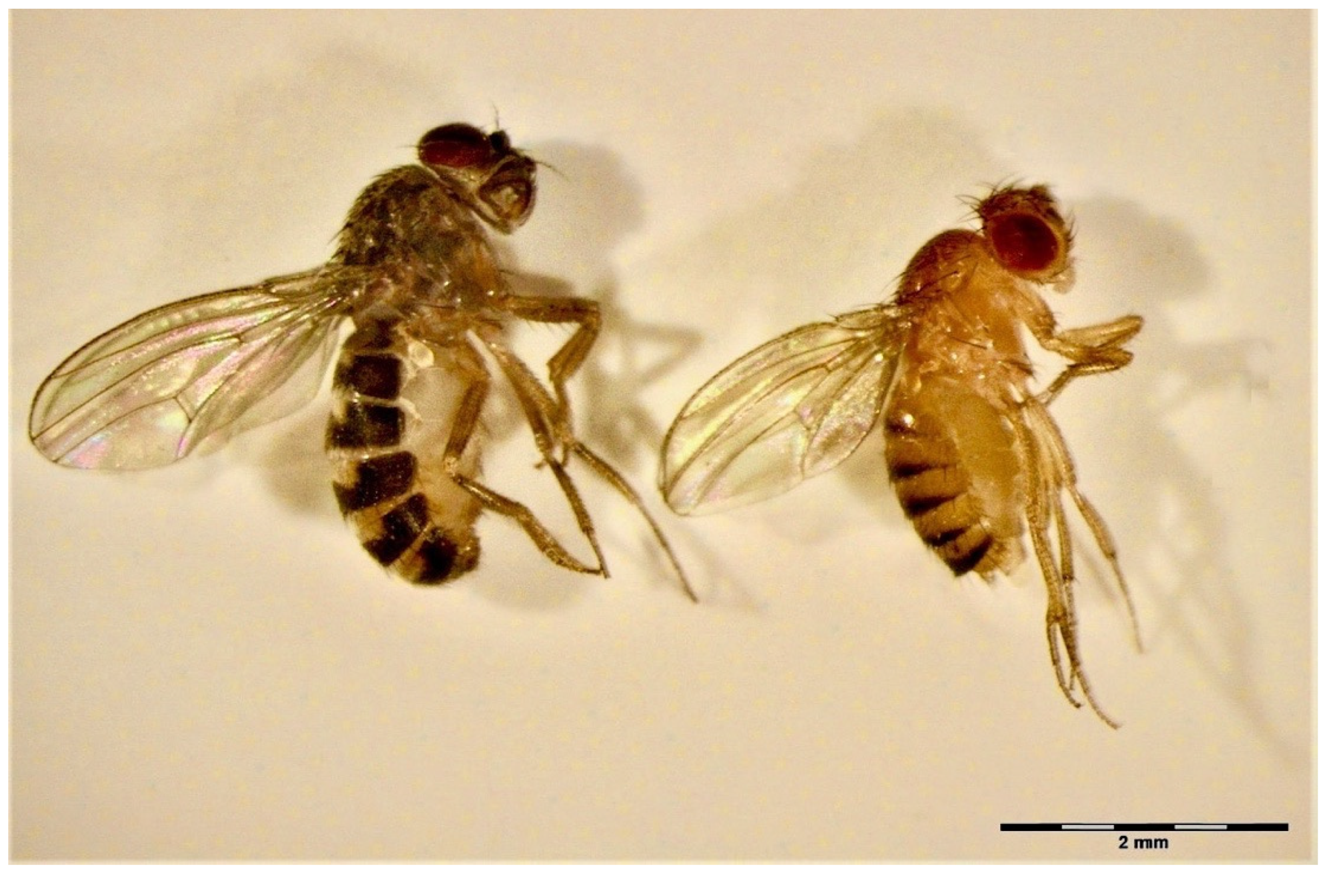
Figure 1. The flightless forms of D. hydei (left) and D. melanogaster (right) used in the experiments.
Insects 14 00146 g001
Figure 2. Percentage of Blattisocius mali females that located on D. melanogaster and D. hydei fruit flies during the first hour of observation using a particular way of getting on the fly’s body. ** 0.001 < p < 0.01.
Insects 14 00146 g002
Figure 3. A female of Blattisocius mali attached to the cervix (neck) of a female of Drosophila hydei (A) and at the coxa III of a female of D. melanogaster (B).
Insects 14 00146 g003
Figure 4. The degree of filling the idiosoma of Blattisocius mali females feeding on fruit flies of (a) Drosophila melanogaster and (b) D. hydei during 24 h observation.
Insects 14 00146 g004
Figure 5. Mortality of Drosophila melanogaster and D. hydei female fruit flies during a 24 h stay with and without Blattisocius mali females in the observation chambers. Number of tested flies in each combination was n = 30. * 0.01 < p ≤ 0.05, *** p < 0.001.
Insects 14 00146 g005
Table 1. Time (mean ± SE, minimum and maximum, n-number of replications) of phases of Blattisocius mali location on the body of Drosophila melanogaster and D. hydei female fruit flies during the first hour of observation. a and b in raw indicate the significant differences between means (p ≤ 0.05).
Fruit Fly SpeciesPhases of the Mite’s Location on the Fly’s Body
1. Attack and First Attachment2. Passage from the Attack Site to the Site of Final Attachment3. Drilling into Fly Integument and Final Attachment
D. melanogaster509.98 ± 86.52 a
(157–2187; n = 25)
249.86 ± 113.50 b
(20–2768; n = 25)
233 ± 81.51 b
(7–2039; n = 25)
D. hydei849.55 ± 269.70 a
(2–2077; n = 11)
83.27 ± 27.07 b
(0–237; n = 11)
269.27 ± 189.42 b
(13–2143; n = 11)
Table 2. Sites of Blattisocius mali attachment to the cervix, thorax, or abdomen of Drosophila melanogaster and D. hydei female fruit flies noted during 24 h observations. For the first hour of observation, only the final attachments (that ended up with drilling) are given.
Fruit Fly SpeciesSite of AttachmentNo of Predators Attached
1st h24 h
D. melanogastercervix, ventral site1013
thorax, dorsal site; at wing joint 1
thorax, at the coxa II 11
thorax, at the coxa III 1317
abdomen, dorsal site—at 5th tergite 11
D. hydeicervix, ventral site25
cervix, lateral site11
thorax, ventral site, between proepisternum and profurcasternum11
thorax, dorsal site; at wing joint 1
junction between thorax and abdomen; dorsal site 12
thorax, at the coxa II 12
thorax, at the coxa III 39
abdomen, ventral site, at 2nd sternite11
abdomen, ventral site, at last sternite 1
abdomen, dorsal site, at 5th tergite 2
abdomen, dorsal site, at 6th tergite 1
abdomen, dorsal site, at last tergite12
Table 3. Number of Blattisocius mali females that drilled their chelicerae into the cervix, at coxa III or other sites on the thorax or abdomen of D. melanogaster and D. hydei according to the site of mite attack.
Site of AttackSite of Drilling and Final Attachment of Chelicerae during 1st Hour of Observation
D. melanogasterD. hydei
CervixCoxa IIIOther SitesCervixCoxa IIIOther Sites
tarsus I71 31
tarsus II131 11
tarsus III27
other body parts 21 14
Disclaimer/Publisher’s Note: The statements, opinions and data contained in all publications are solely those of the individual author(s) and contributor(s) and not of MDPI and/or the editor(s). MDPI and/or the editor(s) disclaim responsibility for any injury to people or property resulting from any ideas, methods, instructions or products referred to in the content.