Article

Development of Enzyme-Linked Immunosorbent and Immunochromatography Assays for Diagnosing Nosema ceranae Infection in Honey Bees

Department of Biology Education, College of Education, Chungbuk National University, Cheongju 28644, Republic of Korea
Insects 2024, 15(1), 59; https://doi.org/10.3390/insects15010059
Submission received: 2 December 2023 / Revised: 7 January 2024 / Accepted: 11 January 2024 / Published: 13 January 2024
(This article belongs to the Section Insect Behavior and Pathology)

Abstract

:

Simple Summary

Nosema ceranae (N. ceranae), a highly aggressive pathogen that affects honeybees, can rapidly spread infection under suboptimal conditions for honeybee breeding, consequently reducing honey production. Preventing and treating Nosema infection remains challenging since symptoms are often manifested in bees only after several million Nosema spores are present in the bee midgut. In this study, specialized methods, namely a sandwich enzyme-linked immunosorbent assay (ELISA) and an immunochromatography assay (ICG), were developed for diagnosing N. ceranae infections, and their effectiveness in diagnosing Nosema infections was evaluated. The sandwich ELISA method effectively detected Nosema infection; comparatively, the ICG approach was less efficient. This comparison between the two diagnostic methods provides insights into their respective strengths and limitations, facilitating future investigations and potential improvements in the development of more effective diagnostic approaches for detecting and managing N. ceranae infections in honeybees.

Abstract

Nosema ceranae (N. ceranae) infection is prevalent globally, causing a decline in bee populations and significant economic losses to apiarists. Although several methods have been proposed for diagnosing Nosema infections, limitations in these methods have hindered their broad applications. Therefore, this current study aimed to develop a specialized method for diagnosing Nosema infections. To achieve this, a sandwich enzyme-linked immunosorbent assay (ELISA) and immunochromatography assay (ICG) were developed, and their effectiveness in screening and diagnosing Nosema infection was assessed. In sandwich ELISA, the combination of the monoclonal antibodies (mAb) 19B2 and biotinylated-19B2 exhibited stronger binding affinity to the antigen than did other combinations of mAbs that were tested. Furthermore, the antigen detection limit achieved with the sandwich ELISA surpassed that previously reported with Western blotting. The ICG was designed using the same antibody combination as that used in sandwich ELISA; however, the assay exhibited a lower diagnostic ability for Nosema infection than the ELISA. The diagnostic models developed in this study offer practical applications for conducting rapid nosemosis detection tests. These innovative techniques will help to improve the timely identification and management of nosemosis.

1. Introduction

Nosema apis and Nosema ceranae are causative agents of nosemosis, one of the most prevalent diseases among honeybees. These unicellular parasites invade the midgut of honeybees, debilitating individual bees and entire colonies, rendering them more susceptible to various pests and diseases [1]. Over the last two decades, N. ceranae has rapidly invaded Apis mellifera populations, establishing a global presence and posing a more substantial threat to honeybee health than that caused by N. apis [2,3,4,5,6]. Moreover, even if the bees are uninfected, millions of N. ceranae spores can persist in beekeeping equipment. Given the ubiquitous presence of this pathogen, infection levels can rapidly escalate under suboptimal honeybee breeding conditions, thereby diminishing honey production and, in severe instances, causing colony collapse.
The primary challenge in preventing and treating Nosema infection is that by the time symptoms are manifested in bees, there are often several million Nosema spores present in the midgut of the bees. Diagnosing nosemosis presents a challenge since distinctive symptoms are absent. While diarrhea is often considered an initial symptom of Nosema infection, it is not exclusive to nosemosis [7]. Therefore, the effect of nosemosis infections may be significantly reduced using specialized diagnostic approaches.
The most frequently employed techniques for diagnosing Nosema infection involve directly observing spores through a microscope and confirming the presence of Nosema genes using a polymerase chain reaction (PCR). The microscope observation method is preferred owing to its simple sample preparation process [8,9]. However, the presence of contaminants, such as tissue fragments, dust, intestinal yeast, and other elements, during spore isolation hinders the achievement of an accurate diagnosis. PCR is a diagnostic technique capable of confirming nosemosis by detecting Nosema spp. genes [3,10]. However, it involves a lengthy procedure for gene isolation and requires specialized experimental procedures and specific equipment, such as a thermocycler [11]. For these reasons, there is a need for more accurate and convenient methods, such as enzyme-linked immunosorbent assay (ELISA), for diagnosing Nosema infections.
A previous study used three monoclonal antibodies (mAbs), namely, 9A4, 14A8, and 19B2 to target Nosema spores [12]. In this current study, we used these antibodies to establish a precise and convenient diagnostic method for N. ceranae-induced nosemosis. Our results demonstrated that sandwich ELISA could effectively detect Nosema infection while immunochromatography (ICG) could not. A sandwich ELISA quantifies the presence of an antigen using a dual-layer of antibodies comprising capture and detection antibodies. The primary benefit of employing a sandwich ELISA lies in its remarkable binding activity to antigens, surpassing that of direct or indirect ELISA. Furthermore, sandwich ELISA has superior specificity since it relies on a pair of antibodies to identify target antigens. The utilization of sandwich ELISA can enable beekeepers to monitor and assess the prevalence of N. ceranae infections in their bee colonies more efficiently. By incorporating this diagnostic tool into routine beekeeping practices, beekeepers can implement timely and targeted interventions, thereby improving the overall health and productivity of their honeybee populations.

2. Materials and Methods

2.1. Isolation of N. ceranae Spores (Preparation of N. ceranae Spore Antigens)

Honeybees infected with N. ceranae were acquired from an apiary in Chungbuk Province, Republic of Korea. Nosema spores were isolated using a previously described method [13]. The honeybee midgut was thoroughly washed with phosphate-buffered saline (PBS) and homogenized using a tissue grinder. A 70-μm mesh filter was utilized to remove large particles. The suspension that passed through the filter was subjected to centrifugation at 250× g and 400× g rcf for 10 min. The isolated spores were identified using specific primers (sense strand: 5′-CGGATAAAAGAGTCCGTTACC-3′, antisense strand: 5′-TGAGCAGGGTTCTAGGGAT-3′, Bioneer Co., Daejeon, Republic of Korea) for N. ceranae, as previously described [9,14,15]. Nosema spore lysates were generated through sonication. To achieve this, Nosema spores were combined with RIPA lysis buffer (50 mM Tris-HCl, 150 mM NaCl, 1% NP-40, 0.5% sodium deoxycholate, and 0.1% sodium dodecyl sulfate (SDS)). The mixture was supplemented with a protease inhibitor cocktail (Sigma, St. Louis, MO, USA), sonicated, and centrifuged at 18,000× g rcf for 10 min.

2.2. Monoclonal Antibody for N. ceranae

The procedure for generating three different N. ceranae-targeting mAbs, specifically 9A4 (IgG1), 14A8 (IgG2b), and 19B2 (IgG3), has been described previously [12]. To obtain substantial quantities of the antibodies (2 mg/mL, total volume 3 mL), hybridoma cells were cultured in Dulbecco’s modified Eagle’s medium (DMEM; Hyclone, Logan, UT, USA) supplemented with 10% fetal bovine serum (FBS; Hyclone Laboratories, Logan, UT, USA), 100 U/mL penicillin (Gibco BRL, Grand Island, NY, USA), and 100 μg/mL streptomycin (Gibco BRL).
The anti-Nosema mAbs were purified by targeting IgG from 20 mL of hybridoma culture supernatants; the final mAb was resuspended in FBS-free DMEM. The process involved using a 5-mL HiTrap desalting column, followed by a 1-mL HiTrap protein G HP column, according to the manufacturer’s instructions. The mAb yield obtained was >2 mg/mL. The purified antibody was confirmed through the presence of 50 and 25 kDa bands on a 12% acrylamide gel via sodium dodecyl sulfate-polyacrylamide gel electrophoresis (SDS-PAGE) under reducing conditions.

2.3. Feasibility Test of Antibody

The reactivity of the mAbs with N. ceranae antigens was assessed following the ELISA protocol described by Anuracpreeda et al. [16]. Briefly, wells were coated with N. ceranae antigen, either in the form of whole spores or spore lysate diluted in a carbonate/bicarbonate coating buffer (15 mM Na2CO3, 35 mM NaHCO3, and pH 9.6). Following a 2 h incubation at 37 °C, the microtiter plate was washed three times with the ELISA buffer (0.05% Tween-20 in PBS). Subsequently, each well was treated with 100 μL of blocking buffer, containing the ELISA buffer and 0.5% bovine serum albumin (Sigma), and incubated for 1 h at 37 °C. After washing, 50 μL of mAb was introduced into the wells and incubated for 2 h at 37 °C. The plate was washed once more, following which 50 μL of horseradish peroxidase (HRP)-conjugated goat anti-mouse IgG (1:10,000 dilution) was added and incubated for an additional 1 h at 37 °C. After washing the plate, 50 μL of 3,3′,5,5′-tetramethyl benzidine (TMB; KPL, Gaithersburg, MD, USA) was added to each well. The process was conducted in the dark for 20–30 min at 25 °C. The reaction was terminated by adding 50 μL of stop solution (1 N HCl), and the optical densities (OD) were measured at 450 nm using a microplate reader (Bio-Rad Laboratories, Hercules, CA, USA).

2.4. Biotinylation of mAbs
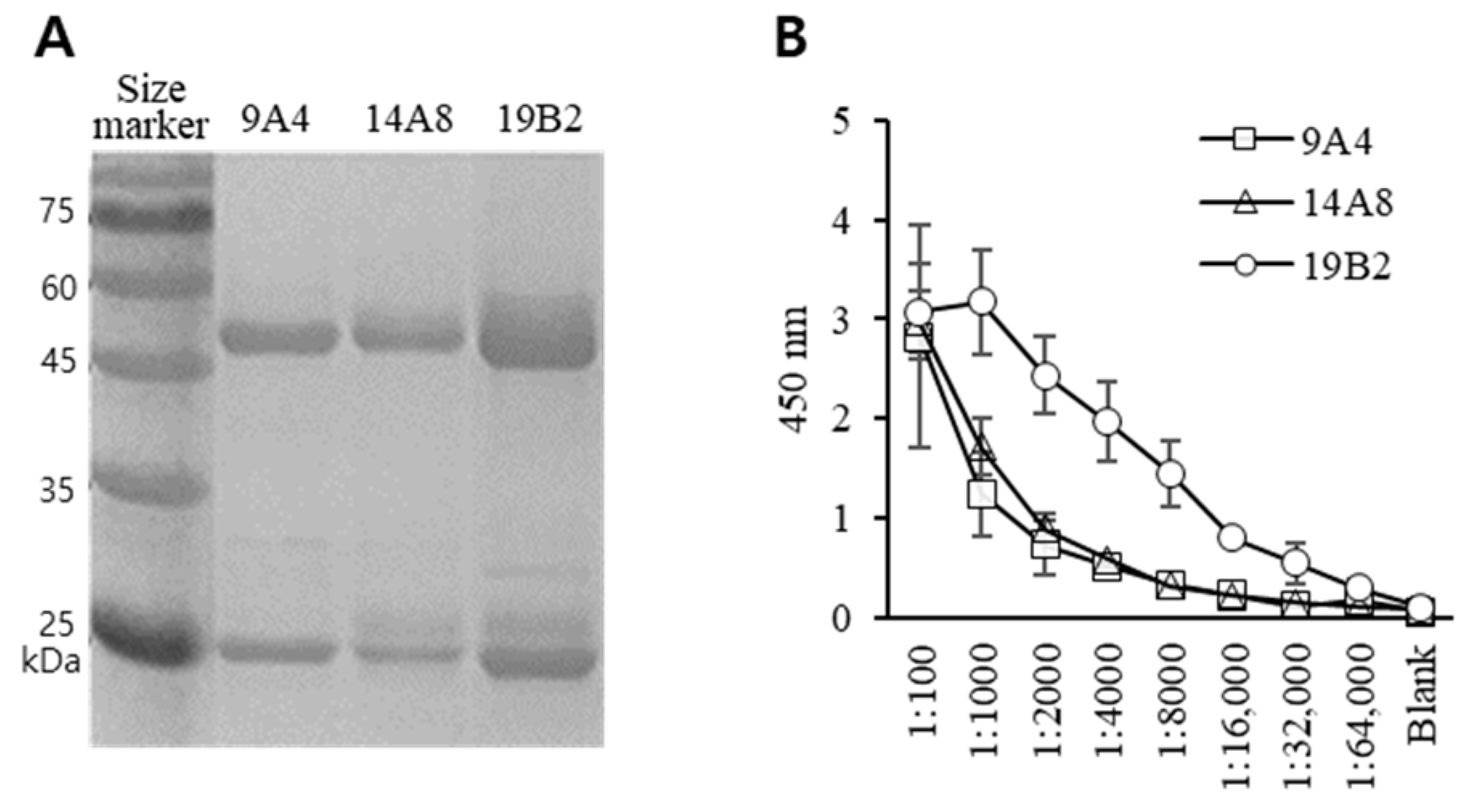
The mAbs were conjugated to biotin molecules using a commercially available antibody biotinylation kit (Abcam, Cambridge, MA, USA), following the manufacturer’s instructions. Briefly, 10 μg of antibody were mixed with a ‘Modifier reagent’ at a 10:1 ratio and introduced to the lyophilized Biotin Conjugation Mix, followed by suspension. After incubating for >15 min in the dark at 25 °C, one-tenth of the volume of the quencher reagent was incorporated. The biotinylated mAbs were used without purification.

2.5. SDS-PAGE

SDS-PAGE was performed using the Laemmli method, as previously described [17]. Proteins were separated on 12% or 15% acrylamide gels, and subsequently, the gel was stained with Coomassie Brilliant Blue (Bio-Rad Laboratories).

2.6. Sandwich ELISA

The capture antibody was diluted to a final concentration of 1–5 μg/mL using carbonate/bicarbonate buffer (pH 7.4), from which 100 µL was added to each well of a microplate (MaxiSorp, Waltham, MA, USA). After overnight incubation at 4 °C, the plate was washed and blocked with 3% skim milk solution. Following a 2 h incubation at 4 °C, the plate was washed, and the diluted samples were introduced using a blocking buffer. After a 2 h incubation at 25 °C, the plate was washed again, and 100 µL of the biotinylated detection antibody (1 μg/mL) was added and incubated for 2 h at 25 °C. Thereafter, the plate was washed again, and a 1:5000 diluted avidin-HRP conjugate was added. After a 30 min incubation at 25 °C, the plate was washed, and 50 μL of TMB solution was added. The reaction was performed in the dark for 20–30 min at 25 °C and terminated by the addition of 50 μL stop solution (1 N HCl). Subsequently, the OD was measured at 450 nm using a microplate reader.

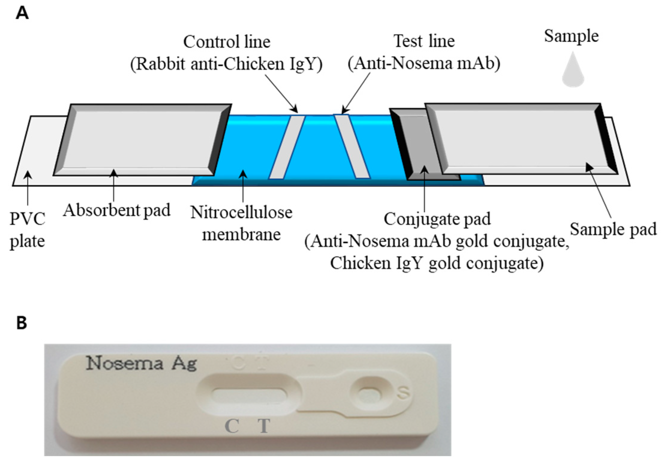
2.7. Immunochromatography (ICG) to Diagnose N. ceranae Infection

The ICG strip used for diagnosing Nosema infection was produced in collaboration with VetAll Laboratories (Goyang-si, Gyeonggi-do, Republic of Korea). Briefly, to conjugate the antibody with gold particles, the 9A4, 14A8, and 19B2 mAbs were mixed with a colloidal gold solution (Abcam) at varying concentrations (6–20 μg/mL) and with a pH range of 6–9; the pH was adjusted by adding 0.1 M K2CO3. Subsequently, the mixture was subjected to centrifugation at 15,000× g for 30 min to remove uncoordinated proteins. 9A4, 14A8, and 19B2 exhibited the most effective conjugation with the colloidal gold at 8 μg/mL (pH 7), 10 μg/mL (pH 6), and 10 μg/mL (pH 6), respectively. A gold-conjugated antibody (anti-Nosema mAb gold conjugate or chicken IgY gold conjugate) with an OD value of 3 was evenly distributed on a segment of the conjugate pad. Purified anti-Nosema mAb and rabbit anti-chicken IgY were then applied to nitrocellulose membranes (Immunopore membrane; Whatman, Maidstone, UK) to create the test and control lines, respectively (Figure 1A). The prepared sample pad and conjugate pad, nitrocellulose membrane, and absorbent pad were successively affixed to a PVC plate. Figure 1B illustrates the complete Nosema Antigen Test Kit.

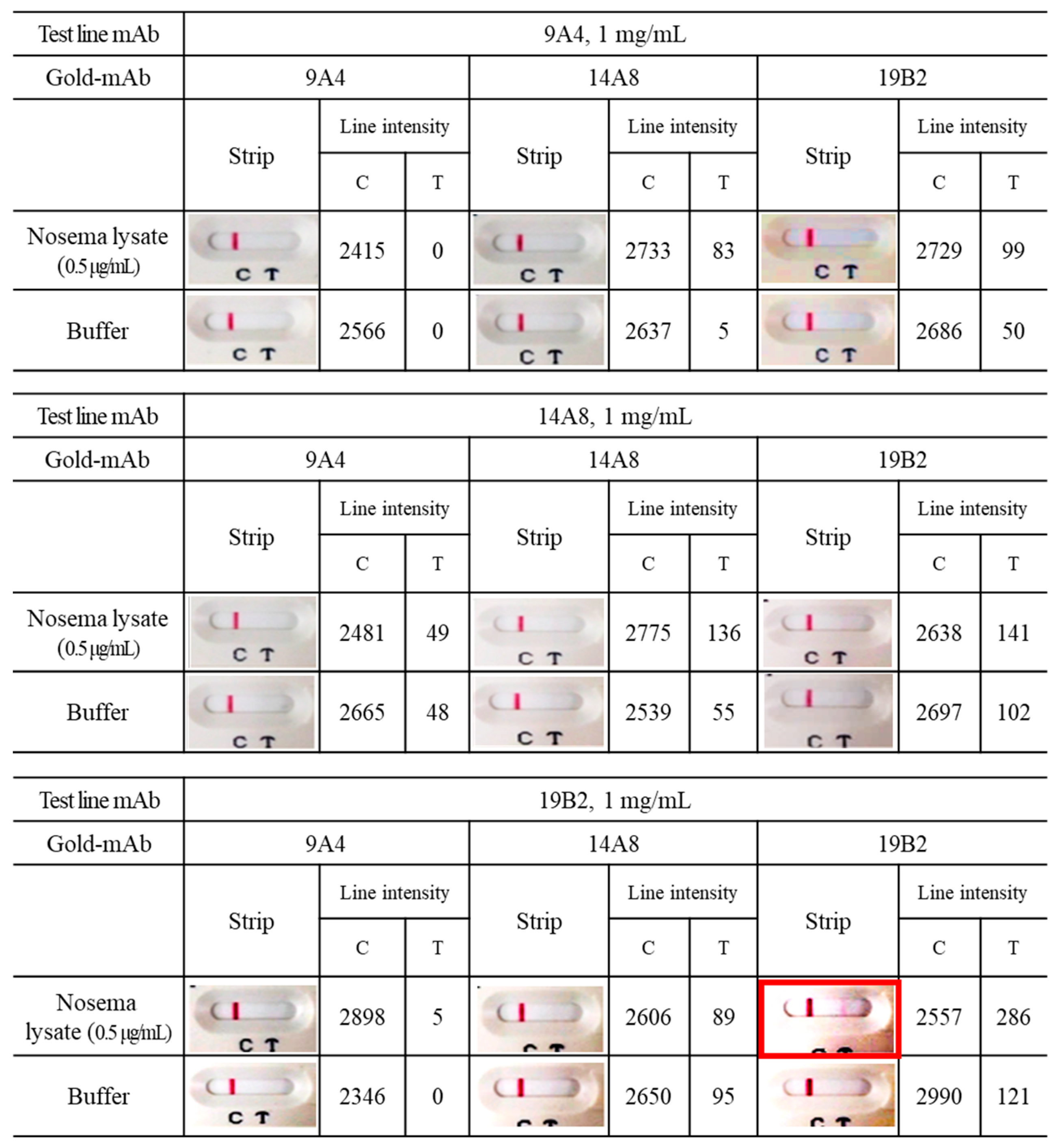
2.8. Pairing test of Colloidal Gold Strip

To assess the activity of the colloidal gold test strip for Nosema infection, 9A4, 14A8, and 19B2 and three different gold conjugated mAbs (9A4, 14A8, and 19B2) were tested using the strips. The diagnostic activity of these test strips was determined based on the test line intensity values. The intensities of the control and test lines were quantified using the Image J Gel Analysis program.

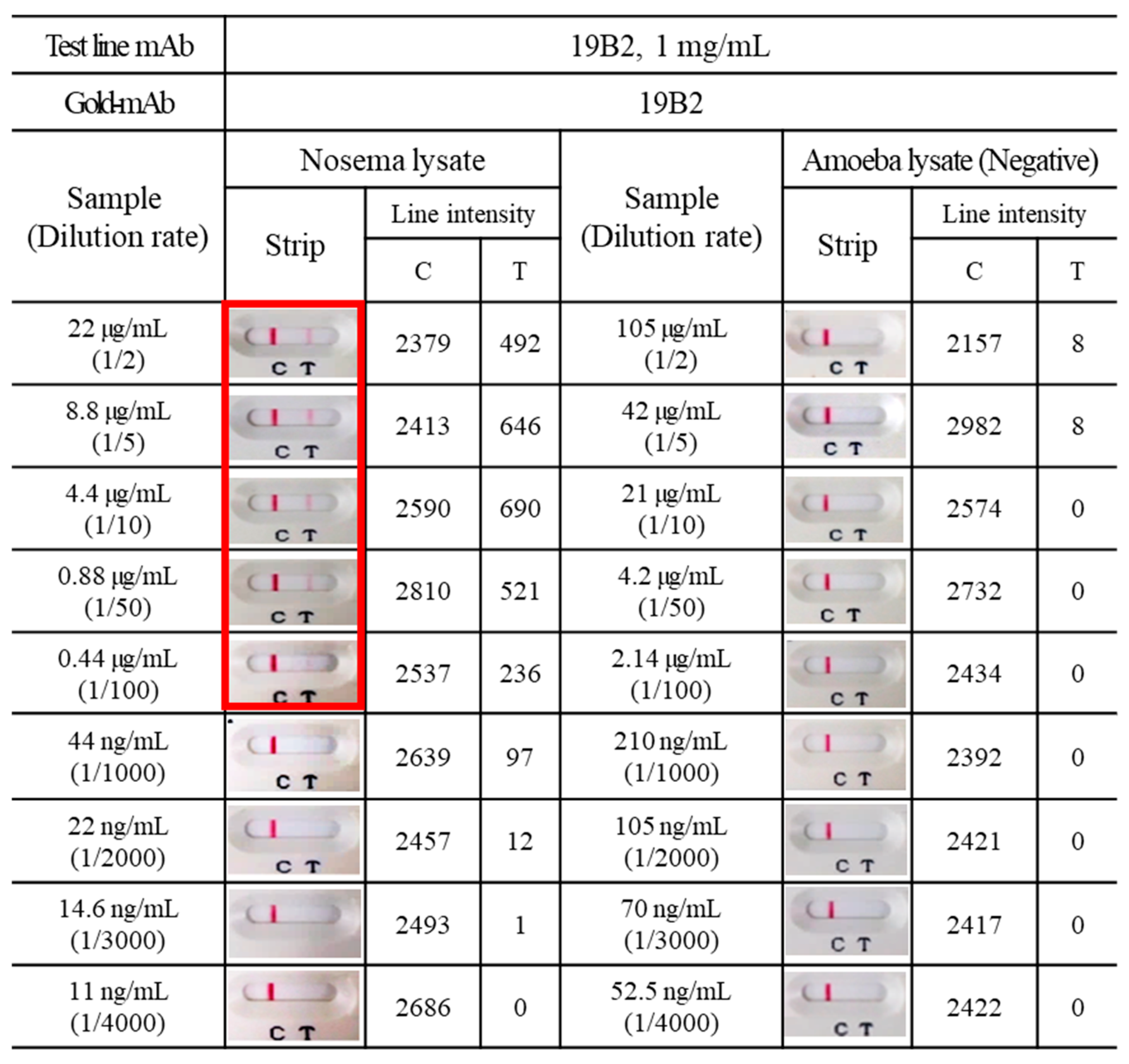
2.9. Detection of Nosema Spores in Experimental Specimens

To assess ICG’s feasibility, honeybees were divided into normal and weak groups, with Nosema infection being confirmed through microscopic observation. To prepare the test samples, we followed the spore isolation procedure previously described [13]. Briefly, Nosema spores were isolated by rinsing the honeybee midgut with PBS and homogenizing it with a tissue grinder. To remove large particles, a 70 µm mesh filter was utilized. Subsequently, the filtered suspension underwent centrifugation at 250× g and 400× g rcf for 10 min each. The separated spores, suspended in the diluent buffer supplied by the kit manufacturer (VetAll Laboratories), were homogenized using a homogenizer pestle (SciLab, Seoul, Republic of Korea). Subsequently, three drops of the diluted sample were dispensed into the circular window of the “Nosema antigen test kit.” Following the manufacturer’s guidelines, the test reaction was left to stand for 10 min, following which a visual assessment was conducted. The appearance of two red lines, which denoted the control (C) and test (T), was regarded as a positive result, indicating the presence of Nosema spore antigens in the sample. In contrast, if only the C line appeared, the result was considered negative; however, if line C did not appear, it was regarded as invalid.

2.10. Statistical Analysis

Data are expressed as means ± standard deviation. SPSS (version 17.0; SPSS Inc., Chicago, IL, USA) was used for data analysis.

3. Results

3.1. Generation of Hybridoma and Characterization of mAbs

To obtain a substantial quantity of antibodies, hybridoma cells were cultured in vitro in DMEM containing 10% FBS. mAbs were obtained from the cell culture supernatants and utilized in experimental diagnostic tests. The mAbs 9A4, 14A8, and 19B2 comprised heavy and light chain fragments of approximately 50 kDa and 25 kDa, respectively (Figure 2A), consistent with the pattern typically associated with an IgG subclass.

3.2. Feasibility Test of Antibody

To demonstrate the binding activity of 9A4, 14A8, and 19B2, spore lysates at a concentration of 10 μg/mL were assessed, adhering to the ELISA protocol with mAb serial dilution. The three mAbs showed reactivity with spore lysate samples, particularly at a high concentration (1:100 dilution of 2 mg/mL purified antibody solution). When diluted to 1:2000, 19B2 exhibited a reactivity of approximately 76% compared with the highest response level (Figure 2B). Moreover, 19B2 demonstrated the highest response when Nosema lysate was diluted to 1:1000. Conversely, 9A4 and 14A8 exhibited reactivity of approximately 44% and 58%, respectively, when exposed to Nosema lysate diluted at 1:1000, compared with the highest response level.

3.3. Effect of mAb Biotinylation on Antibody Binding to Nosema Spore

To enhance the detection activity of mAb for Nosema spores, each mAb was conjugated with biotin. As shown in Table 1, both 14A8 and 19B2, irrespective of biotinylation, exhibited strong reactivity with the Nosema lysate in a concentration-dependent manner. Among the mAbs, both before and after biotinylation, 14A8 exhibited the most potent binding response. Furthermore, biotinylated-19B2 (average OD: 1.07) demonstrated a significant improvement in reactivity against the Nosema lysate than the unbiotinylated-19B2 (average OD: 0.25); a consistent pattern was observed when reacting with whole Nosema spores (Table 2). However, the overall reactivity of mAbs toward whole Nosema spores tended to be slightly lower than that toward the Nosema lysate, because a whole spore cannot adequately expose antigen epitopes.

3.4. Sandwich ELISA for Diagnosis of Nosema Infection

To establish a sandwich ELISA method, pairing tests were conducted using biotinylated and unbiotinylated mAbs. The capture antibody (i.e., unbiotinylated mAb) was coated onto microplates at a concentration of 1 μg/mL; subsequently, Nosema spore lysate at concentrations of 250 and 1000 ng/mL was added and incubated for 2 h, following which a biotinylated detection antibody (1 μg/mL) was added. The paring test (Table 3) findings revealed that Nosema lysate at a concentration of ≤ 1 μg /mL could not be detected under the tested conditions (1 μg /mL capture antibody and 1 μg/mL detection antibody). Therefore, in the subsequent experiment (Table 4), a pairing test was conducted by increasing the concentrations of the capture antibody and Nosema lysate. In the ELISA method, conducted with the concentration of the capture antibody set to 5 μg/mL and the Nosema lysate to 10,000 ng/mL, the combinations of “19B2 and biotinylated-19B2” (average OD: 0.50) and “19B2 and biotinylated-14A8” (average OD: 0.39) demonstrated meaningful binding activity against low concentration Nosema lysate (156.3 ng/mL). The “19B2 and biotinylated-19B2” combination demonstrated superior binding activity to the Nosema lysate of 1000 ng/mL (average OD: 3.05) than did the other tested combinations.

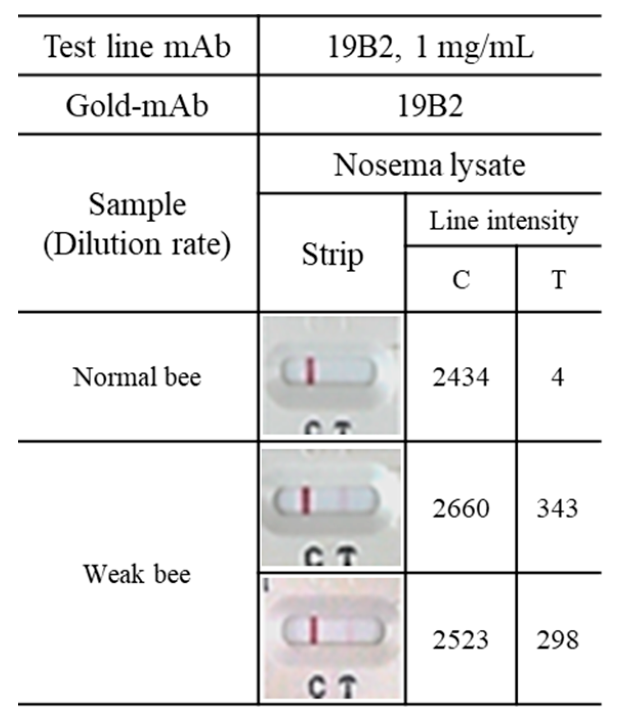
3.5. Application of mAbs to ICG

An ICG was developed for the rapid detection of Nosema infection with the same antibodies (i.e., 9A4, 14A8, and 19B2) used in the sandwich ELISA method. Following the labeling of 9A4, 14A8, and 19B2 with colloidal gold, the binding activity was assessed using the “colloidal gold test strip” to detect Nosema infection. To determine the most effective combination, each gold-conjugated mAb was paired with 9A4, 14A8, and 19B2. The combination of gold-conjugated 19B2 and 19B2 exhibited a stronger reaction with Nosema lysate (0.5 μg/mL) than did other mAb combinations, revealing the most distinct band with an intensity of 286 (Figure 3). Therefore, the 19B2 antibody pair, serving as the capture and gold-conjugated antibody, was selected for the colloidal gold test strip.
Next, to establish the detection limit of Nosema infection using ICG, the presence of Nosema at various sample concentrations (Nosema lysate) was explored. Test strips that contained the gold conjugate-19B2 and 19B2 combination were utilized. Distinct bands corresponding to Nosema lysates were detected in samples with a total protein concentration of up to 440 ng/mL. Amoeba (Malpighamoeba mellificae) is a microorganism found in the honeybee midgut and shares similarities with Nosema spores (Figure 4). Therefore, amoeba lysates were used as analogous but distinct antigens to Nosema spp. The ICG designed for Nosema did not detect the amoeba lysate, even at its highest concentration.
Figure 5 shows the results of Nosema infection testing in bees collected from an apiary in Chungbuk Province. Weak bees exhibited discernible red lines in the Nosema diagnostic kit test, and laboratory microscopy revealed that each weak bee had approximately 3 × 10⁶ Nosema spores.

4. Discussion

Nosemosis remains a major threat to honeybee populations, and beekeepers frequently struggle to achieve accurate diagnoses before administering anti-Nosema medications [3,7]. The diagnosis of Nosema spores serves a dual purpose: safeguarding honeybee colonies against nosemosis and preventing and controlling the spread of diseases. Therefore, nosemosis detection methods must uphold the highest standards of accuracy to prevent false-negative and false-positive results. We have previously created mAbs for diagnosing N. ceranae infections [12] by employing the antibody production technique developed by Kohler and Milstein [18]. Approximately 985 hybridomas were screened, of which three monoclones, namely 9A4, 14A8, and 19B2, demonstrated a robust reaction with the spore antigen of N. ceranae. This method significantly enhanced targeting of Nosema spores. These three mAbs facilitated the identification of N. ceranae spore lysate with a concentration level > 1 μg using Western blotting.
Herein, we assessed the efficacy of 9A4, 14A8, and 19B2 in targeting N. ceranae spores using a sandwich ELISA approach. An ELISA technique utilizing mAbs for detecting Nosema bombycis (N. bombycis) spores has been previously developed [19,20]; however, this method has shown significant limitations in its sensitivity for detecting antigens. Kawarabata and Hayasaka [21] have also developed a highly sensitive ELISA method capable of detecting as little as 600 ng/mL of soluble protein from the lysate of N. bombycis spores. Moreover, Aronstein et al. [22] created polyclonal antibodies based on a single protein (SWP-32; spore wall protein of N. bombycis) and utilized them in an ELISA approach. The advantage of employing the polyclonal antibody targeting SWP-32 in Western blotting lies in its capacity to specifically detect a single Nosema antigen and the shorter sample preparation time required than that required for Nosema detection using PCR. Addressing the limitations associated with polyclonal antibodies is challenging; therefore, in this study, we developed a sandwich ELISA method using biotinylated mAbs to enhance antigen detection activity. Moreover, self-sandwiching antibody pairs (19B2 and biotinylated-19B2, and 14A8 and biotinylated-14A8) showed potent sensitivities. Self-sandwiching antibody pairs can be employed when the target antigen possesses sufficient size to present multiple binding sites or epitopes at appropriate intervals, facilitating effective capture and detection. The self-sandwich method is better suited for cases where the amount of antigen is small [23]. Our findings revealed that the sandwich ELISA method allowed for detection at a minimum concentration of 156 ng/mL, surpassing previously reported detection limits [9].
ICG is commonly employed for early identification and initial screening of infectious diseases before conducting confirmatory real-time PCR tests [24,25]. Antigen tests provide rapid results and are user-friendly, allowing for extensive and swift screening of potential infections on a large scale. Real-time PCR is the most commonly used method for detecting Nosema infections. Although this method offers precise data regarding the infection status, the sample preparation process is time-consuming and complicated. In our previous study, mAbs were developed for Western blotting assays. The sample preparation procedures for these experimental techniques are simpler and faster than those for real-time PCR; however, they require longer overall experimental time and show lower accuracy. The ICG method introduced in this study for Nosema spore detection offers a relatively shorter total experimental time, including sample preparation, than that of Western blotting and ELISA methods. The minimum detection concentration of the ICG was 440 ng/mL, which was higher than that using the sandwich ELISA method (156 ng/mL) in this study. While the diagnostic activity of ICG for Nosema infection may be lower than that of ELISA, it offers the distinct advantage of being faster and more convenient. To date, no attempts have been made to develop an ICG for diagnosing Nosema.
This study has some limitations. The ICG utilized in this study was prepared using antibodies initially developed for Western blot analysis; therefore, whether they are suitable for achieving optimal ICG results is not definitive. While Western blotting and ICG rely on antigen-antibody interactions for analysis, the procedures for antigen detection and signal generator (i.e., gold colloid) are distinct. In addition to the binding activity of antibodies against antigen, factors that may influence the potential of false results in ICG include sample diluent composition, protein extraction method used for sample preparation, inadequate specimen collection, logistical and storage conditions of the test, and potential laboratory errors [26,27]. Therefore, to design an ICG with optimal performance, selecting the most suitable antibody combination from a wide array of antibodies is imperative. Further studies are warranted to determine the optimal sample diluent composition and experimental methods.

5. Conclusions

The results from the sandwich ELISA demonstrated that combining “19B2 and biotinylated-19B2” exhibited the highest binding activity against antigens. Furthermore, the detection limit of the sandwich ELISA surpassed those previously reported using Western blotting. The establishment of a sandwich ELISA method for the diagnosis of Nosema infection is expected to be more precise and reliable than conventional diagnostic methods that rely on microscopic observation. Herein, the sandwich ELISA was superior to ICG, which was designed using the same antibody combination as that used for the sandwich ELISA in diagnosing Nosema infection, necessitating further research to optimize the performance of the ICG. The pioneering diagnostic strategies presented in this study will help facilitate the efficient, reliable, and rapid detection of nosemosis, promoting the timely identification and comprehensive management of nosemosis outbreaks.

Funding

This research was supported by “Regional Innovation Strategy (RIS)” through the National Research Foundation of Korea (NRF) funded by the Ministry of Education (MOE)(2021RIS-001).

Data Availability Statement

The data presented in this study are available on request from the corresponding author. The data are not publicly available due to the excessive data size.

Acknowledgments

The author would like to thank Rangachari Balamurugan for his helpful advice on various technical issues examined in this paper.

Conflicts of Interest

The author declares no conflicts of interest.

References

  1. Fries, I.; Feng, F.; Dasilva, A.; Slemenda, S.B.; Pieniazek, N.J. Nosema ceranae n sp (Microspora, Nosematidae), morphological and molecular characterization of a microsporidian parasite of the Asian honey bee Apis cerana (Hymenoptera, Apidae). Eur. J. Protistol. 1996, 32, 356–365. [Google Scholar] [CrossRef]
  2. Higes, M.; Martin-Hernandez, R.; Botias, C.; Bailon, E.G.; Gonzalez-Porto, A.V.; Barrios, L.; Del Nozal, M.J.; Bernal, J.L.; Jimenez, J.J.; Palencia, P.G.; et al. How natural infection by Nosema ceranae causes honeybee colony collapse. Environ. Microbiol. 2008, 10, 2659–2669. [Google Scholar] [CrossRef]
  3. Klee, J.; Besana, A.M.; Genersch, E.; Gisder, S.; Nanetti, A.; Tam, D.Q.; Chinh, T.X.; Puerta, F.; Ruz, J.M.; Kryger, P.; et al. Widespread dispersal of the microsporidian Nosema ceranae, an emergent pathogen of the western honey bee, Apis mellifera. J. Invertebr. Pathol. 2007, 96, 1–10. [Google Scholar] [CrossRef]
  4. Paxton, R.J.; Klee, J.; Korpela, S.; Fries, I. Nosema ceranae has infected Apis mellifera in Europe since at least 1998 and may be more virulent than Nosema apis. Apidologie 2007, 38, 558–565. [Google Scholar] [CrossRef]
  5. Fries, I. Nosema ceranae in European honey bees (Apis mellifera). J. Invertebr. Pathol. 2010, 103, S73–S79. [Google Scholar] [CrossRef] [PubMed]
  6. Martín-Hernández, R.; Botías, C.; Bailón, E.G.; Martínez-Salvador, A.; Prieto, L.; Meana, A.; Higes, M. Microsporidia infecting Apis mellifera: Coexistence or competition. Is Nosema ceranae replacing Nosema apis? Environ. Microbiol. 2012, 14, 2127–2138. [Google Scholar] [CrossRef] [PubMed]
  7. Michalczyk, M.; Sokot, R. Nosemosis in honey bees. Pol. J. Nat. Sci. 2014, 29, 91–99. [Google Scholar]
  8. Cantwell, G.E. Standard methods for counting nosema spores. Am. Bee J. 1970, 110, 222–223. [Google Scholar]
  9. Kim, J.H.; Park, J.K.; Lee, J.K. Evaluation of antimicrosporidian activity of plant extracts on Nosema ceranae. J. Apic. Sci. 2016, 60, 167–178. [Google Scholar] [CrossRef]
  10. Klee, J.; Tek Tay, W.; Paxton, R.J. Specific and sensitive detection of Nosema bombi (Microsporidia: Nosematidae) in bumble bees (Bombus spp.; Hymenoptera: Apidae) by PCR of partial rRNA gene sequences. J. Invertebr. Pathol. 2006, 91, 98–104. [Google Scholar] [CrossRef]
  11. Carletto, J.; Blanchard, P.; Gauthier, A.; Schurr, F.; Chauzat, M.P.; Ribiere, M. Improving molecular discrimination of Nosema apis and Nosema ceranae. J. Invertebr. Pathol. 2013, 113, 52–55. [Google Scholar] [CrossRef] [PubMed]
  12. Kim, D.Y.; Lee, J.K. Development of monoclonal antibodies against spores of Nosema ceranae for the diagnosis of nosemosis. J. Apic. Res. 2021, 2022, 1–8. [Google Scholar] [CrossRef]
  13. Gisder, S.; Mockel, N.; Linde, A.; Genersch, E. A cell culture model for Nosema ceranae and Nosema apis allows new insights into the life cycle of these important honey bee-pathogenic microsporidia. Environ. Microbiol. 2011, 13, 404–413. [Google Scholar] [CrossRef] [PubMed]
  14. Gisder, S.; Hedtke, K.; Mockel, N.; Frielitz, M.C.; Linde, A.; Genersch, E. Five-year cohort study of Nosema spp. in Germany: Does climate shape virulence and assertiveness of Nosema ceranae? Appl. Environ. Microbiol. 2010, 76, 3032–3038. [Google Scholar] [CrossRef] [PubMed]
  15. Genersch, E.; Evans, J.D.; Fries, I. Honey bee disease overview. J. Invertebr. Pathol. 2010, 103 (Suppl. 1), S2–S4. [Google Scholar] [CrossRef] [PubMed]
  16. Anuracpreeda, P.; Wanichanon, C.; Chawengkirtikul, R.; Chaithirayanon, K.; Sobhon, P. Fasciola gigantica: Immunodiagnosis of fasciolosis by detection of circulating 28.5 kDa tegumental antigen. Exp. Parasitol. 2009, 123, 334–340. [Google Scholar] [CrossRef]
  17. Laemmli, U.K. Cleavage of structural proteins during the assembly of the head of bacteriophage T4. Nautre 1970, 15, 680–685. [Google Scholar] [CrossRef]
  18. Kohler, G.; Milstein, C. Continuous cultures of fused cells secreting antibody of predefined specificity. Nature 1975, 256, 495–497. [Google Scholar] [CrossRef]
  19. Qian, Y.; Guo, X.; Hu, X. Distinguishing the Pebrine spores N. bombycis, of the mulberry silkworm by immunoenzymatic technique. Sci. Seric. 1986, 72, 183–184. [Google Scholar]
  20. Greenstone, M.H. An Enzyme-Linked Immunosorbent-Assay for the Amblyospora Sp of Culex-Salinarius (Microspora, Amblyosporidae). J. Invertebr. Pathol. 1983, 41, 250–255. [Google Scholar] [CrossRef]
  21. Kawarabata, T.; Hayasaka, S. An Enzyme-Linked-Immunosorbent-Assay to Detect Alkali-Soluble Spore Surface-Antigens of Strains of Nosema-Bombycis (Microspora, Nosematidae). J. Invertebr. Pathol. 1978, 50, 118–123. [Google Scholar] [CrossRef]
  22. Aronstein, K.A.; Webster, T.C.; Saldivar, E. A serological method for detection of Nosema ceranae. J. Appl. Microbiol. 2013, 114, 621–625. [Google Scholar] [CrossRef] [PubMed]
  23. Hsu, S.-M.; Ree, H.J. Self-sandwich Method. An Improved Immunoperoxidase Technic for the Detection of Small Amounts of Antigens. Am. J. Clin. Pathol. 1980, 74, 32–40. [Google Scholar] [CrossRef] [PubMed]
  24. Mertens, P.; De Vos, N.; Martiny, D.; Jassoy, C.; Mirazimi, A.; Cuypers, L.; Van den Wijngaert, S.; Monteil, V.; Melin, P.; Stoffels, K.; et al. Development and potential usefulness of the COVID-19 Ag Respi-Strip diagnostic assay in a pandemic context. Front. Med. 2020, 7, 225. [Google Scholar] [CrossRef]
  25. Korenkov, M.; Poopalasingam, N.; Madler, M.; Vanshylla, K.; Eggeling, R.; Wirtz, M.; Fish, I.; Dewald, F.; Gieselmann, L.; Lehmann, C.; et al. Evaluation of a rapid antigen test to detect SARS-CoV-2 infection and identify potentially infectious individuals. J. Clin. Microbiol. 2021, 59, e0089621. [Google Scholar] [CrossRef]
  26. Lippi, G.; Simundic, A.-M.; Plebani, M. Potential preanalytical and analytical vulnerabilities in the laboratory diagnosis of coronavirus disease 2019 (COVID-19). Clin. Chem. Lab. Med. 2020, 58, 1070–1076. [Google Scholar] [CrossRef]
  27. Richard-Greenblatt, M.; Ziegler, M.J.; Bromberg, V.; Huang, E.; Abdallah, H.; Tolomeo, P.; Lautenbach, E.; Glaser, L.; Kelly, B.J. Impact of nasopharyngeal specimen quality on SARS-CoV-2 test sensitivity. Med. Rxiv. 2020; preprint. [Google Scholar]
Figure 1. Schematic diagram of the immunochromatography (ICG) strip. (A) The colloidal gold test strip comprised three pads (sample, conjugate, and absorbent), a nitrocellulose membrane, and a PVC plate. The conjugate pad contained anti-Nosema mAb gold and chicken IgY gold conjugates, manifesting as a distinct red color. The nitrocellulose membrane displayed two lines, namely the control and test. The control and test lines comprised rabbit anti-chicken IgY mAb and mouse anti-Nosema mAb, respectively. (B) The fully packed strips enclosed within cassettes.
Insects 15 00059 g001
Figure 2. Confirmation of immunoglobulin proteins and monoclonal antibody (mAb) suitability. (A) Analysis of three distinct mAbs (9A4, 14A8, and 19B2) using SDS-PAGE with 12% acrylamide gel. Each acrylamide gel was subsequently stained with Coomassie brilliant blue. (B) Purified immunoglobulins were diluted serially and evaluated as primary antibodies, with HRP-conjugated anti-mouse IgG (1:10,000 dilution) utilized as the secondary antibody. All experiments were repeated more than thrice, and representative data are presented. Values are presented as mean ± standard deviation (n = 3).
Insects 15 00059 g002
Figure 3. Selection of the optimal combination between gold-conjugated and unconjugated antibodies for ICG establishment. Each anti-Nosema mAb was reacted with three different gold-conjugated anti-Nosema mAbs (Gold-mAb). The diagnostic activity of the test strip was estimated based on the corresponding band intensity values (C: control line, T: test line). All experiments were repeated more than thrice, and data from one representative experiment are presented.
Insects 15 00059 g003
Figure 4. Limit of detection for the ICG strip using N. ceranae spore lysate. The ICG strip comprises a conjugate pad coated with gold-19B2 and a test line coated with 19B2 (1 mg/mL). The ICG results were obtained by applying various Nosema lysate concentrations (11–22,000 ng/mL) or Amoeba lysate (52.5–105,000 ng/mL) on the ICG strip. The intensities of the control and test lines were analyzed using ImageJ. All experiments were repeated more than thrice, and data from one representative experiment are presented.
Insects 15 00059 g004
Figure 5. Detection of Nosema spore in experimental specimens.
Insects 15 00059 g005
Table 1. Effect of biotinylation on mAb binding activity for Nosema spore lysate.
Coated AntigenNosema Spore Lysate (1 μg/mL)
Primary Antibody9A414A819B29A4-biotin14A8-biotin19B2-biotin
Antibody
Concentration
(ng/mL)
10000.17 ± 0.015 11.99 ± 0.0280.25 ± 0.010.27 ± 0.0271.75 ± 0.2961.07 ± 0.048
1000.11 ± 0.0050.33 ± 0.0230.11 ± 0.0260.24 ± 0.0260.36 ± 0.0470.34 ± 0.016
100.11 ± 0.0020.12 ± 0.0020.09 ± 0.0150.23 ± 0.0150.26 ± 0.0350.23 ± 0.012
00.11 ± 0.0060.09 ± 0.0080.09 ± 0.0120.22 ± 0.0120.23 ± 0.0150.25 ± 0.036
2nd AntibodyAnti-mouse IgG Fc-HRP (1:10,000)Avidin-HRP (1:5000)
1 All experiments were repeated more than thrice. Values are presented as means ± standard deviation (n = 3).
Table 2. Effect of biotinylation on mAb binding activity for Nosema whole spore.
Coated AntigenNosema Spore (1 × 10⁵ Cells/mL)
Primary Antibody9A414A819B29A4-biotin14A8-biotin19B2-biotin
Antibody
Concentration
(ng/mL)
10000.12 ± 0.011 10.37 ± 0.0370.14 ± 0.0070.22 ± 0.0240.51 ± 0.0210.42 ± 0.036
1000.1 ± 0.0120.11 ± 0.0070.08 ± 0.0030.19 ± 0.0170.25 ± 0.0330.25 ± 0.015
100.08 ± 0.0010.08 ± 0.0040.07 ± 0.0060.18 ± 0.0110.22 ± 0.0130.26 ± 0.03
00.08 ± 0.0030.09 ± 0.0080.08 ± 0.0040.21 ± 0.0130.2 ± 0.0030.19 ± 0.002
2nd AntibodyAnti-mouse IgG Fc-HRP (1:10,000)Avidin-HRP (1:5000)
1 All experiments were repeated more than thrice. Values are presented as means ± standard deviation (n = 3).
Table 3. Detection of Nosema antigen (250 and 1000 ng/mL) using a combination of biotinylated and unbiotinylated mAbs (1 μg/mL) in sandwich ELISA.
Capture Antibody9A4 Antibody (1 μg/mL)
Detector Antibody9A4-biotin (1 μg/mL)14A8-biotin (1 μg/mL)19B2-biotin (1 μg/mL)
Nosema spore
lysate (ng/mL)
10000.20 ± 0.028 10.18 ± 0.0060.22 ± 0.027
2500.18 ± 0.0170.16 ± 0.0030.20 ± 0.022
Capture Antibody14A8 Antibody (1 μg/mL)
Detector Antibody9A4-biotin (1 μg/mL)14A8-biotin (1 μg/mL)19B2-biotin (1 μg/mL)
Nosema spore
lysate (ng/mL)
10000.20 ± 0.0160.22 ± 0.0200.19 ± 0.006
2500.18 ± 0.0160.19 ± 0.0080.18 ± 0.008
Capture Antibody19B2 Antibody (1 μg/mL)
Detector Antibody9A4-biotin (1 μg/mL)14A8-biotin (1 μg/mL)19B2-biotin (1 μg/mL)
Nosema spore
lysate (ng/mL)
10000.18 ± 0.0050.31 ± 0.0960.21 ± 0.004
2500.15 ± 0.0110.15 ± 0.0060.18 ± 0.014
1 All experiments were repeated more than thrice. Values are presented as means ± standard deviation (n = 3).
Table 4. Detection of Nosema antigen (up to 10,000 ng/mL) using a combination of biotinylated and unbiotinylated mAbs (5 μg/mL) in sandwich ELISA.
Capture Antibody9A4 (5 μg/mL)
Detector Antibody9A4-biotin (1 μg/mL)14A8-biotin (1 μg/mL)19B2-biotin (1 μg/mL)
Nosema spore
Lysate (ng/mL)
10,0000.25 ± 0.029 10.39 ± 0.0370.49 ± 0.011
25000.24 ± 0.0100.31 ± 0.0200.31 ± 0.015
6250.24 ± 0.0180.29 ± 0.0160.26 ± 0.011
156.30.22 ± 0.0120.29 ± 0.0320.24 ± 0.013
00.19 ± 0.0050.26 ± 0.0060.26 ± 0.015
Capture Antibody14A8 (5 μg/mL)
Detector Antibody9A4-biotin (1 μg/mL)14A8-biotin (1 μg/mL)19B2-biotin (1 μg/mL)
Nosema spore
Lysate (ng/mL)
10,0000.57 ± 0.0202.88 ± 0.0822.32 ± 0.093
25000.37 ± 0.0371.03 ± 0.0210.89 ± 0.112
6250.32 ± 0.0100.56 ± 0.0500.42 ± 0.011
156.30.39 ± 0.0330.42 ± 0.0470.34 ± 0.029
00.31 ± 0.0170.34 ± 0.0130.29 ± 0.015
Capture Antibody19B2 (5 μg/mL)
Detector Antibody9A4-biotin (1 μg/mL)14A8-biotin (1 μg/mL)19B2-biotin (1 μg/mL)
Nosema spore
Lysate (ng/mL)
10,0001.69 ± 0.0903.04 ± 0.0513.05 ± 0.055
25000.59 ± 0.0202.53 ± 0.3482.65 ± 0.117
6250.33 ± 0.0110.97 ± 0.0631.43 ± 0.088
156.30.26 ± 0.0150.39 ± 0.0110.50 ± 0.038
00.14 ± 0.0520.23 ± 0.0120.21 ± 0.007
1 All experiments were repeated more than thrice. Values are presented as means ± standard deviation (n = 3).
Disclaimer/Publisher’s Note: The statements, opinions and data contained in all publications are solely those of the individual author(s) and contributor(s) and not of MDPI and/or the editor(s). MDPI and/or the editor(s) disclaim responsibility for any injury to people or property resulting from any ideas, methods, instructions or products referred to in the content.

