Communication

Monitoring the Season–Prevalence Relationship of Vairimorpha ceranae in Honey Bees (Apis mellifera) over One Year and the Primary Assessment of Probiotic Treatment in Taichung, Taiwan

1
Doctoral Program in Microbial Genomics, National Chung Hsing University and Academia Sinica, Taichung 402202, Taiwan
2
Department of Entomology, National Chung Hsing University, Taichung 402202, Taiwan
*
Authors to whom correspondence should be addressed.
Insects 2024, 15(3), 204; https://doi.org/10.3390/insects15030204
Submission received: 11 February 2024 / Revised: 13 March 2024 / Accepted: 14 March 2024 / Published: 19 March 2024
(This article belongs to the Special Issue Bee Conservation: Behavior, Health and Pollination Ecology)

Abstract

:

Simple Summary

The Vairimorpha ceranae prevalence and the temperature were surveyed and recorded at the National Chung Hsing University apiary for one year. A positive correlation was found between V. ceranae and temperature, suggesting that the prevalence or activity of V. ceranae increases as temperatures rise. Furthermore, the anti-microsporidia activity of the lactic acid bacteria Leuconostoc mesenteroides TBE-8 against V. ceranae was assessed for the management of microsporidiosis.

Abstract

Microsporidiosis, which is caused by the pathogen Vairimorpha ceranae, is a prevalent disease in the honey bee (Apis mellifera) and might lead to significant adult honey bee mortality. In this study, we conducted an annual survey of the mature spore load of V. ceranae in the guts of nurse bees and forager bees in the apiary of National Chung Hsing University (NCHU) in Taiwan. The results indicated that, on average, honey bees hosted approximately 2.13 × 10⁶ mature spore counts (MSCs)/bee in their guts throughout the entire year. The highest number of MSCs was 6.28 × 10⁶ MSCs/bee, which occurred in April 2020, and the lowest number of MSCs was 5.08 × 10⁵ MSCs/bee, which occurred in November 2020. Furthermore, the guts of forager bees had significantly higher (>58%) MSCs than those of nurse bees. To evaluate the potential of the probiotic to treat microsporidiosis, the lactic acid bacterium Leuconostoc mesenteroides TBE-8 was applied to honey bee colonies. A significant reduction (>53%) in MSCs following probiotic treatment was observed, indicating the potential of probiotic treatment for managing microsporidiosis. This research provided information on V. ceranae MSCs in the honey bee gut at NCHU in Taiwan and the MSCs’ correlation with the annual season. Furthermore, a potential probiotic treatment for microsporidiosis was assessed for future management.

1. Introduction

The honey bee (Apis mellifera) plays a crucial role as a primary pollinator in crop production, contributing significantly to the agricultural economy. By 2020, honey bees had contributed approximately USD 6.4 billion to crop yield in the USA, showcasing their immense economic value and extensive benefits for global agriculture [1,2,3,4]. Despite their pivotal role, honey bee colonies face various stressors, including pesticide use, environmental pollution, food scarcity, inadequate nutrition, and multiple diseases, ultimately leading to colony decline [2,5]. The health of honey bee colonies is jeopardized by a range of pathogens, including bacteria, viruses, and fungi, resulting in diseases such as American foulbrood (caused by Paenibacillus larvae) [5], chalkbrood disease (caused by Ascosphaera apis) [6], and microsporidiosis (caused by Vairimorpha spp.). Microsporidiosis, a disease specific to A. mellifera, has been reported in honey bee populations worldwide [7,8,9,10]. The causal agents are Vairimorpha ceranae and Vairimorpha apis, both of which are fungal pathogens. The extensive global trade among beekeepers and honey bee pollination have significantly contributed to the high prevalence and abundance of V. ceranae and V. apis in bee guts [11,12,13].
Regarding the host range, V. ceranae was initially discovered in the Eastern honey bee (Apis cerana) in 1996 [14]. V. ceranae was later identified as a natural infection in A. mellifera colonies in 2005 [15,16]. Over time, the range and prevalence of V. ceranae have significantly expanded, and V. ceranae has become the predominant species in numerous regions, such as Spain, Italy, Israel, Greece, and Turkey [17]. Epidemiological evidence suggests that V. apis was replaced by V. ceranae [9]. Despite the widespread dominance of V. ceranae in A. mellifera colonies, its prevalence demonstrates a seasonal infection pattern that varies across geographical areas. Some areas, such as Serbia, Germany, and New Zealand, showed a higher prevalence in spring. However, other areas, such as Uruguay, exhibited a higher incidence from the winter to the spring [12,18,19]. A preliminary study revealed that V. ceranae has higher loads in the spring and fall and that the intensity of V. ceranae is negatively correlated with low temperature; this phenomenon relating the intensity of V. ceranae and temperature has been described in studies in Taiwan [12].
Vairimorpha sp. is an obligate intracellular fungus that is transmitted to hosts through mature spores [2,20]. Throughout all developmental stages of Vairimorpha sp., diplokaryotic nuclei are present, and a long flexible polar tube appears in the mature spores [21]. Vairimorpha sp. spores germinate in the midgut lumen of bees, extrude polar filaments, and transfer sporoplasm into epithelial cells [17]. The sporoplasm develops into a meront during the merogonic phase, and then matures into spores. These environmental spores are released into the midgut lumen through cell lysis, initiating the reproductive cycle, or are expelled via defecation as free spores [2,17]. The proliferation of V. ceranae causes lesions in the bee gut and may decrease the digestive ability of the bee, which causes further nutritional and energetic stress [17]. Infection by V. ceranae weakens A. mellifera, and severe cases can result in bee mortality, which is often correlated with significant colony losses. These infections are also associated with various physiological impairments, contributing to reduced honey production, malnutrition, shortened lifespans, and increased mortality among adult honey bees [2,22].
The exploration of treatments for microsporidiosis is crucial. Although the chemical fumagillin has been widely utilized for several decades for the treatment of microsporidiosis in honey bees, recent studies suggest its potential ineffectiveness against V. ceranae infections and even suggest that fumagillin may exacerbate the severity of microsporidiosis [2,23,24]. Several extracts and natural supplements, such as Agaricus blazei mushroom extract, Andrographis paniculate decoction, and BEEWELL AminoPlus, have been extensively researched. These treatments have shown promising results in enhancing bee survival and reducing spore loads following oral administration, providing new options for managing V. ceranae infections [24,25,26].
Additionally, several studies have investigated the use of probiotics, such as those in the Lactobacillus, Bacillus, Bifidobacterium, Enterococcus, and Pediococcus genera, for controlling microsporidiosis in honey bees [27]. These probiotics have demonstrated the ability to enhance honey bee survival, reduce Vairimorpha spores, increase adult bee populations, and boost honey production [28,29,30].
As mentioned above, a seasonal increase in V. ceranae infection intensity was observed in Taiwan, with peak pathogen loads reported during spring and fall, suggesting that V. ceranae is suitable for subtropical to tropical climates [12]. However, these data included average temperatures between 15 °C and 30 °C, which were lower than the temperature recorded in Taichung. Previous research has indicated a negative correlation between pathogen load and temperature for V. ceranae; a temperature of 15 °C is associated with the highest V. ceranae spore count recorded, suggesting an association between microsporidiosis and temperature [12]. In this study, we conducted a survey of colonies within the apiary at National Chung Hsing University, Taichung, Taiwan. We collected V. ceranae samples from the apiary and recorded spore counts to assess the pathogen load of V. ceranae in colonies and to determine the correlation between infection intensity across different seasons and temperatures. Additionally, we conducted a preliminary evaluation of the potential effects of the honey bee probiotic, Leuconostoc mesenteroides TBE-8. In this study, the effectiveness of probiotics in controlling V. ceranae was discussed, and this information provided new insights for the treatment of microsporidiosis.

2. Materials and Methods

2.1. Honey Bees

Honey bee adults were collected from honey bee (A. mellifera) colonies via the NCHU apiary (National Chung Hsing University, Taichung, Taiwan). Worker bees were collected weekly from 4–6 colonies throughout the year (for a total of forty-nine weeks) from April 2020 to March 2021. A total of five nurse bees or forager bees were collected as one sample. The temperature of the apiary was recorded weekly. For the probiotic treatment experiment, two colonies were used to be the treatment group, and another two colonies were used to be the control group. Each colony contained a young, normal, egg-laying queen and had a working population of 7–8 frames of combs with larvae, pupae, honey, and pollen.

2.2. Mature Spore Counts

The collected honey bees were anesthetized at −20 °C for midgut tissue dissection. Five midgut tissues were pooled in 500 μL of 1× TE buffer (0.1 M Tris, 0.01 M EDTA, pH 9.0) in 1.7 mL microtubes as one sample and homogenized. The number of mature spores was observed and calculated by using a hemocytometer (Paul Marienfeld GmbH & Co. KG, Lauda-Königshofen, Germany) under 400× light microscopy (WHITED).

2.3. Treatment of Probiotics

For the probiotic treatment, two colonies were treated, and two colonies were used as the control group for five weeks from September to October. The probiotic Leuconostoc mesenteroides TBE-8 bacterial powder was prepared via the lyophilization of a 2 L culture. The viable bacterial count indicated that 1 g of the TBE-8 bacterial powder contained 3 × 10¹² colony-forming units (CFUs). The treatment group honey bee colonies were provided 500 mL of 50% sucrose syrup containing 1 g of the TBE-8 bacterial powder two times a week (on Monday and Thursday), and the control group colonies were provided with 50% sucrose syrup. The probiotic treatment started on 28 September 2020 and continued for one month until 22 October 2020, as this period precedes the anticipated increase in V. ceranae and is suitable for therapeutic evaluation. A total of four bee hives were subjected to the experiment; two bee hives were treated with probiotics (treatment group), and another two bee hives were fed with same food and quantity without probiotics (control group). The monitoring of V. ceranae infection was performed by detecting MSCs and genome copies from September 2020 to December 2020 (early autumn to winter), which were considered before, during, and after probiotic treatment.

2.4. Statistical Analysis

Statistical analysis and plotting were performed with R 4.0.3, and the R packages included ggplot2, ggpubr, and ggpmisc [31,32,33,34]. The statistical analysis of probiotic-detected bar plots was performed using the R package rstatix [35].

3. Results

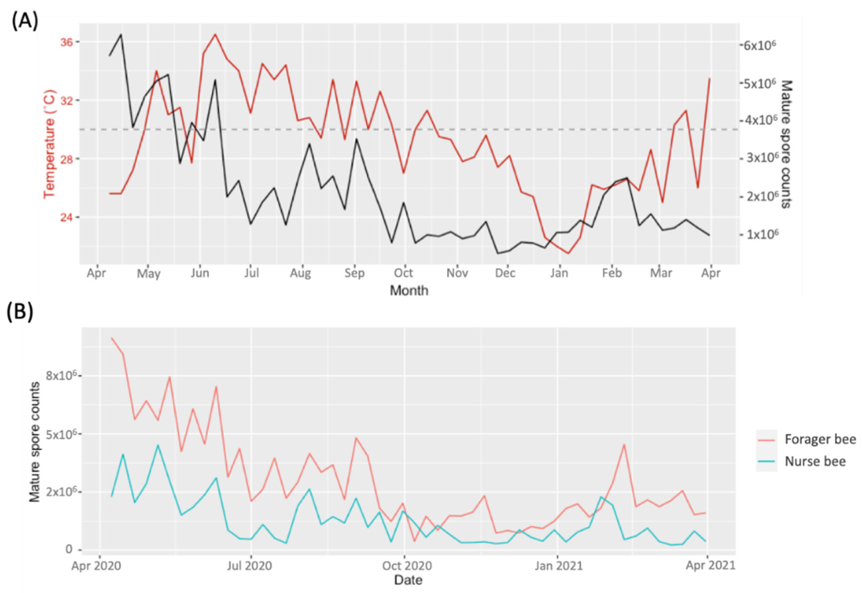
3.1. Season–Prevalence Relationship of V. ceranae

The annual records of the MSC in the NCHU apiary are depicted in Figure 1. The results revealed the continuous presence of MSCs throughout the entire year. The highest peaks in MSC (ranging from 2 × 10⁶~6.28 × 10⁶ MSC/honey bee) were noted from April to June 2020, followed by August to September 2020, ranging from 7.9 × 10⁵~3.53 × 10⁶ MSC/honey bee. Another peak was observed in February 2021, at 2.1 × 10⁶~2.5 × 10⁶ MSCs/bee (Figure 1). Based on our observations, temperatures increased from April (27.2 °C) to July (34.4 °C), while MSC decreased from 6.28 × 10⁶ to 1.26 × 10⁶ MSC/honey bee (Figure 1A). It was further observed that the MSC consistently decreased when the apiary temperature was at or above 30 °C from June to November 2020. However, the MSC increased as the temperature decreased below 30 °C from December 2020 to February 2021 (Figure 1A). Additionally, on average, the detection of MSCs was higher in forager bees than in nurse bees from April 2020 to October 2020 and from January 2021 to March 2021 (Figure 1B).
An analysis of the correlation between temperature and MSC revealed positive correlations between MSC and temperature (Figure 2). The highest MSC was 6.28 × 10⁶ in April 2020, and the lowest MSC was 5.08 × 10⁵ in November 2020 (Figure 2A). This positive correlation was also found for both nurse bees and forager bees (Figure 2B). For the nurse bees, the highest MSC was 4.5 × 10⁶ in May 2020, the lowest MSC was 3 × 10⁵ in July 2020, the difference between the highest and lowest MSC for nurse bees was 4.2 × 10⁶. For the forager bees, the highest MSC was 9.1 × 10⁶ in April 2020, the lowest MSC was 3.8 × 10⁵ in October 2020, and the difference between the highest and lowest MSC was 8.7 × 10⁶ MSC (Figure 2B). Therefore, the difference was more noticeable in forager bees than in nurse bees, and the temperature difference might be a factor that contributes to the MSC in honey bees.

3.2. Efficacy of Probiotic Treatment

For the probiotic treatment of the colonies, in the L. mesenteroides treatment group, the average MSC before treatment was 2.4 × 10⁶. After L. mesenteroides treatment, the MSC experienced a significant decrease by 53.6% from the average load before treatment (p value < 0.05), from 1.38 × 10⁶ (treatment) to 8.86 × 10⁵ (after treatment). From the observations, the highest MSC loads before and after treatment were 1.2 × 10⁷ and 8.3 × 10⁶, respectively, which decreased by 30%. Though the average MSC after treatment (9.01 × 10⁵) was lower than that before treatment (1.87 × 10⁶), there was no significant difference between before and after treatment in the control group. Furthermore, the MSC average load increased from 8.84 × 10⁵ (before treatment) to 9.01 × 10⁵ (after treatment) in the control group, suggesting that L. mesenteroides has anti-mature spore activity to V. ceranae (Figure 3).

4. Discussion

The presence of V. ceranae in A. mellifera colonies in the NCHU apiary, Taichung city, demonstrated fluctuations in MSC throughout the season. The year-round presence of V. ceranae has been reported, with variations in peak spore loads across different climates. A report from 2008–2009 showed a negative correlation between temperature and V. ceranae load; the highest load was observed in the winter, with an MSC of approximately 7 × 10⁶ at 15 °C to 20 °C [12,17]. Our survey revealed that MSC increased as temperature increased, and the highest MSC load we recorded was higher than that in a previous study from 2008 to 2009, which was different from the findings of Chen et al. (2012). Notably, the temperature range recorded in 2008–2009 was 15 to 30 °C, which was lower than our observations from 2020 to 2021 in Taichung (21.5 to 36.5 °C) [12]. This finding suggested that high temperatures led to lower MSCs in our study, indicating that global warming might adversely affect the prevalence of V. ceranae. Consequently, the temperature we recorded during 2020–2021 was higher than that recorded during 2008–2009. Additionally, our data showed different MSC trends than those in the 2008–2009 report, suggesting that temperature caused the different incidences of V. ceranae infection and MSC throughout the year.
Increased MSCs were also observed in forager bees within this study. On average, the MSC in forager bees was 3 × 10⁶, while MSC in nurse bees was 1.2 × 10⁶, and the forager MSC was approximately 58% higher than the nurse bee MSC. Discrepancies in V. ceranae spore loads between nurse bees and forager bees have been documented in earlier research [8,36,37]. Positive correlations between temperature and MSC were evident for forager bees but were less noticeable for nurse bees during the autumn to spring period, suggesting that forager bees might obtain mature spores while collecting nectar and pollen, particularly during the spring season. V. ceranae does not have enough time to develop during the nurse bee stage. Because V. ceranae growth extends from the nurse bee stage, V. ceranae in forager bees has more time to develop, which is a potential reason for the higher MSC in forager bees [8].
A decrease in the MSCs was observed after probiotic treatment, indicating that the effectiveness of the anti-V. ceranae treatment was specific to the mature spore stage in the intracellular phase. According to the genome copy number (GCN) (Supplementary Figure S1), probiotic treatment had a more significant impact on MSC, suggesting that probiotic treatment might influence the mature spore stage of V. ceranae rather than the merogony stage. The intracellular life cycle of V. ceranae includes the proliferative phase (merogony) and the sporogonic phase (sporogony). After infection, the microsporidia were first replicated at the intracellular stage, followed by the formation and release of mature spores from host cells [38]. Upon returning to the environment, there is an opportunity to infect new bees [38]. Therefore, the suppression of the mature spore load within the bee colony can reduce the opportunities of MSCs in the environment, thereby reducing the probability of new emergency honey bees suffering microsporidia infection. This may lead to an overall reduction in the V. ceranae load within the colony. Based on the outcomes of this study, probiotic treatment might reduce the mature spore load of V. ceranae within the bee colony. In the future, a large-scale experiment is needed to further elucidate the efficacy of probiotic treatment on honey bee microsporidiosis.

5. Conclusions

This one-year surveillance of V. ceranae incidence in A. mellifera apiaries at NCHU in Taiwan revealed noteworthy correlations between MSC incidence and environmental factors and the variation in spore loads among different social castes of honey bees. Our data revealed a positive correlation between MSC incidence and temperature, suggesting that temperature differences might impact the development of V. ceranae. However, further experiments are needed to validate these associations. A comparison of the MSCs between nurses and forager bees revealed distinct loads among the different life stages, indicating that the development of MSC loads follows the infection process in various honey bee life stages. Preliminary data on probiotic treatment exhibited efficacy in controlling MSC, suggesting the potential to prevent honey bees from reaching the mature spore stage of V. ceranae infection. Our study delineated the intricate relationship between environmental factors and V. ceranae across different social castes while introducing probiotics as a potential treatment for microsporidiosis control. Future research endeavors should focus on developing a comprehensive microsporidiosis treatment process in apiculture.

Supplementary Materials

The following supporting information can be downloaded at: https://www.mdpi.com/article/10.3390/insects15030204/s1, Figure S1: Total spore counts and genome copy numbers of the control group (A and C) and treatment group (B and D) from September to December.

Author Contributions

Conceptualization, Y.-H.L., M.-C.W. and Y.-S.N.; methodology, Y.-H.L., F.-M.C., Y.-H.C., M.-C.W. and Y.-S.N.; validation, Y.-H.L. and F.-M.C.; investigation, Y.-H.L., F.-M.C. and Y.-H.C.; resources, M.-C.W. and Y.-S.N.; data curation, M.-C.W. and Y.-S.N.; writing—original draft preparation, Y.-H.L., F.-M.C. and Y.-H.C.; writing—review and editing, Y.-H.L., M.-C.W. and Y.-S.N.; visualization, Y.-H.L. supervision, M.-C.W. and Y.-S.N.; project administration, M.-C.W. and Y.-S.N.; All authors have read and agreed to the published version of the manuscript.

Funding

This research was supported by 113AS-5.3.1-VP-01 from the Animal and Plant Health Inspection Agency, Ministry of Agriculture.

Data Availability Statement

The data presented in this study are available upon request from the corresponding authors.

Acknowledgments

We thanks Yu-Wei Weng for his kindly help us to collect the honey bee samples.

Conflicts of Interest

The authors declare no conflicts of interest.

References

  1. Gallai, N.; Salles, J.-M.; Settele, J.; Vaissière, B.E. Economic valuation of the vulnerability of world agriculture confronted with pollinator decline. Ecol. Econ. 2009, 68, 810–821. [Google Scholar] [CrossRef]
  2. Burnham, A.J. Scientific Advances in Controlling Nosema ceranae (Microsporidia) Infections in Honey Bees (Apis mellifera). Front. Vet. Sci. 2019, 6, 79. [Google Scholar] [CrossRef] [PubMed]
  3. Khalifa, S.A.M.; Elshafiey, E.H.; Shetaia, A.A.; El-Wahed, A.A.A.; Algethami, A.F.; Musharraf, S.G.; AlAjmi, M.F.; Zhao, C.; Masry, S.H.D.; Abdel-Daim, M.M.; et al. Overview of Bee Pollination and Its Economic Value for Crop Production. Insects 2021, 12, 688. [Google Scholar] [CrossRef] [PubMed]
  4. Reilly, J.; Artz, D.R.; Biddinger, D.; Bobiwash, K.; Boyle, N.K.; Brittain, C.; Brokaw, J.; Campbell, J.W.; Daniels, J.; Elle, E.; et al. Crop production in the USA is frequently limited by a lack of pollinators. Proc. R. Soc. B 2020, 287, 20200922. [Google Scholar] [CrossRef] [PubMed]
  5. Evans, J.D.; Schwarz, R.S. Bees brought to their knees: Microbes affecting honey bee health. Trends Microbiol. 2011, 19, 614–620. [Google Scholar] [CrossRef] [PubMed]
  6. Aronstein, K.A.; Murray, K.D. Chalkbrood disease in honey bees. J. Invertebr. Pathol. 2010, 103, S20–S29. [Google Scholar] [CrossRef] [PubMed]
  7. Tozkar, C.O.; Kence, M.; Kence, A.; Huang, Q.; Evans, J.D. Metatranscriptomic analyses of honey bee colonies. Front. Genet. 2015, 6, 126955. [Google Scholar] [CrossRef]
  8. Jack, C.J.; Lucas, H.M.; Webster, T.C.; Sagili, R.R. Colony Level Prevalence and Intensity of Nosema ceranae in Honey Bees (Apis mellifera L.). PLoS ONE 2016, 11, e0163522. [Google Scholar] [CrossRef]
  9. Gisder, S.; Schüler, V.; Horchler, L.L.; Groth, D.; Genersch, E. Long-Term Temporal Trends of Nosema spp. Infection Prevalence in Northeast Germany: Continuous Spread of Nosema ceranae, an Emerging Pathogen of Honey Bees (Apis mellifera), but No General Replacement of Nosema apis. Front. Cell. Infect. Microbiol. 2017, 7, 301. [Google Scholar] [CrossRef]
  10. Emsen, B.; Guzman-Novoa, E.; Hamiduzzaman, M.M.; Eccles, L.; Lacey, B.; Ruiz-Pérez, R.A.; Nasr, M. Higher prevalence and levels of Nosema ceranae than Nosema apis infections in Canadian honey bee colonies. Parasitol. Res. 2016, 115, 175–181. [Google Scholar] [CrossRef]
  11. Zhu, X.J.; Zhou, S.J.; Huang, Z.Y. Transportation and pollination service increase abundance and prevalence of Nosema ceranae in honey bees (Apis mellifera). J. Apic. Res. 2014, 53, 469–471. [Google Scholar] [CrossRef]
  12. Chen, Y.W.; Chung, W.-P.; Wang, C.-H.; Solter, L.F.; Huang, W.-F. Nosema ceranae infection intensity highly correlates with temperature. J. Invertebr. Pathol. 2012, 111, 264–267. [Google Scholar] [CrossRef] [PubMed]
  13. Klee, J.; Besana, A.M.; Genersch, E.; Gisder, S.; Nanetti, A.; Tam, D.Q.; Chinh, T.X.; Puerta, F.; Ruz, J.M.; Kryger, P.; et al. Widespread dispersal of the microsporidian Nosema ceranae, an emergent pathogen of the western honey bee, Apis mellifera. J. Invertebr. Pathol. 2007, 96, 1–10. [Google Scholar] [CrossRef] [PubMed]
  14. Fries, I.; Feng, F.; da Silva, A.; Slemenda, S.B.; Pieniazek, N.J. Nosema ceranae n. sp. (Microspora, Nosematidae), morphological and molecular characterization of a microsporidian parasite of the Asian honey bee Apis cerana (Hymenoptera, Apidae). Eur. J. Protistol. 1996, 32, 356–365. [Google Scholar] [CrossRef]
  15. Chen, Y.P.; Huang, Z.Y. Nosema ceranae, a newly identified pathogen of Apis mellifera in the USA and Asia. Apidologie 2010, 41, 364–374. [Google Scholar] [CrossRef]
  16. Huang, W.F.; Jiang, J.H.; Chen, Y.W.; Wang, C.H. A Nosema ceranae isolate from the honeybee Apis mellifera. Apidologie 2007, 38, 30–37. [Google Scholar] [CrossRef]
  17. Martín-Hernández, R.; Bartolomé, C.; Chejanovsky, N.; Le Conte, Y.; Dalmon, A.; Dussaubat, C.; García-Palencia, P.; Meana, A.; Pinto, M.A.; Soroker, V.; et al. Nosema ceranae in Apis mellifera: A 12 years postdetection perspective. Environ. Microbiol. 2018, 20, 1302–1329. [Google Scholar] [CrossRef]
  18. Martin-Hernandez, R.; Meana, A.; Prieto, L.; Salvador, A.M.; Garrido-Bailón, E.; Higes, M. Outcome of colonization of Apis mellifera by Nosema ceranae. Appl. Environ. Microbiol. 2007, 73, 6331–6338. [Google Scholar] [CrossRef]
  19. Jabal-Uriel, C.; Barrios, L.; Bonjour-Dalmon, A.; Caspi-Yona, S.; Chejanovsly, N.; Erez, T.; Henriques, D.; Higes, M.; Le Conte, Y.; Lopes, A.R.; et al. Epidemiology of the Microsporidium Nosema ceranae in Four Mediterranean Countries. Insects 2022, 13, 844. [Google Scholar] [CrossRef]
  20. Fries, I. Nosema ceranae in European honey bees (Apis mellifera). J. Invertebr. Pathol. 2010, 103, S73–S79. [Google Scholar] [CrossRef]
  21. Chen, Y.P.; Evans, J.D.; Murphy, C.; Gutell, R.; Zuker, M.; Gundensen-Rindal, D.; Pettis, J.S. Morphological, Molecular, and Phylogenetic Characterization of Nosema ceranae, a Microsporidian Parasite Isolated from the European Honey Bee, Apis mellifera 1. J. Eukaryot. Microbiol. 2009, 56, 142–147. [Google Scholar] [CrossRef] [PubMed]
  22. Chang, Z.T.; Ko, C.Y.; Yen, M.R.; Chen, Y.W.; Nai, Y.S. Screening of Differentially Expressed Microsporidia Genes from Nosema ceranae Infected Honey Bees by Suppression Subtractive Hybridization. Insects 2020, 11, 199. [Google Scholar] [CrossRef] [PubMed]
  23. Huang, W.-F.; Solter, L.F.; Yau, P.M.; Imai, B.S. Nosema ceranae escapes fumagillin control in honey bees. PLoS Pathog. 2013, 9, e1003185. [Google Scholar] [CrossRef] [PubMed]
  24. Glavinic, U.; Stevanovic, J.; Ristanic, M.; Rajkovic, M.; Davitkov, D.; Lakic, N.; Stanimirovic, Z. Potential of fumagillin and Agaricus blazei mushroom extract to reduce Nosema ceranae in honey bees. Insects 2021, 12, 282. [Google Scholar] [CrossRef] [PubMed]
  25. Glavinic, U.; Stankovic, B.; Draskovic, V.; Stevanovic, J.; Petrovic, T.; Lakic, N.; Stanimirovic, Z. Dietary amino acid and vitamin complex protects honey bee from immunosuppression caused by Nosema ceranae. PLoS ONE 2017, 12, e0187726. [Google Scholar] [CrossRef] [PubMed]
  26. Chen, X.X.; Wang, S.; Xu, Y.; Gong, H.; Wu, Y.; Chen, Y.; Hu, F.; Zheng, H. Protective potential of Chinese herbal extracts against microsporidian Nosema ceranae, an emergent pathogen of western honey bees, Apis mellifera L. J. Asia-Pac. Entomol. 2021, 24, 502–512. [Google Scholar] [CrossRef]
  27. Iorizzo, M.; Letizia, F.; Ganassi, S.; Testa, B.; Petrarca, S.; Albanese, G.; Di Criscio, D.; De Cristofaro, A. Recent Advances in the Biocontrol of Nosemosis in Honey Bees (Apis mellifera L.). J. Fungi 2022, 8, 424. [Google Scholar] [CrossRef]
  28. Iorizzo, M.; Testa, B.; Ganassi, S.; Lombardi, S.J.; Ianiro, M.; Letizia, F.; Succi, M.; Tremonte, P.; Vergalito, F.; Cozzolino, A.; et al. Probiotic properties and potentiality of Lactiplantibacillus plantarum strains for the biological control of chalkbrood disease. J. Fungi 2021, 7, 379. [Google Scholar] [CrossRef]
  29. Royan, M. Mechanisms of probiotic action in the honeybee. Crit. Rev.™ Eukaryot. Gene Expr. 2019, 29, 95–103. [Google Scholar] [CrossRef]
  30. Audisio, M.C. Gram-positive bacteria with probiotic potential for the Apis mellifera L. honey bee: The experience in the northwest of Argentina. Probiotics Antimicrob. Proteins 2017, 9, 22–31. [Google Scholar] [CrossRef]
  31. Team, R.C. R: A Language and Environment for Statistical Computing, R Version 4.0. 3; R Foundation for Statistical Computing: Vienna, Austria, 2021. [Google Scholar]
  32. Wickham, H.; Chang, W.; Wickham, M.H. Package ‘ggplot2’. Creat. Elegant Data Vis. Using Gramm. Graph. 2016, pp. 1–189. Available online: https://www.sciencedirect.com/science/article/pii/S2352711023001607 (accessed on 1 March 2024).
  33. Kassambara, A.; Kassambara, M.A. Package ‘ggpubr’. R Package, Version 0.1. 2020. Available online: https://www.ncbi.nlm.nih.gov/pmc/articles/PMC10281596/ (accessed on 1 March 2024).
  34. Aphalo, P.J. ggpmisc: An R Package. 2016. Available online: https://researchportal.helsinki.fi/en/publications/ggpmisc-an-r-package (accessed on 1 March 2024).
  35. Kassambara, A. Package ‘Rstatix’; R Topics Documented. 2020. Available online: https://www.ncbi.nlm.nih.gov/pmc/articles/PMC8026890/ (accessed on 1 March 2024).
  36. Smart, M.D.; Sheppard, W.S. Nosema ceranae in age cohorts of the western honey bee (Apis mellifera). J. Invertebr. Pathol. 2012, 109, 148–151. [Google Scholar] [CrossRef]
  37. Li, W.; Evans, J.D.; Li, J.; Su, S.; Hamilton, M.; Chen, Y. Spore load and immune response of honey bees naturally infected by Nosema ceranae. Parasitol. Res. 2017, 116, 3265–3274. [Google Scholar] [CrossRef]
  38. Gisder, S.; Möckel, N.; Linde, A.; Genersch, E. A cell culture model for Nosema ceranae and Nosema apis allows new insights into the life cycle of these important honey bee-pathogenic microsporidia. Environ. Microbiol. 2011, 13, 404–413. [Google Scholar] [CrossRef]
Figure 1. From April 2020 to April 2021, the Vairimorpha ceranae mature spore count was measured. (A) Record of spore counts and temperature (°C) and the dotted line indicates 30 °C. (B) Mature spore count record of nurse and forager bees.
Insects 15 00204 g001
Figure 2. Mature Vairimorpha ceranae spore counts with temperature correlations from April 2020 to April 2021. (A) Scatter plots of worker bee temperature; (B) scatter plots of nurse bee and forager bee temperature; n = 52; gray area = 95% confidence limit.
Insects 15 00204 g002
Figure 3. Detection of MSC from V. ceranae following probiotic treatment. Significant differences before and after probiotic treatment were determined using one-way ANOVA with a Bonferroni correction; * p value < 0.05; error bar = SE; n = 52.
Insects 15 00204 g003
Disclaimer/Publisher’s Note: The statements, opinions and data contained in all publications are solely those of the individual author(s) and contributor(s) and not of MDPI and/or the editor(s). MDPI and/or the editor(s) disclaim responsibility for any injury to people or property resulting from any ideas, methods, instructions or products referred to in the content.

