Article

Molecular Identification and Prevalence of the Mite Carpoglyphus lactis (Acarina: Carpoglyphidae) in Apis mellifera in the Republic of Korea

1
Laboratory of Parasitic and Honeybee Diseases, Bacterial Disease Division, Department of Animal and Plant Health Research, Animal and Plant Quarantine Agency, Gimcheon 39660, Republic of Korea
2
Institute of Biotechnology, Vietnam Academy of Science & Technology, Ha Noi 11300, Vietnam
*
Author to whom correspondence should be addressed.
These authors contributed equally to this work.
Insects 2024, 15(4), 271; https://doi.org/10.3390/insects15040271
Submission received: 12 March 2024 / Revised: 8 April 2024 / Accepted: 12 April 2024 / Published: 14 April 2024
(This article belongs to the Section Insect Systematics, Phylogeny and Evolution)

Abstract

:

Simple Summary

Honeybees (Apis mellifera) are crucial for our ecosystem, but they face various threats. This study investigates the detection of a new mite species emerging in Korean honeybees, Carpoglyphus lactis. Using the polymerase chain reaction method to amplify the Cytochrome c oxidase subunit 1 gene region, we examined the presence of C. lactis in honeybee colonies across nine provinces of the Republic of Korea. We found C. lactis in eight provinces in ROK, particularly during winter. This is the first identification of C. lactis in Korean honeybees. Identifying new threats is essential for beekeepers and researchers. Understanding the prevalence of C. lactis mite species and its impact on honeybee health will help develop strategies to protect bee populations.

Abstract

Apis mellifera, especially weak ones, are highly vulnerable to Carpoglyphus lactis mites, which can rapidly infest and consume stored pollen, leading to weakened colonies and potential colony collapse. This study aimed to ascertain and investigate the prevalence of this mite in honeybee colonies across nine provinces in the Republic of Korea (ROK). A total of 615 honeybee colony samples were collected from 66 apiaries during the spring and 58 apiaries during the summer of 2023. A 1242 bp segment of the Cytochrome c oxidase subunit 1 (COI) gene was amplified using the polymerase chain reaction method. The detection levels of C. lactis in the honeybees were compared between winter and summer. Based on the COI sequence analysis, the nucleotide sequence similarity of C. lactis mites isolated in the ROK with those from China (NC048990.1) was found to be 99.5%, and with those from the United Kingdom (KY922482.1) was 99.3%. This study is the first report of C. lactis in Korean apiaries. The findings of this study demonstrate a significantly higher detection rate in winter, which is 4.1 times greater than that in summer (p < 0.001). Furthermore, the results underscore the usefulness of molecular diagnostic techniques for detecting C. lactis mites.

1. Introduction

The honeybee (Apis mellifera), also known as the western honeybee, plays a vital role in the world. These industrious insects act as essential pollinators, ensuring the stability and health of ecosystems [1,2]. Beyond their ecological significance, honeybees provide a wealth of economic and health benefits through the production of honey, royal jelly, propolis, and beeswax [1]. Unfortunately, the rate of honeybee colony losses has been increasing annually in many countries worldwide [3,4,5]. According to a collaborative survey conducted by the Rural Development Administration, Animal and Plant Quarantine Agency, local authorities, and the Korean Beekeeping Association in the Republic of Korea (ROK), 416,409 beehives disappeared between October 2021 and March 2022. The health and survival of honeybee colonies, particularly during the harsh winter months, are of paramount importance. However, winter presents a multitude of challenges that threaten honeybee health throughout this extended period. Consequently, winter colony losses have become a growing concern in temperate climates [3,4,5]. This is primarily due to limited foraging opportunities as temperatures drop and flowers become scarce. As a result, bees rely heavily on pre-stored food reserves within the hive to survive winter. However, the prolonged presence of these stored food resources can also create a potential disease transmission risk within bee colonies. This is primarily due to limited foraging opportunities as temperatures drop and flowers become scarce. As a result, bees rely heavily on pre-stored food reserves within the hive to survive winter. However, the prolonged presence of these stored food resources can also create a potential disease transmission risk within bee colonies. Numerous factors influence the health of honeybee colonies, and one of the devastating factors in colony loss is parasitic infection, especially mite species [1,6,7,8,9,10,11,12]. In the ROK, significant harm to honeybee colonies is attributed to Varroa mite [1,11,13,14]. Research has also documented the presence of other potentially harmful mite species via morphological and/or genetic analysis, including Tropilaelaps mercedesae, Tyrophagus curvipenis, and Acarapis mites in the ROK [12,14]. However, this is the first report of Carpoglyphus lactis within the Korean honeybee population.
Carpoglyphus, commonly known as the cheese mite or milk mite, belongs to the genus Carpoglyphus within the family Carpoglyphidae of the Astigmata order [15,16,17]. This mite is primarily associated with cheese and other milk products [16]. The previous reports mainly focus on the relationship between the mite and milk products, as well as its impact on cheese quality. This species has been observed in the Holarctic, Oriental, Australian, and Indian regions [15,18,19,20,21]. Additionally, C. lactis mites are known to be relatively common in beehives at different positions within the observed hive. They are primarily found in stored pollen and aged beehives, potentially posing a threat to stored honey [22]. Moreover, they are present in beehive debris, in hive materials, on dead bees, and on beehive frames, serving as a source of pollen, honey, and essential bee substances stored in queen bee cells [23,24]. This ability to exploit various resources within the hive highlights their potential to disrupt and harm bee colonies. The detrimental impact of C. lactis becomes particularly severe during the winter. Germany and the United States highlight widespread cases of beehive destruction due to C. lactis mites during winter storage [20,25,26]. In Zander, Germany, 250 beehives were reportedly destroyed by C. lactis mites in a single winter storage area back in 1947 [27]. Similarly, research by Baker and Delfinado in 1978 documented cases in Alabama of the US where beehives were found heavily infested with C. lactis mites following winter storage [21]. These observations reveal a devastating impact, with C. lactis mites consuming stored pollen, polluting the hive with a brownish-yellow powder of mite carcasses, debris, and pollen, and ultimately destroying the combs [20,25,26]. While previous research has focused on the relationship between C. lactis and stored food products, information regarding its detection rate and prevalence within honeybee colonies remains limited. Given the potential role of C. lactis infestation for bee health, understanding its presence and prevalence is crucial for beekeepers to develop effective management strategies. Accurate identification of mite species is crucial for understanding their impact on honeybee health. Based on DNA sequencing, the Cytochrome c oxidase subunit I (COI) gene has proven to be a valuable tool in mite phylogeny studies. In this study, the molecular method was applied to detect the presence of C. lactis mites within honeybee colonies across ROK.
This study aimed to identify the C. lactis mites in honeybee colonies in the ROK in 2023. We conducted a nationwide survey encompassing nine provinces to determine the detection rate of this mite species. By establishing a baseline understanding of C. lactis prevalence in Korean honeybees, this research marks the initiation of investigations for future reference and encourages the development of effective management strategies against C. lactis in honeybee colonies.

2. Materials and Methods

2.1. Collection of Samples and Detection of Astigmatid Mites in Honeybees

Honeybee samples were collected from 9 provinces (Gangwon, Gyeonggi, Chungcheonnam, Chungcheonbuk, Gyeongsangnam, Gyeongsangbuk, Jeollanam, Jeollabuk, and Jeju) in the ROK (Figure 1). The honeybee samples were regularly collected throughout the year 2023 from non-migrating honeybee colonies. Honeybee samples were collected from the hive of each opened colony; subsequently, the comb was taken out after carefully observing the comb without the queen bee. A total of 615 honeybee colonies from 124 apiaries were sampled to investigate the presence of Astigmatid mites. The worker bees (n = 10~30) were collected in 50 mL Falcon tubes from colonies in each apiary. Larvae samples were collected from three random locations within the hive using forceps, checking 10 larvae at each site.
Samples were collected using sterile tools to prevent contamination and directly placed into 50 mL Falcon tubes. Each tube was labeled with a unique identifier to ensure sample traceability throughout the testing process. To preserve the integrity of the collected samples, they were promptly transferred to a temperature-controlled storage facility at −20 °C. The honeybee samples were used to assess temperature-dependent appearance patterns. Concurrently, these samples were used to evaluate the prevalence, diversity, and simultaneous occurrence of pathogens and Astigmatid mites. The samples were divided into two portions; the first was used for Astigmatid mite examination under a dissecting microscope, and the remaining one was used for total nucleic extraction in polymerase chain reaction (PCR). For beehives that tested positive for C. lactis mites, the combs in honeybee colonies were collected during the next sampling round for further laboratory examination. These samples were observed under a dissecting microscope to confirm the presence and morphology of this mite. As this study only involved honeybees, which are invertebrates not covered by most ethical regulations related to animal research, formal ethical approval was not required.

2.2. Total Nucleic Acid Extraction

The honeybee samples were homogenized using a Precellys 24 tissue homogenizer (Bertin Instruments, Montigny-le-Bretonneux, France) in three cycles of 15 s at a speed of 5000 rpm in 1 mL of phosphate-buffered saline solution. Subsequently, a mixture of 200 μL of lysis buffer and 20 μL of proteinase K solution was added to the homogenized sample, and the resulting mixture was incubated at 56 °C for 10 min. The total nucleic acids were extracted using the Maxwell® RSC automated system according to the manufacturer’s instructions. Finally, 60 μL of purified total nucleic acids were employed to detect honeybee pathogens. The total nucleic acids from the honeybee and control samples were stored at −20 °C until further analysis.

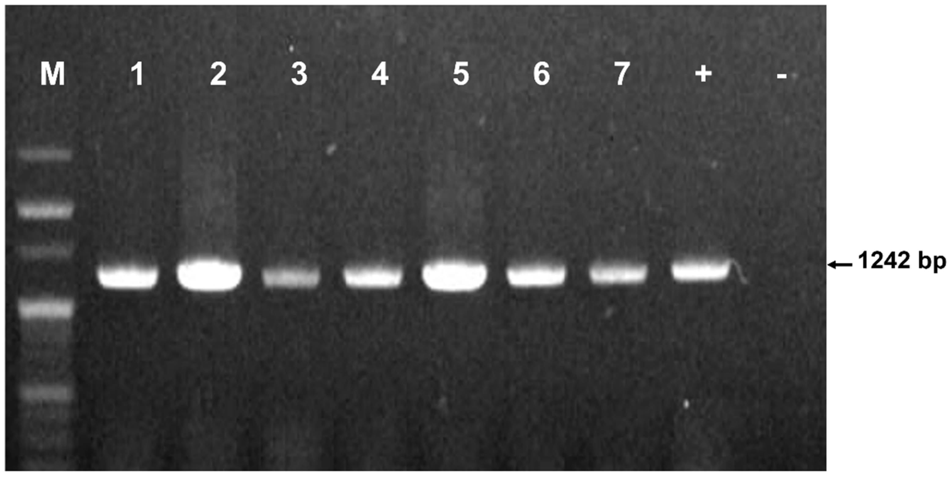
2.3. Amplification of Astigmatid Mite-Specific Genes Using Polymerase Chain Reaction

DNA sequence-based identification has proven to be the most reliable approach for species determination. The COI gene has been successfully used in the species identification and phylogenetic studies of mites [28]. A specific primer pair targeting the COI gene of C. lactis mites was designed and used for PCR amplification. The primer sequence was derived from a reference C. lactis COI gene sequence deposited in GenBank (https://www.ncbi.nlm.nih.gov/, accessed on 18 Mar 2020) under accession number KY922482.1. This approach ensured the amplification of the desired target gene and minimized the possibility of amplifying DNA from other organisms. The primer pairs used in this experiment are described in Table 1.
The total reaction volume (20 µL), including 1 µL of DNA, 1 µL of each primer (10 pmol), and 17 µL ddH2O, was used by AccuPower® PCR PreMix and Master Mix (Bioneer Corp., Daejeon, Republic of Korea). The PCR thermal cycling conditions were as per those by Nguyen et al. [12]: 94 °C (5 min); 5 cycles of 94 °C (20 s), 52 °C (30 s), and 68 °C (30 s); 5 cycles of 94 °C (20 s), 50 °C (30 s), and 68 °C (30 s); 30 cycles of 94 °C (20 s), 48 °C (30 s), and 68 °C (30 s); 30 cycles of 94 °C (20 s), 46 °C (30 s), and 68 °C (30 s); and a final extension step at 68 °C (5 min). The amplified products corresponding to the COI gene segment of C. lactis in the honeybee colonies were visualized using 1% agarose gel electrophoresis, with a determined size of 1242 bp. Subsequently, the PCR products were purified and sequenced by Cosmogenetech Co., Ltd. (Seoul, Republic of Korea). To verify the presence of C. lactis mite and assess its genetic diversity within Korean honeybees, the positive control was specifically designed by cloning the COI gene from a known Korean C. lactis mite.

2.4. Phylogenetic Analysis and Statistical Analysis

The sequences of the COI gene segments of C. lactis mites amplified from different regions were sequenced and compared with the COI gene of this mite available in the National Center for Biotechnology Information (NCBI). Subsequently, the sequences were aligned using ClustalW multiple alignment in BioEdit version 7.2.5 [29]. A species phylogenetic tree was constructed using MEGA11 based on the nucleotide sequences of the COI gene [30]. Simultaneously, a phylogenetic tree was built according to the calculated values obtained from Kimura’s two-parameter distance method by neighbor-joining with 1000 bootstrap replicates and the maximum-likelihood method. Three sequences of C. lactis mites isolated from China (NCBI accession nos.: NC_048990.1 and MN073839.1) and the UK (NCBI accession no. KY922482.1) were chosen as representative sequences.
The values were evaluated using the likelihood ratio chi-square test to measure the strength between the variables of the two groups. Statistical significance was set at p < 0.05.

3. Results

3.1. Molecular Identification of Carpoglyphus lactis Mites in Honeybee Colonies

Among 615 honeybee colonies sampled from 124 apiaries, PCR analysis successfully identified the presence of the C. lactis mite in a subset of colonies. The characteristic 1242 bp PCR product confirmed the presence of the mite (Figure 2).
Comparative analysis of the gene segment sequences of C. lactis mites compared with the GenBank database (blast.ncbi.nlm.nih.gov/Blast.cgi/, accessed on 03 Apr 2023) unveiled a significant level of nucleotide similarity (99.1~99.5%) between those from our study and those from China (NCBI accession No. NC_048990.1) and the UK (NCBI accession no. KY922482.1) (Figure S1). Variations in the COI gene sequence of C. lactis, involving A to G and C to T at identical positions from the UK, were identified. These mutation positions exhibited remarkable consistency among strains isolated nationwide, resembling those from the UK (Figure S1). Analysis of the COI gene sequence revealed a high degree of similarity (between 99.1% and 99.5%) among C. lactis mites isolated from honeybee colonies across ROK (Figure 3). Notably, these sequences exhibited the greatest similarity to strains isolated from China (NCBI accession nos. MN073839.1 and NC_048990.1). Interestingly, one isolate from Gyeonggi province showed a closer match to a strain from the UK (NCBI accession no. KY922482.1), suggesting some potential genetic variation within the Korean C. lactis mites. Our analysis of the COI gene sequence in honeybee samples revealed the widespread presence of C. lactis in honeybee colonies across nine provinces in the ROK.

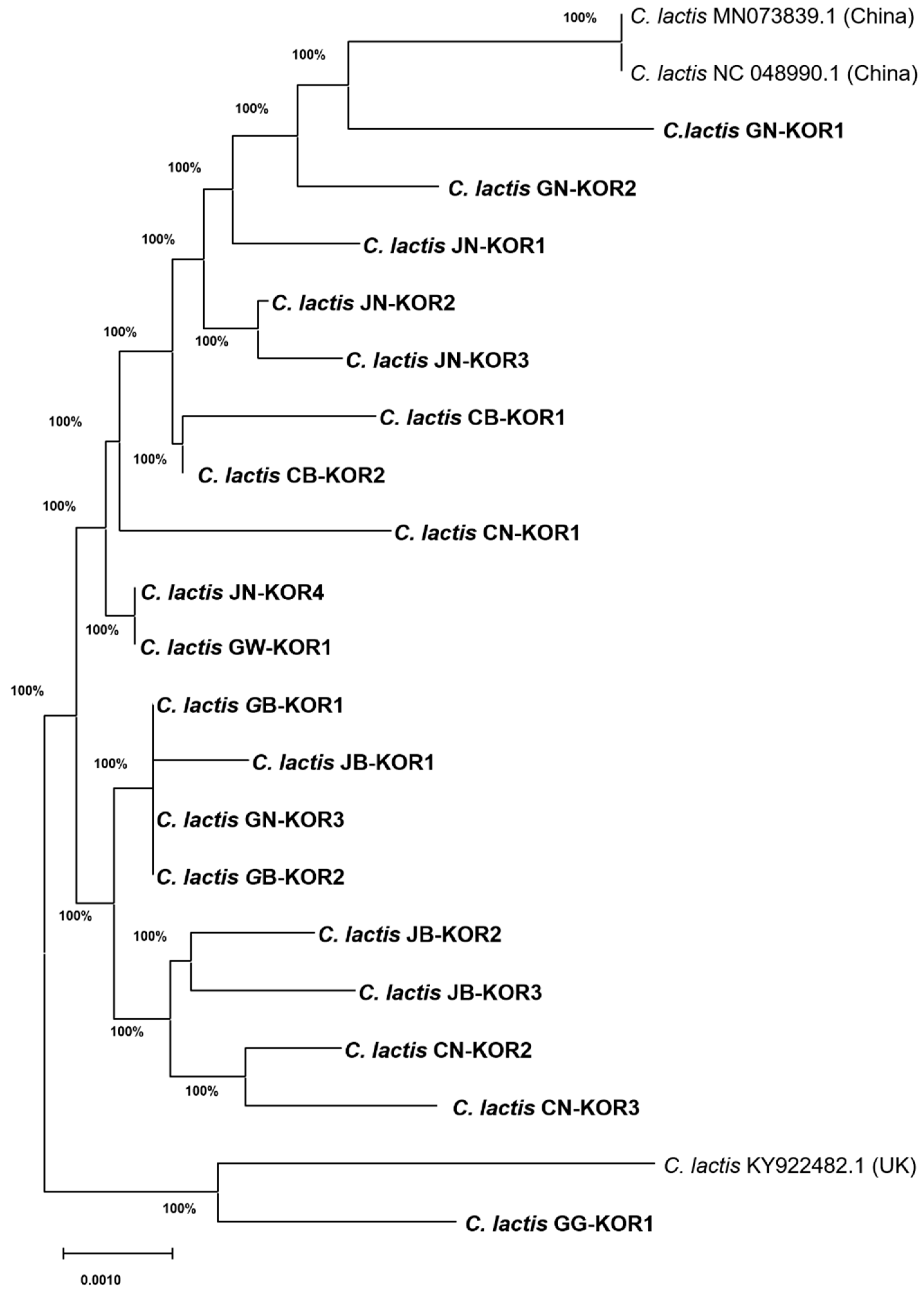
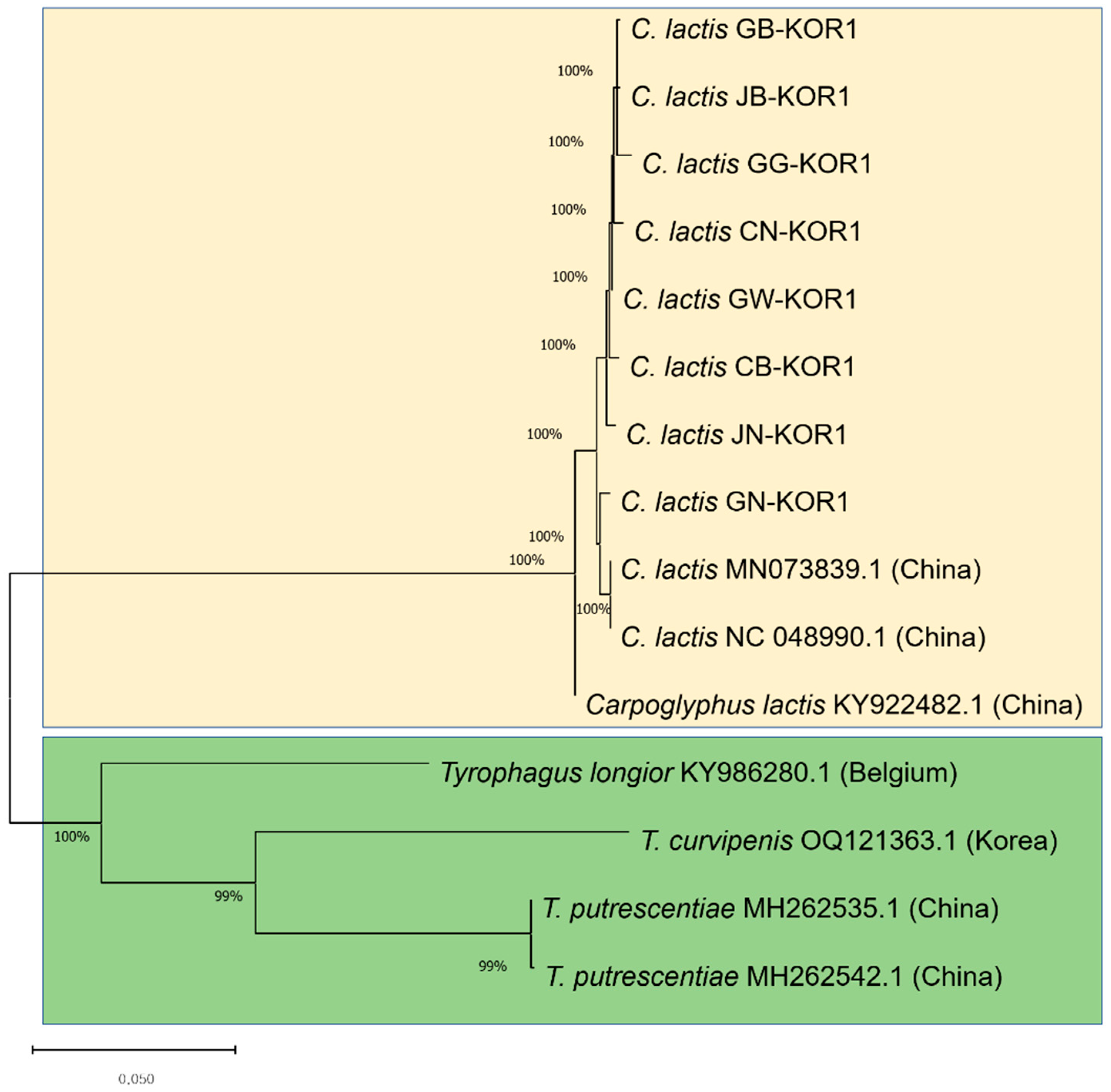
3.2. Similarities of COI Gene Sequence of Astigmatid Mites in Honeybee Colonies

Astigmatid mites are characterized by their tiny size and detrimental impact on honeybee colonies. Species identification was conducted by analyzing the phylogeny, utilizing the COI gene sequences of C. lactis compared to confirm mite species found in honeybee colonies (Figure 4). Our phylogenetic analysis, based on the COI gene sequences belonging to two families, aligns closely with current morphological classifications. The mites found in honeybee apiaries belong to the hyporder Astigmatid; however, within this order, the C. lactis mite forms a distinct and separate branch from Tyrophagus mites. The first brand contains C. lactis of the family Carpodae. The remaining three species cluster together: T. longior from Belgium (NCBI accession no. KY986280.1); T. putrescentiae from China (NCBI accession nos. MH2gyphi62535.1 and MH262542.1); and T. curvipenis from ROK (NCBI accession no. OQ121363.1). The COI nucleotide sequence of C. lactis mites isolated from Korean honeybee colonies exhibited a high nucleotide similarity of 80.2% with Tyrophagus mites.

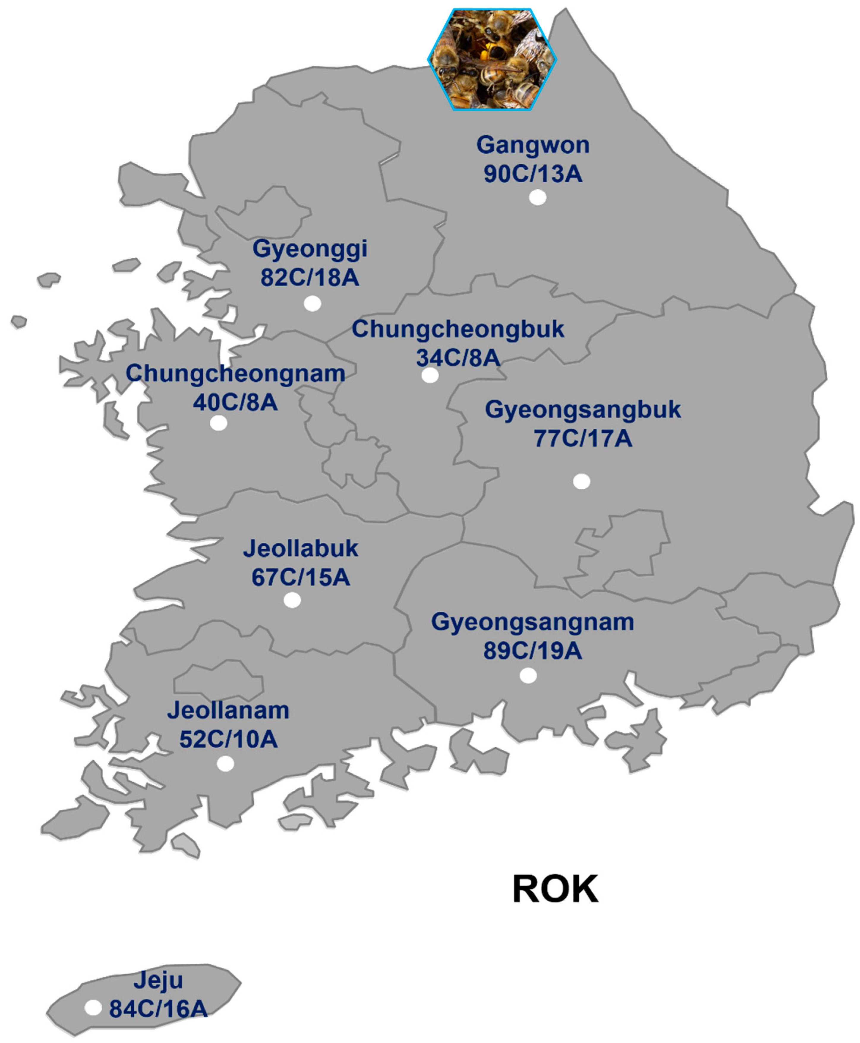
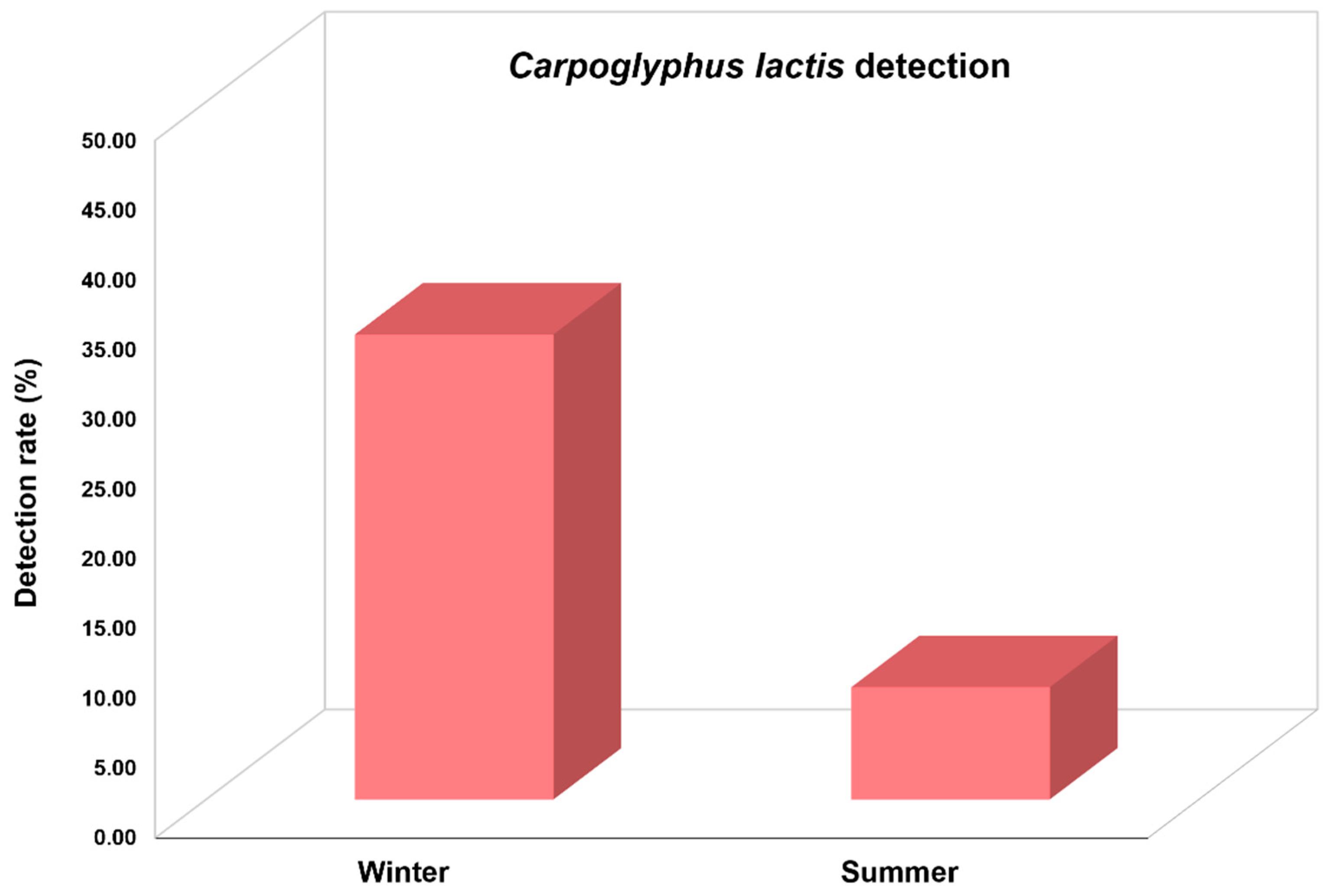
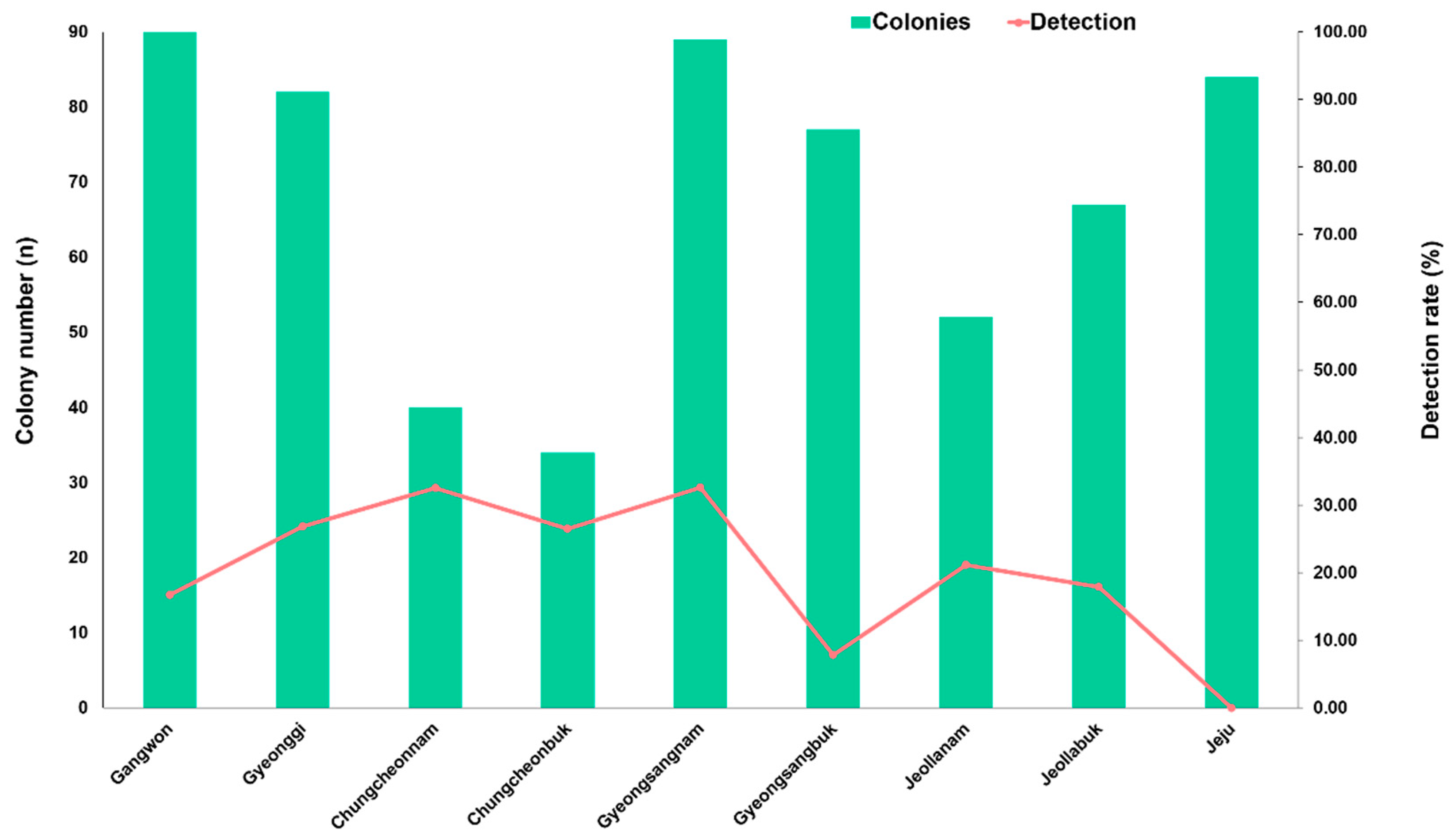
3.3. Prevalence of Carpoglyphus lactis Mite in Korean Honeybee Colonies

We investigated C. lactis mites in honeybee colonies collected during winter and summer in ROK in 2023 (Figure 5). The results indicated a significantly higher detection rate of this mite during winter compared to summer (χ2 = 62.713, df = 1, p < 0.001). The detection rate of C. lactis in 267 colonies from 66 apiaries collected during winter was 33.3%, whereas this rate during summer was only 8.1% in 348 colonies from 58 apiaries. The highest detection rate was observed in March, with a prevalence rate of 74.7% among the 75 honeybee colonies collected from 23 apiaries.
As shown in Figure 6, C. lactis mites were found in eight out of nine provinces. This mite was not detected in any of the surveyed apiaries in the Jeju province. The detection rates of C. lactis in different provinces were as follows: Gangwon at 16.7% (15/90); Gyeonggi at 26.8% (22/82); Chungcheongbuk at 26.5% (9/34); Chungcheongnam at 32.5% (13/40); Gyeongsangbuk at 7.8% (6/77); Gyeongsangnam at 32.6% (29/89); Jeollabuk at 17.9% (12/67); and Jeollanam at 21.2% (11/52). The highest infection rate was observed in the provinces of Chungcheonnam and Gyeongsangnam.

4. Discussion

This study investigated the molecular detection and prevalence of C. lactis mites in honeybee colonies in the ROK. This finding provides valuable insight into the occurrence, prevalence, and phylogenetic position of C. lactis mites in the ROK, a species known to increase mortality rates in honeybee colonies during prolonged winter periods.
We employed molecular techniques to identify C. lactis mites in honeybee colonies. To achieve this, we designed specific primers targeting the COI gene of C. lactis mites with an amplicon size of 1242 bp to minimize non-specific binding of PCR (Table 1). Successful amplification of the targeted COI gene segment confirmed the presence of C. lactis mites in the tested colonies. The subsequent sequencing and comparative analysis of these gene segments with reference sequences in the GenBank database revealed a substantial nucleotide similarity with previously isolated C. lactis strains from China and the UK, further supporting the accurate identification of the mite species. Analysis of the COI gene sequences revealed variations at specific positions in C. lactis mites collected across South Korean provinces (Figure S1). Compared to strains from other countries, these variations suggest a potential for adaptation to distinct environmental conditions within ROK. Further research is necessary to fully understand the functional significance of these variations and their possible impacts on C. lactis biology and behavior in the Korean beekeeping environment.
Honeybee mites constitute one of the major factors causing harm to honey bee colonies globally. They parasitize bees and act as disease vectors within the population. Consequently, they adversely impact the health and alter the healthy behavior of honeybees. As the life cycle of the mite occurs within the beehive, the dense population of these parasites can lead to contaminated honeybee products, reduced food resources, and compromised honeybee health. Recently, a T. curvipenis mite was reported in Korean honeybee apiaries [12]. Carpoglyphus and Tyrophagus mites that primarily feed on stored pollen and other organic materials have been found in beehives [22,27,31]. In this study, we detected C. lactis for the first time in the Korean honeybee in eight of nine provinces in the ROK. The nucleotide of the COI gene of C. lactis mite is similar to that of Tyrophagus mite at 80.2%. Building on prior research by Dabert et al. [32], our analysis of the COI gene further supports the close relationship between C. lactis and Tyrophagus mites within the suborder Astigmata. Notably, Dabert et al. [32] also found these two species to cluster together in their phylogenetic tree of Arachnid mites, constructed using the 18S rDNA gene. In 2023, Bowman reported that C. lactis and Tyrophagus mites could co-occur in UK beehives [15]. The discovery of C. lactis mite in this study, along with the recent report of T. curvipenis by Nguyen et al. (2023), suggests two potential implications regarding the presence of these mite species in Korean honeybee colonies. Firstly, this discovery may indicate a genuine increase in the prevalence of newly introduced mite species in ROK. Alternatively, these mite species may have already been present but remained undetected due to the lack of previous studies specifically targeting them. Our research provides the first report of C. lactis mites in Korean honeybee colonies, contributing to a more comprehensive understanding of the mite fauna associated with these colonies. The spread of diseases within bee populations infested with bee mites is increasing; hence, effective control measures are challenging. Therefore, investigating the types of bee mites and assessing their prevalence is crucial to sustaining honeybee health and preventing the propagation of possible harmful mites to the bee population.
Our prevalence analysis provided valuable insights into the occurrence and distribution of C. lactis mites in honeybee colonies in the ROK. A nationwide survey was conducted in 2023, encompassing all nine provinces of the ROK. Sampling times were strategically chosen to capture potential variations in mite presence throughout the year, with adjustments made based on observed temperature fluctuations. The observed temporal variations in the infection rate with a higher prevalence during winter warrant further investigation into potential drivers. Environmental factors and fluctuations in honeybee populations likely play a role. Previous studies have linked its presence to increased honeybee mortality, particularly during winter months [18,20,27]. The initial detection of C. lactis in overwintering honeybee colonies was first reported in Germany [27], suggesting a robust development of C. lactis mite at lower temperatures, particularly within the beehive, making it an ideal habitat for this mite species. This potential threat is concerning, especially considering the extended periods of food storage necessary for honeybee survival in temperate climates like ROK. Our findings on the high prevalence of C. lactis highlight the need for further research to elucidate its role in honeybee health and potential interactions with winter mortality.
Our survey revealed a significant presence of C. lactis mites across a substantial portion of the honeybee population sampled in the ROK. A striking 88.89% of the samples collected from eight out of nine provinces surveyed tested positive for C. lactis infestation. Interestingly, Jeju province, an island separated from the mainland, emerged as an exception. None of the samples collected from Jeju beehives showed any detectable presence of C. lactis mites, contrasting sharply with the widespread occurrence observed in the other provinces. In addition, identifying C. lactis in both honeybee and beeswax samples indicates that C. lactis mites may have a substantial impact on the honeybees’ health environmentally as well as individually. A more profound investigation into the life cycle of C. lactis mites in honeybee colonies is necessary to better understand the effects of mite infestation on honeybee populations and overall hive health. This knowledge will inform timely strategies and protective measures for the sustainability of beekeeping in the ROK. Until now, research on the detection and prevalence of C. lactis mites in honeybee colonies, as well as the associated potential risks to honeybee health, remains limited.
While our study successfully employed COI gene sequencing to identify C. lactis mites in South Korean honeybee colonies, some limitations warrant consideration for future investigations. The morphology of C. lactis was not observed in our study. Identifying C. lactis mites was not our initial objective, leading to delayed sampling for morphological confirmation during the late winter season. Winter sampling focused solely on bees and excluded mite examination in comb or beehive, thus precluding direct morphological confirmation of C. lactis. Summer sampling across different bee stages and materials also yielded no detections, possibly due to low prevalence in summer or dead mite presence. Furthermore, the morphology of mites within the Astigmatid order is relatively similar, which may lead to confusion during the observation. Further investigation using alternative methods is ongoing. For a more definitive confirmation of C. lactis presence, a complementary approach that combines genetic analysis (utilizing genes like COI and ITS) with traditional morphological examination is recommended. The detected C. lactis mites using PCR solely confirm their presence and do not necessarily reflect active infection. The potential impact of C. lactis on honeybee health and honey production remains an area requiring further investigation. While traditionally not considered a primary pest, its high prevalence and potential for competition for food resources, nest debris generation, and possible involvement in disease transmission at high infection rates warrant further study. These are some limitations; however, our study provides valuable initial insights into the presence of C. lactis in Korean apiaries. Further research addressing these limitations will be crucial for understanding its potential impact on honeybee health and developing effective management strategies.

5. Conclusions

This study provides the first report of C. lactis mites in honeybee colonies within the ROK. Employing molecular detection techniques, we revealed a widespread distribution of C. lactis across eight out of nine provinces surveyed. Furthermore, our results demonstrated a seasonal variation in prevalence, with higher detection rates observed during winter. Phylogenetic analysis confirmed the distinct identity of C. lactis within the suborder Astigmata and its near relationship with Tyrophagus mites. These findings highlight the potential threat posed by C. lactis to Korean honeybee health and emphasize the need for further research. Future investigations should focus on elucidating the impact of C. lactis infestation on bee colony health and productivity, particularly during winter.

Supplementary Materials

The following supporting information can be downloaded at https://www.mdpi.com/article/10.3390/insects15040271/s1, Figure S1: Multiple alignments of the differing nucleotide positions in the COI gene of Carpoglyphus lactis. A comparison was conducted between the COI sequence (1137 nucleotides) of C. lactis mite isolated from the ROK in this study and the published sequence in GenBank. The highlighted blue frame signified shared nucleotide differences between the isolated strains and those from China (NCBI accession nos.: NC_048990.1, MN073839.1) and the UK (NCBI accession no. KY922482.1).

Author Contributions

Conceptualization, T.-T.N., M.-S.Y. and Y.-S.C.; methodology, T.-T.N. and Y.-S.C.; software, T.-T.N.; formal analysis, J.K., H.-S.L. and T.-T.N.; investigation, H.-S.L., T.-T.N. and Y.-S.C.; collected honeybee samples, S.-J.L., T.-T.N. and Y.-S.C.; isolation of total nucleic acids, S.-Y.Y., S.-J.L. and S.-K.S.; writing—original draft preparation, T.-T.N.; writing—review and editing, M.-S.Y., J.K. and Y.-S.C. All authors have read and agreed to the published version of the manuscript.

Funding

This research was funded by the Animal and Plant Quarantine Agency, the Republic of Korea (Project No. N-1543081-2021-25-03).

Data Availability Statement

All data are provided in this manuscript.

Acknowledgments

Our heartfelt thanks go out to the beekeepers who facilitated our sampling process. We extend our appreciation to all the members of our laboratories for their unwavering dedication and diligent efforts.

Conflicts of Interest

The authors declare no conflicts of interest.

References

  1. Truong, A.-T.; Yoo, M.-S.; Seo, S.K.; Hwang, T.J.; Yoon, S.-S.; Cho, Y.S. Prevalence of honey bee pathogens and parasites in South Korea: A five-year surveillance study from 2017 to 2021. Heliyon 2023, 9, e13494. [Google Scholar] [CrossRef] [PubMed]
  2. Klein, A.-M.; Vaissière, B.E.; Cane, J.H.; Steffan-Dewenter, I.; Cunningham, S.A.; Kremen, C.; Tscharntke, T. Importance of pollinators in changing landscapes for world crops. Proc. R. Soc. B 2007, 274, 303–313. [Google Scholar] [CrossRef]
  3. Mutinelli, F.; Pinto, A.; Barzon, L.; Toson, M. Some Considerations about Winter Colony Losses in Italy According to the Coloss Questionnaire. Insects 2022, 13, 1059. [Google Scholar] [CrossRef] [PubMed]
  4. Johannesen, J.; Wöhl, S.; Berg, S.; Otten, C. Annual fluctuations in winter colony losses of Apis mellifera L. are predicted by honey flow dynamics of the preceding year. Insects 2022, 13, 829. [Google Scholar] [CrossRef] [PubMed]
  5. Gray, A.; Adjlane, N.; Arab, A.; Ballis, A.; Brusbardis, V.; Bugeja Douglas, A.; Cadahia, L.; Charriere, J.D.; Chlebo, R.; Coffey, M.F.; et al. Honey bee colony loss rate in 37 countries using the COLOSS survey for winter 2019–2020: The combined effects of operation size, migration and queen replacement. J. Apic. Res. 2023, 62, 204–210. [Google Scholar] [CrossRef]
  6. Chantawannakul, P.; de Guzman, L.I.; Li, J.; Williams, G.R. Parasites, pathogens, and pests of honeybees in Asia. Apidologie 2016, 47, 301–324. [Google Scholar] [CrossRef]
  7. Chantawannakul, P.; Ramsey, S.; Khongphinitbunjong, K.; Phokasem, P. Tropilaelaps mite: An emerging threat to European honey bee. Curr. Opin. Insect Sci. 2018, 26, 69–75. [Google Scholar] [CrossRef] [PubMed]
  8. Denmark, H.A.; Cromroy, H.L.; Sanford, M.T. Honey Bee Tracheal Mite, Acarapis woodi (Rennie) (Arachnida: Acari: Tarsonemidae); University of Florida, IFAS Extension: Gainesville, FL, USA, 2000. [Google Scholar] [CrossRef]
  9. Sammataro, D.; Gerson, U.; Needham, G. Parasitic mites of honey bees: Life history, implications, and impact. Annu. Rev. Entomol. 2000, 45, 519–548. [Google Scholar] [CrossRef] [PubMed]
  10. Emmanouel, N.G.; Pelekassis, C.D.; Santas, L.A. Harmful mesostigmatic mites ectoparasitic to honey bees. Entomol. Hell. 1983, 1, 17–23. [Google Scholar] [CrossRef]
  11. Truong, A.-T.; Yoo, M.-S.; Yun, B.-R.; Kang, J.E.; Noh, J.; Hwang, T.J.; Seo, S.K.; Yoon, S.-S.; Cho, Y.S. Prevalence and pathogen detection of Varroa and Tropilaelaps mites in Apis mellifera (Hymenoptera, Apidae) apiaries in South Korea. J. Apic. Res. 2023, 62, 804–812. [Google Scholar] [CrossRef]
  12. Nguyen, T.-T.; Yoo, M.-S.; Truong, A.-T.; Lee, J.H.; Youn, S.Y.; Lee, S.-J.; Kim, D.-H.; Yoon, S.-S.; Cho, Y.S. First identification of Tyrophagus curvipenis (Acari: Acaridae) and pathogen detection in Apis mellifera colonies in the Republic of Korea. Sci. Rep. 2023, 13, 9469. [Google Scholar] [CrossRef] [PubMed]
  13. Jung, C.; Lee, M.L. Beekeeping in Korea: Past, present, and future challenges. In Asian Beekeeping in the 21st Century; Chantawannakul, P., Williams, G., Neumann, P., Eds.; Springer: Singapore, 2018; pp. 175–197. [Google Scholar]
  14. Ahn, A.J.; Ahn, K.S.; Noh, J.H.; Kim, Y.H.; Yoo, M.S.; Kang, S.W.; Yo, D.H.; Shin, S.S. Molecular prevalence of Acarapis mite infestations in honey bees in Korea. Korean J. Parasitol. 2015, 53, 315–320. [Google Scholar] [CrossRef] [PubMed]
  15. Bowman, C. Variation in the trophic morphology of Astigmatid mites common in UK beehives. Acarologia 2023, 63, 4–16. [Google Scholar] [CrossRef]
  16. Hubert, J.; Nesvorna, M.; Kopecký, J.; Ságová-Marečková, M.; Poltronieri, P. Carpoglyphus lactis (Acari: Astigmata) from various dried fruits differed in associated micro-organisms. J. Appl. Microbiol. 2015, 118, 470–484. [Google Scholar] [CrossRef] [PubMed]
  17. Jiang, Z. A new species of Carpoglyphidae from China (Acarina: Acaroidea). J. Jiang Univ. 1991, 15, 82–86. [Google Scholar]
  18. Chmielewski, W. Morphological and bio-ecological characteristics of Carpoglyphus lactis found in natural honey, warehouses and beehives. Pszczel. Zesz. Naukowe 1970, 14, 109–127. [Google Scholar]
  19. Clark, J.M. A new species of Carpoglyphus (Astigmatina: Carpoglyphidae) from the bark of black beech (Nothofagus) honeydew in New Zealand. Int. J. Acarol. 2010, 36, 453–459. [Google Scholar] [CrossRef]
  20. Haragsim, O.; Samšiňák, K.; Vobrázková, E. The mites inhabiting the beehives in ČSR. Z. Angew. Entomol. 1978, 87, 52–67. [Google Scholar] [CrossRef]
  21. Vijayakumar, K.; Muthuraman, M.; Jayaraj, R. Infestation of Carpoglyphus lactis (Linnaeus)(Acari: Carpoglyphidae) on Trigona iridipennis (Apidae: Meliponinae) from India. Sch. J. Agric. Sci. 2013, 3, 25–28. [Google Scholar]
  22. Baker, E.; Delfinado, M. Notes on the driedfruit mite Carpoglyphus lactis (Acarina: Carpoglyphidae) infesting honeybee combs. J. Apic. Res. 1978, 17, 52–54. [Google Scholar] [CrossRef]
  23. Zhang, Z.-Q. New Zealand records of Carpoglyphus lactis (Acari: Carpoglyphidae). Syst. Appl. Acarol. 2012, 17, 239–240. [Google Scholar] [CrossRef]
  24. Zhang, K.; Zhang, Z.-Q. The dried fruit mite Carpoglyphus lactis (Acari: Carpoglyphidae) is a suitable alternative prey for Amblyseius herbicolus (Acari: Phytoseiidae). Syst. Appl. Acarol. 2021, 26, 2167–2176. [Google Scholar] [CrossRef]
  25. Hughes, A.M. The Mites of Stored Food and Houses, 2nd ed.; Her Majesty’s Stationery Office: London, UK, 1976. [Google Scholar]
  26. Chmielewski, W. Bionomics of Carpoglyphus lactis (Acari: Carpoglyphidae) on honey. In Ecology and Evolution of the Acari, 3rd ed.; Bruin, J., van der Geest, L.P.S., Sabelis, M.W., Eds.; Spinger: Dordrecht, The Netherlands, 1999; pp. 423–424. [Google Scholar]
  27. Zander, E. Handbook of Beekeeping in Individual Representations. Diseases and Pests of Adult Bees; Verlag Eugen Ulmer: Stuttgart, Germany, 1947. [Google Scholar]
  28. Yang, B.; Cai, J.; Cheng, X. Identification of astigmatid mites using ITS2 and COI regions. Parasitol. Res. 2011, 108, 497–503. [Google Scholar] [CrossRef] [PubMed]
  29. Hall, T. BioEdit Version 7.0.0. 2004. Distributed by the Author, Website. Available online: www.mbio.ncsu.edu/BioEdit/bioedit.html (accessed on 7 February 2004).
  30. Kumar, S.; Stecher, G.; Li, M.; Knyaz, C.; Tamura, K. MEGA X: Molecular evolutionary genetics analysis across computing platforms. Mol. Biol. Evol. 2018, 35, 1547–1549. [Google Scholar] [CrossRef]
  31. Chmielewski, W. Stored products mites (Acaroidea) in Polish bee hives. In Modern Acarology, Proceedings of the 8 International Congress of Acarology, held in Ceske Budejovice, Czechoslovakia, 6–11 August 1990; Dusbabek, F., Bukva, V., Eds.; SPB Academic Publishing bv: The Hague, The Netherlands, 1991; pp. 615–619. [Google Scholar]
  32. Dabert, M.; Witalinski, W.; Kazmierski, A.; Olszanowski, Z.; Dabert, J. Molecular phylogeny of acariform mites (Acari, Arachnida): Strong conflict between phylogenetic signal and long-branch attraction artifacts. Mol. Phylogenet. Evol. 2010, 56, 222–241. [Google Scholar] [CrossRef]
Figure 1. Honeybee colonies collected from 124 Korean apiaries in 9 provinces of the ROK in 2023. The number includes adult bees and larvae. C: colony; A: Apiary.
Insects 15 00271 g001
Figure 2. Amplification of COI regions from Carpoglyphus lactis mites in honeybee samples. PCR products were confirmed on 1% agarose gel. Lane M is a 100 bp DNA ladder (Enzymomics Co., Ltd., Daejeon, Republic of Korea); lanes 1–7 showcase PCR products of the honeybee samples; lane “+” indicates a positive control; lane “–” indicates a negative control without a DNA template.
Insects 15 00271 g002
Figure 3. Neighbor-joining phylogenetic tree of Carpoglyphus lactis mite based on sequences of COI gene (1137 bp). C. lactis mite was isolated from each province of the ROK.
Insects 15 00271 g003
Figure 4. Phylogenetic maximum-likelihood tree based on sequences of COI gene of mites. The species name, NCBI accession numbers, and origin of the accession are given for each sequence. Carpoglyphus mites are highlighted in gold, Tyrophagus mites are highlighted in green. Carpoglyphus lactis (C. lactis), Tyrophagus curvipenis (T. curvipenis), and Tyrophagus putrescentiae (T. putrescentiae).
Insects 15 00271 g004
Figure 5. Carpoglyphus lactis detection rate during the winter and summer of 2023 in Korean honeybee colonies.
Insects 15 00271 g005
Figure 6. Detection rate of Carpoglyphus lactis mites in honeybee colonies from 9 provinces in the ROK.
Insects 15 00271 g006
Table 1. Primers used in this study.
Name of PrimersSequences (5’→ 3’)Amplicon Size (bp)NoteReferences
COI–ForGTTTTGGGATATCTCTCATAC377Used for Astigmatid mite detection[28]
COI–RevGAGCAACAACATAARAAGTATC
CL-ForCTTGAATTTGTAGAATGGA1242Used for C. lactis mite detectionThis study
CL-RevCTAATCGAGGTGTCCGAGGT
Disclaimer/Publisher’s Note: The statements, opinions and data contained in all publications are solely those of the individual author(s) and contributor(s) and not of MDPI and/or the editor(s). MDPI and/or the editor(s) disclaim responsibility for any injury to people or property resulting from any ideas, methods, instructions or products referred to in the content.

