Article

Impacts of Combining Steinernema carpocapsae and Bracon hebetor Parasitism on Galleria mellonella Larvae

by
Neama A. Aamer
1,
Zeinab A. El-Moaty
2,3,*,
Maria Augustyniak
4,
Lamia M. El-Samad
3,*,† and
Hanaa S. Hussein
1,†
1
Department of Applied Entomology and Zoology, Faculty of Agriculture, Alexandria University, Alexandria 21545, Egypt
2
Biological Sciences Department, College of Science, King Faisal University, Al-Ahsaa 31982, Saudi Arabia
3
Department of Zoology, Faculty of Science, Alexandria University, Moharam Bey, Alexandria 21511, Egypt
4
Institute of Biology, Biotechnology and Environmental Protection, Faculty of Natural Sciences, University of Silesia in Katowice, Bankowa 9, 40-007 Katowice, Poland
*
Authors to whom correspondence should be addressed.
These authors contributed equally to this work.
Insects 2024, 15(8), 588; https://doi.org/10.3390/insects15080588
Submission received: 1 July 2024 / Revised: 25 July 2024 / Accepted: 25 July 2024 / Published: 1 August 2024

Abstract

:

Simple Summary

The greater wax moth, Galleria mellonella, is a significant pest in apiculture. This investigation explores the individual and combined effects of the ectoparasite Bracon hebetor (B.h.) and the entomopathogenic nematode Steinernema carpocapsae (S.c.) on G. mellonella larvae. We found that control larvae exhibited higher glutathione peroxidase (GPx) and glutathione S-transferase (GST) activities compared to those treated with B.h., S.c., or the B.h. + S.c. combination. Conversely, malondialdehyde (MDA) levels displayed an opposite trend. Superoxide dismutase (SOD) activity was reduced in the B.h. and S.c. groups but significantly higher in the combined treatment. Cytochrome P450 activity increased in response to parasitism by B. hebetor. The Annexin V-FITC assay revealed decreased cell viability in parasitized groups (B.h. 79.4%, S.c. 77.3%, B.h. + S.c. 70.1%) compared to controls. DNA damage analysis demonstrated significant differences between groups, and SEM observations confirmed severe cuticle abnormalities or malformations in G. mellonella larvae.

Abstract

The greater wax moth, Galleria mellonella, is a significant pest in apiculture and a well-established model organism for immunological and ecotoxicological studies. This investigation explores the individual and combined effects of the ectoparasite Bracon hebetor (B.h.) and the entomopathogenic nematode Steinernema carpocapsae (S.c.) on G. mellonella larvae. We evaluated the activity of oxidative stress enzymes, including superoxide dismutase (SOD), glutathione peroxidase (GPx), glutathione S-transferase (GST), malondialdehyde (MDA) levels, cytochrome P450 activity, cell viability using Annexin V-FITC, DNA damage via comet assay, and larval morphology through scanning electron microscopy (SEM). Control larvae exhibited higher GPx and GST activities compared to those treated with B.h., S.c., or the B.h. + S.c. combination. Conversely, MDA levels displayed the opposite trend. SOD activity was reduced in the B.h. and S.c. groups but significantly higher in the combined treatment. Cytochrome P450 activity increased in response to parasitism by B. hebetor. The Annexin V-FITC assay revealed decreased cell viability in parasitized groups (B.h. 79.4%, S.c. 77.3%, B.h. + S.c. 70.1%) compared to controls. DNA damage analysis demonstrated significant differences between groups, and SEM observations confirmed severe cuticle abnormalities or malformations in G. mellonella larvae. These findings highlight the complex interactions between B. hebetor, S. carpocapsae, and their host, G. mellonella. Additionally, they illuminate the intricate physiological responses triggered within the host larvae.

1. Introduction

The greater wax moth, Galleria mellonella (Linnaeus, 1758) (Pyralidae: Lepidoptera), is a globally recognized pest in apiculture, causing significant economic losses due to its larvae consuming honeycombs [1]. Because it is easy to handle, thrives in labs, and is inexpensive to raise, this organism is perfect for immune system and toxin studies [2,3]. This in vivo model offers several advantages, including low biohazard risk, ease of manipulation, affordability, and minimal space or infrastructure requirements, making it ethically acceptable and widely used in research [4]. Current approaches utilizing G. mellonella larvae primarily focus on host–pathogen interactions, with these larvae frequently employed as infection models to evaluate the efficacy of biological control agents before pre-clinical mammalian research [5].
Decades of widespread insecticide application have selected for resistant insect populations [6,7,8]. Various methods, including chemical treatments (e.g., aluminum phosphide, paradichlorobenzene, ethylene dibromide, carbon dioxide, and sulfur) and physical techniques (e.g., heat and cold treatments), have been employed to protect honeycombs from G. mellonella infestation [9]. However, biological control strategies utilizing natural enemies such as parasitic wasps are considered eco-friendly, sustainable, and harmless approaches for integrated pest management (IPM) [10,11]. These natural enemies can continuously breed as long as hosts are available, ensuring long-term pest population control [12].
Parasitic wasps, like Bracon hebetor (Say) (Hymenoptera: Braconidae), exhibit high dispersal rates and can locate hosts in hidden areas. They employ various strategies to regulate host physiology, including idiobiont and koinobiont lifestyles. Idiobionts permanently paralyze their hosts during parasitism, halting host growth and development, while koinobionts allow hosts to continue developing post-parasitism [13]. The host regulation process is complex, often resulting in significant physiological changes without causing immediate host death [14]. Venoms from at least six insect orders serve multiple purposes, including communication, protection, predation, and paralyzing hosts for progeny feeding [15,16,17,18,19,20]. In parasitoids, venom injections facilitate reproduction by preventing host interference during oviposition [21,22].
Within the Hymenoptera, parasitoids employ various host regulatory tactics, ranging from immune suppression to neuroendocrine disruptions affecting reproduction and development [23,24,25,26]. Parasitic wasps induce significant physiological changes in hosts through toxic secretions during egg laying, which can cause paralysis and rapid host death [21,26,27]. The idiobiont ectoparasitoid B. hebetor is particularly effective in the biocontrol of lepidopterous pests, triggering host defense mechanisms upon injury and penetration [28].
Entomopathogenic nematodes (EPNs), particularly those in the Steinernematidae and Heterorhabditidae families, also serve as biological control agents. These obligate parasites kill their insect hosts during part of their life cycle [29]. EPNs release symbiotic bacteria upon penetrating the host, with the bacteria proliferating and killing the host within 24–48 h [30]. EPNs are environmentally safe, pose no threat to humans, animals, plants, or earthworms, and thus offer a viable alternative to chemical pesticides [31]. EPNs go through complex processes to determine the suitability of a host [32]. Beyond host selection, successful infection by EPNs is influenced by factors such as the host’s immune response, population dynamics, and interactions with other organisms [33] Co-infection with other insect pathogens, including viruses, bacteria, other nematodes, or hymenopteran parasitoids, can enhance the success rate of these infections [34]. The interaction between nematodes and parasitoids favored the nematodes when they were introduced during the parasitoid’s young larval stage, enhancing the nematodes’ growth and reproduction, leading to the death of the parasitoid larvae. However, when nematodes were introduced during the parasitoid’s late larval stage, the competition somewhat favored the wasps, allowing about 50% of them to develop, emerge, and reproduce. This indicates that both nematodes and wasps can reproduce in the same host [35,36].
Insects encounter various stressors, both chemical (e.g., pesticides, heavy metals) and physical (e.g., radiation, temperature), leading to oxidative stress and cellular damage [37,38,39]. To combat reactive oxygen species (ROS) and prevent cellular damage, insects activate antioxidant enzymes (such as glutathione peroxidase (GPx), glutathione S-transferase (GST), and superoxide dismutase (SOD)) [40,41]. Malondialdehyde (MDA) is a common marker of oxidative stress, while GPx, GSTs, and SODs play critical roles in detoxification and antioxidative defense [42,43,44,45]. Cytochrome P450 enzymes, part of a heme-thiolate protein superfamily, are crucial for insect oxidative metabolism and detoxification [46,47]. They are involved in the metabolism of ecdysterone, juvenile hormones, and cuticular hydrocarbons, which are essential for insect development and growth [48].
Annexins, a large protein family, are involved in essential biological functions such as calcium metabolism, cell adhesion, growth, differentiation, subcellular transport, and membrane repair [49,50]. Flow cytometry using Annexin-V is a common method for detecting apoptosis at the cytological level [51]. Genotoxicity assays in insects, such as the comet assay, are valuable tools for studying DNA damage mechanisms and monitoring environmental contaminants [52]. The comet assay is a sensitive and reliable method for detecting DNA damage induced by physical agents or xenobiotics in insect tissues [53,54].
Building on our understanding of the biological control agents and the host response mechanisms, this study aims to comprehensively investigate the physiological, molecular, and morphological effects of the entomopathogenic nematode Steinernema carpocapsae (Weiser) (Rhabditida: Steinernematidae) (all strains) and the ectoparasite B. hebetor on G. mellonella larvae. Both individual and combined effects will be examined. This research offers a unique perspective by utilizing scanning electron microscopy to elucidate the morphological alterations induced by these bioagents. Additionally, we will explore the physiological and molecular changes occurring in the host larvae, providing a holistic understanding of these interactions.

2. Materials and Methods

2.1. Insect and Nematode Cultures

Greater wax moth (Galleria mellonella): Larvae were obtained from infested beehives at the main farm of Alexandria University’s Faculty of Agriculture (31.2001° N latitude and 29.9187° E longitude). A laboratory colony was established and maintained at 25 °C with 50–70% relative humidity (RH) in complete darkness. Larvae were reared on a combined diet of honeycombs and an artificial mixture containing wheat flour (2.5 kg), wheat bran (2.5 kg), honey (750 g), medical dried yeast (500 g), low-fat milk powder (350 g), glycerin (800 g), methyl p-hydroxybenzoate (4 g), and freeze-dried beeswax powder (250 g), according to the methods described by Singh (1994) [55] (Figure 1A).
Entomopathogenic nematode (EPN)—Steinernema carpocapsae (all strains): Cultures were acquired from Dr. R. Gaugler’s laboratory at Rutgers University, New Jersey, USA. These cultures were maintained in the laboratory using G. mellonella larvae following the methods outlined by Woodring (1988) [56]. Specifically, last instar G. mellonella larvae were used for mass rearing the nematodes. Infective juveniles (IJs) of S. carpocapsae were harvested using White traps [57] at 25 ± 1 °C and stored at 10 °C until needed. Only IJs less than two weeks old were utilized for experiments (Figure 1B).
Parasitoid wasp—Bracon hebetor: The B. hebetor strain used originated from wild adults that emerged from parasitized larvae of Plodia interpunctella (Hubner) (Lepidoptera: Pyralidae). A laboratory colony was established and maintained on late-stage (25-day-old) G. mellonella larvae at 25 °C with 50–70% RH. Larvae, pupae, and adult wasps were initially collected from infested beehives at Alexandria University’s main farm. The host and parasitoid cultures were maintained in separate glass jars, with B. hebetor reared under a 12:12-h light/dark photoperiod (Figure 1C).
Experiment 1: Parasitism by B. hebetor (B.h.) on G. mellonella larvae.
Mating and parasitism: Newly emerged (two-day-old) male and female B. hebetor wasps (10 per group) were paired in glass vials (2 cm × 10 cm) for mating over 24 h. Last-instar G. mellonella larvae (20 larvae per group) were placed in separate Petri dishes (8.5 cm × 1.3 cm). Each dish was replicated three times and furnished with small pieces of corrugated cardboard to provide shelter for the larvae. After allowing the larvae to settle for 2–3 h, mated female wasps (2 days old, 5 per dish) were introduced and allowed to parasitize the hosts. Four days later, the parasitoid larvae were collected for subsequent analyses (Figure 1D–F).
Experiment 2: Parasitism by S. carpocapsae (S.c.) nematodes on G. mellonella larvae.
Nematode exposure: Twenty late-instar G. mellonella larvae were introduced to individual plastic Petri dishes (9 cm × 1.5 cm) previously inoculated with S. carpocapsae nematodes (100 ij/larva). The dishes were then incubated for 24 h in a climate-controlled chamber (ICB-CC-3 Series) maintained at 25 °C, 50–70% relative humidity (RH), and a 16:8 h light/dark cycle. Following incubation, the larvae were transferred to cups and collected after 48 h for various assessments.
Experiment 3: Combined parasitism by B. hebetor and S. carpocapsae (B.h. + S.c.) on G. mellonella larvae.
Sequential parasitism: G. mellonella larvae that had been parasitized by B. hebetor for two days were then transferred to dishes inoculated with S. carpocapsae nematodes. These dishes were incubated for 24 h under the same conditions as Experiment 2 (25 °C, 50–70% RH, 16:8-h light/dark cycle). Larvae exposed to combined parasitism were collected after three days for assessment. A control group consisted of G. mellonella larvae placed in Petri dishes and inoculated with 1 mL of distilled water. Neither nematodes nor B. hebetor were introduced to the control group.

2.2. Sample Processing and Analysis

Twenty larvae from each control and parasitism experiment were designated for various analyses. Half of the larvae were stored at −80 °C in vials for subsequent biochemical and molecular analyses. The remaining larvae were immediately fixed in 4% formaldehyde and 1% glutaraldehyde (4F1G) in 0.1 M phosphate buffer (pH 7.2) at 4 °C for three hours. These fixed larvae were then post-fixed with 2% osmium tetroxide (OsO4) in the same buffer for two hours to prepare them for SEM.
For enzymatic assays, both treated and control larvae were homogenized in Tris-HCl buffer (pH 7.4) at a tissue-to-buffer ratio of 1:5 for 2 min. The homogenate was then centrifuged at 15,000× g for 60 min at 4 °C. The resulting supernatant was carefully collected and stored cold until further analysis.

2.3. Biochemical Analyses

MDA content was determined following the method described in [58]. The reaction mixture, containing supernatant, 8% sodium dodecyl sulfate (SDS), 20% acetic acid, 0.8% thiobarbituric acid (TBA), and ultrapure water, was prepared in a 1:2:15:15:7 (v/v/v/v/v) ratio. This mixture was then incubated at 95 °C for one hour. After cooling, the sample was mixed with ultrapure water and butanol-pyridine (15:1 v/v), shaken for 10 min, and centrifuged at 15,000× g for 10 min. The absorbance of the top butanol-pyridine layer was measured at 532 nm. The MDA content was expressed as nmol/mg of tissue.
Antioxidant enzyme activities:
GPx activity was determined using the method described in [59], with cumene hydroperoxide (cumOOH) as the substrate. The reaction mixture contained the prepared supernatant, 1.25 mM EDTA, 1 mM sodium azide (NaN3, to inhibit catalase activity), 1 unit (IU) of glutathione reductase, 0.05 M phosphate buffer (pH 7.0), and 0.1 mM NADPH. The reaction was initiated by adding 0.2 mM cumOOH, and the absorbance was measured at 340 nm for two minutes. GPx activity was expressed as mU/mg protein.
GST activity was assessed following the methods outlined in [60,61], using 1-chloro-2,4-dinitrobenzene (CDNB) as the substrate. The reaction mixture contained the prepared supernatant, 33 mM Tris-HCl buffer (pH 9.0), and 7.5 mM GSH. The reaction was initiated by adding 15 mM CDNB, and the formation of the glutathione–CDNB complex was monitored spectrophotometrically at 340 nm. GST activity was expressed as mU/mg protein.
SOD activity was measured according to the method described in [62], which involves monitoring the auto-oxidation rate of adrenaline. The SOD assay utilized a reaction mixture containing the prepared supernatant and a 200 mM sodium carbonate buffer at pH 10.0. SOD activity was quantified in mU/mg based on the reaction mixture’s absorbance.
Cytochrome P450 activity was determined using insect homogenates according to the method described in [63]. The activity was expressed as milliunits of cytochrome P450 per milligram of protein (mU/mg protein).

2.4. DNA Damage Assessment

Comet assay: DNA damage was assessed using the comet assay in an alkaline environment, following the protocol established by Singh et al. (1988) [64]. Testicular cells were isolated by gently macerating the tissue in cold Hank’s balanced salt solution containing 10% dimethyl sulfoxide (DMSO) and 20 mM ethylenediaminetetraacetic acid (EDTA). The cell suspension was mixed with low-melting-point agarose and applied to microscope slides. After solidification, the slides underwent lysis, rinsing, and electrophoresis steps to separate damaged DNA fragments. The slides were then stained with a fluorescent dye, and comets (indicators of DNA damage) were analyzed under a microscope. One hundred comets per slide were scored for analysis.

2.5. Cell Viability Evaluation

Annexin V-FITC assay: Cell viability was evaluated using a commercially available Annexin V-FITC test kit (Sigma-Aldrich, Darmstadt, Germany) according to the recommendations of the manufacturer. The tissue was homogenized in a cold PBS buffer (pH 7.4), and the cell suspension was washed and resuspended in a binding buffer. The suspension was then incubated with Annexin V-FITC conjugate, a fluorescent marker for apoptotic cells, and propidium iodide, a marker for necrotic cells. Fluorescence intensity was measured using flow cytometry to assess cell viability and the extent of cell death (apoptosis and necrosis). Data were analyzed using Cell Quest Pro software version 5.2.1 (BD Biosciences, San Jose, CA, USA).

2.6. Morphological Analysis

Morphological differences between G. mellonella larvae from different treatment groups were examined using a JEOL JSM-5300 SEM (JEOL Ltd., Tokyo, Japan) at the Electron Microscope Unit (EMU) of Alexandria University’s Faculty of Science, Egypt. Last-instar larvae were prepared for SEM analysis by following a fixation, post-fixation, and dehydration protocol involving an ethanol series.

2.7. Statistical Analysis

All data were statistically analyzed to ensure validity and identify significant differences between groups. Homogeneity of variance, a crucial assumption for analysis of variance (ANOVA), was assessed using Levene’s test. Normality of the data distribution was assessed using the Kolmogorov–Smirnov and Lilliefors tests. Based on these tests, a one-way ANOVA followed by Tukey’s post hoc test (p < 0.05) was chosen for groups with equal sample sizes. Mean values and standard deviations (SDs) were presented in the figures, with identical letters indicating groups that were not statistically different from each other based on the Tukey’s test. Additionally, principal component analysis (PCA) was employed to explore potential correlations and patterns among the various biochemical parameters measured in the study. Statistical analysis was performed using Statistica 13.3 software.

3. Results

3.1. Stress Biomarkers

Significant differences were observed in stress biomarkers among the different treatment groups. The final product of polyunsaturated fatty acid peroxidation, MDA, was three times higher in larvae from the combination group (B.h. + S.c.) (36.21 nmol/mg protein) compared to the control group (11.4 nmol/mg protein) (Figure 2A). Conversely, SOD activity, an enzyme crucial for converting superoxide into hydrogen peroxide, was found to be lower in the B.h. and S.c. groups (1.18 and 1.92 U/mg protein, respectively) but highest in the combination group (5.91 U/mg protein) (Figure 2B). The activities of GPx and GST were higher in the control larvae (0.39 and 3.3 U/mg protein, respectively) compared to the infected groups (Figure 2C,D).

3.2. Cytochrome P450 and Principal Component Analysis (PCA)

Cytochrome P450 enzymes, linked to detoxification in insects, showed notable activity in response to G. mellonella parasitism (Figure 2E). PCA analysis revealed that the first two principal components (PC1 and PC2) explained 95.57% of the data variability. PC1 explained 78.51%, while PC2 accounted for 17.06%. PC1 displayed a negative correlation with GST and GPx activity, while SOD and, cytochrome P450 activity and MDA content positively correlated with PC1 (Figure 2F).
Cell Death Analysis: Following staining with Annexin V-FITC and propidium iodide, flow cytometric analysis of G. mellonella cells from the B.h., S.c., and B.h. + S.c. groups demonstrated a significant increase in the proportion of apoptotic cells, particularly those exhibiting early apoptotic characteristics (Figure 3A–D). The proportion of cells undergoing late apoptosis and necrosis was also higher in parasitized insects compared to the control group.
Parasites reduced the viability of cells, as shown by the Annexin V-FITC assay (Figure 3E–H). Compared to the control group, the percentages of viable cells in the B.h., S.c., and B.h. + S.c. groups were 79.4%, 77.3%, and 70.1%, respectively. Differences were also observed in the percentages of necrotic and apoptotic cells between the groups. Insect groups with parasites had more necrotic and apoptotic cells compared to the control group. Necrosis rose from 1.2% in the control group to 6.8% in the B.h. group. Early apoptosis jumped from 0.8% in the control to 14.6% in the B.h. + S.c. group, and late apoptosis increased from 4.5% in the control to 13.2% in the S.c. group.
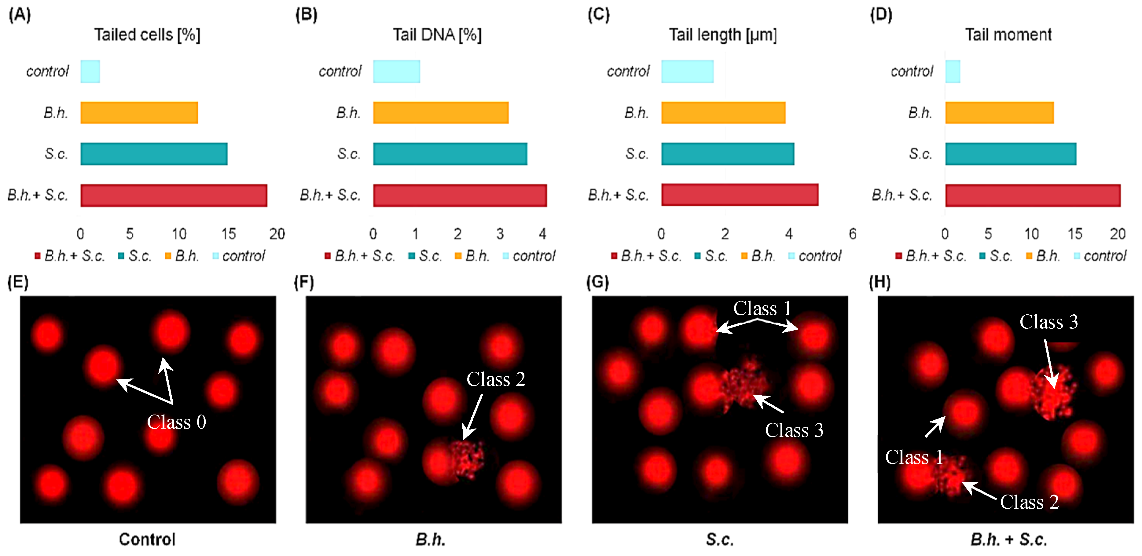
DNA Damage Analysis: DNA damage was increased in the B.h.-, S.c.-, and B.h. + S.c.-treated groups compared to the control group, as evidenced by the strand breaks appearing as comet tails in the comet assay (Figure 4E–H). Additionally, the DNA-damaged cells classified in classes 1, 2, and 3 were more prevalent in the treated groups compared to the control (class 0).
Tail length (TL) fluorescence increased in classes 2 and 3 of the treated groups. Classes 1, 2, and 3 around the head indicated the frequency of cell death. In the control comet cells (class “0”), DNA migration from the nucleoid core was negligible (Figure 4A–D). Analysis of DNA damage revealed clear differences between insects from different treatments (Figure 4). In control insects, the amount of DNA in the comet tail (TDNA) did not exceed 1.12%, and the average tail length (TL) was just under 2 µm, indicating minimal damage. In contrast, insects from the treated groups exhibited significant DNA damage, reflected in the amount of fragmented DNA in the comet tail (mean TDNA ranged from 3.22% to 4.12%) and tail length (mean TL ranged from 3.91 µm to 4.94 µm).
Morphological Analysis of G. mellonella Larvae: The final larval instar of the Greater Wax Moth (G. mellonella) was infected with the entomopathogenic nematode S. carpocapsae and the ectoparasite B. hebetor, both individually and in combination. The morphology of these treated larvae was then compared to a control group. Figure 5 illustrates the observed larval aberrations following infection. The control larva (Figure 5A) displayed a normal appearance with a light creamy color, similar to the B.h. group larvae (Figure 5B). In contrast, larvae in the S.c. group exhibited shrinkage and a dark crimson coloration (Figure 5C), while those in the B.h. + S.c. group shrank and turned black (Figure 5D).
Uninfected G. mellonella Larval Morphology (Figure 6, Figure 7 and Figure 8): Figure 6, Figure 7 and Figure 8 depict the morphology of the uninfected last larval instar of G. mellonella. These larvae possess a hardened head capsule (sclerotized). The thorax is composed of three segments, each bearing a pair of segmented legs. Additionally, a pair of spiracles (breathing pores) can be found on the lateral sides (pleura) of the prothorax and metathorax segments. The abdomen consists of ten segments, each with a hardened upper plate (tergum) and a lower plate (sternum) connected by a membranous pleural region. The first eight abdominal segments each have a pair of spiracles, while segments 3, 4, 5, 6, and 10 have five pairs of prolegs (fleshy appendages) ending in hooks (crochets) used for locomotion.

3.3. SEM Analysis

SEM revealed several abnormalities and malformations in the cuticle and associated sensilla of G. mellonella larvae from the treated groups (B.h., S.c., and B.h. + S.c.) compared to the control group. Obvious distortions and damage to the cuticle were observed in various body segments (Figure 6, Figure 7 and Figure 8).
Figure 6 presents SEM photomicrographs showcasing the dorsal morphology of the larvae. Panels a-d depict the dorsal anterior view, while panels A-D represent the dorsal posterior end. Unlike the smooth cuticles in the control group, the B.h. and S.c. groups had ridges and tears on their tergum cuticle (Figure 6B,b,C,c). Notably, larvae in the B.h. + S.c. group displayed a rough and wrinkled cuticle (rugose) (Figure 6D,d).
The ventral side of the larvae also exhibited numerous abnormalities as revealed by SEM analysis (Figure 7). These abnormalities included a ripped sternum cuticle, a ridged cuticular surface, disorganized hair-like sensory structures (trichoid sensilla), and indistinct segmentation.
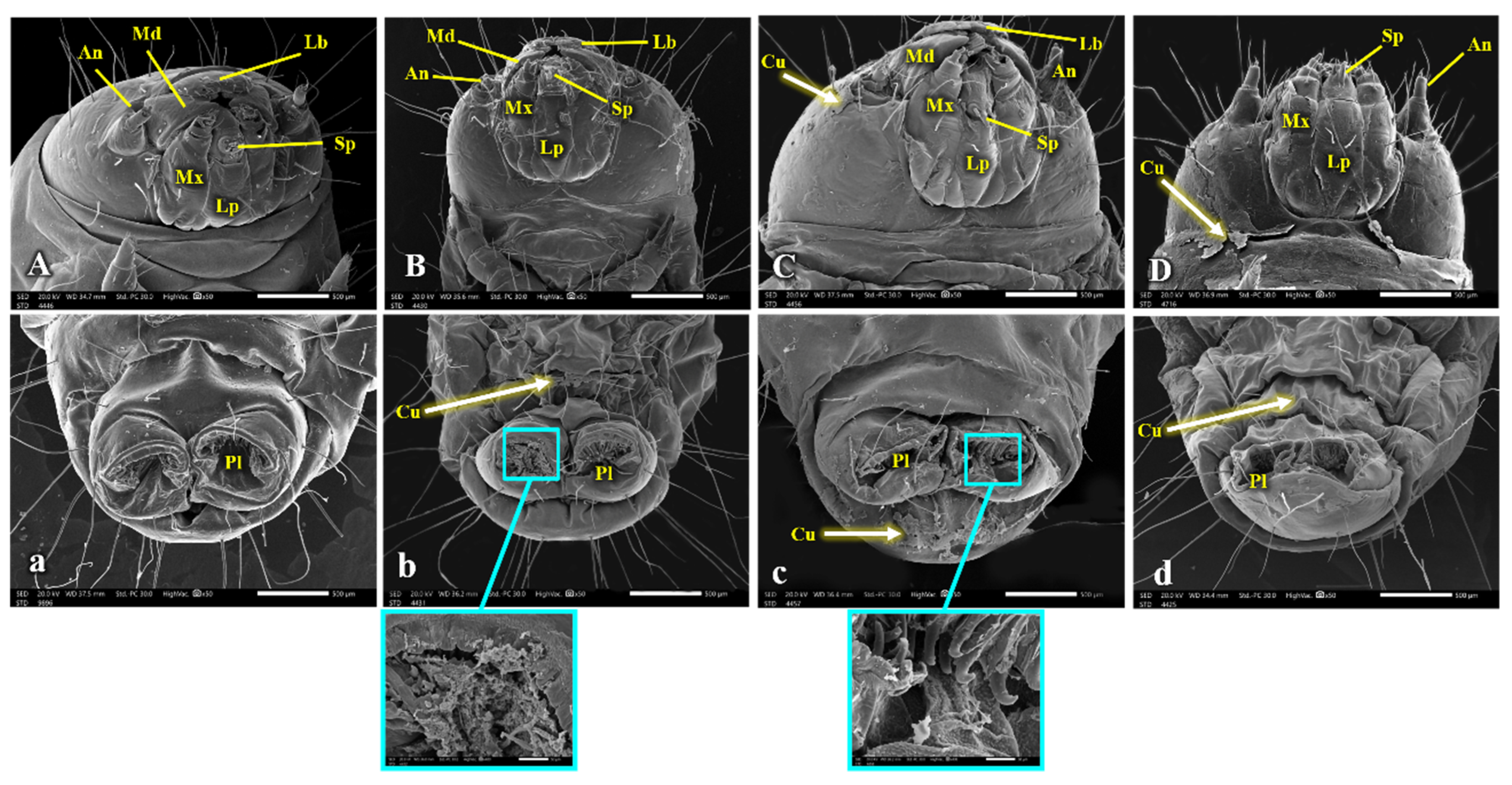
Figure 8 focuses on the head capsule and caudal segment of the larvae. Observations of the caudal segment revealed sunken prolegs and missing crochets (Figure 8d), along with a ruptured sternum cuticle at the abdominal end (Figure 8c). Additionally, the SEM images show that the cuticle of the head capsule shed separately from the rest of the body (Figure 8C,D).

4. Discussion

The greater wax moth, G. mellonella, is a major global pest of beehives. Its larvae are also widely used as a model organism to study insect infections and evaluate the effectiveness of biocontrol agents. This study provides valuable insights into the molecular mechanisms underlying G. mellonella’s responses to parasitic infections by the idiobiont ectoparasitoid B. hebetor and the entomopathogenic nematode S. carpocapsae. Understanding the ecophysiological effects of B. hebetor on pest insects’ metabolism, immunity, and genome is crucial for developing environmentally friendly bioinsecticides. Such knowledge can contribute to sustainable integrated biological control strategies for managing G. mellonella populations [65].
Insects rely on antioxidant metabolism to maintain physiological homeostasis and respond to various stress stimuli, including parasitoid attack. Parasites generate reactive oxygen species (ROS) that lead to oxidative stress and, tissue damage, and exacerbate infection pathology. Nematode attacks can also cause significant metabolic stress in host insects by inducing enzymatic changes [66]. In response to these stressors, G. mellonella typically increases the activity of antioxidant enzymes such as SOD, GST, and GPx. Studies have shown that parasitoids can affect these enzymes in their host insects, influencing their immune and defense systems [67,68].
Oxidative stress in biological systems arises from superoxide anion radicals, hydroxyl radicals, and hydrogen peroxide produced during normal cellular activities. These ROS are toxic to cellular processes, but their role in insect immune defense is becoming increasingly recognized. Our investigation found a significant rise in SOD levels six hours after infection, which aligns with previous findings [69]. SOD plays a critical role in mitigating oxidative stress induced by various stressors and is involved in the innate immune response of many invertebrates [70].
Our results demonstrate that parasitization by B. hebetor and S. carpocapsae, both individually and in combination (B.h. and S.c. groups), led to a downregulation of SOD, GPx, and GST activity compared to control larvae. These findings are consistent with previous studies suggesting changes in antioxidant gene levels following parasitism [71,72]. For example, Shafeeq et al. (2017) [73] reported that envenomation by B. hebetor downregulated SOD activity in the host Plodia interpunctella. Similarly, parasitization by P. turionellae significantly decreased SOD activity in host hemolymph [74].
GPx detoxifies lipid peroxides and protects cell membranes from oxidative damage. Our results regarding GPx activity differ from those of Duman Erbas & Altuntas (2021) [75], who observed no significant changes in GPx activity in G. mellonella larvae treated with juglone. GST plays another crucial role in insect detoxification. While our study showed increased GST activity in control larvae compared to infected groups, Çim & Altuntaş (2021) [74] reported a decrease in GST activity in parasitized host hemolymph. These discrepancies highlight the potential influence of different parasitoid species, venom composition, and experimental conditions on host insect antioxidant enzyme regulation. Interestingly, some studies suggest that idiobiont endoparasitoid venom may reduce oxidative stress in the host-parasite system, potentially explaining the observed downregulation of antioxidant enzymes in our study. MDA concentration, a byproduct of lipid peroxidation and a marker of oxidative stress, was significantly higher in the combination (B.h. + S.c.) group compared to control larvae. This finding suggests increased oxidative damage in response to the combined parasitism. It contrasts with the decreased MDA levels reported by Çim & Altuntaş (2021) [74] following parasitization by P. turionellae, highlighting potential differences in host responses depending on the specific parasite species. Additionally, studies have shown that force-feeding okadaic acid to G. mellonella larvae increases MDA levels, leading to tissue damage and oxidative stress [67].
Our study also revealed activation of cytochrome P450 enzymes in response to G. mellonella parasitism. This aligns with the findings of Rewitz et al. (2006a, 2006b, 2006c) [76,77,78], who demonstrated the role of P450s in insect detoxification. However, the specific activities of these enzymes in parasitoid wasps remain understudied. Notably, research indicates that conserved P450s often play developmental roles, such as those involved in 20E biosynthesis in Drosophila melanogaster and Manduca sexta.
Annexins are multifunctional proteins with roles in both cell biology and pathology. They can be expressed in parasite structures and secretions, as well as in host cells infected by parasites [79]. Our results, demonstrating increased apoptotic cells in parasitized groups, support findings that suggest annexin targeting can influence host defense during infections [80]. Targeting or inhibiting these proteins may have various consequences during illnesses and infections, including parasitic diseases. Potential benefits include reducing infection-induced acute inflammation or enhancing host defense mechanisms [80,81].
DNA damage resulting from parasitism can be evaluated using the comet assay, which analyzes tail migration to assess the extent of DNA strand fragmentation. Studies on the DNA-damaging effects of various parasites on insects using this technique are limited. Our study demonstrates that parasitism by B. hebetor, S. carpocapsae, or their combination induces different levels of DNA damage in G. mellonella larvae. These findings are consistent with previous work by Çim & Altuntaş (2021) [74] who investigated the genotoxic effects of P. turionellae venom on G. mellonella. The study showed that wasp venom damaged the DNA of host pupae more severely at all tested concentrations. This damage increased over time. The results suggest that wasp venom creates oxidative stress in the host, harming its enzymes and DNA. These changes might create a suitable environment for the wasp larvae to develop successfully.
Chitin, a polymer of N-acetyl-D- glucosamine, is a major component of the insect cuticle and is primarily produced by nematodes and constitutes a large portion of the insect cuticle. Breaches in these cuticular barriers trigger the activation of G. mellonella’s defensive mechanisms. Nematode infective juveniles (IJs) typically penetrate the host’s body through natural openings or areas with a thinner cuticle [82]. Our SEM results revealed that parasitism by B. hebetor, S. carpocapsae, or their combination (B.h. + S.c.) caused qualitative differences in the external morphology between control and affected G. mellonella larvae. These results support previous research, which indicates that parasitization by B. hebetor and entomopathogenic nematodes can cause morphological abnormalities in the host cuticle. In response to nematode infection, insect pests have evolved various morphological and immunological defense strategies [83]. G. mellonella relies on a chitin-based integument for protection. This integument also shields vital ectodermal organs like the trachea, foregut, and hindgut from infections [84].

5. Conclusions

Entomopathogenic nematodes and parasitoid wasps play a vital role in biological pest control strategies. This study has demonstrated significant physiological, molecular, and morphological impacts on G. mellonella larvae following parasitism by B. hebetor, S. carpocapsae, or both (B.h. + S.c.). These findings not only validate previous research on the individual effects of these biocontrol agents but also highlight the potential of a novel and effective strategy: the combined parasitism of B. hebetor and entomopathogenic nematodes for controlling G. mellonella populations (integrated pest management approach) (Figure 9). This combined approach presents a promising path for developing sustainable and eco-friendly pest management practices.

Author Contributions

Conceptualization, N.A.A., Z.A.E-M., L.M.E-S. and H.S.H.; methodology, N.A.A. and Z.A.E-M.; validation, N.A.A. and Z.A.E-M.; formal analysis, M.A., Z.A.E-M., L.M.E-S. and H.S.H.; investigation Z.A.E-M., L.M.E-S. and H.S.H.; data curation, M.A.; writing—original draft preparation, L.M.E-S., Z.A.E-M. and H.S.H.; writing—review and editing, L.M.E-S., H.S.H., Z.A.E-M., N.A.A. and M.A.; visualization, N.A.A. and Z.A.E-M.; supervision Z.A.E-M., L.M.E-S. and H.S.H. All authors have read and agreed to the published version of the manuscript.

Funding

This research received no external funding.

Data Availability Statement

Data is contained within the article.

Conflicts of Interest

The authors declare no conflicts of interest.

References

  1. Singkum, P.; Suwanmanee, S.; Pumeesat, P.; Luplertlop, N. A powerful in vivo alternative model in scientific research: Galleria mellonella. Acta Microbiol. Immunol. Hung. 2019, 66, 31–55. [Google Scholar] [CrossRef] [PubMed]
  2. Hickin, M.; Nadel, H.; Schal, C.; Cohen, A.C. Optimization of a diet for the greater wax moth (Lepidoptera: Pyralidae) using full factorial and mixture design. J. Econ. Entomol. 2021, 114, 1091–1103. [Google Scholar] [CrossRef]
  3. Serrano, I.; Verdial, C.; Tavares, L.; Oliveira, M. The virtuous Galleria mellonella model for scientific experimentation. Antibiotics 2023, 12, 505. [Google Scholar] [CrossRef] [PubMed]
  4. Champion, O.L.; Wagley, S.; Titball, R.W. Galleria mellonella as a model host for microbiological and toxin research. Virulence 2016, 7, 840–845. [Google Scholar] [CrossRef]
  5. Wright, C.L.; Kavanagh, O. Galleria mellonella as a novel in vivo model to screen natural product-derived modulators of innate immunity. Appl. Sci. 2022, 12, 6587. [Google Scholar] [CrossRef]
  6. Natarajan, V.; Anusha, N.; Govindarajan, S.; Priya, S. Chapter: 10 insect resistance to pesticides. In Entomology Chronicles: A Collaborative Insight Volume 1; Stella Publications: Kurukshetra, India, 2024. [Google Scholar]
  7. Zhang, Y.; Xu, H.; Tu, C.; Han, R.; Luo, J.; Xu, L. Enhanced capacity of a leaf beetle to combat dual stress from entomopathogens and herbicides mediated by associated microbiota. Integr. Zool. 2024, 27, 63. [Google Scholar] [CrossRef]
  8. Garcia, F.R.M.; Ovruski, S.M.; Suárez, L.; Cancino, J.; Liburd, O.E. Biological Control of Tephritid Fruit Flies in the Americas and Hawaii: A Review of the Use of Parasitoids and Predators. Insects 2020, 11, 662. [Google Scholar] [CrossRef]
  9. Kwadha, C.A.; Ong’amo, G.O.; Ndegwa, P.N.; Raina, S.K.; Fombong, A.T. The biology and control of the greater wax moth, Galleria mellonella. Insects 2017, 8, 61. [Google Scholar] [CrossRef] [PubMed]
  10. Dai, M.L.; Ye, W.T.; Jiang, X.J.; Feng, P.; Zhu, Q.Y.; Sun, H.N.; Li, F.C.; Wei, J.; Li, B. Effect of tachinid parasitoid Exorista japonica on the larval development and pupation of the host silkworm Bombyx mori. Front. Physiol. Invertebr. Physiol. 2022, 13, 824203. [Google Scholar] [CrossRef]
  11. Zhang, Y.; Zhang, S.; Xu, L. The pivotal roles of gut microbiota in insect plant interactions for sustainable pest management. NPJ Biofilms Microbiomes 2023, 9, 66. [Google Scholar] [CrossRef]
  12. Dieckhoff, C.; Tatman, K.M.; Hoelmer, K.A. Natural biological control of Halyomorpha halys by native egg parasitoids: A multi-year survey in northern Delaware. J. Pest Sci. 2017, 90, 1143–1158. [Google Scholar] [CrossRef]
  13. Resh, V.H.; Cardé, R.T. Encyclopedia of Insects; Academic Press: Cambridge, MA, USA, 2009. [Google Scholar] [CrossRef]
  14. Cuny, M.A.; Poelman, E.H. Evolution of koinobiont parasitoid host regulation and consequences for indirect plant defence. Evol. Ecol. 2022, 36, 299–319. [Google Scholar] [CrossRef]
  15. Arbuckle, K. Evolutionary context of venom in animals. In Evolution of Venomous Animals and Their Toxins; Springer: Dordrecht, The Netherlands, 2017; Volume 24, pp. 3–31. [Google Scholar] [CrossRef]
  16. Battaglia, D.; Colella, T.; Laurino, S.; Grossi, G.; Salvia, R.; Riviello, L.; Falabella, P. The effect of Leptomastix dactylopii parasitism and venom injection on host Planococcus citri. Invertebr. Surviv. J. 2014, 11, 273–285. [Google Scholar]
  17. Laurino, S.; Grossi, G.; Pucci, P.; Flagiello, A.; Bufo, S.A.; Bianco, G.; Falabella, P. Identification of major Toxoneuron nigriceps venom proteins using an integrated transcriptomic/proteomic approach. Insect Biochem. Mol. Biol. 2016, 76, 49–61. [Google Scholar] [CrossRef] [PubMed]
  18. Schendel, V.; Rash, L.D.; Jenner, R.A.; Undheim, E.A. The diversity of venom: The importance of behavior and venom system morphology in understanding its ecology and evolution. Toxins 2019, 11, 666. [Google Scholar] [CrossRef] [PubMed]
  19. Schmidt, J.O. Biochemistry of insect venoms. Ann. Rev. Entomol. 1982, 27, 339–368. [Google Scholar] [CrossRef] [PubMed]
  20. Scieuzo, C.; Salvia, R.; Franco, A.; Pezzi, M.; Cozzolino, F.; Chicca, M.; Ferracini, C. An integrated transcriptomic and proteomic approach to identify the main Torymus sinensis venom components. Sci. Rep. 2021, 11, 5032. [Google Scholar] [CrossRef] [PubMed]
  21. Bhatti, F.; Hussain, F.; Arshad, M.; Abbas, S.K.; Shabbir, M.Z. Effects of envenomation on survival and development of the host-parasitoid system of Galleria mellonella (Lepidoptera: Pyralidae) and Bracon hebetor (Hymenoptera: Braconidae). Pak. J. Zool. 2023, 55, 1073. [Google Scholar] [CrossRef]
  22. Cusumano, A.; Duvic, B.; Jouan, V.; Ravallec, M.; Legeai, F.; Peri, E.; Volkoff, A.-N. First extensive characterization of the venom gland from an egg parasitoid: Structure, transcriptome and functional role. J. Insect Physiol. 2018, 107, 68–80. [Google Scholar] [CrossRef]
  23. Falabella, P. The mechanism utilized by Toxoneuron nigriceps in inhibiting the host immune system. Invertebr. Surviv. J. 2018, 15, 240–255. [Google Scholar] [CrossRef]
  24. Gu, Q.; Wu, Z.; Zhou, Y.; Wang, Z.; Shi, M.; Huang, J.; Chen, X. A teratocyte-specific serpin from the endoparasitoid wasp Cotesia vestalis inhibits the prophenoloxidase-activating system of its host Plutella xylostella. Insect Mol. Biol. 2022, 31, 202–215. [Google Scholar] [CrossRef] [PubMed]
  25. Pennacchio, F.; Caccia, S.; Digilio, M.C. Host regulation and nutritional exploitation by parasitic wasps. Curr. Opin. Insect Sci. 2014, 6, 74–79. [Google Scholar] [CrossRef] [PubMed]
  26. Moreau, S.J.; Asgari, S. Venom proteins from parasitoid wasps and their biological functions. Toxins 2015, 7, 2385–2412. [Google Scholar] [CrossRef] [PubMed]
  27. Varricchio, P.; Falabella, P.; Sordetti, R.; Graziani, F.; Malva, C.; Pennacchio, F. Cardiochiles nigriceps polydnavirus: Molecular characterization and gene expression in parasitized Heliothis virescens larvae. Insect Biochem. Mol. Biol. 1999, 29, 1087–1096. [Google Scholar] [CrossRef] [PubMed]
  28. Fathy, R.; Zohdy, N.; Abd-El-Samie, E.M.; Abou-Elfadl, H.; Moamen, S.; Younes, A. Effects of parasitism by the braconid wasp, Bracon hebetor (Hymenoptera: Braconidae), on the host hemolymph and phenoloxidase activation of the rice moth, Corcyra cephalonica larvae (Lepidoptera: Pyralidae). Egypt. J. Biol. Pest Control 2023, 33, 30. [Google Scholar] [CrossRef]
  29. Adams, B.J.; Fodor, A.; Koppenhöfer, H.S.; Stackebrandt, E.; Stock, S.P.; Klein, M.G. Reprint of “Biodiversity and systematics of nematode–bacterium entomopathogens” [Biol. Control 37 (2006) 32–49]. Biol. Control 2006, 38, 4–21. [Google Scholar] [CrossRef]
  30. Shapiro-Ilan, D.; Hazir, S.; Glazer, I. Advances in use of entomopathogenic nematodes in integrated pest management. In Integrated Management of Insect Pests; Burleigh Dodds Science Publishing: Cambridge, UK, 2019; pp. 649–678. [Google Scholar]
  31. Le Vieux, P.; Malan, A.P. The potential use of entomopathogenic nematodes to control Planococcus ficus (Signoret) (Hemiptera: Pseudococcidae). S. Afr. J. Ecol. Vitic. 2013, 34, 269–306. [Google Scholar] [CrossRef]
  32. Lewis, E.E.; Ricci, M.; Gaugler, R. Host recognition behavior reflects host suitability for the entomopathogenic nematode, Steinernema carpocapsae. Parasitology 1996, 113, 573–579. [Google Scholar] [CrossRef] [PubMed]
  33. Shapiro-Ilan, D.I.; Hiltpold, I.; Lewis, E.E. Ecology of invertebrate pathogens: Nematodes. In Ecology of Invertebrate Diseases; Hajek, A.E., Shapiro-Ilan, D.I., Eds.; John Wiley & Sons: Hoboken, NJ, USA, 2018; pp. 415–440. [Google Scholar]
  34. Koppenhöfer, A.M.; Grewal, P.S. Compatibility and interactions with agrochemicals and other biocontrol agents. In Nematodes as Biocontrol Agents; Grewal, P.S., Ehlers, R.-U., Shapiro-Ilan, D.I., Eds.; CABI Publishing: Wallingford, UK, 2005; pp. 363–381. [Google Scholar]
  35. Atwa, A.A.; Hegazi, E.M.; Khafagi, W.E.; Abd El-Aziz, G.M. Interaction of the koinobiont parasitoid Microplitis rufiventris of the cotton leafworm, Spodoptera littoralis, with two entomopathogenic rhabditids, Heterorhabditis bacteriophora and Steinernema carpocapsae. J. Insect Sci. 2013, 13, 84. [Google Scholar] [CrossRef]
  36. Aamer, N.A. Effect of parasitism by Bracon hebetor (Say) on Nematode yields of Steinernema carpocapsae in wax moth larvae, Galleria mellonella (L.). Egypt. J. Biol. Pest Control 2017, 27, 129–134. [Google Scholar]
  37. Farooqui, T.; Farooqui, A.A. Oxidative Stress in Vertebrates and Invertebrates: Molecular Aspects of Cell Signaling; Wiley-Blackwell: Hoboken, NJ, USA, 2012; pp. 377–384. [Google Scholar] [CrossRef]
  38. Halliwell, B.; Gutteridge, J.M. Free Radicals in Biology and Medicine; Oxford University Press: New York, NY, USA, 2015. [Google Scholar]
  39. Özcan, O.; Erdal, H.; Çakırca, G.; Yönden, Z. Oksidatif stres ve hücre içi lipit, protein ve DNA yapıları üzerine etkileri. J. Clin. Exp. Investig. 2015, 6, 331–336. [Google Scholar] [CrossRef]
  40. Ahmad, S. Biochemical defence of pro-oxidant plant allelochemicals by herbivorous insects. Biochem. Syst. Ecol. 1992, 20, 269–296. [Google Scholar] [CrossRef]
  41. Altuntaş, H.; Duman, E.; Demirci, S.N.Ş.; Ergin, E. Toxicological and physiological effects of ethephon on the model organism, Galleria mellonella L. 1758 (Lepidoptera: Pyralidae). Turk. J. Entomol. 2016, 40, 413–423. [Google Scholar] [CrossRef]
  42. Büyükgüzel, E.; Kalender, Y. Exposure to streptomycin alters oxidative and antioxidative response in larval midgut tissues of Galleria mellonella. Pest. Biochem. Physiol. 2009, 94, 112–118. [Google Scholar] [CrossRef]
  43. Güneş, E.; Büyükgüzel, E. Oxidative effects of boric acid on different developmental stages of Drosophila melanogaster Meigen, 1830 (Diptera: Drosophilidae). Turk. J. Entomol. 2017, 41, 3–15. [Google Scholar] [CrossRef]
  44. Kodrík, D.; Bednářová, A.; Zemanová, M.; Krishnan, N. Hormonal regulation of response to oxidative stress in insects—An update. Int. J. Mol. Sci. 2015, 16, 25788–25816. [Google Scholar] [CrossRef]
  45. Surai, P.F. Silymarin as a natural antioxidant: An overview of the current evidence and perspectives. Antioxidants 2015, 4, 204–247. [Google Scholar] [CrossRef]
  46. R Core Team. R A Language and Environment for Statistical Computing; R Foundation for Statistical Computing: Vienna, Austria, 2016. [Google Scholar] [CrossRef]
  47. Qian, X. Experimental study on the control effect of 24% chlorfenapyr suspending agent on the mulberry pest, Glyphodes pyloalis (Walker). China Seric 2012, 33, 48–49. [Google Scholar] [CrossRef]
  48. Feyereisen, R. Insect CYP genes and P450 enzymes. In Insect Molecular Biology and Biochemistry; Elsevier: Amsterdam, The Netherlands, 2012; pp. 236–316. [Google Scholar]
  49. Draeger, A.; Monastyrskaya, K.; Babiychuk, E.B. Plasma membrane repair and cellular damage control: The annexin survival kit. Biochem. Pharmacol. 2011, 81, 703–712. [Google Scholar] [CrossRef]
  50. Gerke, V.; Moss, S.E. Annexins: From structure to function. Physiol. Rev. 2002, 82, 331–371. [Google Scholar] [CrossRef] [PubMed]
  51. El-Ashram, S.; Kheirallah, D.A.M.; El-Samad, L.M.; Toto, N.A. Relative expression of microRNAs, apoptosis, and ultrastructure anomalies induced by gold nanoparticles in Trachyderma hispida (Coleoptera: Tenebrionidae). PLoS ONE 2020, 15, e0241837. [Google Scholar] [CrossRef]
  52. Mishra, N.; Srivastava, R.; Agrawal, U.R.; Tewari, R.R. An insight into the genotoxicity assessment studies in dipterans. Mutat. Res./Rev. Mutat. Res. 2017, 773, 220–229. [Google Scholar] [CrossRef]
  53. Augustyniak, M.; Gladysz, M.; Dziewięcka, M. The Comet assay in insects—Status, prospects and benefits for science. Mutat. Res./Rev. Mutat. Res. 2016, 767, 67–76. [Google Scholar] [CrossRef] [PubMed]
  54. Kaur, M.; Chadha, P.; Kaur, S.; Kaur, A.; Kaur, R.; Yadav, A.K.; Kaur, R. Schizophyllum commune induced genotoxic and cytotoxic effects in Spodoptera litura. Sci. Rep. 2018, 8, 4693. [Google Scholar] [CrossRef] [PubMed]
  55. Singh, S. Technology for Production of Natural Enemies; Project Directorate of Biological Control: Bangalore, India, 1994. [Google Scholar]
  56. Woodring, J.L. Steinernematid and Heterorhabditid Nematodes: A Handbook of Biology and Techniques; Southern Cooperative Series Bulletin; Arkansas Agricultural Experiment Station: Fayetteville, AR, USA, 1988; Volume 331, 30p. [Google Scholar]
  57. White, G. A method for obtaining infective nematode larvae from cultures. Science 1927, 66, 302–303. [Google Scholar] [CrossRef] [PubMed]
  58. Ohkawa, H.; Ohishi, N.; Yagi, K. Assay for lipid peroxides in animal tissues by thiobarbituric acid reaction. Anal. Biochem. 1979, 95, 351–358. [Google Scholar] [CrossRef] [PubMed]
  59. Paglia, D.E.; Valentine, W.N. Studies on the quantitative and qualitative characterization of erythrocyte glutathione peroxidase. J. Lab. Clin. Med. 1967, 70, 158–169. [Google Scholar] [PubMed]
  60. Yu, S. Host plant induction of glutathione S-transferase in the fall armyworm. Pest. Biochem. Physiol. 1982, 18, 101–106. [Google Scholar] [CrossRef]
  61. Yu, S.J.; Hsu, E.L. Induction of detoxification enzymes in phytophagous insects: Role of insecticide synergists, larval age, and species. Arch. Insect Biochem. Physiol. 1993, 24, 21–32. [Google Scholar] [CrossRef]
  62. Misra, H.P.; Fridovich, I. The role of superoxide anion in the autoxidation of epinephrine and a simple assay for superoxide dismutase. J. Biol. Chem. 1972, 247, 3170–3175. [Google Scholar] [CrossRef]
  63. Brogdon, W.; Chan, A. Guidelines for Evaluating Insecticide Resistance in Vectors Using the CDC Bottle Bioassay/Methods in Anopheles Research; CDC Technical Report; CDC: Atlanta, GA, USA, 2010. Available online: https://stacks.cdc.gov/view/cdc/21777/cdc_21777_DS1.pdf (accessed on 24 July 2024).
  64. Singh, N.P.; McCoy, M.T.; Tice, R.R.; Schneider, E.L. A simple technique for quantitation of low levels of DNA damage in individual cells. Exp. Cell Res. 1988, 175, 184–191. [Google Scholar] [CrossRef]
  65. Dmochowska-Ślęzak, K.; Giejdasz, K.; Fliszkiewicz, M.; Żółtowska, K. Variations in antioxidant defense during the development of the solitary bee Osmia bicornis. Apidologie 2015, 46, 432–444. [Google Scholar] [CrossRef]
  66. Ibrahim, S.A.; Salem, H.H.; Taha, M. Dual application of entomopathogenic nematodes and fungi on immune and antioxidant enzymes of the greater wax moth, Galleria mellonella L. Egypt. J. Biol. Pest Control 2019, 29, 1–7. [Google Scholar] [CrossRef]
  67. Coates, C.J.; Lim, J.; Harman, K.; Rowley, A.F.; Griffiths, D.J.; Emery, H.; Layton, W. The insect, Galleria mellonella, is a compatible model for evaluating the toxicology of okadaic acid. Cell Biol. Toxicol. 2019, 35, 219–232. [Google Scholar] [CrossRef] [PubMed]
  68. Lalitha, K.; Karthi, S.; Vengateswari, G.; Karthikraja, R.; Perumal, P.; Shivakumar, M. Effect of entomopathogenic nematode of Heterorhabditis indica infection on immune and antioxidant system in lepidopteran pest Spodoptera litura (Lepidoptera: Noctuidae). J. Parasit. Dis. 2018, 42, 204–211. [Google Scholar] [CrossRef] [PubMed]
  69. Zóltowska, K.; Grochla, P.; Lopienska-Biernat, E. Activity of superoxide dismutase in Galleria mellonella larvae infected with entomopathogenic nematodes Steinernema affinis and S. feltiae. Ann. Parasitol. 2006, 52, 283–286. [Google Scholar]
  70. Zhu, J.; Ze, S.Z.; Stanley, D.W.; Yang, B. Parasitization by Scleroderma guani influences expression of superoxide dismutase genes in Tenebrio molitor. Arch. Insect Biochem. Physiol. 2014, 87, 40–52. [Google Scholar] [CrossRef] [PubMed]
  71. Nguyen, T.; Boudreault, S.; Michaud, D.; Cloutier, C. Proteomes of the aphid Macrosiphum euphorbiae in its resistance and susceptibility responses to differently compatible parasitoids. Insect Biochem. Mol. Biol. 2008, 38, 730–739. [Google Scholar] [CrossRef] [PubMed]
  72. Zhu, J.; Yang, P.; Zhang, Z.; Wu, G.-X.; Yang, B. Transcriptomic immune response of Tenebrio molitor pupae to parasitization by Scleroderma guani. PLoS ONE 2013, 8, e54411. [Google Scholar] [CrossRef] [PubMed]
  73. Shafeeq, T.; Ulabdin, Z.; Lee, K.Y. Induction of stress and immune associated genes in the Indian meal moth Plodia interpunctella against envenomation by the ectoparasitoid Bracon hebetor. Arch. Insect Biochem. Physiol. 2017, 96, e21405. [Google Scholar] [CrossRef]
  74. Çim, S.; Altuntaş, H. Anti-oxidative, genotoxic and mutagenic effects of idiobiont, endoparasitoid, Pimpla turionellae L. (Hymenoptera: Ichneumonidae) venom on its host Galleria mellonella L. (Lepidoptera: Pyralidae). Biol. Control 2021, 158, 104595. [Google Scholar] [CrossRef]
  75. Duman Erbas, E.; Altuntas, H. Effects of Juglone on the Antioxidant Metabolism in the Larval Hemolymph of the Greater Wax Moth Galleria mellonella L. (Lepidoptera: Pyralidae). Karadeniz Bilim. Derg. 2021, 11, 18–28. [Google Scholar] [CrossRef]
  76. Rewitz, K.F.; Rybczynski, R.; Warren, J.T.; Gilbert, L.I. Developmental expression of Manduca shade, the P450 mediating the final step in molting hormone synthesis. Mol. Cell. Endocrinol. 2006, 247, 166–174. [Google Scholar] [CrossRef] [PubMed]
  77. Rewitz, K.F.; Rybczynski, R.; Warren, J.T.; Gilbert, L.I. The Halloween genes code for cytochrome P450 enzymes mediating synthesis of the insect moulting hormone. Biochem. Soc. Trans. 2006, 34, 1256–1260. [Google Scholar] [CrossRef] [PubMed]
  78. Rewitz, K.F.; Rybczynski, R.; Warren, J.T.; Gilbert, L.I. Identification, characterization and developmental expression of Halloween genes encoding P450 enzymes mediating ecdysone biosynthesis in the tobacco hornworm, Manduca sexta. Insect Biochem. Mol. Biol. 2006, 36, 188–199. [Google Scholar] [CrossRef] [PubMed]
  79. Rashidi, S.; Mansouri, R.; Ali-Hassanzadeh, M.; Muro, A.; Nguewa, P.; Manzano-Román, R. The most prominent modulated annexins during parasitic infections. Acta Trop. 2023, 243, 106942. [Google Scholar] [CrossRef] [PubMed]
  80. de Araújo, S.; de Melo Costa, V.R.; Santos, F.M.; de Sousa, C.D.F.; Moreira, T.P.; Gonçalves, M.R.; Nogueira, M.L. Annexin A1-FPR2/ALX signaling Axis regulates acute inflammation during chikungunya virus infection. Cells 2022, 11, 2717. [Google Scholar] [CrossRef] [PubMed]
  81. Vago, J.P.; Tavares, L.P.; Riccardi, C.; Teixeira, M.M.; Sousa, L.P. Exploiting the pro-resolving actions of glucocorticoid-induced proteins Annexin A1 and GILZ in infectious diseases. Biomed. Pharmacother. 2021, 133, 111033. [Google Scholar] [CrossRef] [PubMed]
  82. Lacey, L.A.; Georgis, R. Entomopathogenic nematodes for control of insect pests above and below ground with comments on commercial production. J. Nematol. 2012, 44, 218. [Google Scholar] [PubMed]
  83. Michalková, V.; Valigurová, A.; Dindo, M.L.; Vaňhara, J. Larval Morphology and Anatomy of the Parasitoid Exorista larvarum (Diptera: Tachinidae), with an Emphasis on Cephalopharyngeal Skeleton and Digestive Tract. J. Parasitol. 2009, 95, 544–554. [Google Scholar] [CrossRef]
  84. Wojda, I. Immunity of the greater wax moth Galleria mellonella. Insect Sci. 2017, 24, 342–357. [Google Scholar] [CrossRef]
Figure 1. Galleria mellonela rearing (A), culture of parasites: Steinernema carpocapsae (B) and Bracon hebetor (C), and parasitism by Bracon hebetor (B.h.) on Galleria mellonella larvae (DF).
Insects 15 00588 g001
Figure 2. Mean ± SD of (A) Malondialdehyde (MDA) content and activity of (B) Superoxide dismutase (SOD), (C) Glutathione peroxidase (GPx), (D) Glutathione S-transferase (GST), and (E) Cytochrome P450, as well as (F) Principal Component Analysis (PCA) on all variables measured in larvae of Galleria mellonella from the control group or those infected separately with the parasites Bracon hebetor (B.h.) or nematode Steinernema carpocapsae (S.c.) or combined parasitism (B.h. + S.c.). Groups with the same letters indicate no significant difference between them (Tukey’s post hoc, p < 0.05).
Insects 15 00588 g002
Figure 3. The percentages of (A) viable, (B) early apoptotic, (C) late apoptotic, and (D) necrotic cells, determined using the Annexin V-FITC test kit in larvae of Galleria mellonella from the control group or those infected separately with the parasites Bracon hebetor (B.h.) or Nematode Steinernema carpocapsae (S.c.), or combined parasitism (B.h. + S.c.). (EH) examples of cytometric plots for control and experimental groups. The diagram of flow cytometric analysis was divided into four quadrants: necrotic cells (Q1: upper left quadrant), late apoptotic cells (Q2: upper right quadrant), living cells (Q3: lower left quadrant), and early apoptotic cells (Q4: lower right quadrant).
Insects 15 00588 g003
Figure 4. The percentages of (A) tailed cells, (B) amount of DNA in the comet tail, (C) tail length, and (D) tail moment, measured by the Comet assay in larvae of Galleria mellonella from the control group or those infected separately with the parasites Bracon hebetor (B.h.) or Nematode Steinernema carpocapsae (S.c.) or combined parasitism (B.h. + S.c.). (EH) examples of DNA-damaged nuclei for control and experimental groups.
Insects 15 00588 g004
Figure 5. Observed color changes in larvae of Galleria mellonella: (A) control larva with normal appearance characterized by light creamy color, (B) B.h. group (creamy), (C) S.c. group (dark crimson), and (D) B.h. + S.c. group (black).
Insects 15 00588 g005
Figure 6. SEM photography for dorsal anterior view (ad) and the dorsal posterior end (AD) of control and parasitic Galleria mellonella last instar larvae, showing: Hc, Head capsule; Th.S, Thorax segments; Ab, Abdomen; Cu, cuticle.
Insects 15 00588 g006
Figure 7. SEM photography for anterior ventral view (ad) and posterior ventral view (AD) of control and parasitic Galleria mellonella last instar larvae, showing: Hc, head capsule; Th.L, thoracic lags; Pr.L, abdominal prolegs; Cu, cuticle.
Insects 15 00588 g007
Figure 8. SEM photography for head capsule (AD) and posterior end (ad) of the control and parasitic Galleria mellonella last larval instar, showing: An, antenna; Lr, labrum; Lp, labium; Md, mandible; Mx, maxilla; Sp, spinneret; Cu, cuticle; Pl, abdominal prolegs.
Insects 15 00588 g008
Figure 9. Summary of the effects of Bracon hebetor and Steinernema carpocapsae on Galleria mellonella.
Insects 15 00588 g009
Disclaimer/Publisher’s Note: The statements, opinions and data contained in all publications are solely those of the individual author(s) and contributor(s) and not of MDPI and/or the editor(s). MDPI and/or the editor(s) disclaim responsibility for any injury to people or property resulting from any ideas, methods, instructions or products referred to in the content.