Article

Identification and Functional Insights of Knickkopf Genes in the Larval Cuticle of Leptinotarsa decemlineata

1
State Key Laboratory of Resource Insects, College of Sericulture, Textile, and Biomass Sciences, Southwest University, Chongqing 400715, China
2
Institute of Plant Protection Xinjiang Academy of Agricultural Sciences/Key Laboratory of Integrated Pest Management on Crops in Northwestern Oasis, Ministry of Agriculture/Xinjiang Key Laboratory of Agricultural Biosafety, Urumqi 830091, China
*
Author to whom correspondence should be addressed.
These authors contributed equally to this work.
Insects 2024, 15(8), 623; https://doi.org/10.3390/insects15080623
Submission received: 30 July 2024 / Revised: 12 August 2024 / Accepted: 16 August 2024 / Published: 19 August 2024
(This article belongs to the Section Insect Physiology, Reproduction and Development)

Abstract

:

Simple Summary

The Colorado potato beetle, Leptinotarsa decemlineata, poses a significant threat to potato crops worldwide, causing substantial agricultural damage. To better understand how these pests develop and potentially find ways to control them, we studied a group of genes called Knickkopf (Knk) genes in the larvae of the beetle. These genes are crucial for forming and maintaining the insect’s outer shell, known as the cuticle, which protects them from the environment. We identified four LdKnk-family genes in the beetle and analyzed when and where these genes are expressed during the insect’s growth stages. By using RNA interference (RNAi) to silence these genes, we observed that larvae became weaker, with damaged cuticles and higher death rates. These findings indicate that LdKnk-family genes are vital for the beetle’s survival and development. Targeting LdKnk-family genes could provide a new strategy for pest control, potentially weakening the beetles’ defenses and making them more susceptible to environmental factors and insecticides, ultimately helping to protect potato crops.

Abstract

The Colorado potato beetle (Leptinotarsa decemlineata) is a major pest of potato crops. While Knickkopf (Knk) genes are essential for insect cuticle formation, their roles in pests like L. decemlineata remain unclear. This study aims to identify and characterize Knk genes in L. decemlineata and explore their functions in larval development and cuticle integrity. We used genomic and transcriptomic databases to identify LdKnk-family genes, validated through RT-PCR and RACE. Gene expression was analyzed at various developmental stages and tissues using qRT-PCR. RNA interference (RNAi) and Transmission electron microscopy (TEM) were applied to determine the functional roles of these genes. Four LdKnk-family genes were identified. Spatio-temporal expression analysis indicated significant gene expression during larval molting and pupal stages, especially in the epidermis. RNAi experiments showed that silencing LdKnk and LdKnk3-5′ led to reduced larval weight, cuticle thinning, and increased mortality, while LdKnk3-FL knockdown caused abnormal cuticle thickening and molting disruptions. LdKnk2 knockdown increased epicuticle and endocuticle thickness without visible phenotypic changes. The study highlights the essential roles of LdKnk-family genes in maintaining cuticle structure and integrity, suggesting their potential as targets for RNAi-based pest control.

Graphical Abstract

1. Introduction

The insect cuticle is essential for maintaining an insect’s shape and providing protection against dehydration, pathogens, and environmental damage [1,2]. To accommodate growth, insects periodically shed their cuticle through molting, replacing the old cuticle with a new one [3,4,5,6]. TEM has been crucial for studying the ultrastructure of the insect cuticle, revealing its multi-layered architecture and the organization of chitin microfibrils. TEM studies have shown that the cuticle consists of three layers forming an extracellular matrix. The outer waxy envelope (env) acts as the primary barrier against water loss and foreign substances. Beneath it, the protein-rich epicuticle (epi) contributes to the insect’s overall body shape. The innermost procuticle (pro), composed of a protein-chitin matrix, provides stability [7,8,9].
Chitin, a homopolymer of β-1,4-linked N-acetylglucosamine units, is the main structural component of the insect cuticle [10,11,12,13,14,15]. When associated with proteins, chitin arranges into microfibrils that align parallel to each other, forming stacked helicoidal laminae [16,17,18,19]. Cuticle-associated proteins significantly influence chitin organization [1,20,21]. Among these, the chitin-binding protein Knickkopf (Knk) has been extensively studied in Drosophila melanogaster, Tribolium castaneum, and Locusta migratoria [22]. In D. melanogaster, Knk is crucial for chitin organization within the tracheal system, the larval body, and the adult wing cuticle [23,24]. Knk proteins comprise a signal peptide, two DM13 domains of unknown function, and a dopamine-monooxygenase (DOMON) domain essential for its activity [25]. It interacts with the chitin-binding protein Obstructor-A (Obst-A) and the chitin deacetylase Serpentine (Serp) to form a complex at the apical membrane, facilitating the deposition and organization of chitin fibers [26,27].
Studies on T. castaneum have revealed that Knk proteins bind to chitin, protecting it from enzymatic degradation during molting and ensuring proper laminar organization, playing roles in cuticle formation at different developmental stages and in different parts of the insect anatomy [28,29,30]. Additionally, in L. migratoria, LmKnk contributes to correct chitin content, pore canal formation, and lipid deposition during molting, with LmKnk3-5′ being essential for cuticle formation [31,32]. These findings suggest that Knk-family proteins may exhibit functional diversity across different insect species.
The Colorado potato beetle, L. decemlineata, is a significant agricultural pest that poses a severe threat to potato crops and farmers’ livelihoods [33,34]. Although insecticides have historically been effective in suppressing L. decemlineata populations, the emergence of insecticide-resistant populations has created significant management challenges [35,36,37]. Understanding the genetic and molecular mechanisms of cuticle formation in this species could offer valuable insights for pest management.
In this study, we aim to identify and characterize Knk genes in L. decemlineata and investigate their roles in larval development and cuticle integrity. We identified four Knk-family genes: LdKnk, LdKnk2, LdKnk3-full length (LdKnk3-FL), and LdKnk3-5′. Using qRT-PCR, we analyzed their expression across various developmental stages and tissues. RNAi experiments assessed their impact on larval development and cuticle structure. Our results demonstrate that Knk-family genes exhibit developmentally regulated and tissue-specific expression patterns. Specifically, LdKnk, LdKnk3-FL, and LdKnk3-5′ are essential for larval development in L. decemlineata, as confirmed by TEM analysis. These findings highlight potential targets for pest management and contribute to understanding the function of Knk genes in insects.

2. Materials and Methods

2.1. Experimental Insect

The rearing of L. decemlineata followed standardized protocols as previously described [38]. Adults were collected from potato fields in Urumqi, Xinjiang (43.82° N, 87.61° E) during their spring emergence. The beetles were kept at 28 ± 1 °C with a 14:10 light-dark cycle and 50–60% humidity and were fed fresh potato leaves. Eggs were laid on the underside of potato leaves and hatched into larvae within seven days. The larvae underwent four instars, with the first three instars, each lasting two days and the fourth instar lasting 3.5 days. After the larval period, the fourth-instar larvae ceased feeding, burrowed into the soil for pupation, and subsequently emerged as adults.

2.2. Identification of LdKnk-Family Genes

Using the known amino acid sequences of Knk proteins from T. castaneum, D. melanogaster, and L. migratoria as queries, a genome-wide TBLASTN search was conducted on the L. decemlineata genome data (https://www.hgsc.bcm.edu/arthropods/colorado-potato-beetle-genome-project (accessed on 1 January 2024), PRJNA854273). This search resulted in the identification of four putative Knk-like genes in L. decemlineata. These sequences were further used to identify additional Knk-like genes from the nonredundant (nr) NCBI nucleotide database and transcriptome databases (PRJNA464380 and our unpublished database) of L. decemlineata.
The sequences were verified via polymerase chain reaction (PCR) using primers listed in Table S1. They were then completed by 5′- and 3′-RACE using the SMARTer RACE cDNA amplification kit (Takara Bio., Dalian, China). Table S1 provides the gene-specific and nested primers for the 5′ and 3′ ends. The full-length Knk-family gene sequences have been submitted to GenBank, with Accession numbers listed in Table 1.

2.3. Bioinformatics and Phylogenetic Analysis

Knk proteins open reading frames (ORFs) and amino acid sequences were identified, and their theoretical isoelectric points and molecular weights were calculated using ExPASy’s online server (https://web.expasy.org/peptide_mass/, accessed on 1 January 2024). The results are detailed in Table 1. The exon-intron organization was predicted by aligning ORFs with their corresponding genomic sequences, and these organizations were illustrated using Adobe Illustrator CS6. Domain analysis was performed using SMART software (http://smart.embl-heidelberg.de/ (accessed on 1 January 2024)).
To investigate evolutionary relationships, a phylogenetic tree based on Knk amino acid sequences from ten insect species was constructed using MEGA6 software. The unrooted phylogenetic tree was generated using the neighbor-joining method and the Jones-Taylor-Thornton (JTT) substitution model, based on full-length protein sequence alignments. Bootstrap analysis with 1000 replications was performed, and bootstrap values greater than 50% are indicated on the tree. Sequence accession numbers for Knk genes from various insects are provided in Table S2.

2.4. Expression Analysis of LdKnk-Family Genes

Quantitative real-time PCR (qRT-PCR) was employed to evaluate the temporal and spatial expression patterns of LdKnk-family genes. Gene-specific primers (Table S1) were designed using the PRIMER PREMIER 5 program. cDNA templates were generated from different developmental stages and tissues.
For developmental stage expression analysis, cDNA templates were obtained from eggs, first- to third-instar larvae, wandering larvae, prepupae, and pupae at one-day intervals, and from fourth-instar larvae at twelve-hour intervals. For tissue-specific expression analysis, RNA samples were extracted from the epidermis, foregut, midgut, hindgut, Malpighian tubules, fat body, hemocytes, and trachea of day-3 fourth-instar larvae. Each sample consisted of RNA from 5–30 individuals and was processed in triplicate.
RNA extraction was performed using the SV Total RNA Isolation System (Promega), followed by DNase I treatment to remove residual DNA. mRNA expression levels were quantified using the ABI 7500 Real-Time PCR System (Applied Biosystems, Foster City, CA, USA) and the TaKaRa SYBR Premix Ex Taq kit, following a standard PCR protocol. Internal control genes (Table S1) were used for normalization as per our published results [39]. RT negative controls (without reverse transcriptase) and non-template negative controls were included to ensure the absence of genomic DNA and contamination. Each sample was tested in triplicate. Data analysis was performed using the 2−ΔΔCT method, normalized to the geometric mean of the internal control genes, and validated according to the MIQE guidelines [40].

2.5. Preparation of dsRNA Using Bacterial Expression

To enhance the reliability of RNAi experiments, two different dsRNA fragments were designed for each LdKnk-family gene. The dsRNAs were prepared as described previously [41]. Unique regions of each gene were amplified using gene-specific primers (Table S1) and introduced into a T7 dual promoter expression system in Escherichia coli HT115 (DE3) cells, which lack RNase III, to produce dsRNA. As a control, dsRNA targeting the enhanced green fluorescent protein (EGFP) gene from Aequorea victoria was produced to monitor the nonspecific effects of dsRNA.
Bacterial cultures were inoculated and grown to an OD600 of 1.0. dsRNA expression and were then induced by adding isopropyl β-D-1-thiogalactopyranoside (IPTG) to a final concentration of 0.1 mM. The dsRNA was extracted and confirmed by electrophoresis on a 1% agarose gel. Post-verification, the bacteria were harvested by centrifugation at 5000× g for 10 min and resuspended in 0.05 M phosphate-buffered saline (PBS, pH 7.4) at a 1:1 ratio for bioassays. The final concentration of dsRNA in the bacterial suspension was approximately 0.5 g/mL.

2.6. Dietary dsRNA Bioassays

Bioassays were conducted as previously described [14,15]. Newly ecdysed second-instar larvae were selected to assess the impact of dsRNA ingestion. Ten larvae per treatment group were fed leaves immersed in specific dsRNA targeting LdKnk-family genes for three days, following a four-hour starvation period. Control groups included a blank control (PBS buffer) and a negative control (dsGFP). Each treatment was replicated 15 times.
On the third day, total RNA was extracted from three replicates of each treatment group (10 larvae per replicate) to assess target gene transcript levels using qRT-PCR. The remaining 12 replicates per treatment were provided with untreated leaves under standard conditions for further observation. This included phenotypic analysis, weight measurements on days 3 and 6, and larval survival rates on day 6. Additionally, cuticle structure analysis was performed using TEM, and quantifications of overall procuticle, epicuticle, envcuticle, and layered procuticle thickness were measured using ImageJ software based on larval cuticle TEM images. Each bioassay was performed with three biological replicates to ensure accuracy and reproducibility.

2.7. Transmission Electron Microscopy (TEM) Analysis

For TEM analysis, larvae were dissected on the third day of the fourth instar following the LdKnk-family genes knockdown to obtain integument samples. The dorsal integument was excised and fixed overnight in 3% glutaraldehyde at 4 °C. Samples were then rinsed three times with PBS buffer (pH 7.2) for 15 min each and postfixed with 1% osmium tetroxide (OsO4) solution for 1–2 h. Following postfixation, the samples were rinsed in 0.1 M PBS (pH 7.2) and dehydrated through a graded ethanol series (30%, 50%, 70%, 80%, 95%, and 100%).
The dehydrated samples were embedded in Epon 812 at room temperature for an appropriate duration. The resin blocks were then trimmed to 60–70 nm thickness using an ultramicrotome (Leica UCT, Wetzlar, Germany) and transferred onto copper grids. Sections were stained sequentially with uranyl acetate and lead citrate. After drying, images were captured using a Transmission Electron Microscope (Hitachi HT-7800, Tokyo, Japan) at 80 kV.

2.8. Data Analysis

Data from three biological replicates were pooled and presented as means ± SE. Statistical analyses were conducted using One-way ANOVA with the Tukey-Kramer test to determine significant differences (* p < 0.05, ** p < 0.01, *** p < 0.001), utilizing Prism 10 software. No significant differences were found between dsRNAs targeting two distinct regions of each of the four genes: dsKnk-1 and dsKnk-2, dsKnk2-1 and dsKnk2-2, dsKnk3-FL-1 and dsKnk3-FL-2, dsKnk3-5′-1 and dsKnk3-5′-2. Consequently, data from the RNAi treatments for each gene were combined for further analysis.

3. Results

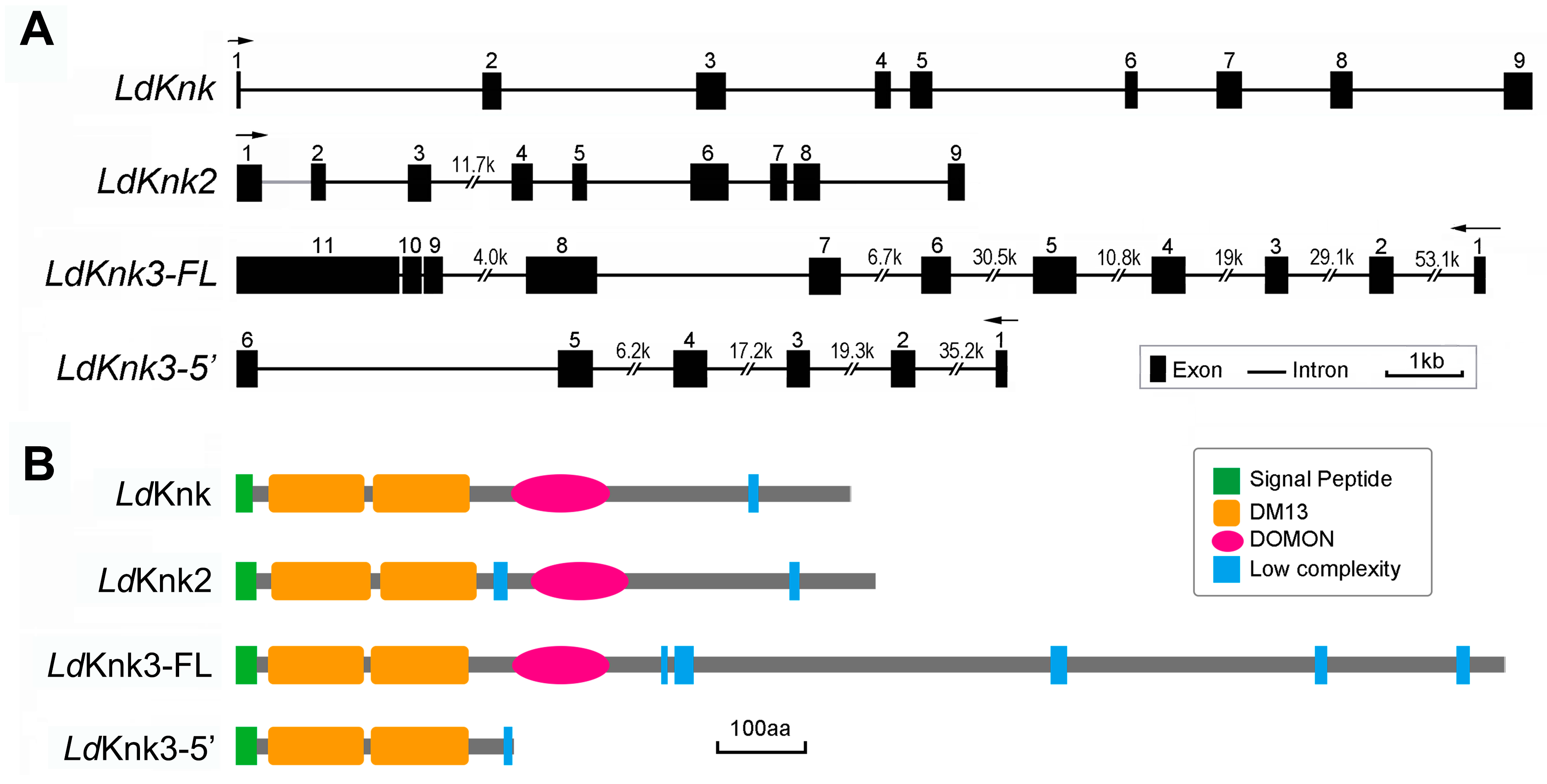
3.1. Identification and Characterization of LdKnk-Family Genes in L.decemlineata

To identify Knk genes in L. decemlineata, comprehensive searches were conducted using genomes [42,43] and transcriptome databases [44]. Amino acid sequences of Knk genes from T. castaneum, D. melanogaster, and L. migratoria were used as queries in BLAST searches. which resulted in the identification of four candidate gene fragments encoding LdKnk-like proteins.
These gene fragments were validated through reverse-transcription PCR (RT-PCR) followed by sequencing. The application of rapid amplification of cDNA ends (RACE) subsequently identified four full-length LdKnk gene sequences in L. decemlineata, named LdKnk, LdKnk2, LdKnk3-FL, and LdKnk3-5′. The accession numbers are listed in Table 1.
The open reading frames (ORFs) and protein characteristics of these genes are as follows. LdKnk comprises 2028 nucleotides encoding a 76.03 kDa protein with 675 amino acids and an isoelectric point (pI) of 6.35. LdKnk2 comprises 2109 nucleotides encoding a 78.90 kDa protein with 702 amino acids and a pI of 5.09. LdKnk3-FL comprises 4188 nucleotides encoding a 156.48 kDa protein with 1395 amino acids and a pI of 7.41. LdKnk3-5′ comprises 915 nucleotides encoding a 33.81 kDa protein with 304 amino acids and a pI of 7.65. Additionally, two alternative splicing transcripts of LdKnk3 were identified. LdKnk3-FL encodes a full-length protein, while LdKnk3-5′ encodes a C-terminally truncated protein (Table 1).
Comparing the full-length cDNA sequences with the genomic DNA sequences of L. decemlineata allowed us to construct gene structures. Both LdKnk and LdKnk2 contain nine exons, LdKnk3-FL contains eleven exons, and LdKnk3-5′ contains six exons. Notably, the first 281 nucleotides from the 5′-end of the LdKnk2 cDNA sequence could not be located in the genome database, preventing the determination of the first intron location (indicated by a grey line). Exons 1–5 of LdKnk3-5′ are identical to the 5′ end sequence of LdKnk3-FL (Figure 1A).
The domain architectures of these four Knk proteins were predicted using their amino acid sequences and SMART software. All four predicted proteins contain a signal peptide and two N-terminal DM13 domains. Additionally, LdKnk, LdKnk2, and LdKnk3-FL contain a dopamine monnoxygenase (DOMON) domain in the middle, which is absent in LdKnk3-5′ (Figure 1B).
A phylogenetic analysis was conducted to identify and classify the putative Knk-like proteins in L. decemlineata. This analysis involved aligning the amino acid sequences of Knk-like proteins from ten representative insect species. As a result, four Knk proteins were identified in L. decemlineata and were organized into three clades (Figure 2). LdKnk and LdKnk2 belong to the Knk and Knk2 protein clades, respectively, while both LdKnk3-FL and LdKnk3-5′ belong to the Knk3 protein clade (Figure 2). This suggests the functional diversification of Knk proteins within L. decemlineata.

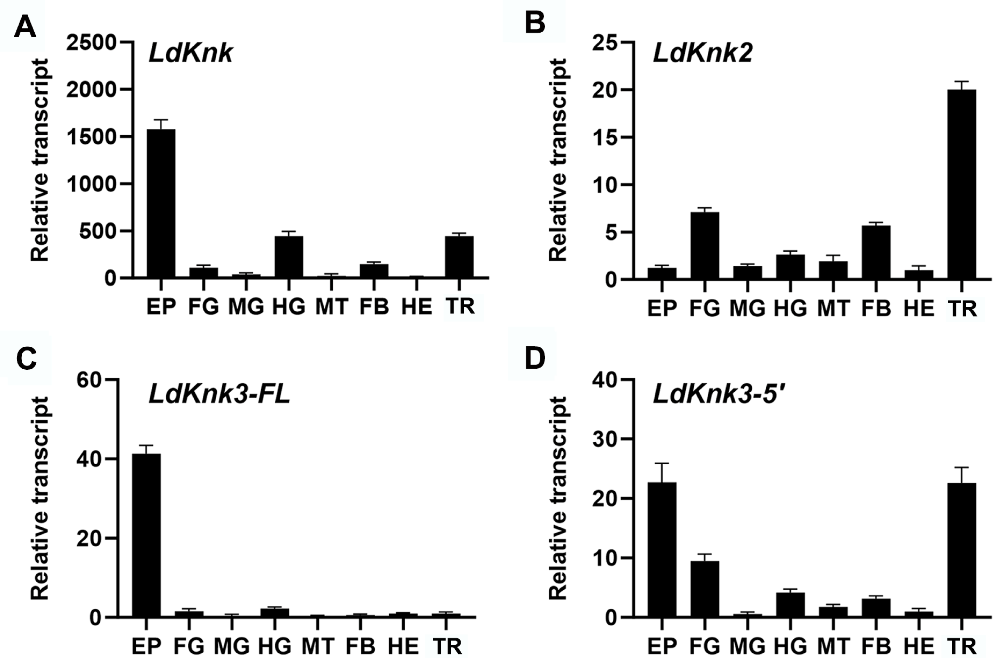
3.2. Spatio-Temporal Expression Patterns of LdKnk-Family Genes

The expression patterns of LdKnk-family gene transcripts were analyzed at various developmental stages, including egg, larvae (first to fourth instar), wandering larvae, prepupae, and pupae, using qRT-PCR. The results indicate significant differences in gene expression (Figure 3). LdKnk exhibited relatively stable expression levels throughout the larval stages, with high expression at the end of the first to third instars and prepupal stages. A noticeable increase was observed during the prepupal and pupal stages, peaking in the mid-pupal stage (day 3). LdKnk2 was expressed in all developmental stages, with its highest expression observed during the pupal stage. LdKnk3-FL displayed distinct expression patterns, with high transcript levels sharply increasing during the end stages of the first to third instars, prepupal, and mid-to-late pupal stages. Its expression was low or undetectable during other periods. LdKnk3-5′ exhibited elevated levels during the late stages of the first to third instars and the prepupal stage, with the highest expression observed from days 1 to 3 of the pupal stage. Unlike LdKnk3-FL, LdKnk3-5′ also exhibited high expression at the early third instar and 24 h into the fourth instar (Figure 3).
The tissue-specific expression patterns of the LdKnk-family genes were analyzed in fourth-instar larvae. Relative transcript levels were measured in various tissues, including the epidermis (EP), foregut (FG), midgut (MG), hindgut (HG), Malpighian tubules (MT), fat body (FB), hemocytes (HE), and trachea (TR) (Figure 4). LdKnk, LdKnk3-FL, and LdKnk3-5′ showed predominant expression in the epidermis. However, LdKnk2 was highly expressed in the trachea. Additionally, LdKnk and LdKnk3-5′ exhibited moderate expression in the trachea, and all four LdKnk-family genes were expressed in the digestive system, particularly in the foregut and/or hindgut. These findings suggest that the LdKnk-family genes play potential roles in cuticle formation and molting processes.

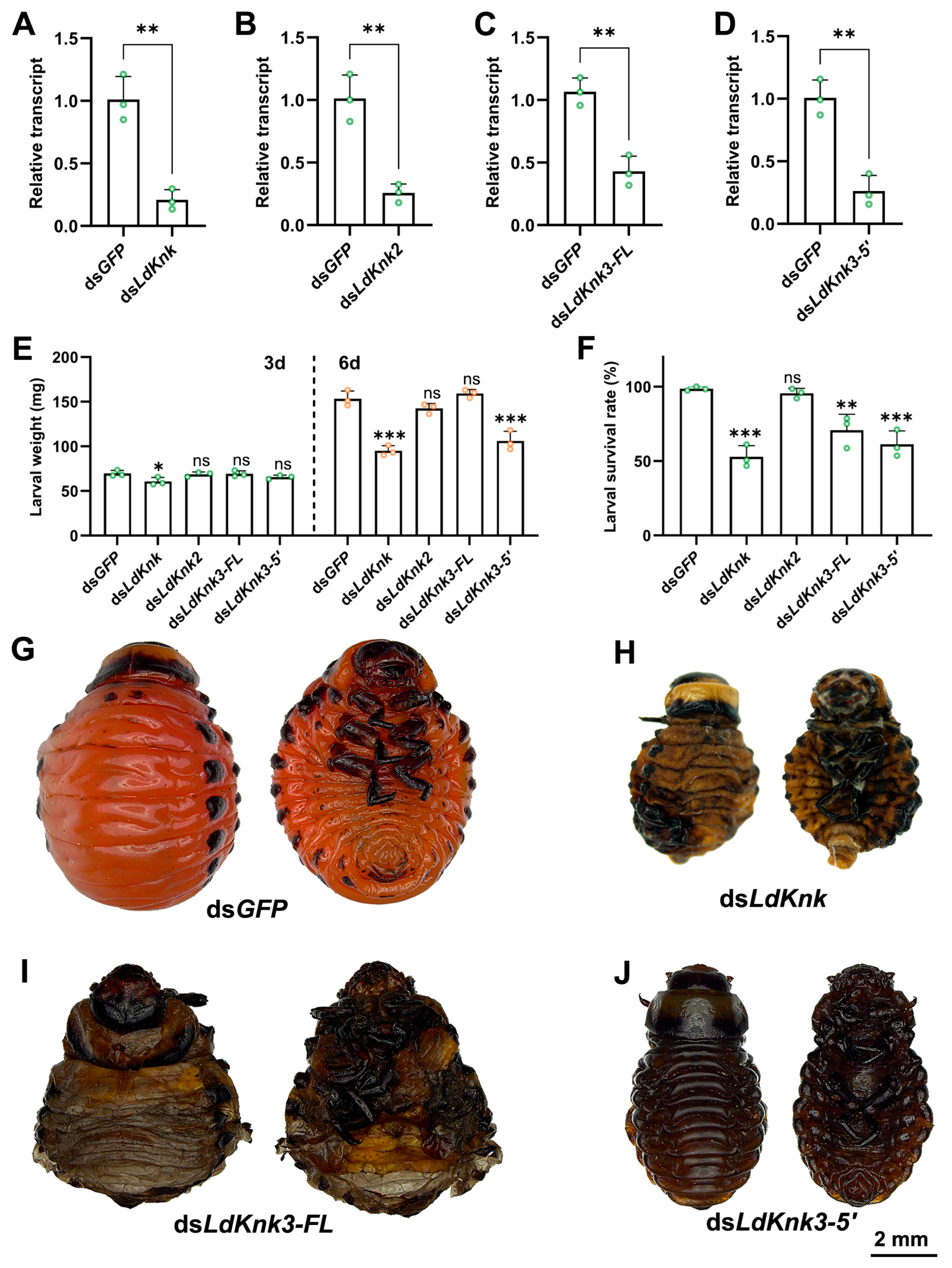
3.3. Effects of Silencing LdKnk Genes on Larval Development and Epidermis Structure

RNAi experiments were conducted to investigate the biological functions of four LdKnk-family genes in L. decemlineata larvae. dsRNA specific to each LdKnk-family gene was produced using Escherichia coli HT115 and administered to newly molted second-instar larvae through ingestion of treated potato leaves. The larvae were fed dsRNA-soaked leaves for three days, followed by fresh potato leaves until pupation. dsGFP was used as the control. RNA was extracted from larvae three days after ingestion of dsRNA, and qRT-PCR was used to measure the transcript levels of the targeted genes. Results indicated significant gene silencing for the four LdKnk-family genes, evidenced by reduced transcript levels in treated larvae compared to controls (Figure 5A–D).
Silencing LdKnk2 in larvae did not result in visible phenotypic changes. There were no significant differences in body weight or mortality between the LdKnk2-silenced group and the control group (Figure 5E,F). The larvae developed normally, molting and pupating successfully, similar to those in the control group (Figure 5G).
In contrast, knockdown of LdKnk caused significant larval weight reduction in larvae on the third and sixth days post-dsRNA feeding (Figure 5E). By the early fourth instar, about 50% of these larvae exhibited localized cuticle damage, and desiccation, leading to death (Figure 5F,H). Silencing LdKnk3-5′ also caused significant changes. on the sixth day post-treatment, these larvae had notable body weight reductions, and some exhibited melanization and death during the early fourth instar (Figure 5E,F,J).
Interestingly, the knockdown of LdKnk3-FL led to a slight increase in body weight on the sixth day, with no significant difference compared to controls (Figure 5E). However, the survival rate was significantly lower (Figure 5F). These larvae stopped developing and died on the soil surface before pupation, with the pupal cuticle trapped within the larval cuticle (Figure 5I). These results highlight the essential roles of LdKnk-family genes in the development and maintenance of the larval epidermis in L. decemlineata.
To investigate the roles of LdKnk-family genes in the structural organization of the larval epidermis, we conducted TEM analysis on the larval cuticle. In the control group treated with dsGFP-dsRNA, TEM revealed that the larval cuticle comprises three distinct layers: the outermost non-conductive transparent waxy envelope layer, the middle epicuticle, and the inner procuticle. The procuticle displayed a clear laminar structure, with a lighter-colored inner endocuticle (endo) and a darker-colored outer exocuticle (exo) (Figure 6(A1–A3)).
RNAi-mediated silencing of LdKnk resulted in significant thinning of the procuticle, including both the endocuticle and exocuticle, and disrupted the laminar organization, causing uneven thickness and localized expansion (Figure 6(B1,B2),F,G vs. Figure S1A,B). Silencing LdKnk3-5′ caused the larval epidermis to become thinner, with both the procuticle and its endocuticle and exocuticle significantly reduced in thickness (Figure 6(E1,E2),F,G vs. Figure S1A,B). The chitin layers were also markedly thinner, with a blurred layer structure (Figure 6(D2)). In contrast, silencing LdKnk3-FL led to a significantly thicker larval procuticle, nearly twice that of the control, with both the endocuticle and exocuticle substantially thicker, while the chitin layer thickness remained similar to the control (Figure 6(D1,D2),F,G vs. Figure S1A,B). Knockdown of LdKnk2 resulted in larvae with procuticle morphology and thickness similar to the control group, with only an increase in procuticle thickness due to a thicker exocuticle (Figure 6(B1,B2),F,G vs. Figure S1A,B).
Measurements of the epicuticle and envcuticle thickness revealed that LdKnk2 knockdown significantly increased the thickness of both layers compared to the control group (Figure 6(B3) vs. Figure S1C,D). In contrast, knockdowns of LdKnk, LdKnk3-FL, and LdKnk3-5′ resulted in significantly thinner epicuticle and envcuticle layers (Figure 6(B3–E3) vs. Figure S1C,D). These findings suggest that LdKnk-family genes play distinct roles in maintaining the structural integrity of the larval cuticle.

4. Discussion

Knickkopf genes are essential for the formation and maintenance of insect cuticles, particularly in organizing and protecting chitin within the exoskeleton [10]. However, research on Knk genes has primarily focused on a few model species, such as D. melanogaster [9,23,26], T. castaneum [28,29,30], and L. migratoria [22,31,32], leaving their broader roles in insects largely unexplored. In this study, we identified and characterized Knk genes in L. decemlineata, a globally destructive pest of potatoes. Our findings reveal the distinct and essential functions of LdKnk-family genes in the larval cuticle, thereby enhancing our understanding of the molecular mechanisms underlying cuticle formation and maintenance and elucidating the developmental roles these genes play.
In our study, we identified and characterized four LdKnk-family coding gene transcripts in the genome and transcriptome of L. decemlineata. Phylogenetic analysis revealed that the LdKnk-family proteins are divided into three distinct clades: Knk, Knk2, and Knk3. We found single transcripts for LdKnk and LdKnk2 genes, whereas LdKnk3 gave rise to two alternatively spliced transcripts, coding for a full-length protein (LdKnk3-FL) and a C-terminally truncated version (LdKnk3-5′). This pattern in the L. decemlineata is somewhat similar to that in T. castaneum, D. melanogaster, and the hemimetabolous insect L. migratoria. Domain analyses showed that LdKnk, LdKnk2, and LdKnk3-FL each consist of a signal peptide, two tandem DM13 domains, and a DOMON domain, whereas LdKnk3-5′ lacks one DOMON domain, which is essential for Knk function in D. melanogaster [25]. These findings suggest that, despite the presence of three Knk coding genes across different insect species, there may be functional differences among them.
To further investigate the function of LdKnk-family genes in L. decemlineata, we used qRT-PCR to analyze the expression of the four LdKnk-family genes at different developmental stages and in various tissues. The results showed that all four LdKnk-family genes were consistently highly expressed before larval molting, at the late prepupal stage, and throughout the pupal stages, with expression patterns closely mirroring those of 20-hydroxyecdysone (20E). In terms of tissue expression, with the exception of LdKnk2, which was predominantly expressed in the trachea, the other LdKnk-family genes were significantly expressed in the larval epidermis (Figure 3 vs. Figure 4). Similarly, in L. migratoria and T. castaneum, Knk genes (LmKnk, LmKnk3-FL, LmKnk3-5′, TcKnk, TcKnk2, and TcKnk3-FL) showed continuous and high expression in late instar larvae, particularly in the epidermis, with LmKnk-family genes in L. migratoria confirmed as downstream targets of 20E [28,31,32]. These patterns indicate that insect Knk genes are significantly upregulated during cuticle formation and molting processes, highlighting their crucial roles in these stages.
The insect protein Knk is conserved across various insect species and plays a crucial role in cuticle formation and maintenance. Our RNAi experiments demonstrate that silencing LdKnk significantly impacts larval development and cuticle integrity. Knockdown of LdKnk in larvae results in significant weight loss, with more than half dying before the prepupal stage. Most larvae exhibit localized epidermal damage, melanization, dehydration, and death. TEM analysis supports these observations, showing that silencing LdKnk significantly reduces the thickness of the procuticle and its layers, leading to thinner epicuticle and endocuticle layers and compromised cuticle integrity (Figure 5 and Figure 6). Similarly, in L. migratoria, LmKnk localizes to the chitin layer of the cuticle. Injecting dsLmKnk reduces chitin content, disrupts crystalline chitin organization, and lowers lipid content, impairing the cuticle’s barrier function [31]. Previous research in D. melanogaster shows that DmKnk is crucial for cuticle formation and epithelial tube morphogenesis [24,25]. RNAi knockdown results in malformed wings and an amorphous procuticle, while overexpression maintains normal structure [23]. DmKnk, along with Dmkkv and Dmcystic, is essential for organizing chitin in tracheal tubes. Rescue experiments confirm that proper Dmknk expression restores tube integrity [9,45]. In T. castaneum, TcKnk proteins protect newly synthesized chitin from degradation and ensure proper laminar organization. RNAi for TcKnk causes developmental arrest at each molt, highlighting its role in chitin stability [28]. The Retroactive (Rtv) protein is essential for trafficking TcKnk into the procuticle. TcRtv localizes within epidermal cells and promotes TcKnk movement, protecting the new cuticle from chitinases. Loss of TcRtv leads to TcKnk entrapment and molting defects [7,29,30]. The conserved function of Knk protein across insects underscores its essential role in chitin organization and cuticle integrity.
Knk3 exhibits multiple alternative splicing variants that vary across species. LdKnk3 has two transcripts, similar to LmKnk3, while TcKnk3 produces three [28,32]. In L. decemlineata, the roles of LdKnk3-FL and LdKnk3-5′ are particularly notable due to their opposing effects on cuticle thickness. Silencing LdKnk3-FL results in a thicker cuticle, leading to molting disruption before the prepupal stage, while silencing LdKnk3-5′ results in a thinner cuticle, causing melanization and death (Figure 5 vs. Figure 6). This suggests that proper procuticle formation is crucial for insect molting and development. Similarly, in L. migratoria, silencing LmKnk3-5′ causes molting defects and high mortality, significantly decreasing chitin content without affecting cuticle structure and impeding lipid deposition on the cuticle surface [32]. In T. castaneum, dsRNA targeting multiple exons of TcKnk3 had varied effects. Injection of exon 9-specific dsRNA, which depletes TcKnk3-FL transcripts, caused 100% mortality at the pharate adult stage. Silencing TcKnk3-3′ specifically led to molting arrest at the same stage. Knocking down all three TcKnk-family genes significantly reduced total chitin content to varying degrees. TcKnk3 transcripts are crucial for the integrity of body wall denticles and tracheal taenidia but are not essential for the elytral and body wall procuticles [28]. These findings highlight the complex roles of Knk3 isoforms in different insect species and their critical functions in maintaining cuticle structure and integrity.
In this study, silencing LdKnk2 in larvae did not result in visible phenotypic changes. However, measurements revealed that LdKnk2 knockdown significantly increased the thickness of the epicuticle and endocuticle compared to the control group. Similarly, in L. migratoria, no visible phenotypic changes were observed after LmKnk2 was silenced by RNAi [32]. In contrast, silencing TcKnk2 in T. castaneum caused molting arrest at the pharate adult stage, indicating that TcKnk2 is crucial for the integrity of body wall denticles and tracheal taenidia, but not for the elytral and body wall procuticles [28].
Understanding the specific roles of LdKnk-family genes in L. decemlineata provides valuable insights for developing targeted pest management strategies. The essential functions of LdKnk, LdKnk3-FL, and LdKnk3-5′ in maintaining cuticle integrity make them promising targets for RNAi-based pest control. For instance, silencing LmKnk3-5′ in L. migratoria not only reduced procuticle thickness but also increased susceptibility to four insecticides from three different classes. Disrupting these LdKnk-family genes could therefore weaken the protective cuticle barrier, making insects more vulnerable to environmental stressors and enhancing insecticide efficacy. Our findings on the opposite effects of LdKnk3-FL and LdKnk3-5′ on cuticle thickness further underscore the importance of precise regulation of the cuticle in pest control applications. This research enhances our understanding of insect cuticle biology and offers new genetic insights for improving pest management strategies.

5. Conclusions

Our study has successfully identified and analyzed four LdKnk-family genes in the larval epidermis of L. decemlineata. The genes LdKnk, LdKnk2, LdKnk3-FL, and LdKnk3-5′ are integral to cuticle formation and maintenance. RNAi experiments revealed that silencing these genes results in significant changes in cuticle structure, larval development, and survival rates. Specifically, LdKnk and LdKnk3-5′ are essential for maintaining cuticle integrity and preventing damage, while LdKnk3-FL regulates cuticle thickness, ensuring proper molting. These findings suggest that targeting LdKnk-family genes could be an effective strategy for developing RNAi-based pest control methods, potentially reducing the impact of the beetle on potato crops. Future research should further clarify these genes’ roles and optimize RNAi techniques for practical agricultural use.

Supplementary Materials

The following supporting information can be downloaded at: www.mdpi.com/article//s1, Figure S1. Cuticle thickness in fourth-instar L. decemlineata larvae following RNAi of LdKnk-family genes. Table S1. Primers used in RT-PCR, RACE, ORF verification, dsRNA synthesis, and qRT-PCR. Table S2. Names and accession numbers of the proteins used for phylogenetic tree construction. Table S3. Significance analysis of LdKnk-family gene expression across developmental stages using one-way ANOVA and Tukey-Kramer test. Table S4. Significance analysis of LdKnk-family gene across tissues using one-way ANOVA and Tukey-Kramer test.

Author Contributions

Conceptualization, M.-Z.Z., W.Z., S.-S.W., W.-C.G. and J.-F.S.; methodology, M.-Z.Z., S.-S.W., H.W. and J.-F.S.; software, M.-Z.Z., S.-S.W. and J.-F.S.; formal analysis, M.-Z.Z., S.-S.W., W.Z., Q.Z. and J.-F.S.; resources, K.-Y.F. and W.-C.G.; writing—original draft preparation, M.-Z.Z. and J.-F.S.; writing—review and editing, W.Z., S.-S.W., H.W., Q.Z., K.-Y.F. and J.-F.S.; project administration, K.-Y.F., W.-C.G. and J.-F.S. All authors have read and agreed to the published version of the manuscript.

Funding

This research was supported by the National Natural Science Foundation of China (No. 32000335), Natural Science Foundation Project of Chongqing (No. CSTB2022NSCQ-MSX0663), Major Science and Technology Projects in Xinjiang (No. 2023A02006), and Project of Fund for Stable Support to Agricultural Sci-Tech Renovation (No. xjnkywdzc-2023004).

Data Availability Statement

All data generated in association with this study have been made available in the Supplementary Materials published online with this article.

Conflicts of Interest

The authors declared no conflicts of interest.

References

  1. Moussian, B. Recent advances in understanding mechanisms of insect cuticle differentiation. Insect Biochem. Mol. Biol. 2010, 40, 363–375. [Google Scholar] [CrossRef]
  2. Ren, Y.; Li, Y.; Ju, Y.; Zhang, W.; Wang, Y. Insect cuticle and insecticide development. Arch. Insect Biochem. Physiol. 2023, 114, e22057. [Google Scholar] [CrossRef] [PubMed]
  3. Qu, M.; Guo, X.; Tian, S.; Yang, Q.; Kim, M.; Mun, S.; Noh, M.Y.; Kramer, K.J.; Muthukrishnan, S.; Arakane, Y. AA15 lytic polysaccharide monooxygenase is required for efficient chitinous cuticle turnover during insect molting. Commun. Biol. 2022, 5, 518. [Google Scholar] [CrossRef] [PubMed]
  4. Riddiford, L.M. A Life’s Journey Through Insect Metamorphosis. Annu. Rev. Entomol. 2020, 65, 1–16. [Google Scholar] [CrossRef]
  5. Zhang, Y.; Li, H.; Liu, X.; Li, H.; Lan, Q.; Wu, H.; Wang, Y.; Zhang, J.; Zhao, X. Nuclear Receptor FTZ-F1 Controls Locust Molt by Regulating the Molting Process of Locusta migratoria. Insects 2024, 15, 237. [Google Scholar] [CrossRef]
  6. Yang, W.J.; Xu, K.K.; Yan, Y.; Li, C.; Jin, D.C. Role of Chitin Deacetylase 1 in the Molting and Metamorphosis of the Cigarette Beetle Lasioderma serricorne. Int. J. Mol. Sci. 2020, 21, 2449. [Google Scholar] [CrossRef] [PubMed]
  7. Chaudhari, S.S.; Noh, M.Y.; Moussian, B.; Specht, C.A.; Kramer, K.J.; Beeman, R.W.; Arakane, Y.; Muthukrishnan, S. Knickkopf and retroactive proteins are required for formation of laminar serosal procuticle during embryonic development of Tribolium castaneum. Insect Biochem. Mol. Biol. 2015, 60, 1–6. [Google Scholar] [CrossRef]
  8. Tonning, A.; Helms, S.; Schwarz, H.; Uv, A.E.; Moussian, B. Hormonal regulation of mummy is needed for apical extracellular matrix formation and epithelial morphogenesis in Drosophila. Development 2006, 133, 331–341. [Google Scholar] [CrossRef]
  9. Moussian, B.; Tang, E.; Tonning, A.; Helms, S.; Schwarz, H.; Nusslein-Volhard, C.; Uv, A.E. Drosophila Knickkopf and Retroactive are needed for epithelial tube growth and cuticle differentiation through their specific requirement for chitin filament organization. Development 2006, 133, 163–171. [Google Scholar] [CrossRef]
  10. Zhu, K.Y.; Merzendorfer, H.; Zhang, W.; Zhang, J.; Muthukrishnan, S. Biosynthesis, Turnover, and Functions of Chitin in Insects. Annu. Rev. Entomol. 2016, 61, 177–196. [Google Scholar] [CrossRef]
  11. Noh, M.Y.; Muthukrishnan, S.; Kramer, K.J.; Arakane, Y. Cuticle formation and pigmentation in beetles. Curr. Opin. Insect Sci. 2016, 17, 1–9. [Google Scholar] [CrossRef] [PubMed]
  12. Kaya, M.; Sofi, K.; Sargin, I.; Mujtaba, M. Changes in physicochemical properties of chitin at developmental stages (larvae, pupa and adult) of Vespa crabro (wasp). Carbohydr. Polym. 2016, 145, 64–70. [Google Scholar] [CrossRef]
  13. Mun, S.; Noh, M.Y.; Geisbrecht, E.R.; Kramer, K.J.; Muthukrishnan, S.; Arakane, Y. Chitin deacetylases are necessary for insect femur muscle attachment and mobility. Proc. Natl. Acad. Sci. USA 2022, 119, e2120853119. [Google Scholar] [CrossRef] [PubMed]
  14. Shi, J.F.; Mu, L.L.; Chen, X.; Guo, W.C.; Li, G.Q. RNA interference of chitin synthase genes inhibits chitin biosynthesis and affects larval performance in Leptinotarsa decemlineata (Say). Int. J. Biol. Sci. 2016, 12, 1319–1331. [Google Scholar] [CrossRef]
  15. Shi, J.F.; Fu, J.; Mu, L.L.; Guo, W.C.; Li, G.Q. Two Leptinotarsa uridine diphosphate N-acetylglucosamine pyrophosphorylases are specialized for chitin synthesis in larval epidermal cuticle and midgut peritrophic matrix. Insect Biochem. Mol. Biol. 2016, 68, 1–12. [Google Scholar] [CrossRef] [PubMed]
  16. Noh, M.Y.; Muthukrishnan, S.; Kramer, K.J.; Arakane, Y. A chitinase with two catalytic domains is required for organization of the cuticular extracellular matrix of a beetle. PLoS Genet. 2018, 14, e1007307. [Google Scholar] [CrossRef]
  17. Noh, M.Y.; Kramer, K.J.; Muthukrishnan, S.; Kanost, M.R.; Beeman, R.W.; Arakane, Y. Two major cuticular proteins are required for assembly of horizontal laminae and vertical pore canals in rigid cuticle of Tribolium castaneum. Insect Biochem. Mol. Biol. 2014, 53, 22–29. [Google Scholar] [CrossRef]
  18. Noh, M.Y.; Muthukrishnan, S.; Kramer, K.J.; Arakane, Y. Tribolium castaneum RR-1 cuticular protein TcCPR4 is required for formation of pore canals in rigid cuticle. PLoS Genet. 2015, 11, e1004963. [Google Scholar] [CrossRef]
  19. Mun, S.; Noh, M.Y.; Dittmer, N.T.; Muthukrishnan, S.; Kramer, K.J.; Kanost, M.R.; Arakane, Y. Cuticular protein with a low complexity sequence becomes cross-linked during insect cuticle sclerotization and is required for the adult molt. Sci. Rep. 2015, 5, 10484. [Google Scholar] [CrossRef] [PubMed]
  20. Yu, H.; Yi, L.; Lu, Z. Silencing of Chitin-Binding Protein with PYPV-Rich Domain Impairs Cuticle and Wing Development in the Asian Citrus Psyllid, Diaphorina citri. Insects 2022, 13, 353. [Google Scholar] [CrossRef]
  21. Muthukrishnan, S.; Merzendorfer, H.; Arakane, Y.; Yang, Q. Chitin Organizing and Modifying Enzymes and Proteins Involved In Remodeling of the Insect Cuticle. Adv. Exp. Med. Biol. 2019, 1142, 83–114. [Google Scholar] [CrossRef]
  22. Yu, R.R.; Duan, J.Q.; Zhao, X.M.; Abbas, M.; Zhang, Y.P.; Shi, X.K.; Chen, N.; Zhang, J.Z. Knickkopf (LmKnk) is required for chitin organization in the foregut of Locusta migratoria. Insect Sci. 2024. [Google Scholar] [CrossRef] [PubMed]
  23. Li, K.; Zhang, X.; Zuo, Y.; Liu, W.; Zhang, J.; Moussian, B. Timed Knickkopf function is essential for wing cuticle formation in Drosophila melanogaster. Insect Biochem. Mol. Biol. 2017, 89, 1–10. [Google Scholar] [CrossRef] [PubMed]
  24. Devine, W.P.; Lubarsky, B.; Shaw, K.; Luschnig, S.; Messina, L.; Krasnow, M.A. Requirement for chitin biosynthesis in epithelial tube morphogenesis. Proc. Natl. Acad. Sci. USA 2005, 102, 17014–17019. [Google Scholar] [CrossRef] [PubMed]
  25. Shaik, K.S.; Wang, Y.; Aravind, L.; Moussian, B. The Knickkopf DOMON domain is essential for cuticle differentiation in Drosophila melanogaster. Arch. Insect Biochem. Physiol. 2014, 86, 100–106. [Google Scholar] [CrossRef] [PubMed]
  26. Pesch, Y.Y.; Riedel, D.; Behr, M. Obstructor A organizes matrix assembly at the apical cell surface to promote enzymatic cuticle maturation in Drosophila. J. Biol. Chem. 2015, 290, 10071–10082. [Google Scholar] [CrossRef]
  27. Petkau, G.; Wingen, C.; Jussen, L.C.; Radtke, T.; Behr, M. Obstructor-A is required for epithelial extracellular matrix dynamics, exoskeleton function, and tubulogenesis. J. Biol. Chem. 2012, 287, 21396–21405. [Google Scholar] [CrossRef]
  28. Chaudhari, S.S.; Moussian, B.; Specht, C.A.; Arakane, Y.; Kramer, K.J.; Beeman, R.W.; Muthukrishnan, S. Functional specialization among members of Knickkopf family of proteins in insect cuticle organization. PLoS Genet. 2014, 10, e1004537. [Google Scholar] [CrossRef] [PubMed]
  29. Chaudhari, S.S.; Arakane, Y.; Specht, C.A.; Moussian, B.; Kramer, K.J.; Muthukrishnan, S.; Beeman, R.W. Retroactive maintains cuticle integrity by promoting the trafficking of Knickkopf into the procuticle of Tribolium castaneum. PLoS Genet. 2013, 9, e1003268. [Google Scholar] [CrossRef] [PubMed]
  30. Chaudhari, S.S.; Arakane, Y.; Specht, C.A.; Moussian, B.; Boyle, D.L.; Park, Y.; Kramer, K.J.; Beeman, R.W.; Muthukrishnan, S. Knickkopf protein protects and organizes chitin in the newly synthesized insect exoskeleton. Proc. Natl. Acad. Sci. USA 2011, 108, 17028–17033. [Google Scholar] [CrossRef]
  31. Yu, R.R.; Zhang, R.; Liu, W.M.; Zhao, X.M.; Zhu, K.Y.; Moussian, B.; Zhang, J.Z. The DOMON domain protein LmKnk contributes to correct chitin content, pore canal formation and lipid deposition in the cuticle of Locusta migratoria during moulting. Insect Mol. Biol. 2022, 31, 127–138. [Google Scholar] [CrossRef] [PubMed]
  32. Zhang, R.; Zhao, X.; Liu, X.; Zhang, X.; Yu, R.; Ma, E.; Moussian, B.; Zhu, K.; Zhang, J. Effect of RNAi-mediated silencing of two Knickkopf family genes (LmKnk2 and LmKnk3) on cuticle formation and insecticide susceptibility in Locusta migratoria. Pest Manag. Sci. 2020, 76, 2907–2917. [Google Scholar] [CrossRef] [PubMed]
  33. Pelissie, B.; Chen, Y.H.; Cohen, Z.P.; Crossley, M.S.; Hawthorne, D.J.; Izzo, V.; Schoville, S.D. Genome Resequencing Reveals Rapid, Repeated Evolution in the Colorado Potato Beetle. Mol. Biol. Evol. 2022, 39, msac016. [Google Scholar] [CrossRef]
  34. Ma, M.Q.; He, W.W.; Xu, S.J.; Xu, L.T.; Zhang, J. RNA interference in Colorado potato beetle (Leptinotarsa decemlineata): A potential strategy for pest control. J. Integr. Agric. 2020, 19, 428–437. [Google Scholar] [CrossRef]
  35. Szendrei, Z.; Grafius, E.; Byrne, A.; Ziegler, A. Resistance to neonicotinoid insecticides in field populations of the Colorado potato beetle (Coleoptera: Chrysomelidae). Pest Manag. Sci. 2012, 68, 941–946. [Google Scholar] [CrossRef]
  36. Scott, I.M.; Tolman, J.H.; MacArthur, D.C. Insecticide resistance and cross-resistance development in Colorado potato beetle Leptinotarsa decemlineata Say (Coleoptera: Chrysomelidae) populations in Canada 2008–2011. Pest Manag. Sci. 2015, 71, 712–721. [Google Scholar] [CrossRef] [PubMed]
  37. Kadoic Balasko, M.; Mikac, K.M.; Bazok, R.; Lemic, D. Modern Techniques in Colorado Potato Beetle (Leptinotarsa decemlineata Say) Control and Resistance Management: History Review and Future Perspectives. Insects 2020, 11, 581. [Google Scholar] [CrossRef]
  38. Shi, J.F.; Cheng, M.H.; Zhou, W.; Zeng, M.Z.; Chen, Y.; Yang, J.X.; Wu, H.; Ye, Q.H.; Tang, H.; Zhang, Q.; et al. Crucial roles of specialized chitinases in elytral and hindwing cuticles construction in Leptinotarsa decemlineata. Pest Manag. Sci. 2024, 80, 4437–4449. [Google Scholar] [CrossRef]
  39. Shi, X.Q.; Guo, W.C.; Wan, P.J.; Zhou, L.T.; Ren, X.L.; Ahmat, T.; Fu, K.Y.; Li, G.Q. Validation of reference genes for expression analysis by quantitative real-time PCR in Leptinotarsa decemlineata (Say). BMC Res. Notes 2013, 6, 93. [Google Scholar] [CrossRef] [PubMed]
  40. Bustin, S.A.; Benes, V.; Garson, J.A.; Hellemans, J.; Huggett, J.; Kubista, M.; Mueller, R.; Nolan, T.; Pfaffl, M.W.; Shipley, G.L.; et al. The MIQE guidelines: Minimum information for publication of quantitative real-time PCR experiments. Clin. Chem. 2009, 55, 611–622. [Google Scholar] [CrossRef]
  41. Chen, Y.; Tang, H.; Zhou, W.; Li, C.; Chen, Y.N.; Zhang, Q.; Fu, K.Y.; Guo, W.C.; Shi, J.F. Identification of chitinase genes and roles in the larval-pupal transition of Leptinotarsa decemlineata. Pest Manag. Sci. 2024, 80, 282–295. [Google Scholar] [CrossRef] [PubMed]
  42. Schoville, S.D.; Chen, Y.H.; Andersson, M.N.; Benoit, J.B.; Bhandari, A.; Bowsher, J.H.; Brevik, K.; Cappelle, K.; Chen, M.M.; Childers, A.K.; et al. A model species for agricultural pest genomics: The genome of the Colorado potato beetle, Leptinotarsa decemlineata (Coleoptera: Chrysomelidae). Sci. Rep. 2018, 8, 1931. [Google Scholar] [CrossRef] [PubMed]
  43. Yan, J.; Zhang, C.; Zhang, M.; Zhou, H.; Zuo, Z.; Ding, X.; Zhang, R.; Li, F.; Gao, Y. Chromosome-level genome assembly of the Colorado potato beetle, Leptinotarsa decemlineata. Sci. Data 2023, 10, 36. [Google Scholar] [CrossRef] [PubMed]
  44. Kumar, A.; Congiu, L.; Lindstrom, L.; Piiroinen, S.; Vidotto, M.; Grapputo, A. Sequencing, De Novo assembly and annotation of the Colorado Potato Beetle, Leptinotarsa decemlineata, Transcriptome. PLoS ONE 2014, 9, e86012. [Google Scholar] [CrossRef]
  45. De Giorgio, E.; Giannios, P.; Espinas, M.L.; Llimargas, M. A dynamic interplay between chitin synthase and the proteins Expansion/Rebuf reveals that chitin polymerisation and translocation are uncoupled in Drosophila. PLoS Biol. 2023, 21, e3001978. [Google Scholar] [CrossRef]
Figure 1. The Exon-intron and Domain architecture of the putative LdKnk-family proteins from L. decemlineata. (A) A schematic diagram of the exon-intron organization of each LdKnk-family gene was determined by sequence comparison between the genomic sequence and the Complete cDNA sequence. Black closed boxes mark exons and lines mark introns. Since the first 281 nucleotides of LdKnk2 cDNA cannot be found in the L. decemlineata genome database, we cannot reveal the exact location of the first intron as shown in the grey line. (B) Domain architecture of deduced amino acid sequences of LdKnk-family proteins as predicted using SMART protein. The green bar is the signal peptide; the orange bar is the DM13 domain; the rose oval is the dopamine monooxygenase N-terminal-like (DOMON) domain; the pink is the coiled-coil region; and the blue bar is the low complexity region.
Insects 15 00623 g001
Figure 2. Phylogenetic tree of known insect Knk proteins. The unrooted phylogenetic trees were constructed using the neighbor-joining method with the Jones-TaylorThornton (JTT) substitution model, based on full-length protein sequence alignments. Bootstrap analyses with 1000 replications were performed, and bootstrap values >50% were shown on the tree. Knk proteins are derived from ten insect species: two Coleoptera T. castaneum (Tc) and L. decemlineata (Ld); an Orthoptera L. migratoria (Lm); an Anoplura Pediculus humanus corporis (Phc); a Hemiptera Acyrthosiphon pisum (Ap); two Hymenoptera Apis mellifera (Am) and Nasonia vitripennis (Nv); and three Diptera D. melanogaster (Dm), Anopheles gambiae (Ag) and Aedes aegypti (Aa). The accession numbers of the proteins are listed in Table S2.
Insects 15 00623 g002
Figure 3. Temporal expression patterns of LdKnk-family genes during various developmental stages of L. decemlineata. The cDNA templates were prepared from pooled samples of mixed eggs (1–3 days old), first- to third-instar larvae, wandering larvae, prepupae, and pupae, collected daily. Additionally, fourth-instar larvae were sampled at 12-h intervals (D0/H0 indicated newly ecdysed larvae). Quantitative real-time PCR (qRT-PCR) was performed in triplicate, using technical replicates and three independent sample pools, each containing 5–10 individuals. Normalized expression values were determined using the 2−ΔΔCt method, with the geometric mean of housekeeping genes for reference. Relative transcript levels are presented as ratios compared to wandering larvae (set to 1). Significant differences were analyzed using One-way ANOVA followed by the Tukey-Kramer test (p < 0.05) in Prism 10 software, with results listed in Table S3.
Insects 15 00623 g003
Figure 4. Tissue-specific expression profiles of LdKnk-family genes in fourth-instar larvae of L. decemlineata (AD). The cDNA was obtained from epidermis (EP), foregut (FG), midgut (MG), hindgut (HG), malpighian tubules (MT), fat body (FB), hemocyte (HE), and tracheae (TR) of fourth-instar larvae. Tissue samples were organized into three independent pools, each containing 5–30 individuals, and analyzed in technical triplicates using qRT-PCR. Expression levels were quantified using the 2−ΔΔCt method (±SE) and normalized to the geometric mean of housekeeping genes. The relative expression levels are presented with hemocytes set as 1. Significant differences were analyzed using one-way ANOVA followed by the Tukey-Kramer test (p < 0.05) in Prism 10 software, with results listed in Table S4.
Insects 15 00623 g004
Figure 5. Effects of dietary ingestion of dsLdKnk-family genes on the second-instar larvae of L. decemlineata. Newly molted second-instar larvae were fed dsRNA-treated foliage for three days and then transferred to untreated leaves. Expression levels of LdKnk-family genes were analyzed on day 3 (AD). Larval fresh weight was recorded on days 3 and 6 (I), and survival rates were recorded on day 6 (J). Data are displayed as bar charts with standard errors (±SE). Asterisks indicate significant differences (* p < 0.05, ** p < 0.01, *** p < 0.001). dsGFP-fed larvae reached the end of the fourth instar six days after the bioassay began (E). In contrast, LdKnk-family genes RNAi larvae displayed various abnormalities, including localized epidermal deformities, shrinkage, molting failure, and melanization, ultimately leading to mortality in some fourth-instar larvae (FH).
Insects 15 00623 g005
Figure 6. Impact of LdKnk genes Knockdown on Larval Cuticular Structure of L. decemlineata. TEM analysis of the epidermal structure in three-day-old fourth instar larvae following LdKnks knockdown. Panels (A1E1) exhibit low-magnification views of the endocuticle and exocuticle within the procuticle (Pro). Panels (A2E2) offer detailed views of the procuticle layer from the green boxes in A1–E1, with green dotted lines indicating the layer boundary. Panels (A3E3) depict detailed views of the epicuticle and envelope cuticle from the orange boxes in A1–E1. Panels (F,G) present quantifications of overall and layered procuticle thickness, respectively, indicating significant (*** p < 0.001) and non-significant (ns) differences between the dsGFP control and treatment groups.
Insects 15 00623 g006
Table 1. Characteristics of four LdKnk-family genes in the L. decemlineata.
Gene NameAccession NumbercDNA (bp)Coding Region5′-UTR (bp)3′-UTR (bp)aaplMw(kDa)
LdKnkPP962378211861–208860306756.3576.03
LdKnk2PP96237921421–21090337025.0978.90
LdKnk3-FLPP962380510049–42364886413957.41156.48
LdKnk3-5PP962381104261–97560673047.6533.81
UTR–untranslated region; aa–amino acid; pl–isoelectric point; Mw–molecular weight.
Disclaimer/Publisher’s Note: The statements, opinions and data contained in all publications are solely those of the individual author(s) and contributor(s) and not of MDPI and/or the editor(s). MDPI and/or the editor(s) disclaim responsibility for any injury to people or property resulting from any ideas, methods, instructions or products referred to in the content.

