Article

Selective Laser Melting of 316L Stainless Steel: Influence of Co-Cr-Mo-W Addition on Corrosion Resistance

School of Mechanical & Electrical and Information Engineering, Shandong University at Weihai, Weihai 264209, China
*
Author to whom correspondence should be addressed.
Metals 2021, 11(4), 597; https://doi.org/10.3390/met11040597
Submission received: 10 March 2021 / Revised: 24 March 2021 / Accepted: 26 March 2021 / Published: 6 April 2021
(This article belongs to the Section Additive Manufacturing)

Abstract

:
The selective laser melting (SLM) of o-Cr-Mo-W/316L composite with 10 wt% Co-Cr-Mo-W addition to 316L stainless steel (SS) powder is produced to explore it’s the corrosion behavior. The tensile experiment of SLM composites is also measured to investigate the difference between the two samples. The optimum parameters of SLM 316L SS and it’s composite samples are obtained by adjusting laser power and scanning speed with the relative density of 99.04 ± 0.69 and 99.15 ± 0.43. The yield strength of samples is increased from 731.96 MPa to 784.09 MPa after doping, and no obvious crack or fracture failure in the tensile samples are observed, indicating that the excellent plasticity is still maintained. The corrosion resistance of samples is improved largely with an order of magnitude lower corrosion current density than that of 316L SS and increasing of 277 mv of epit Ep. The addition of Cr element in the doped powder contributes to the formation of the passivated film containing Cr. The different pitting corrosion pit occurs mainly around the pre-existing pores of the powder and further extends outward to form pits with the increase of voltage.

1. Introduction

316L stainless steel (SS) bears the feature of high toughness and excellent corrosion resistance with relatively a low cost as a result of its room temperature mechanical strength, thus is broadly used in oil and gas industries, refineries, chemical plants construction and automotive industries, etc. [1]. Although the casting fabricated 316L SS have been already used in above-mentioned fields, the personalized production with high accuracy is still not available. Selective laser melting (SLM) is widely used in fabricating complicated shapes by means of building an object layer-by-layer from a powder, which, compared to traditional forming technique, will save the cost and reduce the weight [2]. The microstructure and properties of SLM 316L has been widely reported in the literature. Calleja et al. [3]. Use laser cladding technology to manufacture complex parts. The effect of scanning strategy on the microstructure and mechanical properties of 316L SS by SLM have been explored by Ya’nan Song et al. [4]. It indicates that the scanning strategy with rotation between layers breaks the growth of columnar grains and contributes to the grain refinement, which shows great effect on their mechanical properties. Most of research focus on the solidification process dependence of the microstructure and mechanical properties, while the flexibility of Addtive Manufacturing (AM) technology makes it possible in the application of biomedicine [5,6]. For example, products with complex structures such as bridges, crowns and surgical devices are prepared by metal 3Dprinting technology [7,8]. The application environment of 316L SS exhibits a higher requirement for its corrosion resistance in both biomedical and industrial fields [9,10,11]. Stainless steel has a tendency to exhibit localized corrosion, hampering its structural applications, particularly within biomedical industry, and 24% of the implant failures are caused by this corrosion phenomenon [12]. Laleh et al. [13] further illustrated that the SLM 316L SS works worse than its commercial counterpart on the re-passivation ability. The SLM 316L SS in a 0.6 M NaCl solution has a re-passivation potential as approximentely lower than 100 mVSCE, which is because of the micro-pores within the specimens. The metal ions that initiates inflammatory and adverse cellular will be released as a result of corrosion [14]. Therefore, higher corrosion resistance of the SLM forming 316L SS is necessary to broaden its application field and promote the development of biomedical materials. The strength and ductility of stainless steel also has a great influence on its application. Han et al. [15] investigated the microstructural features and mechanical properties of graphene nanoplatelets (GNPs) reinforced 316L composite parts via SLM and found that GNPs reinforcement improves the strength of the 316L SS matrix without affecting the ductility. Co-based alloys are widely used in aerospace and biological field due to their excellent thermal corrosion resistance and corrosion resistance [8,16]. Co-Cr-Mo-W alloy powder has been approved by Japan in 2018 as a Class II medical device manufacturing and sales material, making 3D printed Co-Cr alloy dentures officially put into use [17]. Three-point bending experiment [16] is performed to evaluate the metal-ceramic bonding strength of Co-Cr alloys which is prepared by SLM technology. The founding indicates that the cermet prepared by SLM shows a higher bond strength than that-obtained by tradition casting methods. As strong carbide forming element, Mo and W can be added to stainless steel to improve the toughness of the alloy without greatly reducing the plasticity, becoming a common addition element for strengthening metals. 316L shows less strength and more plastic toughness due to it’s austenite matrix, therefore Mo and W elements are added to improve the strength of the 316L SS. In this paper, 10 wt% Co-Cr-Mo-W alloy powder is mixed up with 316L SS powder and then the samples are formed by SLM. The objective of present work is to obtain the optimal molding process of SLM 316L SS through adjusting laser power and scanning speed, the higher corrosion resistance as well as good mechanical properties of SLM 316L with addition of Co-Cr-Mo-W powder.

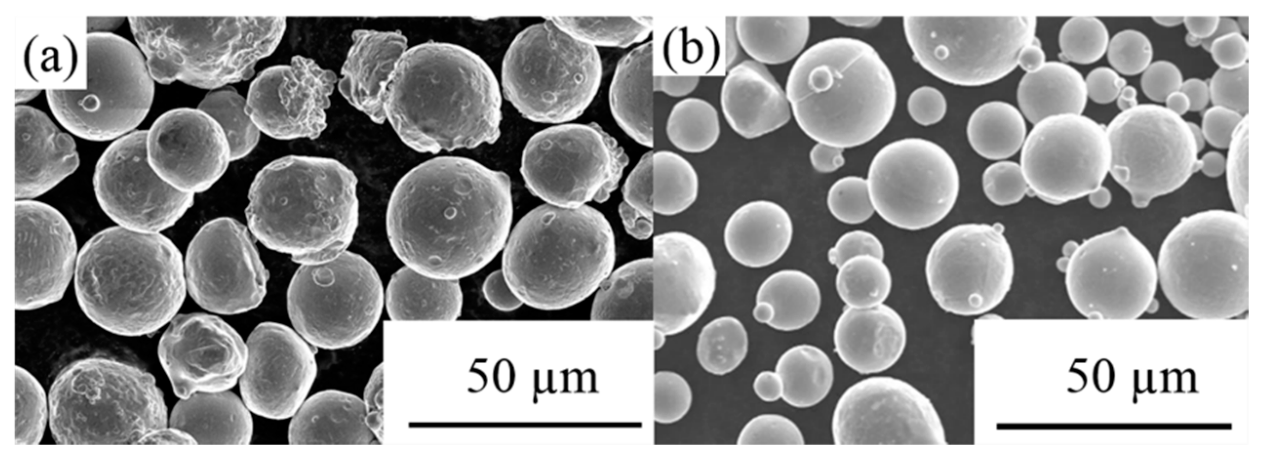
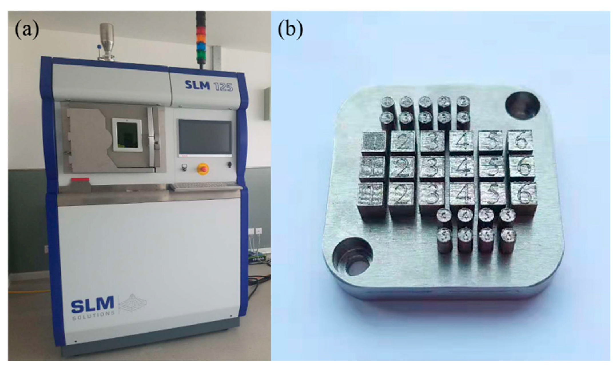
2. Materials and Methods

316L SS powder bears a particle size ranging from 15 to 45 µm and is highly sphericized a smooth surface (Figure 1a). It is produced by the gas atomization process. Figure 1b shows that the Co-Cr-Mo-W powder bares with regular spheres and sized between 15 and 53 µm. Being mixed with 10 wt% Co-Cr-Mo-W powder, 316L SS powder is used in this paper as, namely, 316L + 10Co-Cr-Mo-W samples. Figure 2 displays the image of 316L + 10Co-Cr-Mo-W powder along with an energy-dispersive X-ray spectroscopy (EDS, TESCAN Inc, Czech, Brno) spectrum. The chemical compositions of materials are listed in Table 1.
The samples are made on stainless alloy substrates which adopts an SLM 125HL (SLM Solutions Inc., Lubeck, Germany) equipment (Figure 3a). A 400W Yb: YAG (Y3Al5O12) fiber lazer is equipped with a spot size of 80 µm. In order to be preserved from oxidation, the whole processing chamber is settled in a high-purity Air atmosphere. Scanning speed can be used to control the energy density. A continuous laser mode running in a zigzag pattern are used to scan the layers between which a 33° rotation is applied. Dimensions of 5 × 5 × 5 mm3 for electrochemical measurements are shown in cubic samples (Figure 3b).
Before the microstructural examination, the SLM-processed samples are polished with SiO2 solution. The scanning electron microscopy (SEM; Nova NanoSEM450, FEI compony, Hillsboro, OR, USA) are used to observe the micro-structure while the phase identification is performed by X-ray diffraction (XRD, Rigaku, Tokyo, Honshu, Japan). The scattering angular (2θ) varies in a range from 20° to 80°. The relative density is monitored by an electronic densitometer (ZMD-2, Fangrui Co. Ltd., Shanghai, China) based on Archimedes’ principle. The mechanical properties of the specimens are evaluated for tensile test. The tensile tests run under the universal testing machine (Exceed E45, MTS systems Co. Ltd., Shenzhen, China). A CHI660E electrochemical station (Chenhua Instrument Co. Ltd., Shanghai, China) is used in conducting the potentiodynamic polarization. It is composed by three-electrode cell, the 316L + 10Co-Cr-Mo-W sample as the working electrode, a saturated calomel electrode (Ag/AgCl2) in saturated KCl as the reference electrode, and platinum sheet as the counter electrode. The potentiodynamic polarization scanned from −2.5 V to 2.5 V with a rate of 3 mV s−1. EIS spectra are obtained after immersed for 0.5 h with a frequency range of 10 KHz~0.01 Hz and an amplitude sinusoidal voltage of 0.05 V. To ensure the reproducibility, all the measurements are carried out at 35 ± 2 °C and are repeated for at least 3 times.

3. Results and Discussion

3.1. Optimization of Process Parameters

Table 2 shows the forming process and relative density of the 316L SS and 316L + 10Co-Cr-Mo-W alloy fabricated by SLM. The layer thickness and scanning distance are set as 0.03 mm and 0.12 mm respectively for all samples. The relative densities of all samples are higher than 96% but less than 100%, indicating the presence of defects in the obtained parts, such as pores and cracks. The relative density shows highest value of 99.04 ± 0.69% at laser power of 200 W, scanning speed of 800 mm/s, namely an energy density of 69 J/mm³; compared to that of 316L + 10Co-Cr-Mo-W alloy, the highest relative density is 99.15 ± 0.43% at laser power of 170 W, scanning speed of 680 mm/s, energy density of 69 J/mm³.
The 316L powder will melt in its melting temperature as about 1200~1300 °C, where the applied the SLM process’s energy density is high enough to melt it down sufficiently. However, Co-Cr-Mo-W bears higher melting temperature (about 1400 °C) than that of 316L, which results in holes in the surface because of the lower energy input that leaves residual unmelted Co-Cr-Mo-W powder in the matrix and thus leads to a poor surface bonding of 316L + 10Co-Cr-Mo-W powder. The effect of energy on the relative density, in fact, is twofold: on one hand, the quality of the upper surface of the melting pool would be destroyed by the high energy because of the liquid convection in the pool. The next layer of powder would not spread well under the inhibition by those irregular surfaces, which would be ascribed to those holes on it. On the other hand, the relative density would be declined by higher energy density because of the vaporization, the pores trapped in the matrix or following-formed irregular surface, which also inhibits the spread of the next layer of powder. The fact that a higher energy would lead to more time and energy consumption in the SLM process convince us that the fine process for dense alloy with high density allows an energy of 69 J/mm³.

3.2. Mechanical Properties and Phase Characteristic

Figure 4 shows the stress-strain curves of SLM-printed 316L and 316L + 10Co-Cr-Mo-W with the optimum forming process-at room temperature. The tensile strength of the 316L + 10Co-Cr-Mo-W is 784.09 Mpa, 7.1% higher than that of 316L. More importantly, SLM-printed 316L + 10 Co-Cr-Mo-W still shows high elongation of ∼40%, a little bit decreasing compared to that of 316L.
XRD patterns of 316L and 316L + 10Co-Cr-Mo-W are presented in Figure 4b. Both samples contained face centered cubic (fcc) gamma (γ) austenite phase without the presence of any new phase. Table 3 lists the variation of 2θ locations and intensities of the γ-Fe diffraction peaks in SLM-processed 316L and 316L/Co-Cr-Mo-W composites.
The fracture surfaces after tensile tests for the 316L and 316L + 10Co-Cr-Mo-W were investigated by SEM, as shown in Figure 5. A long defect is observed in Figure 5b, which was ascribed to the incompletely fused materials. Also, as shown in Figure 5a,b, many large pores exist in the fracture surface, which can be attributed to the entrapped gas bubble during the shaping process [18]. The tensile samples are close to shear fracture at an angle of 45° with the tensile axis from a macroscopic view. Some pores and cracks can be observed from the morphology of the tensile fracture. These lack of fusion defects will cause stress concentration and void nucleation, thereby driving the initiation and propagation of cracks. The fracture surfaces of samples exhibit a large amount of small dimples (Figure 5c,d), with nonuniform size, revealing ductile fracture pattern for both samples. The composite material sample is mainly plastic deformation of cellular sub-grains in the initial stage of deformation, and defects such as dislocations accumulate in the cell boundary in a large amount. Increasing strain hardening effect, the amount of deformation and stress causes dislocations to start at the dendritic subgrain boundary, and the strain hardening rate decreases at the same time. The final fracture showed a mixed morphology of intergranular ductile fracture and dimples.

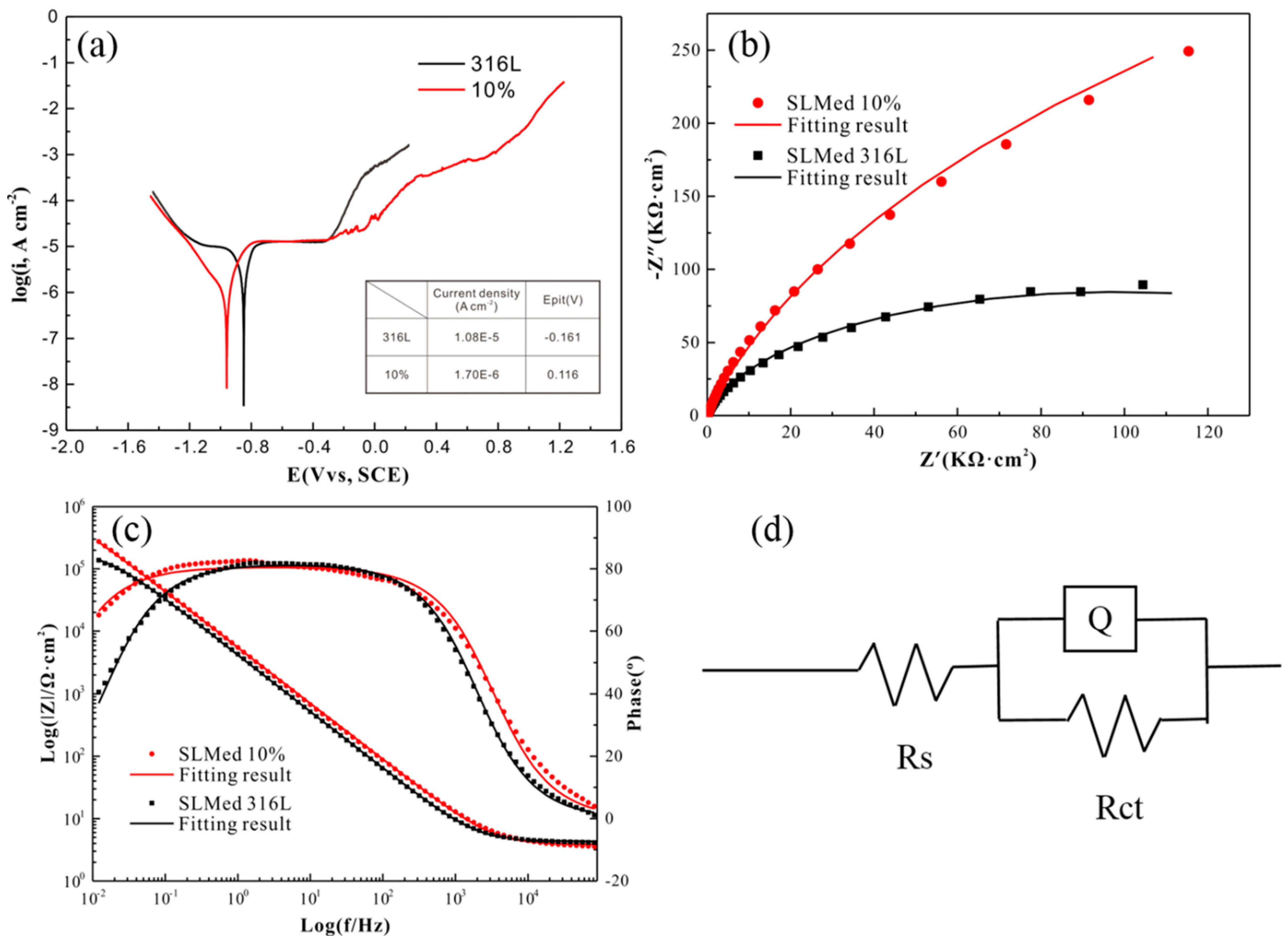
3.3. Corrosion Behavior of SLM-Processed Alloy

Figure 6 shows the results of the electrochemical polarization curve and impedance spectroscopy of 316L and 316L + 10Co-Cr-Mo-W composites, respectively. The corrosion current density (icorr) of 316L decreases from 1.08 × 10⁻⁵ to 1.70 × 10⁻⁶ A/cm2 pitting potential Ep increases by 277 mV, indicating increasing corrosion resistance. The addition of Cr element in the doped powder contributes to the formation of passivated film containing Cr oxide. Figure 6b shows the EIS in the form of the Nyquist plot for the SLM specimens. These Nyquist plots can be fitted by equivalent circuit diagram, as show in Figure 6d and the fitting parameters of impedance spectrum of samples is listed in Table 4. The equivalent circuit is selected according to the principle of least fitting error, and the fitting error of the two samples is less than 10%. Here, Rs and Rct represent solution resistance, the charge transfer resistance, respectively. It can be seen that the Rct of 316L composites is significantly higher than that of 316L SS, indicating its slower charge transfer rate of in solution and lower dissolution rate, as a result of higher corrosion resistance. Compared to 316L samples, 316L composite shows larger impedance values Rct in both low and high frequency regions, which is consistent with the results obtained by Tafel curve (Figure 6a).

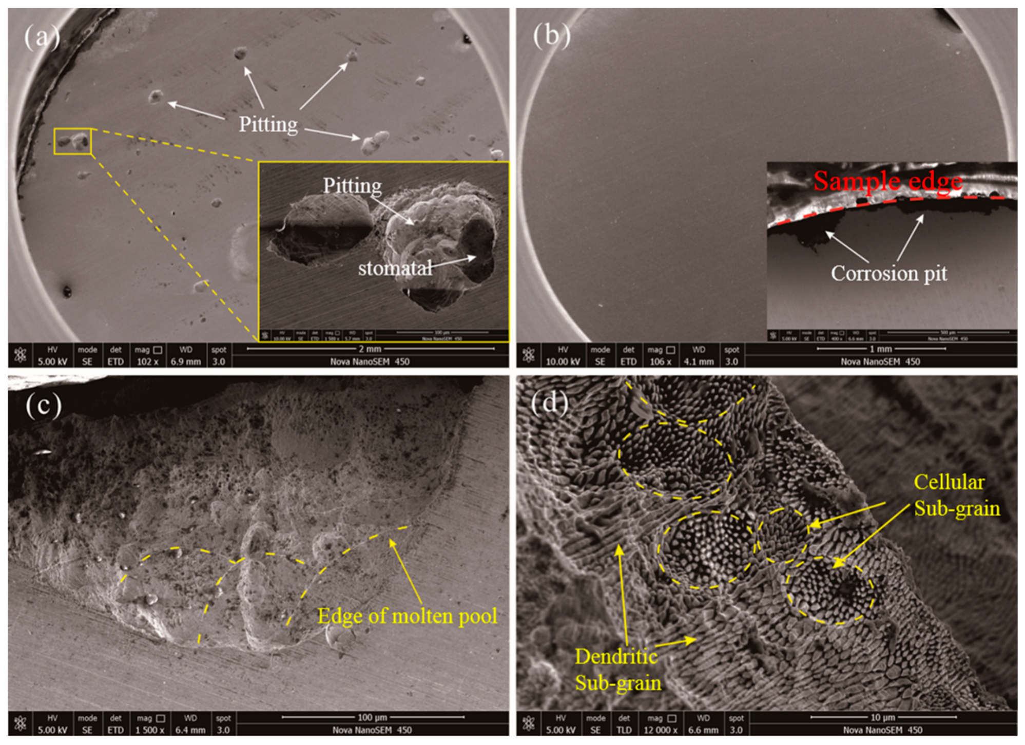
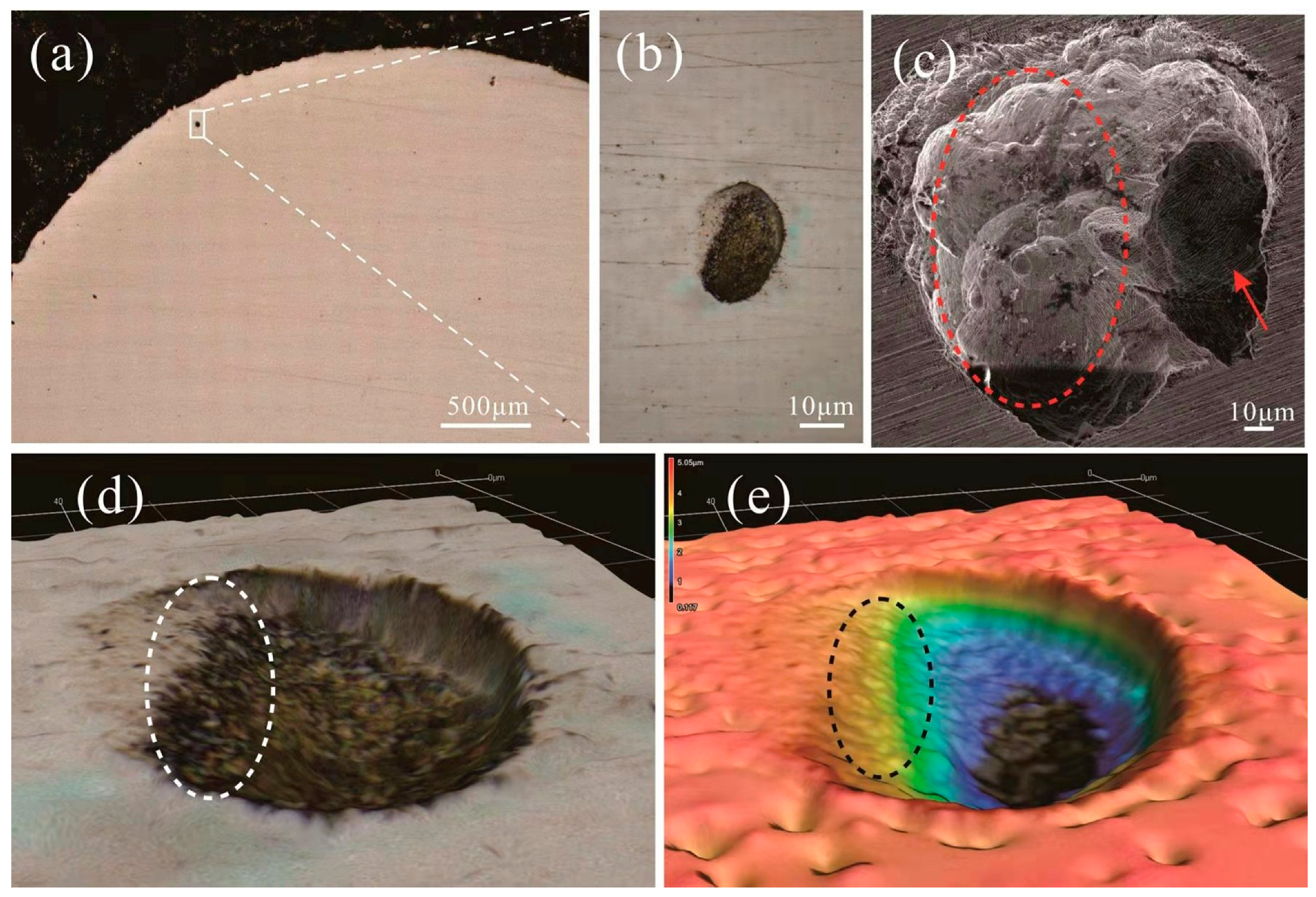
3.4. Microstructural Characterization of SLM-Processed Alloy

The breakdown of the passive film directly results in the localized corrosion. In stainless steels, the most usual localized mechanisms is pitting corrosion, where the attack is confined to small pits. The pitting corrosion of wrought stainless steels always initiates at the inclusions (MnS) [19,20]. Dissolution of sulphide inclusions is the most commonly postulated reason for pit initiation. There is 0.1–10 s for the nucleation and diffusional growth of MnS precipitates. Due to the rapid solidification rates in SLM (typically ~107 K/s), the time for Mn and S diffusing to the oxides decreases about 105 order of magnitude, and it is reasonable to claim that the MnS precipitates are sufficient reduced or completely eliminated. Some researchers proposed that the higher porosity resulted in the weaker corrosion resistance [21,22]. The pitting corrosion was initiated at the manufactured defects, among which the gas pores are the most common sites [23]. SEM images of the two samples after electrochemical test are illustrated in Figure 7. It reveals that there are amount of pitting corrosion under the surface of the sample, as shown in Figure 7a and the pitting corrosion behavior happens around the gas pores for SLM 316L SS samples. While, it can be observed mainly at the edges of 316L + 10Co-Cr-Mo-W sample, as shown in Figure 7b. Only a few pitting regions occur around the pores at the center of the composite samples. The pitting corrosion is related to the stability of the passive film formed on the bottom of the gas pores [24]. The pit growth initiated at the gas pore occurs mainly in the form of electrochemical dissolution of the passive film under this condition. The corrosion resistance of stainless steels can be drastically improved by suitable alloying. Mischler et al. showed that alloying with molybdenum reduced the surface enrichment of chlorides and sulphates [25]. The composition and properties of the passive films depend on the alloy composition. The corrosion resistance of 316L SS has been improved by doping Co-Cr-Mo-W powder, due to the passivation film with Cr element on the surface [26,27,28,29]. The passivity is also enhanced with addition of Mo [30,31,32,33]. This is attributed to the fact that donor and acceptor densities could be decreased by addition of Mo in passive film. The pitting corrosion is also inhibited with Mo in Cl containing solutions [34,35]. The existence of Mo also can increase the thickness of the passive film, as well as the content of Cr2O3 in the passive film. Passive films composes of oxide nanoparticles, and it’s formation contains nucleation, three-dimensional growth, and particle connection [36,37]. The nucleation preferentially takes place at the defects, because of the higher activation energies than that of grains [38]. Elements with larger atomic radius, such as W and Mo, dissolved into the stainless steel austenite grains can lead to lattice distortion. Such a sub-grain structure is formed by the compositional fluctuation due to the slow kinetics of homogeneous alloying of large Mo atoms during rapid solidification. Sun et al. noted that in the case of localized lattice distortion and metal oxide reactions, high activation energy occurs in regions with high concentrations of dislocation [39]. Therefore, dislocations could also act as nucleation sites like grain boundaries and sub-grain boundaries. Mo element are enriched at the sub-grain boundaries accompanied with high dislocation concentrations [40]. The density of dislocations in the 316L + 10Co-Cr-Mo-W is much higher than that in the 316L SS. Therefore, the higher density of dislocation in the 316L + 10Co-Cr-Mo-W should also be a reason that results in the formation of a thicker passive film on the 316L + 10Co-Cr-Mo-W. In this work, the amount of nucleation sites in the 316L + 10Co-Cr-Mo-W is more than that of 316L SS sample, which would promote the formation process of passive film of the 316L + 10Co-Cr-Mo-W. As shown in Figure 7b, the multi-point corrosion of 316L SS sample leads to the larger corrosion current and smaller impedance value. It can be seen that there are many fish scales-like molten pool boundaries in the corrosion pit, which is caused by the different corrosion rates of sub-grains with different orientations on both sides of the Molten Pool boundaries (MPBs), as shown in Figure 7c. Similarly, the morphology differences caused by different corrosion rates can also be found in Figure 7d, and most of the pitting occurs preferring at the sub-grain boundary. More grain boundaries of cellular sub-grains are exposed in the solution than that of dendritic sub-grains, the cellular sub-grains become sparse and dark-colored due to a large amount of corrosion.
W also has a certain positive effect on the corrosion resistance of 316L SS. Belfrouh et al. [41]. indicated that when used in combination with Mo, W can improve pitting corrosion resistance. Tomashov et al. [42]. studied the influence of W additions from 0.18 to 2.67 wt% to austenitic stainless steels. Experiments show that 0.545 wt% of w element can make 316L SS obtain lower corrosion rates and higher pitting potentials (EP). The final solidification area generally occurs around the pores, where some impurity elements or element segregation always can be found. The corrosion pits of the 316L SS sample are analyzed by Laser Scanning Confocal Microscope (LSCM), and it can be observed that the edges of the pores is rather rough by SLM process, which increases the contact area between sample and corrosion liquid (Figure 8d,c). In electrochemical experiments, it is difficult to form a dense passivation film around the pores due to the presence of impurities and element segregation. With increasing the corrosion voltage, the passive film is broken down before it can be formed, indicating that the stable corrosion occurs, the larger current is generated at the junction between the pores and the metal substrate with the interaction of corrosive liquid. (Figure 8 dashed circle) and then, the corrosion gradually expands from the connection to the surroundings, and the pitting morphology can be formed. Meanwhile, the matrix is still protected from further corrosion by the relatively dense passivation film on the flat surface.

4. Conclusions

(1)
316L SS and 316L + 10Co-Cr-Mo-W samples are fabricated using the SLM process. The optimization parameters of 316L SS and 316L + 10Co-Cr-Mo-W are 200 W, 170 W of laser power and 800 mm/s, 680 mm/s of scanning speed, respectively. Both relative densities reach to more than 99%.
(2)
Corrosion resistance of 316L is enhanced without loss of the strength. Although no new phase can be found in the matrix, the lattice distortion is caused by the dissolution of Co, Cr, Mo and W elements into the austenite grains. The pitting corrosion is related to the stability of the passive film formed on the bottom of the gas pores. The increasing corrosion resistance of 316L stainless steel is attributed to the addition of Cr and Mo element in the doped powder which contributes to the formation of the passivated film. The existence of Mo can increase the thickness of the passive film, as well as the content of Cr2O3 in the passive film. The higher density of dislocation in the 316L + 10Co-Cr-Mo-W should also be a reason that results in the formation of a thicker passive film on the 316L + 10Co-Cr-Mo-W.
(3)
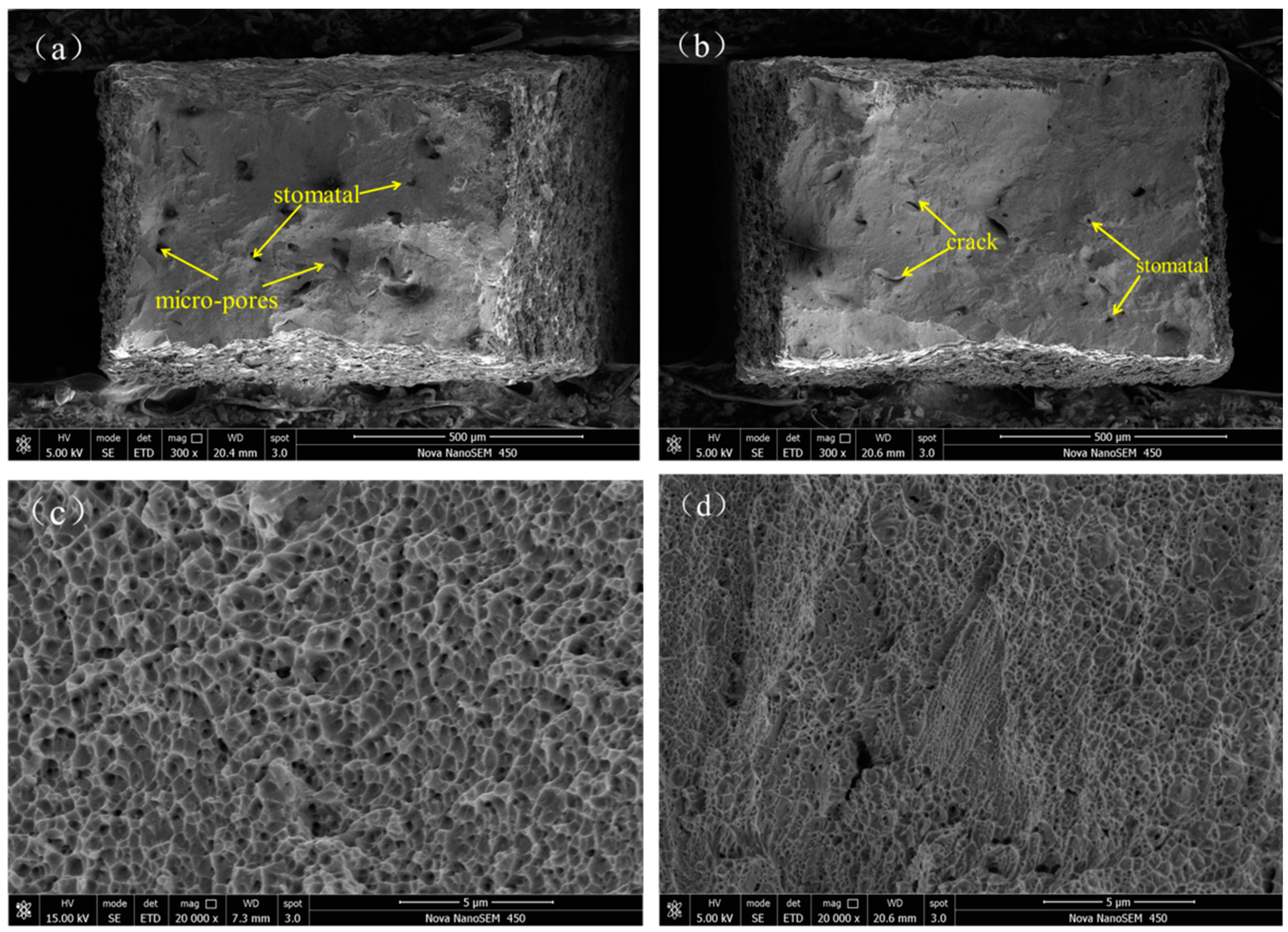
The SEM analysis of corrosion surface of 316L and 316L + 10Co-Cr-Mo-W demonstrate that most of the pitting occurs preferring at the sub-grain boundary. More grain boundaries of cellular sub-grains are exposed in the solution than that of dendritic sub-grains, a larger amount of corrosion takes place and thus the cellular sub-grains become sparse and dark-coloured.
The corrosion resistance and strength of the composite material can meet the requirements of lsome applications, and it has a lower cost compared with titanium alloy. Composite materials can provide some inspiration for the future research of medical materials.

Author Contributions

Conceptualization, P.L. and T.W.; methodology, P.L.; software, P.L.; validation, B.L. and P.L.; formal analysis, T.W.; investigation, T.W.; resources, L.W.; data curation, B.L.; writing—original draft preparation, B.L.; writing—review and editing, B.L.; visualization, S.W.; supervision, T.W.; project administration, L.W.; funding acquisition, L.W. All authors have read and agreed to the published version of the manuscript.

Funding

This research was funded by the Shandong Provincial Key Research and Development Program, grant number 2019JZZY010337 and Natural Science Foundation of Shandong Province grant number ZR2019MEM040, ZR2020ME008.

Data Availability Statement

The data presented in this study are available on request from the corresponding author. The data is not publicly available due to ongoing research based on it.

Acknowledgments

The authors are grateful to Qiuhong Huo (Experimentalist), Yanqing Xin (Experimentalist) and Jun Mi (Experimentalist) for technical assistance. The authors are also grateful for the Physical-Chemical Test & Analysis Center of Shandong University at Weihai.

Conflicts of Interest

The authors declare no conflict of interest.

References

  1. Li, J.; Qin, W.; Peng, P.; Chen, M.; Mao, Q.; Yue, W.; Kang, J.; Meng, D.; She, D.; Zhu, X.; et al. Effects of geometric dimension and grain size on impact properties of 316L stainless steel. Mater. Lett. 2021, 284, 128908. [Google Scholar] [CrossRef]
  2. Huang, S.H.; Liu, P.; Mokasdar, A.; Hou, L. Additive manufacturing and its societal impact: A literature review. Int. J. Adv. Manuf. Technol. 2013, 67, 1191–1203. [Google Scholar] [CrossRef]
  3. Calleja, A.; Tabernero, I.; Ealo, J.A.; Campa, F.J.; Lamikiz, A.; De Lacalle, L.N.L. Feed rate calculation algorithm for the homogeneous material deposition of blisk blades by 5-axis laser cladding. Int. J. Adv. Manuf. Technol. 2014, 74, 1219–1228. [Google Scholar] [CrossRef]
  4. Song, Y.; Sun, Q.; Guo, K.; Wang, X.; Liu, J.; Sun, J. Effect of scanning strategies on the microstructure and mechanical behavior of 316L stainless steel fabricated by selective laser melting. Mater. Sci. Eng. A 2020, 793, 139879. [Google Scholar] [CrossRef]
  5. Kong, D.; Ni, X.; Dong, C.; Lei, X.; Zhang, L.; Man, C.; Yao, J.; Cheng, X.; Li, X. Bio-functional and anti-corrosive 3D printing 316L stainless steel fabricated by selective laser melting. Mater. Des. 2018, 152, 88–101. [Google Scholar] [CrossRef]
  6. Bains, P.S.; Sidhu, S.S.; Bahraminasab, M.; Prakash, C. Biomaterials in Orthopaedics and Bone Regeneration: Design and Synthesis; Springer: Singapore, 2019. [Google Scholar]
  7. Silva, M.; Felismina, R.; Mateus, A.; Parreira, P.; Malça, C. Application of a hybrid additive manufacturing methodology to produce a metal/polymer customized dental implant. Procedia Manuf. 2017, 12, 150–155. [Google Scholar] [CrossRef]
  8. Koutsoukis, T.; Zinelis, S.; Eliades, G.; Al-Wazzan, K.; Al Rifaiy, M.; Al Jabbari, Y.S. Selective laser melting technique of co-Cr dental alloys: A review of structure and properties and comparative analysis with other available techniques. J. Prosthodont. 2015, 24, 303–312. [Google Scholar] [CrossRef]
  9. Shih, C.-C.; Su, Y.-Y.; Su, L.H.J.; Chang, M.-S.; Lin, S.-J. Effect of surface oxide properties on corrosion resistance of 316L stainless steel for biomedical applications. Corros. Sci. 2004, 46, 427–441. [Google Scholar] [CrossRef]
  10. Gnanavel, S.; Ponnusamy, S.; Loganathan, M.; Muthamizhchelvan, C. In vitro corrosion behaviour of Ti–6Al–4V and 316L stainless steel alloys for biomedical implant applications. J. Bio-Tribo-Corros. 2017, 4, 1–8. [Google Scholar] [CrossRef]
  11. Ryan, M.P.; Williams, D.E.; Chater, R.J.; Hutton, B.M.; McPhail, D.S. Why stainless steel corrodes. Nat. Cell Biol. 2002, 415, 770–774. [Google Scholar] [CrossRef]
  12. Sailer, I.; Philipp, A.; Zembic, A.; Pjetursson, B.E.; Hämmerle, C.H.F.; Zwahlen, M. A systematic review of the performance of ceramic and metal implant abutments supporting fixed implant reconstructions. Clin. Oral Implant. Res. 2009, 20, 4–31. [Google Scholar] [CrossRef] [PubMed] [Green Version]
  13. Laleh, M.; Hughes, A.E.; Xu, W.; Gibson, I.; Tan, M.Y. Unexpected erosion-corrosion behaviour of 316L stainless steel produced by selective laser melting. Corros. Sci. 2019, 155, 67–74. [Google Scholar] [CrossRef]
  14. Trelewicz, J.R.; Halada, G.P.; Donaldson, O.K.; Manogharan, G. Microstructure and corrosion resistance of laser additively manufactured 316L stainless steel. JOM 2016, 68, 850–859. [Google Scholar] [CrossRef]
  15. Han, Y.; Zhang, Y.; Jing, H.; Lin, D.; Zhao, L.; Xu, L.; Xin, P. Selective laser melting of low-content graphene nanoplatelets reinforced 316L austenitic stainless steel matrix: Strength enhancement without affecting ductility. Addit. Manuf. 2020, 34, 101381. [Google Scholar] [CrossRef]
  16. Xiang, N.; Xin, X.-Z.; Chen, J.; Wei, B. Metal–ceramic bond strength of Co–Cr alloy fabricated by selective laser melting. J. Dent. 2012, 40, 453–457. [Google Scholar] [CrossRef]
  17. Wei, W.; Zhou, Y.; Liu, W.; Li, N.; Yan, J.; Li, H. Microstructural Characterization, Mechanical Properties, and Corrosion Resistance of Dental Co-Cr-Mo-W Alloys Manufactured by Selective Laser Melting. J. Mater. Eng. Perform. 2018, 27, 5312–5320. [Google Scholar] [CrossRef]
  18. Zhou, X.; Wang, D.; Liu, X.; Zhang, D.; Qu, S.; Ma, J.; London, G.; Shen, Z.; Liu, W. 3D-imaging of selective laser melting defects in a Co–Cr–Mo alloy by synchrotron radiation micro-CT. Acta Mater. 2015, 98, 1–16. [Google Scholar] [CrossRef]
  19. Wijesinghe, T.S.L.; Blackwood, D.J. Real time pit initiation studies on stainless steels: The effect of sulphide inclusions. Corros. Sci. 2007, 49, 1755–1764. [Google Scholar] [CrossRef]
  20. Hara, N.; Sugawara, Y.; Muto, I. The role of MnS inclusions and passive films in the initiation of pitting corrosion of stainless steels. ECS Meet. Abstr. 2012. [Google Scholar] [CrossRef]
  21. Duan, Z.; Man, C.; Dong, C.; Cui, Z.; Kong, D.; Wang, L.; Wang, X. Pitting behavior of SLM 316L stainless steel exposed to chloride environments with different aggressiveness: Pitting mechanism induced by gas pores. Corros. Sci. 2020, 167, 108520. [Google Scholar] [CrossRef]
  22. Man, C.; Dong, C.; Liu, T.; Kong, D.; Wang, D.; Li, X. The enhancement of microstructure on the passive and pitting behaviors of selective laser melting 316L SS in simulated body fluid. Appl. Surf. Sci. 2019, 467-468, 193–205. [Google Scholar] [CrossRef]
  23. Schaller, R.F.; Mishra, A.; Rodelas, J.M.; Taylor, J.M.; Schindelholz, E.J. The Role of Microstructure and surface finish on the corrosion of selective laser melted 304L. J. Electrochem. Soc. 2018, 165, C234–C242. [Google Scholar] [CrossRef]
  24. Man, C.; Dong, C.; Liang, J.; Xiao, K.; Yu, Q.; Li, X. Characterization of the passive film and corrosion of martensitic AM355 stainless steel. Anal. Lett. 2016, 50, 1091–1111. [Google Scholar] [CrossRef]
  25. Mischler, S.; Vogel, A.; Mathieu, H.J.; Landolt, D. ChemInform abstract: Chemical composition of the passive film on Fe-24Cr and Fe-24Cr-11Mo studied by AES, XPS, and SIMS. ChemInform 2010, 22. [Google Scholar] [CrossRef]
  26. Bhasker-Ranganath, S.; Wick, C.; Ramachandran, B. Computational insights into the molecular mechanisms for chromium passivation of stainless-steel surfaces. Mater. Today Chem. 2020, 17, 100298. [Google Scholar] [CrossRef]
  27. Lv, J.; Meng, Y.; Miura, H.; Liang, T. The effect of surface enriched chromium and grain refinement by ball milling on corrosion resistance of 316L stainless steel. Mater. Res. Bull. 2017, 91, 91–97. [Google Scholar] [CrossRef]
  28. Olsson, C.-O.; Landolt, D. Passive films on stainless steels—Chemistry, structure and growth. Electrochim. Acta 2003, 48, 1093–1104. [Google Scholar] [CrossRef]
  29. Dong, Z.; Zhou, T.; Liu, J.; Zhang, X.; Shen, B.; Hu, W.; Liu, L. Effects of pack chromizing on the microstructure and anticorrosion properties of 316L stainless steel. Surf. Coat. Technol. 2019, 366, 86–96. [Google Scholar] [CrossRef]
  30. Nath, S.D.; Clinning, E.; Gupta, G.; Wuelfrath-Poirier, V.; L’Espérance, G.; Gulsoy, O.; Kearns, M.; Atre, S.V. Effects of Nb and Mo on the microstructure and properties of 420 stainless steel processed by laser-powder bed fusion. Addit. Manuf. 2019, 28, 682–691. [Google Scholar] [CrossRef]
  31. Saidi, D.; Zaid, B.; Souami, N.; Saoula, N.; Siad, M.; Ahmed, A.S.; Bibérian, J. AES depth profiles in Mo-coated 304L stainless steel achieved by RF-magnetron sputtering and influence of Mo on the corrosion in 3.5% NaCl solution. J. Alloys Compd. 2015, 645, 45–50. [Google Scholar] [CrossRef]
  32. Lv, J.; Liang, T.; Wang, C. Surface enriched molybdenum enhancing the corrosion resistance of 316L stainless steel. Mater. Lett. 2016, 171, 38–41. [Google Scholar] [CrossRef]
  33. Li, D.; Wang, J.; Chen, D.; Liang, P. Molybdenum addition enhancing the corrosion behaviors of 316L stainless steel in the simulated cathodic environment of proton exchange membrane fuel cell. Int. J. Hydrogen Energy 2015, 40, 5947–5957. [Google Scholar] [CrossRef]
  34. Tobler, W.; Virtanen, S. Effect of Mo species on metastable pitting of Fe18Cr alloys—A current transient analysis. Corros. Sci. 2006, 48, 1585–1607. [Google Scholar] [CrossRef]
  35. Kawakita, J.; Kuroda, S.; Fukushima, T.; Kodama, T. Improvement of corrosion resistance of high-velocity oxyfuel-sprayed stainless steel coatings by addition of molybdenum. J. Therm. Spray Technol. 2005, 14, 224–230. [Google Scholar] [CrossRef]
  36. Li, T.; Liu, L.; Zhang, B.; Li, Y.; Wang, F. An investigation on the continuous and uniform thin membrane passive film formed on sputtered nanocrystalline stainless steel. Corros. Sci. 2016, 104, 71–83. [Google Scholar] [CrossRef]
  37. Man, C.; Dong, C.; Cui, Z.; Xiao, K.; Yu, Q.; Li, X. Characterization of the outer layer nanostructure in the electrochemical response of stainless steel in aqueous sodium hydroxide. Anal. Lett. 2018, 51, 1384–1399. [Google Scholar] [CrossRef]
  38. Gaberšček, M.; Pejovnik, S. Impedance spectroscopy as a technique for studying the spontaneous passivation of metals in electrolytes. Electrochim. Acta 1996, 41, 1137–1142. [Google Scholar] [CrossRef]
  39. Sun, M.; Xiao, K.; Dong, C.; Li, X.; Zhong, P. Effect of stress on electrochemical characteristics of pre-cracked ultrahigh strength stainless steel in acid sodium sulphate solution. Corros. Sci. 2014, 89, 137–145. [Google Scholar] [CrossRef]
  40. Saeidi, K.; Gao, X.; Zhong, Y.; Shen, Z. Hardened austenite steel with columnar sub-grain structure formed by laser melting. Mater. Sci. Eng. A 2015, 625, 221–229. [Google Scholar] [CrossRef]
  41. Belfrouh, A.; Masson, C.; Vouagner, D.; De Becdelievre, A.; Prakash, N.; Audouard, J. The cumulative effect of alloying elements N, W, Mo and Cu on the corrosion behaviour of 17Cr-13Ni stainless steel in 2N H2SO4. Corros. Sci. 1996, 38, 1639–1648. [Google Scholar] [CrossRef]
  42. Tomashov, N.D.; Chernova, G.P.; Marcova, O.N. Effect of supplementary alloying elements on pitting corrosion susceptibility of 18Cr-14Ni stainless steel. Corrosion 1964, 20, 166t–173t. [Google Scholar] [CrossRef]
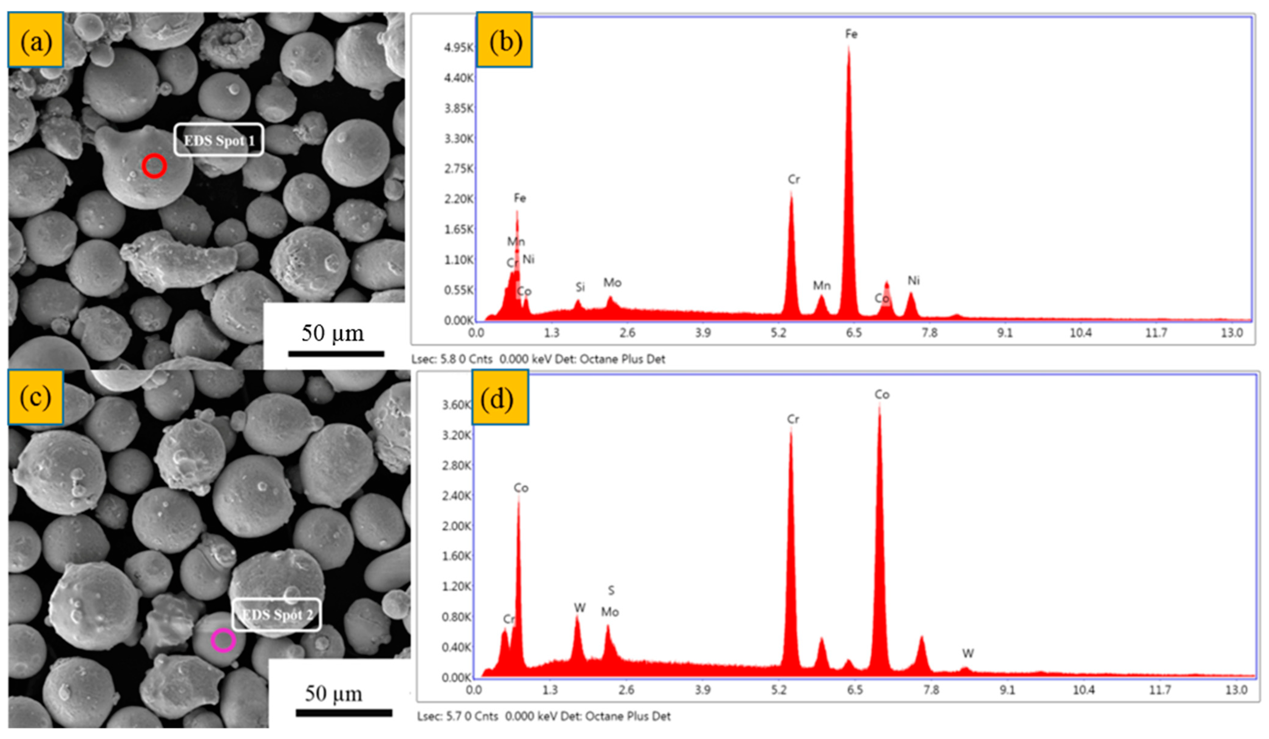
Figure 1. Scanning electron microscopy (SEM) image of powder. (a) 316L stainless steel (SS); (b) Co-Cr-Mo-W.
Metals 11 00597 g001
Figure 2. (a,c) SEM image of 316L SS + 10Co-Cr-Mo-W. (b,d) EDS spectrum of (a,c), respectively.
Metals 11 00597 g002
Figure 3. (a) SLM 125HL; (b) produced samples.
Metals 11 00597 g003
Figure 4. Tensile stress-strain curve (a) and XRD pattern (b) of 316L SS and 316L + 10Co-Cr-Mo-W under optimal laser power.
Metals 11 00597 g004
Figure 5. Tensile fracture topography. (a) low magnification morphology of 316L SS: (b) Low-magnification image of composite materials; (c) High-power micrograph of 316L SS; (d) High-power micrograph of composite materials.
Metals 11 00597 g005
Figure 6. The anodic polarization curves and impedance spectra of 316L SS and it’s composites under the optimal laser parameters. (a) Tafel curve; (b) Nyquist plot; (c) Bode plot; (d) Equivalent circuit.
Metals 11 00597 g006
Figure 7. Scanning electron microscope topography of electrochemical sample, (a) 316L SS; (b) Stainless steel composite; (c) Edge corrosion pit morphology; (d) High magnification of corrosion pit.
Metals 11 00597 g007
Figure 8. Pitting pit morphology of 316L SS electrochemical sample. (a) OM low power image; (b) OM high power image; (c) SEM Topography; (d) Confocal laser micrograph; (e) Confocal laser rendering.
Metals 11 00597 g008
Table 1. Chemical composition of 316L SS and 316L + 10Co-Cr-Mo-W.
Element (wt%)CrCoMoWMnCPNiFeSiS
316L17.1-2.3-1.130.0180.0210.6Bal.0.630.009
Co-Cr-Mo-W24.12Bal.4.955.45-----0.95-
Table 2. Processing parameters for SLM-fabricated 316L and 316L + 10Co-Cr-Mo-W alloy
Laser Power
(w)
Scanning
Speed (mm/s)
Laser Energy
Density (J/mm3)
Relative Density of 316L (%)Relative Density of 316L + 10 Co-Cr-Mo-W (%)
1705908097.01 ± 1.1697.20 ± 0.84
2006948098.29 ± 0.8896.55 ± 1.63
2308008097.39 ± 0.9398.30 ± 0.63
1706806997.11 ± 1.1099.15 ± 0.43
2008006999.04 ± 0.6997.85 ± 1.50
2309206998.73 ± 0.5097.49 ± 1.53
1707876097.14 ± 0.8997.40 ± 0.96
2009266096.57 ± 1.3297.69 ± 1.11
23010656098.22 ± 0.4797.81 ± 1.00
Table 3. Variation of 2θ locations and intensities of the γ-Fe diffraction peaks in SLM-processed 316L and 316L + 10Co-Cr-Mo-W.
Sample2θ (°)Intensity (cts)FWHM
(rad)
2θ (°)Intensity (cts)FWHM
(rad)
2θ (°)Intensity (cts)FWHM
(rad)
316L SS43.66033660.50250.64021200.49474.60143870.426
316L+10Co-Cr-Mo-W43.58230190.43950.67919660.45874.63953330.381
Table 4. The impedance spectrum fitting parameters of 316L SS and it’s 316L + 10Co-Cr-Mo-W under the optimal laser parameters.
SampleRct (KΩ·cm2)Rs (KΩ·cm2)Q × 10−5, Y0
(S·Secn/cm2)
n
316L SS205.64.2794.5240.906
316L + 10Co-Cr-Mo-W10773.8363.4890.896
Publisher’s Note: MDPI stays neutral with regard to jurisdictional claims in published maps and institutional affiliations.

