Review

Nano-Biosensors Based on Noble Metal and Semiconductor Materials: Emerging Trends and Future Prospects

1
College of Engineering, Nanjing Agricultural University, Nanjing 210031, China
2
College of Artificial Intelligence, Nanjing Agricultural University, Nanjing 210031, China
3
Key Laboratory of Agricultural Sensors, Ministry of Agriculture and Rural Affairs, Beijing 100097, China
4
Research Center of Intelligent Equipment, Beijing Academy of Agriculture and Forestry Sciences, Beijing 100097, China
*
Authors to whom correspondence should be addressed.
Metals 2023, 13(4), 792; https://doi.org/10.3390/met13040792
Submission received: 22 March 2023 / Revised: 12 April 2023 / Accepted: 13 April 2023 / Published: 17 April 2023
(This article belongs to the Special Issue Metallic Nanomaterials with Biomedical Applications)

Abstract

:
The aim of this review is to gather current researches into sensors based on noble metal and semiconductor nanomaterials in biomedical detection and elucidate the basic principle and applications of different sorts of semiconductor nanomaterials, i.e., metal oxide NPs, transition metal dichalcogenides (TMDs), metal-organic frameworks (MOFs) and magnetic metal oxide (MMO) NPs. Based on the classifications of nano-biosensors, they can be summarized as electrochemical nano-biosensors, optical nano-biosensors, calorimetric nano-biosensors, and piezoelectric nano-biosensors, wherein, electrochemical and optical nano-biosensors acting as most popular study objects are focused on to excavate the great improvements in excellent sensitivity, selectivity and stability based on fabrication techniques.

1. Introduction

The boosting demands for accurately and sensitively detecting a variety of bio-molecules at low or even trace concentrations provide an impetus for inventing advanced and sophisticated sensors. Biosensors utilize specific interactions between biological recognition elements (e.g., enzymes, antibodies, etc.) and target small molecules and are therefore highly sensitive compared with traditional physicochemical sensors. By introducing nanomaterials decorated with polymers and biometric elements, the sensitivity, specificity, stability, and interference resistance of the biosensor are further enhanced [1,2,3]. The biometric elements can be divided into: some lower molecular weight molecules: uric acid, urea; nucleic acids: DNA, RNA; protein: immunoglobulin, BSA; cells: cancer cells, bacteria, and viruses [4]. Transducer is a signal conversion device that converts the interaction between bio-sensitive elements and target analyte into different signals, such as biological enzymes catalyzing chemical reactions of specific substances and converting them into corresponding electrical signals. Bio-catalysis and bio-affinity are the two main types of bio-detection according to the bio-recognition mechanism. Bio-catalysis refers to the process of using enzymes or biological organisms (whole cells, organelles, tissues, etc.) as catalysts for chemical transformation, while bio-affinity-based sensor uses bio-receptor to bind with the target analyte [5]. Based on different transduction pathways, biosensors can be classified as electrochemical, optical, and calorimetric biosensors.
In fact, advancements in nanobiotechnology can be largely attributed to new discoveries and progress in the nanomaterials field [6]. Nanomaterials can be defined as materials possessing, one external dimension measuring 1–100 nm at minimum. The definition given by the European Commission demonstrates that the particle size of at least half of the particles in the number size distribution must measure 100 nm or below. The main characteristics of nanomaterials resulting from nanoscale dimensions can be summarized as follows: high surface-to-volume ratio, quantum size effect, small-size effect, surface and interface effects, quantum tunnel effect, and enhanced biological and chemical activity [1,7,8].
This review mainly summarizes the advances in nano-biosensors based on noble metal and semiconductor materials. Semiconductor nanomaterials include metallic oxides, TMDs, MOFs, and MMONPs. Noble metal and semiconductor materials possess unique optical, electrical, and biological properties, which have attracted significant attention due to their potential use in many applications, such as bio-imaging, biological sensors, and drug delivery [9,10,11]. The development and applications of nano-biosensors are of great significance for timely prevention, accurate diagnosis, and effective treatments of various diseases. The roles of noble metal and semiconductor nanomaterials in biosensors mainly include increasing the changes in refractive index, accelerating electron transfer, and improving the catalytic performance between chemiluminescence and substrates, thus helping the accurate and even ultra-sensitive detection of neurotransmitters and specific markers for diseases such as cancer and Alzheimer’s disease [12,13]. Gold NPs, silver NPs, and platinum NPs are the most representative and popular noble metal NPs with good biocompatibility, abundant surface modification properties, and unique optical properties, i.e., Surface plasmon Resonance (SPR) and surface-enhanced Raman scatting (SERS), which display unique properties in biomedical detection [14,15]. Metal oxides have a wide range of applications in the biomedical fields. The methods used to synthesize metal oxide nanomaterials are chemical, physical, and biosynthetic, such as chemical vapor deposition (CVD), sol-gel, hydrothermal, and green deposition methods [16,17]. Since the beginning of research on biosensors in 1954, metal oxides have been important candidates for sensor applications. Titanium dioxide and zinc oxide as representative metal oxides have been emphatically introduced due to their wide application in biosensors [18]. MMONPs are a special type of nanomaterials, possessing superparamagnetism, biocompatibility, and low toxicity, that can be well applied in the field of biomedicine [19,20]. Additionally, the current usage and development of Fe3O4 and Fe-based binary mixed metal oxides are introduced in detail, and we stress on the summary of the drawbacks and limitations overcame by MMONPs in biomedical applications. Apart from metal oxides, 2D transition metal dichalcogenides (TMDs), in this review, i.e., MoS2 and WS2, have received widespread attention due to their graphene-like layered structure [21,22]. The use of MOFs and MOFs-based nanomaterials in biomedical applications has been greatly explored owing to their precision tunability, high surface areas, and high loading capacities, and they are highly expected to be substitutes for conventional enzymes in enzymatic reactions, providing a new direction for design and applications in biomedical fields [23,24].

2. Noble Metal Nanoparticles

2.1. Gold Nanoparticles

Gold nanoparticles (AuNPs) have good biocompatibility, abundant surface modification properties, and unique optical properties, which are related to the surfactant, shape, size, and structure of the nanoparticles [14]. AuNPs can be modified in covalent and non-covalent ways [25]. Covalent modification is generally performed by sodium borohydride reduction and ligand substitution. The surface of the nanoparticles can be easily chemically modified with functional group-containing ligands such as thiols, phosphines, and amines [26]. For example, reducing chloroauric acid with silyl mercaptan as a protecting agent in ethanol solution or encapsulating gold nanoparticles with thiopolyethylene glycol may help the application in various fields of nucleic acids, proteins, and immunoassays. The high surface free energy of gold nanoparticles allows them to adsorb surrounding molecules through non-covalent interactions, reducing the surface free energy of surface ligand functional groups, thus allowing some other parts to attach, such as proteins, nucleic acids, and antibodies to improve their properties [27]. Depending on their characteristics, they can be used in various biomedical fields, such as medical testing, imaging and therapy, photochemotherapy, photodynamic therapy, and photothermal therapy. For example, in Tasneem’s research, they immobilized the Human epidermal growth factor receptor 2 on the screen-printed electrode surface by electrostatic adsorption and modified with gold nanoparticles (~20 nm in diameter) to support aptamer immobilization [28]. It took only 5 min binding time to receive a response and showed a log-linear response in a wide concentration range.
Gold nanoparticles with surface plasmon resonance (SPR) phenomena are used in the field of nucleic acid construction and analysis of protein detection [15]. SPR refers to the resonance phenomenon that may occur when light forms an extinction wave into the light sparse medium when the total reflection phenomenon occurs on the prism and metal film surface, and a certain plasma wave exists in the medium when the two waves meet based on the premise of energy conservation. They can convert the biological recognition reflection into optical or electrical signals, so they have been combined with DNA, RNA, and amino acids, which is very effective in the detection of nucleic acids and proteins [29,30,31]. Additionally, the detection method has many advantages, such as simplicity of operation, good anti-oxidation, and biocompatibility. A number of colorimetric sensors have been developed given that the shortening of the distance between AuNPs caused by the plasma coupling of the surface between particles makes significant color changes in different aggregation states and can be applied for visual inspection [32,33]. In a recent study, the stochastic DNA dual-walkers for ultrafast colorimetric bacteria detection method was proposed. The proposed method can detect bacteria sensitively and specifically within 15 min owing to its ultrafast reaction kinetics and color change mechanism, displaying a linear response ranging from 100 to 105 CFU/mL with a limit of detection of 1 CFU/mL [34].
The sensitivity of SPR biosensors is proportional to the overlapping integral of the modal electromagnetic field with the ambient medium [35]. As a result, appropriate surface treatment in improving the performance of these sensors merits. Assembling low refractive index materials (AuNPs) on the surface of high refractive index dielectric film materials (graphene oxide, TiO2, Al2O3, and other organic materials) plays an important part in improving the sensitivity, resolution, and specificity of biosensors in detection [36,37]. As shown in Figure 1, Zhu and coworkers developed a bio-nanonetwork by pyridinium porphyrin mediated calixarene-functionalized AuNPs composites (Apt/PyP-pSC4-AuNPs) acting as an SPR signal amplification tag for the sensitive and rapid Brain Natriuretic Peptide (BNP) assay [31], acting as a quantitative plasma biomarker in detecting the existence and severity of heart failure. A wide linear concentration range (1–10,000 pg/mL (R2 = 0.9852)) and low limit of detection (0.3 pg/mL) were obtained. However, the enhanced sensitivity of biosensors may bring about the problem of lack of reliability due to vulnerability from spectral signal contaminations or other analytes. Therefore, some researchers were trying to solve this problem with other optical phenomena. Surface-enhanced Raman scattering (SERS) is a phenomenon in which the Raman signal of adsorbed molecules is enhanced due to the enhancement of the electromagnetic field when molecules are adsorbed on metal surfaces, which can be utilized to make the bio-detection process more sensitive and easier. Recently, Song and coworkers proposed a novel SPR/SERS dual-mode plasmonic biosensor based on a catalytic hairpin assembly (CHA)-induced AuNPs network aimed at both high sensitivity and reliable detection of cancer-related miRNA-652 [38]. As depicted in Figure 2, the proposed biosensor is composed of capture DNA-functionalized AuNPs (Probe 1), H1 and 4-mercaptobenzoic acid (4-MBA) co-modified AuNPs (Probe 2), and 6-carboxyl-Xrhodamine (ROX)-labeled H2 (fuel strands). Then, the networks composed of Probe 1–Probe 2 were formed via the target-triggered CHA reactions, thus resulting in a color change in dark-field microscopy (DFM) images and an enhanced SERS effect. Additionally, the SPR sensing mode can be achieved by extracting the integral optical density of dark-field color in the DFM images.
At present, gold nanomaterials-modified biosensors have been widely used in bioanalysis, environmental monitoring, medical diagnosis, and other fields. In Table 1, we show some examples of the application of AuNPs. We need to make continuous efforts to explore and optimize this technique to solve various problems encountered in practical applications. First, the preparation methods of gold nanoparticles are usually small batches or laboratory level, so more large-scale preparation methods need to be explored. This is critical for translating AuNPs biosensors technology into realistic clinical applications. Second, the sensitivity, specificity, and response speed of AuNPs biosensors need to be further optimized to meet the requirements of more stringent and complex biomedical detection. Last, gold nanomaterials themselves may have toxic effects on the human body, so a comprehensive and in-depth evaluation of their biocompatibility and related safety studies should be carried out when using AuNPs biosensors.

2.2. Silver Nanoparticles

Silver nanoparticles (NPs) are currently popular metal nanomaterials with wide applications in the biomedical field, including diagnostics [39], imaging [40], and drug delivery [41]. AgNPs are usually synthesized by reducing silver salts and adding polyvinyl pyrrolidone (PVP) or citrate to improve the stability of AgNPs, thus benefiting their applications in biosensors [42]. AgNPs possess a large specific surface area, good catalytic activity, and high conductivity. Moreover, AgNPs have the optical property of surface plasmon resonance (SPR), which is helpful for Raman signal enhancement. The SPR of AgNPs can produce electromagnetic field enhancement, thus benefiting biosensors detection by the surface-enhanced Raman scatting (SERS) phenomenon [43]. Biosensors using AgNPs are often used to detect glucose, cholesterol, dopamine, DNA and other substances via using fluorescence, colorimetry, electrochemistry, and Raman spectroscopy.
Glucose detection is usually performed by oxidizing glucose to gluconic acid and hydrogen peroxide (H2O2) and then calculating the glucose concentration by detecting the amount of generated H2O2 or the changed pH caused by gluconic acid. Prabha Soundharaj et al. synthesized a silica and silver (SiO2-Ag) colloidal nanoparticles to detect glucose [44]. During detection, the sample solution and SiO2-Ag colloidal nanoparticles were mixed and excited at 260 nm, and the fluorescence spectrum at 325 nm was recorded. The fluorescence quenching during the detection process is shown in Figure 3. When the excited SiO2-Ag colloidal nanoparticles released fluorescence, part of the energy was absorbed by biomolecules and then reduced the released fluorescence intensity. Therefore, they can obtain the glucose concentration by reducing the fluorescence intensity. The biosensor can efficiently detect glucose in human urine and serum, and eliminate the interference of other biomolecules, which can meet clinical needs. In addition to this direct detection method using SiO2-Ag colloidal nanoparticles, methods for the indirect detection of glucose have been developed. Wu et al. successfully established a colorimetric biosensor to detect glucose in blood [45]. They deposited AgNPs on the surface of MIL-101 (Fe) to form a peroxidase mimic. In the detection process, glucose was oxidized to H2O2 with glucose oxidase (GOx). Then they used AgNPs@MIL-101 (Fe) and H2O2 to oxidize the colorless substance 3,3′,5,5′-Tetramethylbenzidine (TMB), turning it into blue product oxTMB. Finally, the concentration of glucose was calculated by the change in absorbance of the product detected by UV-vis spectroscopy. The detection limit of the biosensor is 0.23 μM with a satisfying sensitivity in detecting glucose. Compared with the direct detection method, the indirect detection method can detect glucose more sensitively, and it can reduce the interference of other substances. However, the biological enzymes used are greatly affected by the environment, which can be a new area to explore for researchers.
Most biosensors for cholesterol detection are combined with cholesterol oxidase (ChOx). ChOx can break down cholesterol into H2O2, and then the concentration of cholesterol can be obtained by detecting the concentration of H2O2. Wu et al. used AgNPs@MIL-101 (Fe), which is used as the peroxidase and substrate of SERS [46]. As depicted in Figure 4, they first oxidized cholesterol to H2O2 with ChOx, and then colorless leucomalachite green (LMG) with no Raman activity was oxidized to malachite green (MG) with the combined action of H2O2 and AgNPs@MIL-101 (Fe). Finally, they obtained the concentration of cholesterol from the enhanced Raman signal. The detection limit of AgNPs@MIL-101 (Fe)-based SERS biosensor is 0.36 μM, which enables ultra-sensitive detection of cholesterol.
Dopamine is a well-known neurotransmitter. The low content of dopamine in the human body may lead to Alzheimer’s disease, depression and other diseases, while the high concentration of dopamine may also lead to hypertension, nerve overexcitation, and other problems [47]. Therefore, accurate and non-toxic detection of dopamine is an important research direction. Isa Anshori et al. reported a sensitive biosensor that detected dopamine in human urine [48]. As Figure 5 shows, they synthesized functionalized multi-walled carbon nanotubes (f-MWCNT) and AgNPs as biosensing materials modified on glassy carbon electrodes to detect the concentration of dopamine based on differential pulse voltammetry (DPV). The biosensor has good stability and high sensitivity with a minimum detection limit of 0.28 mM, which is far lower than the normal dopamine concentration in human urine and can be used in routine detection.
DNA detection has recently become a very popular research direction. During gene expression, DNA is transcribed and translated into proteins, which have different functions and shapes. The process of genes controlling traits is mainly divided into two ways: one is by controlling the shapes of the proteins, and the other is by synthesizing enzymes to control metabolic processes. Therefore, we can study the process of some diseases by detecting DNA, helping the process of prevention and efficient treatment. Kais Daoudi et al. synthesized AgNPs/SiNPs hybrid arrays that can detect DNA sensitively [49]. The biosensor utilized the SERS phenomenon to detect DNA, based on the fact that different Raman peaks on the Raman spectrum represent different kinds of DNA. The AgNPs/SiNPs biosensor not only displays good stability and reproducibility, but also performs ultra-sensitive detection ability of DNA with a satisfying detection limit of 1.52 pg/L.
As far as the current research is concerned, biosensors using AgNPs are often used for the detection of daily indicators, such as glucose detection, cholesterol detection displayed in Table 1, and most of them can reduce the originally expensive detection costs. Moreover, other study committed to detecting some early signs of serious diseases helps a lot in diagnosis due to the sensitivity in detecting abnormal indicators.

2.3. Platinum Nanoparticles

Platinum nanoparticles (NPs), which possess excellent biocompatibility, high specific surface area, and excellent electrocatalytic performance, have been a hot metal nanomaterial. Generally, PtNPs are synthesized with H2PtCl6 as the raw material, ethylene glycol as the solvent, NaOH as the reducing agent, and polyethylene pyrrolidone (PVP) as the stabilizer [50]. In view of the strong strength of precious metals such as gold and silver in biomedicine, people have also begun to study the applications of PtNPs in various fields. In the field of treatment, compared with AuNPs and AgNPs, PtNPs have stronger cytotoxicity [51]. Therefore, PtNPs are often used to treat cancer. At present, PtNPs have been used in the targeted treatment of liver cancer [52]. In addition to treatment, PtNPs also have many applications in biosensors. In the field of biosensors, most biosensors use PtNPs to detect target analytes by electrochemical methods. PtNPs are a kind of special metal nanoparticles with strong electrocatalytic activity for H2O2. During the detection process, certain enzymes combined with PtNPs can oxidize the target analyte to H2O2 and other substances, and then PtNPs can promote the decomposition process of H2O2. Finally, we can calculate the concentration of the target analyte by the current resulting from the decomposition of H2O2. In this way, more sensitive and convenient detection of target analytes can be achieved, and interference of most interfering substances can be prevented.
With the increasing number of patients with Alzheimer’s disease, an increasing number of studies have focused on the diagnosis and treatment of Alzheimer’s disease. Acetylcholine is an excitatory neurotransmitter that plays an important role in signal transmission between neurons, so the decrease of acetylcholine content in the human body may lead to Alzheimer’s disease. Therefore, detecting the content of acetylcholine in the human body has become one of the diagnostic methods. N. Chauhan et al. constructed a biosensor using dual-enzyme to detect acetylcholine [53]. As shown in Figure 6, they co-immobilized acetylcholinesterase and choline oxidase on PtNPs and metallic organic frameworks to modify Au electrode. During the detection process, acetylcholine was first oxidized to choline by acetylcholinesterase, and then choline was oxidized to H2O2 and other substances by choline oxidase. Finally, H2O2 was decomposed on the surface of the gold electrode. The current generated during this process is linearly dependent on the concentration of acetylcholine. This biosensor helps a lot in the area of diagnosis and treatment of Alzheimer’s disease.
Apart from this, Parkinson’s disease, which is a neurodegenerative disease similar to Alzheimer’s disease, is related to the content of dopamine [54]. Dopamine, as an indispensable part of the human brain nervous system, controls human mood and movement. When the level of dopamine in the human brain decreases, he will be depressed and his motor ability will decrease. Therefore, the detection of dopamine has also become an important research direction. Jing Li et al. developed a non-enzyme biosensor for the real-time detection of dopamine [55]. They first combined PtNPs onto f-MWCNT, and then modified them on the screen-printed electrode (SPE). Finally, they successfully assembled a convenient and sensitive biosensor. At the same time, the strong biocompatibility of PtNPs enables the biosensor to avoid interference from other substances.
However, there are few studies on biosensors using PtNPs based on optical methods, but optical methods are still a convenient and fast detection method that cannot be ignored. Khan et al. reported a label-free electrochemical luminescence (ECL) biosensor [56]. The PtNPs were deposited on the graphene oxide sheets and modified with luminol to produce a strong ECL signal, and then the antibody of prostate-specific antigen (PSA) was attached to the surface of PtNPs. They obtained a linear relationship between PSA content and ECL reduction. The biosensor uses ECL to achieve the ultra-sensitive detection of PSA, which has great clinical potential. As mentioned above, the optical method is indeed a sensitive and convenient way to detect target analytes, which has lots of areas to develop in biosensor using PtNPs.
In recent years, most biosensors have used PtNPs to detect target analytes via electrochemical methods. Subsequent research on PtNPs may continuously improve the detection efficiency and sensitivity on the basis of electrochemistry. In Table 1, we list recent applications of PtNPs in biosensors. Apart from this, most biosensors using PtNPs are used to detect conventional analytes. There is a lack of research on the detection of specific biomolecules, which appear only when humans have a serious disease. This may be a blank area to be filled by future research.
Table 1. Metallic nanomaterials biosensors for biomedical applications.
NanomaterialDetection MethodLODLinear RangeAnalyteRef.
CdS-Au nanorod arraysECL0.6 ppb1.0–12 ppbPSA[57]
AuNP-based 3D trackColorimetric method1 CFU/mL1–105 CFU/mLStaphylococcus aureus[34]
GO/AgNPsLSV0.33 ppt5–2 × 10⁴ pptPSA[58]
AgNPs modified gold electrodeLSV0.1 pM0.1–100 pMp53[59]
AgNPs and ChOxColorimetric method0.04 mM0.1–1.5 mMcholesterol[60]
Ag/MoS2CV and DPV0.2 μM0.2–50 μMdopamine[61]
GNR/AgNPsLSPR0.46 μM
1.2 μM
0.25–1.1 μM
1–75 μM
dopamine
glutathione
[62]
CNF-AgNPsUV-vis-5–35 μMglucose[63]
AgNPs/CNFSWV0.39 nM0.1–10 nMbisphenol A[64]
GONRs/Ag@AuNPsCV100 aM100 aM−1 × 10⁶ aMHPV-16[65]
GC/PtNPs-MWCNTs/PPy/GlUtOxAmperometric
measurement
0.88 μM10–100 μML-glutamic acid[66]
UOx/BSA/BLG-MWCNTs-PtNPsCA0.8 μM0.02–0.5 mMuric acid[67]
Zn-MOF-74-rGO-
PtNPs-GOx
LSV1.8 × 10−3 mM0.006–6 mMglucose[68]
LSG/PBSE/PtNPsCV2.57 mM
1.8 × 10⁻⁵ μM
5–3200 mM
5–480 mM
glucose
uric acid
[69]
SOx/Naf/Pt/MTP/GCEAmperometric
measurement
0.4 μM1–71 μMsarcosine[70]

3. Metallic Oxide

3.1. TiO2

High surface-to-volume ratio and excellent biocompatibility of TiO2 make it easier to fix with biometric elements such as TiO2 electrochemical enzyme biosensor. TiO2 NPs have a strong ability to immobilize enzymes by forming chemical bonds with amino groups and carboxyl groups of enzymes, thus fixing enzymes on the surface of TiO2 or inside the nanostructure [71]. According to recent studies, glucose oxidase (GOx), d-amino acid oxidase (DAAO) [72], thrombin [73], and dehydrogenase enzymes [74] can be easily fixed by TiO2, thus enhancing the ability of biosensors to analyze target analytes. Bhawna Batra et al. proposed a method to immobilize cholesterol oxidase on TiO2 NPs and adsorb them to a modified pencil graphite electrode to establish an amperometric biosensor to detect cholesterol in the blood. Experiments displayed that the enzyme sensor responded relatively quickly (2 s) with a wide linear range (3–10 mM), reproducibility, and strong stability (4 months at 4 °C) [75].
To date, researchers have extensively synthesized and reported a variety of nanomaterials, among which nano-hybrids have received widespread attention, and nanomaterials are combined by hydrogen bonding, van der Waals forces, and electrostatic forces to obtain hybrid materials aimed at enhancing properties benefiting for biomedical detection such as electrical conductivity, stability and sensitivity [76,77]. In this review, discussions of TiO2 nanocomposite biosensors have been divided into TiO2-organic sensors and TiO2-inorganic sensors.

3.1.1. TiO2-Organic Nanocomposite

Most of the organic compounds of titanium dioxide composite are conductive polymers, such as polyaniline, polypyrrole, and polythiophene. These organic materials are associated with TiO2 for the effective diagnosis of biosensors. Table 2 lists the organic materials mixed with TiO2. The composite of conductive polymer with TiO2 greatly changes the electron transfer efficiency and shortens the reaction time. Hongin Jeong et al. synthesized high-purity TiO2 NPs in a hot plasma, deposited them directly on the matrix, and then electrodeposited the conductive polymer chitosan-polypyrrole (CS-PPY) to construct a non-enzymatic glucose sensor. As shown in Figure 7, FTO (fluorine-doped tin oxide-coated glass slide) fixed with TiO2 NPs was added to the prepared reaction solution. A three-electrode cell system was used to deposit the CS-PPY conductive polymer film on the TiO2 layer with potentials ranging from −1 to +1.2 V (vs. Ag/AgCl) with a scan rate of 50 mV s−1 [78]. In addition, to overcome the limitations of traditional infectious disease detection and achieve rapid, accurate, and low-cost detection of COVID-19, MA Sadique et al. developed an electrochemical immunosensor based on chitosan-functionalized titanium dioxide nanoparticles (TiO2-CS nanocomposites) to detect severe acute respiratory syndromecoronavirus2 (SARS-CoV-2). It can be seen from the electrochemical characterization that the GCE/TiO2-CS/Antigen/BSA electrochemical probe was successfully prepared for the recognition of SARS-CoV-2 antibody. The sensor showed excellent performance with a limit of detection of 3.42 ag mL−1 and a linear range from 50 ag mL−1 to 1 ng mL−1 [79].

3.1.2. TiO2-Inorganic Nanocomposite

Au, Pt, Mn, Cu and other noble metals and TiO2 composite nanomaterials have been widely used in biosensors to reduce the recombination rate of electrons and holes, enhancing the quantum yield and conductivity serving for the expected detection sensitivity [84]. Table 3 summarizes different nanostructures of TiO2 and their applications in biosensing in recent years. Jiao Yang et al. assembled gold nanorods (AuNRs) that grow TiO2 at both ends on the surface of fluoride tin oxide (FTO) electrodes by hydrophobic action to form an optical microRNA-21 biosensor modified with carbon dots (CDs) as photosensitizers and dumbbell-like heterostructures (AuNRs @ end-TiO2). Hairpin probes (HPs) are fixed on the electrode surface by Au-S bonds, then HPs were binded to miRNA to trigger double-stranded specific nucleases in order to shear their complementary parts. The sheared HPs expose sequences complementary to the photosensitive probe cDNA and trap the photosensitive probes to the electrode surface to increase the photocurrent, and the detection limit is 96 aM in the linear range of 0.1 fM~100 pM [85].
Graphene has been widely used in biosensors due to its unique physical properties, high specific surface area and excellent electrical conductivity. Recently, TiO2-graphene biosensors have attracted widespread attention. Qi Yan Siewet et al. used graphene/titanium dioxide (G/TiO2) nanocomposites modified screen-printed electrode carbon electrodes to construct an electrochemical immunosensor for the detection of dengue virus (DENV) IgG antibodies. Using dengue envelope domain III (EDIII) protein as the antigen probe, the bilayer structure of G/TiO2 provides an excellent matrix for the fixation of EDIII and directly promotes the transfer of electrons. This electrochemical immunosensor exhibits high sensitivity for IgG detection with a detection limit of 5.2 ng/mL in the linear range of 2.81 ng/mL to 62 μg/mL [86]. Figure 8 displays the step-by-step assembly of the mentioned immune sensor. They first prepared functionalized PSE nanocomposites to modify the electrodes. Subsequently, 5 μL of a diluted solution of EDIII was deposited on each electrode and incubated at room temperature. Finally, 5 μL of diluted antibody solution was incubated on the working electrode for detection of target antibodies (DENV 1–4 IgG antibodies).
Siraprapa Boobphahom et al. reported a new TiO2 sol/graphene-Lox electrochemical sensor, which was decorated by 3D porous Ni foam coated with TiO2 sol/graphene nanocomposites. Compared to the un-modified Ni foam electrode, the modification of TiO2 sol/graphene greatly improves the sensitivity of the sensor to hydrogen peroxide. In addition, its stability is also enhanced, and it can effectively avoid interference in the presence of dopamine and glucose. Under optimal conditions, the linear range of lactic acid is 50~10 mM and the detection limit is 19 μM [87].
Table 3. Shows some TiO2-inorganics used in biosensor analysis.
Hybrid MaterialDetection MethodAnalyteLODRef.
Pt/TiO2PECADA0.019 UL−1[88]
GQDs-TiO2 NTsPECDEHP0.1 ng/L[89]
AuNPs/g-C3N4@TiO2PECDNA2.2 aM[90]
TiO2-NGO/Au@PdCVHE413.33 fM[91]
Au-VG/TiO2CVCA1250.0001 mU∙mL−1[92]

3.2. ZnO

ZnO nanostructures (NSMs) due to their unique structure, in the field of biomedical sensing attract abundant attentions. With the development of nanotechnology, ZnO crystals can already synthesize approximately 10 types of ZnO NSMs in the field of growth, such as nanowires [93], nanorods [94], nanoparticles [95], quantum dots [96] and thin films [97]. The NSM and properties of ZnO crystals directly determine the immobilization of the detection substance. Additionally, the biometric elements can be divided into: some lower molecular weight molecules: uric acid, urea; nucleic acids: DNA, RNA; protein: immunoglobulin, BSA; cells: cancer cells, bacteria, viruses. This section focuses on biosensors for the detection of infectious diseases, small molecules and early cancer detection. Table 4 summarizes the nanostructures of ZnO and their applications in biosensing in recent years. Figure 9 shows SEM images of several nanostructures of ZnO.
The 1-D nanometer geometry has received much attention because it can guide charge carriers during transmission such as nanorods and nanofibers. The 1-D nanometer has been widely used in biosensors for the detection of infectious diseases and small molecules. Ling Zhu et al. developed a new photoelectrochemical (PEC) immunodetection method to detect prostate-specific antigen (PSA), which uses the ion exchange reaction between silver ions and ZnO/CdS nanorods on the photosensitive electrode to generate ZnO/CdS/Ag2S nanohybrids, thus generating a strong photocurrent. PEC immunodetection has good reproducibility and high specificity, and the detection limit is 0.018 ng within the specified detection range of 0.05~50 ng mL−1, which provides new perspectives on the detection of other disease markers [98]. Fernanda L. Migliorini et al. proposed a biosensor for the detection of urea, fabricated using polymeric electrospun nanofibers of polyamide 6 (PA6) and polypyrrole (PPY) deposited on a fluorine doped tin oxide (FTO) electrode. Moreover, ZnO was used to modify the electrode. The FTO/PA6/PPY/ZnO/urease modified electrode showed good performance with immobilized urease and high sensitivity in the concentration range of 0.1~250 mg dL−1 with a detection limit of 0.011 mg dL−1 [99].
Early cancer diagnosis has always been a problem to be solved, however, available diagnostic approaches cannot achieve both low cost and high analytical performance. To address this, Thevendran Ramesh et al. developed an improved nano-biosensor based on an interdigitated electrode (IDE) for biorecognition of Human Papillomavirus-16 (HPV-16) infected cervical cancer cells by electrochemical impedance spectroscopy. The IDE was coated with gold-doped zinc oxide nanorods and HPV-16 viral DNA bioreceptors. Due to the improved sensitivity and biocompatibility of the designed nanohybrid film, Au decorated ZnO-Nanorod biosensors showed good detection of the HPV-16 E6 oncogene with a sensitivity as low as 1 fM [100].
Figure 9. SEM images of (a) nanorods (Reprinted with permission from Ref. [94]. Copyright 2018, Elsevier); (b) nanobelts (Reprinted with permission from Ref. [101]. Copyright 2017, Elsevier); FESEM image of (c) nanosheets (Reprinted with permission from Ref. [102]. Copyright 2018, Elsevier); FESEM images of deposited (d) ZnO NWs (Reprinted with permission from Ref. [103]. Copyright 2018, Elsevier).
Metals 13 00792 g009
Table 4. Recent findings on ZnO nanostructures for the detection of biomolecules.
NanomaterialStructureDetection MethodLODLinear RangeAnalyteRef.
Co3O4-ZnOnanorodCV and EIS0.03 µM0.05–50 µMaspartic acid[104]
NCDs@CuO/ZnOnanoflowersPEC1.81 × 10−7 nM10−6–7.5 × 10⁻¹ nMDNA[105]
ZnO/CNOnano-onionCV-0.1–15 mMglucose[106]
MWCNT-ZnOnanofibersCV and EIS5.368 zM10–1 × 10¹⁵ zMatrazine[107]
ZnO/MXenenanoflakesCV17 μM50–700 μMglucose[108]
ZnO nanorodsnanorodAmperometry1 μM-glucose[109]
Zn0.5Cd0.5nanoparticlePEC0.22 pg/mL1.0–10,000 pg/mLPSA[110]
IGZOnanosheetFET0.0066 ng/mL0.01–1000 ng/mLcTnI[111]
ZnO-g-Ru-C3N4nanorodsCV, DPV and
chronoamperometry
3.5 × 10⁻⁶ mM2–28 mMglucose[112]
ZnO/ZnIn2S4nanorodsPEC0.03 ng/mL0.1–100 ng/mLAFP[113]
Chit-ZnONPnanoparticlesCV and EIS1.34 fM4.08 fMBCR/ABL[114]
Although ZnO nano-biosensors have been well developed in biosensing recently, there are still many problems worth deeply studying. First, how to achieve efficient and stable preparation of P-type ZnO nanomaterials is still a bottleneck problem. This requires better control of defect self-compensation and improved solubility of acceptor dopant in ZnO and understands the mechanism of p-type doping in ZnO. Second, how to further improve the response and sensitivity of the sensor is still worth studying. Last, sensor detection and identification are not the ultimate goal, and how to use the unique properties of nanomaterials such as photoelectric characteristics, photodynamics and other treatments of diseases, is worth further research.

4. Magnetic Metal Oxide Nanoparticles

4.1. Magnetic Iron Oxide Nanoparticles

With the continuous development of nanotechnology, magnetic iron oxide nanoparticles (MIONPs) have also begun to attract attention. MIONPs generally refer to Fe3O4 and γ-Fe2O3, which can be synthesized by coprecipitation and hydrothermal synthesis [115]. Normally, we hope MIONPs to combine with other substances to prevent them from coalescing due to magnetism and oxidizing by oxygen because of their strong surface activity [116]. In the process of modification, not only can the original properties of MIONPs be maintained, but other functions can also be added by using the modifier. They have the advantages of flexible structure, small size, low toxicity, easy synthesis, similar activity to biological enzymes, superparamagnetism and biocompatibility. Thus, they have broad prospects in biosensors [117].
Electrochemical biosensor is a very common sensor, which has the advantages of high sensitivity, simple operation, stable operation and strong selectivity. Thus, MIONPs are often combined with electrochemical detection methods to attain satisfying catalytic activity, superparamagnetism and biocompatibility to achieve the detection of different analytes. Lincai Peng et al. reported a glucose biosensor modified with chitosan, β-cyclodextrin and MWCNTs on spherical Fe3O4 [118]. Unlike using GOx to detect glucose, they established a direct electron transfer (DET) between the enzyme and the electrode. Normally, DET is difficult to be established because the redox center of the enzyme is located in the center of the enzyme, but the addition of Fe3O4 can promote the electron transfer between the enzyme and the electrode, thus helping to establish DET. The biosensor can achieve ultra-sensitive detection of glucose concentrations ranging from 40 μM to 1.04 mM with excellent reproducibility and stability.
MIONPs are also used in optical biosensors due to their small size effect and optical characteristics such as SPR, which can be well combined with optical detection methods. The surface that is easy to modify and the magnetic properties of MIONPs also facilitate the detection. The main methods used in optical biosensors are colorimetry, fluorescence detection, SPR technology and SERS phenomenon. Xiaoyu Qi et al. assembled a biosensor based on the SERS phenomenon with Teicoplanin (Tcp) functionalized gold coated Fe3O4 as a capture probe and oligonucleotide (Apt) functionalized silver coated gold nanoparticles as a signal probe [119]. Tcp can recognize gram-positive bacteria and Apt can specifically capture Staphylococcus aureus. Therefore, as a gram-positive bacterium, Staphylococcus aureus can be easily detected by the biosensor. During the detection process, when the capture probe and signal probe are connected with pieces of Staphylococcus aureus, a sandwich structure is formed, which can largely improve the Raman signal. This biosensor can be used to detect other analytes by changing the capture probe and signal probe as well.
The enzyme activity of MIONPs can be applied to construct nano-enzyme biosensors. However, because it is impossible to detect signals only by enzyme activity, nano-enzyme biosensors are often combined with electrochemical and optical methods. Yudum Tepeli Buyuksunetci et al. developed a colorimetric sensor for detecting SARS-CoV-2 based on γ-Fe2O3 as a peroxidase [120]. Due to the existence of the S-protein on the surface of SARS-CoV-2, it can interact with angiotensin-converting enzyme 2 (ACE2). During the test, γ-Fe2O3 first oxidized colorless TMB to blue oxTMB, and then ACE2 and the test sample were added. If there is S-protein in the sample, the solution will become colorless, otherwise it will not change. This biosensor realizes the rapid and accurate detection of SARS-CoV-2, and can be used to make a kit for routine detection.
A variety of detection methods have enriched the application of MIONPs in biosensors. In addition to the detection of conventional analytes, biosensors of MIONPs can carry out ultra-sensitive detection of specific biological molecules of major diseases such as cancer, providing convenience for the diagnosis of cancer. Preeyanut Butmee et al. reported a biosensor for the detection of carcinoembryonic antigen [121]. Carcinoembryonic antigen (CEA) is one of the most important biomarkers in clinical cancer, and it is associated with a variety of cancers. Core-shell Fe3O4@Au was used to immobilize CEA antibody and MnO2 modified graphene nanosheets were used to enhance the electrocatalytic activity. Finally, they modified the composite material onto the screen-printed carbon electrode. During detection, CEA was first bound to the antibody on the electrode surface. They can obtain the concentration of CEA by comparing the signal response in the [Fe(CN)6]3−/4− redox process before and after interaction with CEA. Fe3O4@Au unique magnetism can be good at adsorbing CEA antibody to achieve sensitive detection of CEA.
As far as the current research is concerned, MIONPs are mostly used in electrochemical biosensors and nano-enzyme biosensors, while optical biosensors are less studied. However, convenient and fast optical detection methods still have great development space. Table 5 displays some biosensors using MIONPs, which can be used as a reference for research achievements in recent years. In the future, there may be more research on different shapes of MIONPs. Moreover, the unique properties of MIONPs could also be useful in detecting specific biomolecules for major diseases.

4.2. Magnetic Transition Metal Ferrites (MFe2O4, M = Co, Ni, etc.)

CoFe2O4 nanomaterials, as a representative member of magnetic transition metal ferrites, have a sharp stone type crystal structure. Owing to a large magneto-optical deflection angle in the visible light region, stable chemical properties, mild saturation magnetization field and large coercivity making CoFe2O4 fascinating materials [130,131]. Moreover, benefiting from high coercivity property, CoFe2O4 nanomaterials displayed improved cell separation properties without influencing the iron oxides properties. Compared with the undesirable low purity cell separation between hemoglobin and the iron atoms in Fe3O4, CoFe2O4 nanomaterials are hoped to overcome the defects in Fe3O4 via strong Co-Fe interactions [132]. Additionally, due to their high saturation magnetization strength and magnetic anisotropy, CoFe2O4 nanomaterials are highly expected to be used in magnetic resonance imaging, magnetothermal therapy and other medical fields. Proper coating and modification on the surface of CoFe2O4 nanomaterials can increase their biocompatibility. However, for NiFe2O4 and MgFe2O4 nanomaterials, though relevant researches in verifying the biomedical activity and antibacterial activity have been performed, studies using them in biodetection are still a blank area that needs to be explored [133,134].
The advantages of utilizing magnetic nanomaterials in biosensing include but are not limited to a high surface-to-volume ratio benefiting the detection limit due to more binding sites for cells, dynamic interactions owing to the magnetic nanomaterials improving the capability of binding cells and a decline in the possibility of cell solution pollution via magnetic separation, in Vajhadin’s study, i.e., decreasing the electrode biofouling with blood matrix and even impeding false positive responses via cell separation. In Vajhadin’s work, they demonstrated CoFe2O4@Ag magnetic nanohybrids combined with the HB5 aptamer using a Mxene-based cytosensor for the electrochemical detection of HER2-positive cancer cells, where the HB5 aptamer possessing electrostatic interactions with HER-2 positive cells was immobilized on the MXene layers (2 nm thickness and 1.5 μm lateral size), as depicted in Figure 10 [135]. Then, HER-2 positive circulating tumor cells were sensitively and efficiently detected via cell isolation instead of cell capture using CoFe2O4@Ag magnetic nanohybrids combined with the HB5 aptamer, and a rather wide detection linear range of 102–106 cells/mL and low detection limit (47 cells/mL) were attained.
In Xu’s work, near-infrared (NIR) fluorescent quantum dots (QDs), i.e., Ag2S QDs doped with CoFe2O4 nanomaterials were proposed as a new method to detect and image Cry1Ab, which is widely known as a member of Bacillus thuringiensis in genetically modified crops [136]. In vitro, the enhanced fluorescence intensity consistent with the Langmuir binding isotherm equation in the range of 0–200 ng/mL of Cry1Ab proteins with a low detection limit of 0.2 ng/mL. In vivo imaging was benefited from the extended fluorescence wavelength to the second NIR. The innovation breakthrough lies in this study lies in the available technique for the sensitive direct visual detection method of Cry1Ab both in vitro and vivo, thus providing potential prospects for biomedical applications.
Figure 10. Schematic illustration of an MXene-based cytosensor for the detection of SK-BR-3 cells: (a) Magnetic cell isolation using CoFe2O4@Ag-HB5, (b) Electrochemical cell detection on a functionalized MXene-based surface (Reprinted with permission from Ref. [137]. Copyright 2022, Elsevier).
Metals 13 00792 g010

5. Metallic Sulfide

5.1. MoS2

With the rapid development of nanotechnology, 2D transition metal dichalcogenides have received widespread attention, including compounds that can form stable two-dimensional structures, including metals, semiconductors, and superconductors. As a graphene-like layered structure, it has a wide range of research prospects in biomedical fields because of its unique bandgap structure and photoelectric properties, as depicted in Figure 11. Among them, nanosheets are one of the materials with the best-known optoelectronic performance of two-dimensional semiconductors. Due to its atomic-level thin planar structure, large surface area, rather low band gap (1.8 eV), good biocompatibility and excellent catalytic performance, MoS2 has higher sensitivity than graphene and has been widely used in biosensors.
In the past 5 years, the research team has developed a number of new biosensors with good biometric capabilities based on MoS2 (Table 5). Siyuan Wu et al. developed a new PEC biosensor with MoS2 nanosheets for sensitive glucose detection. With the help of C3N4 sacrificial template, ultra-thin MoS2 nanosheets with high PEC performance were generated by thermal decomposition to form a three-dimensional porous skeleton. The three-dimensional porous structure has a uniform porous distribution and a large surface area, which is conducive to the fixation of glucose oxidase. As a photocatalytic material, ultrathin MoS2 nanosheets have good photocatalytic activity against glucose in neutral buffer, and their detection limit is 0.61 nM, which is much lower than the detection limit of similar structures previously reported [138].
However, enzymes may fall off during fixation and are affected by ambient temperature and pH. Therefore, researchers began to use noble metals, metal oxides and alloys, etc., as catalytic materials instead of biological enzymes immobilized on the electrode surface. The surface defects of MoS2 nanosheets affect their electrocatalytic properties, so the development of new biosensors should be combined with a variety of high-quality materials, which is difficult to meet with a single material. MoS2 nanosheets have a high degree of anisotropy and unique crystal structure and can be easily functionalized by modifying their surface with chemical and physical methods, showing good biocompatibility and providing a basis for the development of composite materials [139]. For example, some researchers found that the synergistic effect of MoS2 nanosheets and Au nanoparticles is the fundamental reason for promoting the enhancement of electrocatalytic activity. Li, F. et al. used MoS2/Au NPs as substrate-modified naked glassy carbon electrodes to construct novel ultrasensitive sandwich-type electrochemical immunosensors, and cuprous oxide decorated with titanium dioxide octahedral composites composite (Cu2O@TiO2-NH2) were prepared to load platinum-copper nanoparticles (Pt-Cu NPs). Under optimal conditions, the immunosensor is in the linear range of 0.1~100 ng/mL, and the minimum detection limit is 0.024 pg/mL [137].
Recently, biosensors based on MoS2 nanosheets for early cancer diagnosis have attracted much attention. Karaman, C. et al. used AuNPs modified molybdenum disulfide and reduced graphene oxide (AuNPs@MoS2/rGO) as the electrode platform and CoFe2O4 @Ag nanometers as the signal amplification to construct an electrochemical nanostructures (NSE) immune sensor for small cell lung cancer(SCLC) detection and early cancer diagnosis. AuNPs@MoS2/rGO captures anti-NSE secondary antibody, and conjugation on anti-NSE secondary antibody was successfully achieved by esterification with a detection minimum detection limit of 3.00 fg mL−1 [140]. Song, Y. et al. fixed molybdenum disulfide-based graphitic phase carbon nitride (MoS2/g-C3N4) on Pt-CuNPs and synthesized MoS2/g-C3N4-PtCu in visible light with a 5-fold lower electrode impedance than under dark conditions, improving the detection ability of carcinoembryonic antigen (CEA) with a detection limit of 33 fg mL−1 [141].
Due to its good biocompatibility, high specific surface area, high switching ratio, and good catalytic activity, 2D MoS2 is widely regarded as an important material for biosensors to convert biological signals into electricity or optics, solving some bottlenecks in medicine, such as the detection of cancer cells. Table 6 summarizes the recent applications of MoS2 in biosensing. However, there are still many problems in the preparation of 2D MoS2. First, it is difficult to synthesize 2D MoS2 with a layered structure with good dispersion properties and uniform thickness. Second, the potential drawbacks of its biocompatibility also limit its application in detection and analysis. Therefore, it is important to find suitable methods for the synthesis and functionalization of 2D MoS2. As a result, researchers can change the structure of MoS2 nanomaterials and their functionalization in the future. Due to the large modulus of elasticity of 2D MoS2, it has broad application prospects for flexible wearable biosensors.
Figure 11. (a) The structure of a single-layered MoS2 or WS2. The Mo or W atoms are in black and the sulfur atoms are in yellow. (b) The structure of GO showing rich oxygenated groups: the carbon atoms in black, oxygen in red, and hydrogen in grey (Reprinted with permission from Ref. [142]. Copyright 2021, Elsevier).
Metals 13 00792 g011
Table 6. Biosensors based on MoS2 NPs.
Hybrid MaterialDetection MethodAnalyteLODRef.
AuNPs@MoS2@Ti3C2TxSWVNSCLC0.03 pg mL−1[142]
MoS2-graphenePECglucose-[143]
MoS2@Cu2O-AgECLcTnI2.90 fg mL−1[144]
Au/MoS2-PAADPVLPS29 ag mL−1[145]

5.2. WS2

Recently, WS2 nanomaterials such as WS2 nanotubes, nanoparticles, quantum dots, and WS2-based nanocomposites have been used in medical and biological science research [146,147]. WS2 acting as a member of TMDs has attracted wide attention due to the similar excellent two-dimensional layered nanomaterials analogous to graphene, as shown in Figure 10. Monolayer WS2 (1.69 eV) is a straight-gap semiconductor material with a high carrier migration rate [148]. Additionally, due to the presence of heavy atom W, the spin-orbit coupling effect (SOC) in the WS2 structure brings about an obvious valence band energy level split, and the valence band splitting value (426 meV) is approximately three times the valence band splitting value of MoS2 (150 meV), thus making its valley hall effect more obvious [149]. TMDs nanosheets can be easily synthesized at scale and dispersed directly in aqueous solutions without surfactants or oxidation treatments, thus preventing the denaturation of proteins and unexpected structural changes in semiconductors [150]. Additionally, thiophenolic compounds can be added to the surface of WS2 nanosheets by a simple self-assembly process. These discoveries indicate promising prospects in biomedical detection. WS2 nanomaterials have been successfully used to observe DNA hybridization, enzymes and proteins, as well as in biosensors and nanomedicine such as environmental pollution and medical diagnostics [147]. Simple DNA oligonucleotides, aptamers and thiolated DNA combined with WS2 and MoS2 have been reported for DNA-based sensing applications. The main reason for the adsorption of DNA bases by TMDs is the van der Waals force. Lu and coworkers reported the comparison of MoS2, WS2 and graphene oxide for DNA adsorption and sensing [21]. They demonstrated that MoS2, WS2 and GO can all adsorb single-stranded DNA while repelling double-stranded DNA, which may be beneficial for electrochemical or fluorescent sensors for biomedical applications. Zhang fabricated electrodes with WS2 nanosheets and graphite microfibers via in situ synthesis of WS2 nanosheets on the surface of graphite microfibers to realize high sensitivity and selectivity for the detection of adenine and guanine [151]. Additionally, the proposed hybrid WS2 nanosheet combined with graphite microfiber electrode has the potential to be applied in harsh environments and even in vivo, providing a new vision for biomedical applications. Similarly, as shown in Figure 12, Xi and coworkers proposed a highly sensitive and selective strategy for MicroRNA detection based on WS2 nanosheet-mediated fluorescence quenching and duplex-specific nuclease signal amplification, wherein, a DNA/RNA heteroduplex was formed by hybridizing target miRNA with ssDNA [152]. It is worth noting that Xi’s strategy displayed a detection limit of 300 fM and can even differentiate single-base from miRNA family members [152].
In addition to relying on the single-stranded DNA as probe biomolecules, Sun and coworkers demonstrated that WS2 and MoS2 can absorb Arg amino acids and especially for Arg-rich peptides [22]. Depending on the adsorption ability, they designed a novel WS2 and MoS2-based platform using peptides as probe biomolecules for the detection of collagen. Selective adsorption of a fluorescent Arg-rich probe peptide was observed using WS2 and MoS2 nanosheets, making the fluorescence quenching of the dye. The highly specific WS2-based platforms to target collagen peptides can be employed in quantitative detection for complex biological fluids and have promising prospects in biomedical applications.
Due to its good biocompatibility, high specific surface area, high switching ratio and good catalytic activity, metallic sulfide is widely regarded as an important material for biosensors to convert biological signals into electricity or optics, solving some bottlenecks in medicine, such as the detection of cancer cells. However, there are still many problems in the preparation of metallic sulfide. First, it is difficult to synthesize a layered structure of metallic sulfide with good dispersion properties and uniform thickness. Second, the potential drawbacks of its biocompatibility also limit its application in detection and analysis. Therefore, it is important to find suitable methods for the synthesis and functionalization of metallic sulfide. Researchers can change the structure of metallic sulfide and their functionalization in the future. Due to the large modulus of elasticity of metallic sulfide, it has broad application prospects for flexible wearable biosensors.

6. MOFs

Metal-organic frameworks (MOFs) are porous crystalline materials, composed of metal ions or metal clusters (transition metals and lanthanides) coordinated with organic ligands (carboxylates, phosphonates, imidazole, and phenolates), forming a one-, two-, or three-dimensional (1D, 2D, or 3D) extended coordination network [24]. It is worth noting that the pore size, shape, and surface area of MOFs can be adjusted via changing suitable organic linkers, combinations of metals and organic ligands, and synthesis reaction conditions. This adjustment allows MOFs to bind with various bioactive molecules for biomedical sensing and detection, providing great potential in biomedical applications due to the easy synthesis, functionalization, and good biocompatibility [154]. Table 7 summarizes the recent applications of MOFs in biosensing. Luminescent metal organic framework materials (LMOFs) have attracted great interest recently as a member of MOFs. Wang proposed a ratiometric fluorescent probe for the detection of dopamine and reduced glutathione using a member of the LMOFs called UiO-66-NH2 MOF [155]. UiO-66-NH2 MOF possessing a fluorescence emission wavelength of 450 nm was synthesized by using a hydrothermal process. Dopamine and reduced glutathione can be simultaneously detected by comparing the ratiometric fluorescence intensity because dopamine can be oxidized to polyethyleneimine solution to form a copolymer, i.e., PDA-PEI, which quenches the fluorescence of UiO-66-NH2 MOF and increases at 530 nm. The ratiometric fluorescent method has a satisfying detection limit, 0.68 μM and 0.57 μM for dopamine and reduced glutathione, respectively. Limin Zhou et al. proposed a sensitive ECL immunosensing method. They used Cu-doped terbium MOF as ECL emitter to detect CYFRA21-1. An immunosensor was prepared by immobilizing the capture antibody on a Pd nanoparticle-modified Ni-Co layered double hydroxide (Pd-ZIF-67@LDH) nanocartridge, which exhibited strong electrocatalytic activity against the reduction of S2O82−, thereby amplifying the ECL signal. The linear range of this method is 0.01–100 ng/mL, and the detection limit is 2.6 pg/mL [156].
The SERS effect, as discussed in Section 2.1, could greatly amplify the ordinary Raman scattering signal due to the enhancement of the electromagnetic field on the sample surface or near the surface in the excitation region on the surface or sol of some specially prepared metal conductors or sols. Therefore, many researchers have combined MOFs with SERS technology to further improve biosensing performance [153]. In Fu’s research, an ultrasensitive and multiplex detection strategy of volatile organic compounds (VOCs) using an SERS-active MIL-100 (Fe) sensory array was proposed, where the SERS-active substrate was composed of Fe clusters and 1,3,5-benzenetricarboxylic acid with deposited concentrated AuNPs onto the substrate [157]. This strategy has the ability to detect the gaseous indicators of lung cancer with a ppM detection limit and promising prospects for early lung cancer diagnosis in vivo. Similarly, Huang and coworkers demonstrated a noninvasive diagnosis of gastric cancer using a tubular SERS sensor based on breath analysis [130]. A silver particle core modified with a uniform zeolitic imidazolate framework-67 (ZIF-67) shell was modified with 4-ATP. It was then introduced into a glass capillary, where the end holes were functionalized to act as air inlets and outlets, as well as flow channels and detection chambers for capturing target molecules as shown in Figure 13. The improvement of gas enrichment with the coating of the ZIF-67 layer was tested by pumping fluorescent gas molecules encapsulated with Ag@ZIF-67/4-ATP and Ag/4-ATP. In Figure 13d–f, a brighter red intensity was displayed and calculated, indicating that the ZIF-67 layer has a greater gas absorption capacity benefiting from the nanoporous MOFs structure. As depicted in Figure 14, a barcode output diagnosis performed with a smartphone is shown in Figure 13a,b based on the model established with SERS spectra of 57 gastric cancer patients and 61 healthy subjects. The proposed method paves a broad way for the noninvasive detection of gastric cancer and other diseases.
Table 7. MOFs biosensors for biomedical applications.
NanomaterialDetection MethodLODLinear RangeAnalyteRef.
Cu-MOFColorimetry
or fluorometry
0.04 ng/mL0.1–50 ng/mLCRP[158]
Dy-MOFECL0.3 fg/mL1.0–1.0 × 109 fg/mLkanamycin[159]
Zn-MOFs-NPsPL0.145 fg/mL0.1–2.0 × 104 fg/mLPSA[160]
Cu QD-SH-SiO2@Cu-MOFSWV-0.2–34285.0 μMpiroxicam[161]
J-aggregate K-ptc MOF ECL7.4 10−3 ng/mL10–50 ng/mLSCLC[162]
Cu2+@Zr-MOF@TiO2 NRsPEC8.6 pg/mL-cTnI[163]
Mn/Fe-MIL(53) MOFcolorimetry2.8 nM
0.95 nM
10–120 nM
5–50 nM
Parathion
chlorpyrifos
[164]

7. Emerging Trends and Future Prospects

With the rapid development of nanotechnology, nanoparticles have been widely used in biomedical fields such as medical imaging, disease treatment, drug transportation, and biosensors. Due to the excellent electrochemical and optical properties of noble metal and semiconductor nanomaterials, this review focuses on electrochemical and optical biosensors. The combination of biosensors with noble metal and semiconductor nanomaterials can achieve sensitive and accurate detection of target analytes, improving the sensitivity and selectivity of detection and reduce the response time of signals by doping and modifying different substances with noble metal and semiconductor nanomaterials. Based on the phenomena of plasma resonance and surface Raman spectroscopy enhancement, researchers can also continue to develop many optical biosensors with higher accuracy.
The gradual maturation of noble metal and semiconductor nanomaterials provides new insight for research on electrochemical sensors. In the future, with the progress of synthesis technology of nanomaterials and the discovery of new metal-based nanomaterials, the following are the research directions worth exploring for biosensors: (1) Set out to develop sensors that are simple, convenient, and can respond quickly in natural conditions. (2) Taking advantage of the properties of noble metal and semiconductor nanomaterials (surface effect, small size, macroscopic tunneling effect) to build micron-sized sensors suitable for intracellular use. In addition, attention can also be paid to constructing a bionic interface, which is conducive to the study of neural activity. (3) Biosensors can be used to detect life-threatening diseases in the heart, brain, kidneys, and other critical parts of the body. Therefore, the development of flexible wearable electrochemical biosensors has broad prospects. Due to the presence of interference in human body fluids (such as glucose, lactic acid, and metal ions), the specific detection of target substances is particularly critical. By combining with other high-quality materials, noble metal and semiconductor nanomaterials can realize the specific recognition of the detection substance and reduce the detection limit.

8. Conclusions

With the rapid development of nanotechnology, nanoparticles have been widely used in biomedical fields such as medical imaging, disease treatment, drug transportation, and biosensors. This review focuses on electrochemical and optical biosensors achieving sensitive and accurate detection of target analytes based on excellent electrochemical and optical properties of noble metal and semiconductor nanomaterials. In fact, numerous research teams have devoted themselves to the study of noble metal and semiconductor nanomaterials and their synthetic method for the specific detection of electrochemical biosensors. In earlier studies, metallic nanomaterials such as gold, silver, and platinum as well as metal oxides such as TiO2 and ZnO have been widely explored due to their high surface-to-volume ratio, high conductivity, excellent catalytic activity and biocompatibility. Recently, optical properties such as SPR and SERS of noble metal nanomaterials have been widely used, and the electromagnetic field of metal nanomaterials was enhanced after absorbing photons, thus improving the sensitivity of biosensors by increasing the Raman signal of absorbed molecules. The composite of various metal and metal compounds, such as alloys and metal or metal oxide composites, has attracted widespread attention because of its synergistic effect and excellent electrocatalytic activity. Conductive polymer and metal compound composites are also used for the modification of electrode surfaces due to their high selectivity and biocompatibility. Additionally, with the vigorous development of nanotechnology, TMDs are also well known as a graphene-like layered structure, which has high carrier mobility compared to graphene. TMDs nanosheets are more sensitive than graphene, and can be directly dispersed in aqueous solutions without surfactants or oxidation treatment. At the same time, functionalized MOFs show great potential in biosensing because they can bind various bioactive substances by changing organic connectomes and thus changing their pore size, shape and surface area. Compared to other materials used in biosensors, magnetic metal oxide nanoparticles have strong biocompatibility and minimal biological toxicity, which can not only avoid causing immune reactions in the human body, but also have no toxic effects on the living body. Therefore, it is possible to further develop highly sensitive in vivo biosensors.
Although novel electrochemical biosensors have been improved in various parameters such as linear detection range, detection limit, and stability, there are still many problems and challenges. The biocompatibility and stability of noble metal and semiconductor nanomaterials need to be improved, for example, MOFs-based drug delivery systems still have potential toxicity in clinical applications. The research is still in the laboratory, and the actual promotion of the user side has not yet been carried out. In addition, the development of biosensor technology requires the close cooperation of many researchers to achieve continuous innovation and common progress through mutual communication.

Author Contributions

Conceptualization, G.Z.; methodology, G.Z. and X.Z.; investigation, L.F., S.S., H.L., S.C., J.W. and R.H.; writing—original draft preparation, L.F., S.S., H.L. and R.H.; writing—review and editing, L.F., G.Z. and X.Z.; supervision, G.Z.; project administration, G.Z. All authors have read and agreed to the published version of the manuscript.

Funding

The authors acknowledge the support provided by the Natural Science Foundation of Jiangsu Province (No. BK20200546), the National Natural Science Foundation of China (No. 32001411), the Fundamental Research Funds for the Central Universities (No. KYLH2022001) and the National Natural Science Foundation of China (32271977).

Data Availability Statement

Not applicable.

Conflicts of Interest

The authors declare no conflict of interest.

References

  1. Chen, A.C.; Chatterjee, S. Nanomaterials based electrochemical sensors for biomedical applications. Chem. Soc. Rev. 2013, 42, 5425–5438. [Google Scholar] [CrossRef] [PubMed]
  2. Feng, W.; Han, X.J. Nanozymes and Their Application Progress in Biomedical Detection. Chin. J. Anal. Chem. 2021, 49, 581–590. [Google Scholar] [CrossRef]
  3. Jeerapan, I.; Sonsa-ard, T.; Nacapricha, D. Applying Nanomaterials to Modern Biomedical Electrochemical Detection of Metabolites, Electrolytes, and Pathogens. Chemosensors 2020, 8, 71. [Google Scholar] [CrossRef]
  4. Shafiee, A.; Ghadiri, E.; Kassis, J.; Zarandi, N.P.; Atala, A. Biosensing Technologies for Medical Applications, Manufacturing, and Regenerative Medicine. Curr. Stem Cell Rep. 2018, 4, 105–115. [Google Scholar] [CrossRef]
  5. Malekzad, H.; Zangabad, P.S.; Mirshekari, H.; Karimi, M.; Hamblin, M.R. Noble metal nanoparticles in biosensors: Recent studies and applications. Nanotechnol. Rev. 2017, 6, 301–329. [Google Scholar] [CrossRef]
  6. Yuan, P.Y.; Ding, X.; Yang, Y.Y.; Xu, Q.H. Metal Nanoparticles for Diagnosis and Therapy of Bacterial Infection. Adv. Healthc. Mater. 2018, 7, 1701392. [Google Scholar] [CrossRef]
  7. Koczorowski, T.; Glowacka-Sobotta, A.; Sysak, S.; Mlynarczyk, D.T.; Lesyk, R.; Goslinski, T.; Sobotta, L. BODIPY-Based Nanomaterials-Sensing and Biomedical Applications. Appl. Sci. 2022, 12, 7815. [Google Scholar] [CrossRef]
  8. Wen, Y.L.; Li, L.Y.; Wang, L.L.; Xu, L.; Liang, W.; Ren, S.Z.; Liu, G. Biomedical Applications of DNA-Nanomaterials Based on Metallic Nanoparticles and DNA Self-Assembled Nanostructures. Chin. J. Chem. 2016, 34, 283–290. [Google Scholar] [CrossRef]
  9. Doria, G.; Conde, J.; Veigas, B.; Giestas, L.; Almeida, C.; Assuncao, M.; Rosa, J.; Baptista, P.V. Noble Metal Nanoparticles for Biosensing Applications. Sensors 2012, 12, 1657–1687. [Google Scholar] [CrossRef]
  10. Harish, V.; Tewari, D.; Gaur, M.; Yadav, A.B.; Swaroop, S.; Bechelany, M.; Barhoum, A. Review on Nanoparticles and Nanostructured Materials: Bioimaging, Biosensing, Drug Delivery, Tissue Engineering, Antimicrobial, and Agro-Food Applications. Nanomaterials 2022, 12, 457. [Google Scholar] [CrossRef]
  11. Khursheed, R.; Dua, K.; Vishwas, S.; Gulati, M.; Jha, N.K.; Aldhafeeri, G.M.; Alanazi, F.G.; Goh, B.H.; Gupta, G.; Paudel, K.R.; et al. Biomedical applications of metallic nanoparticles in cancer: Current status and future perspectives. Biomed. Pharmacother. 2022, 150, 112951. [Google Scholar] [CrossRef]
  12. Rebelo, R.; Barbosa, A.I.; Kundu, S.C.; Reis, R.L.; Correlo, V.M. Chapter 26—Biodetection and sensing for cancer diagnostics. In Biomaterials for 3D Tumor Modeling; Kundu, S.C., Reis, R.L., Eds.; Elsevier: Amsterdam, The Netherlands, 2020; pp. 643–660. [Google Scholar]
  13. Liu, Q.; Wang, H.; Yang, Q.; Tong, Y.; He, W. Metal–organic frameworks loaded Au nanozymes with enhanced peroxidase-like activity for multi-targeted biodetection. Mater. Adv. 2022, 3, 8557–8566. [Google Scholar] [CrossRef]
  14. Wang, J.; Zhang, H.Z.; Li, R.S.; Huang, C.Z. Localized surface plasmon resonance of gold nanorods and assemblies in the view of biomedical analysis. Trac-Trends Anal. Chem. 2016, 80, 429–443. [Google Scholar] [CrossRef]
  15. Wang, Q.; Ren, Z.H.; Zhao, W.M.; Wang, L.; Yan, X.; Zhu, A.S.; Qiu, F.M.; Zhang, K.K. Research advances on surface plasmon resonance biosensors. Nanoscale 2022, 14, 564–591. [Google Scholar] [CrossRef]
  16. Eka Putri, G.; Rilda, Y.; Syukri, S.; Labanni, A.; Arief, S. Highly antimicrobial activity of cerium oxide nanoparticles synthesized using Moringa oleifera leaf extract by a rapid green precipitation method. J. Mater. Res. Technol. 2021, 15, 2355–2364. [Google Scholar] [CrossRef]
  17. Nandagudi, A.; Nagarajarao, S.H.; Santosh, M.S.; Basavaraja, B.M.; Malode, S.J.; Mascarenhas, R.J.; Shetti, N.P. Hydrothermal synthesis of transition metal oxides, transition metal oxide/carbonaceous material nanocomposites for supercapacitor applications. Mater. Today Sustain. 2022, 19, 100214. [Google Scholar] [CrossRef]
  18. Bukkitgar, S.D.; Kumar, S.; Pratibha; Singh, S.; Singh, V.; Reddy, K.R.; Sadhu, V.; Bagihalli, G.B.; Shetti, N.P.; Reddy, C.V.; et al. Functional nanostructured metal oxides and its hybrid electrodes—Recent advancements in electrochemical biosensing applications. Microchem. J. 2020, 159, 105522. [Google Scholar] [CrossRef]
  19. Masud, M.K.; Na, J.; Younus, M.; Hossain, M.S.A.; Bando, Y.; Shiddiky, M.J.A.; Yamauchi, Y. Superparamagnetic nanoarchitectures for disease-specific biomarker detection. Chem. Soc. Rev. 2019, 48, 5717–5751. [Google Scholar] [CrossRef]
  20. Sanchez, L.M.; Alvarez, V.A. Advances in Magnetic Noble Metal/Iron-Based Oxide Hybrid Nanoparticles as Biomedical Devices. Bioengineering 2019, 6, 75. [Google Scholar] [CrossRef]
  21. Lu, C.; Liu, Y.B.; Ying, Y.B.; Liu, J.W. Comparison of MoS2, WS2, and Graphene Oxide for DNA Adsorption and Sensing. Langmuir 2017, 33, 630–637. [Google Scholar] [CrossRef]
  22. Sun, X.X.; Fan, J.; Fu, C.H.; Yao, L.Y.; Zhao, S.; Wang, J.; Xiao, J.X. WS2 and MoS2 biosensing platforms using peptides as probe biomolecules. Sci. Rep. 2017, 7, 10290. [Google Scholar] [CrossRef] [PubMed]
  23. Liu, A.Q.; Huang, H.J.; Chin, L.K.; Yu, Y.F.; Li, X.C. Label-free detection with micro optical fluidic systems (MOFS): A review. Anal. Bioanal. Chem. 2008, 391, 2443–2452. [Google Scholar] [CrossRef] [PubMed]
  24. Qiu, Y.Z.; Tan, G.J.; Fang, Y.Q.; Liu, S.; Zhou, Y.B.; Kumar, A.; Trivedi, M.; Liu, D.; Liu, J.Q. Biomedical applications of metal-organic framework (MOF)-based nano-enzymes. New J. Chem. 2021, 45, 20987–21000. [Google Scholar] [CrossRef]
  25. Jazayeri, M.H.; Amani, H.; Pourfatollah, A.A.; Pazoki-Toroudi, H.; Sedighimoghaddam, B. Various methods of gold nanoparticles (GNPs) conjugation to antibodies. Sens. Bio-Sens. Res. 2016, 9, 17–22. [Google Scholar] [CrossRef]
  26. Zhao, P.X.; Li, N.; Astruc, D. State of the art in gold nanoparticle synthesis. Coord. Chem. Rev. 2013, 257, 638–665. [Google Scholar] [CrossRef]
  27. Carnerero, J.M.; Jimenez-Ruiz, A.; Castillo, P.M.; Prado-Gotor, R. Covalent and Non-Covalent DNA-Gold-Nanoparticle Interactions: New Avenues of Research. Chemphyschem 2017, 18, 17–33. [Google Scholar] [CrossRef]
  28. Ali, T.; Ahmed, A.; Alam, U.; Uddin, I.; Tripathi, P.; Muneer, M. Enhanced photocatalytic and antibacterial activities of Ag-doped TiO2 nanoparticles under visible light. Mater. Chem. Phys. 2018, 212, 325–335. [Google Scholar] [CrossRef]
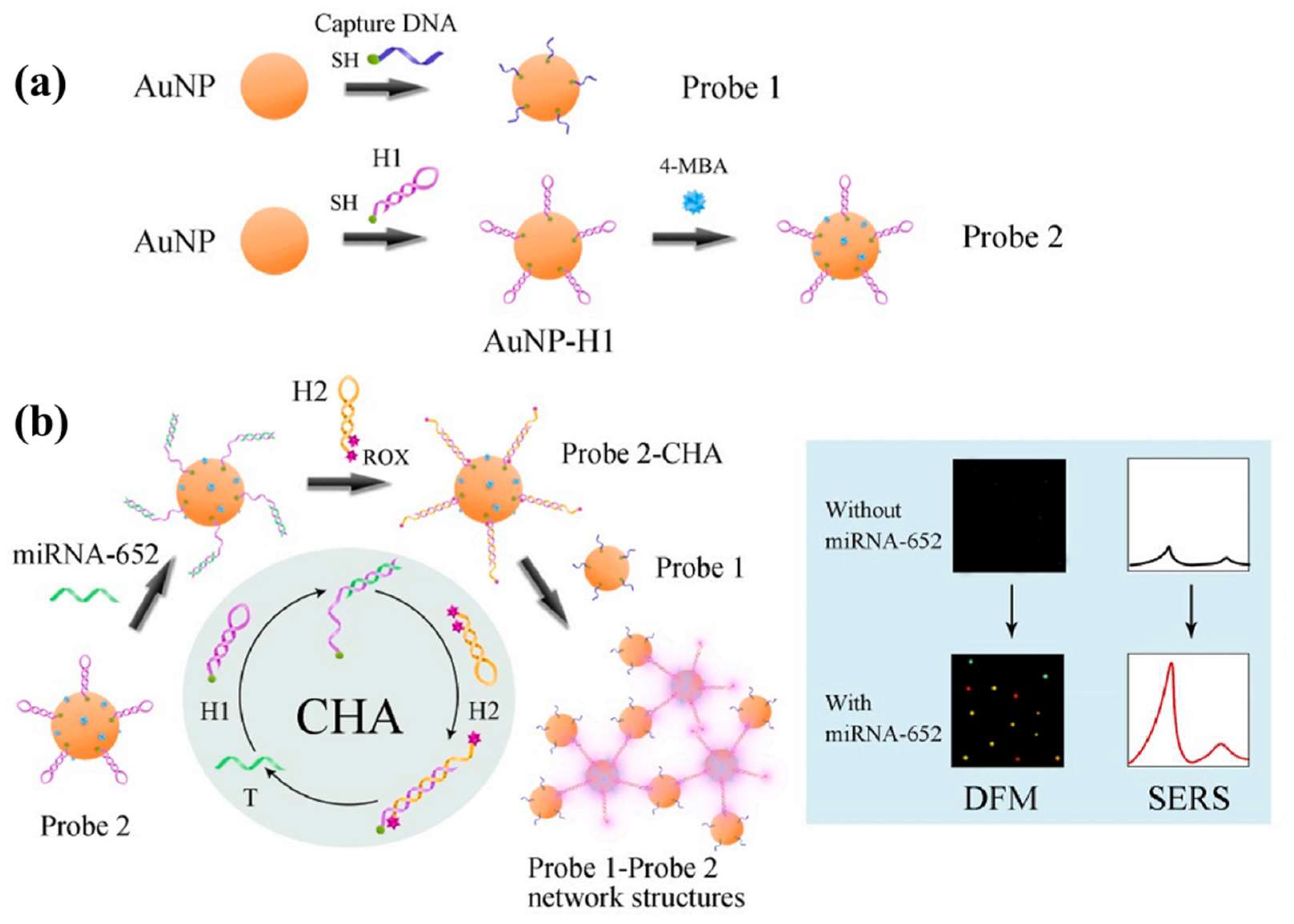
  29. Sun, Y.D.; Li, T. Composition-Tunable Hollow Au/Ag SERS Nanoprobes Coupled with Target-Catalyzed Hairpin Assembly for Triple-Amplification Detection of miRNA. Anal. Chem. 2018, 90, 11614–11621. [Google Scholar] [CrossRef]
  30. Daniel, M.C.; Grow, M.E.; Pan, H.M.; Bednarek, M.; Ghann, W.E.; Zabetakis, K.; Cornish, J. Gold nanoparticle-cored poly(propyleneimine) dendrimers as a new platform for multifunctional drug delivery systems. New J. Chem. 2011, 35, 2366–2374. [Google Scholar] [CrossRef]
  31. Zhu, Z.K.; Li, H.J.; Xiang, Y.Q.; Koh, W.N.; Hu, X.J.; Chen, H.X. Pyridinium porphyrins and AuNPs mediated bionetworks as SPR signal amplification tags for the ultrasensitive assay of brain natriuretic peptide. Microchim. Acta 2020, 187, 327. [Google Scholar] [CrossRef]
  32. Cordeiro, M.; Carlos, F.F.; Pedrosa, P.; Lopez, A.; Baptista, P.V. Gold Nanoparticles for Diagnostics: Advances towards Points of Care. Diagnostics 2016, 6, 43. [Google Scholar] [CrossRef]
  33. Elahi, N.; Kamali, M.; Baghersad, M.H. Recent biomedical applications of gold nanoparticles: A review. Talanta 2018, 184, 537–556. [Google Scholar] [CrossRef]
  34. Yang, H.H.; Xiao, M.S.; Lai, W.; Wan, Y.; Li, L.; Pei, H. Stochastic DNA Dual-Walkers for Ultrafast Colorimetric Bacteria Detection. Anal. Chem. 2020, 92, 4990–4995. [Google Scholar] [CrossRef]
  35. Shalabney, A.; Abdulhalim, I. Sensitivity-enhancement methods for surface plasmon sensors. Laser Photonics Rev. 2011, 5, 571–606. [Google Scholar] [CrossRef]
  36. Usman, F.; Dennis, J.O.; Aljameel, A.I.; Ali, M.K.M.; Aldaghri, O.; Ibnaouf, K.H.; Zango, Z.U.; Beygisangchin, M.; Alsadig, A.; Meriaudeau, F. Plasmonic Biosensors for the Detection of Lung Cancer Biomarkers: A Review. Chemosensors 2021, 9, 326. [Google Scholar] [CrossRef]
  37. Loste, J.; Lopez-Cuesta, J.M.; Billon, L.; Garay, H.; Save, M. Transparent polymer nanocomposites: An overview on their synthesis and advanced properties. Prog. Polym. Sci. 2019, 89, 133–158. [Google Scholar] [CrossRef]
  38. Song, C.Y.; Zhang, J.J.; Jiang, X.Y.; Gan, H.Y.; Zhu, Y.F.; Peng, Q.; Fang, X.Y.; Guo, Y.; Wang, L.H. SPR/SERS dual-mode plasmonic biosensor via catalytic hairpin assembly-induced AuNP network. Biosens. Bioelectron. 2021, 190, 113376. [Google Scholar] [CrossRef]
  39. Devi, R.S.; Girigoswami, A.; Siddharth, M.; Girigoswami, K. Applications of Gold and Silver Nanoparticles in Theranostics. Appl. Biochem. Biotechnol. 2022, 194, 4187–4219. [Google Scholar] [CrossRef]
  40. Ray, A.; Mukundan, A.; Xie, Z.X.; Karamchand, L.; Wang, X.D.; Kopelman, R. Highly stable polymer coated nano-clustered silver plates: A multimodal optical contrast agent for biomedical imaging. Nanotechnology 2014, 25, 445104. [Google Scholar] [CrossRef]
  41. Wu, M.Y.; Guo, H.B.; Liu, L.; Liu, Y.; Xie, L.M. Size-dependent cellular uptake and localization profiles of silver nanoparticles. Int. J. Nanomed. 2019, 14, 4247–4259. [Google Scholar] [CrossRef]
  42. de Souza, T.A.J.; Souza, L.R.R.; Franchi, L.P. Silver nanoparticles: An integrated view of green synthesis methods, transformation in the environment, and toxicity. Ecotoxicol. Environ. Saf. 2019, 171, 691–700. [Google Scholar] [CrossRef] [PubMed]
  43. Liu, C.; Song, Q.C.; Chen, J.N.; Li, X.H.; Cai, J.X.; Lu, Z.G.; Li, W.D.; Fang, N.X.; Feng, S.P. Electromagnetic and Chemical Enhancements of Surface-Enhanced Raman Scattering Spectra from Cu2O Hexagonal Nanoplates. Adv. Mater. Interfaces 2019, 6, 1900534. [Google Scholar] [CrossRef]
  44. Soundharraj, P.; Dhinasekaran, D.; Aruna, P.; Ganesan, S. Facile synthesis of biomass silica-silver colloidal nanoparticles and its application as highly sensitive fluorescent biosensor. Surf. Interfaces 2021, 23, 101010. [Google Scholar] [CrossRef]
  45. Wu, Y.; Zhou, J.M.; Jiang, Y.S.; Li, W.; He, M.J.; Xiao, Y.; Chen, J.Y. Silver nanoparticles@metal-organic framework as peroxidase mimics for colorimetric determination of hydrogen peroxide and blood glucose. Chin. J. Anal. Chem. 2022, 50, 100187. [Google Scholar] [CrossRef]
  46. Wu, Y.; Chen, J.Y.; He, W.M. Surface-enhanced Raman spectroscopy biosensor based on silver nanoparticles@metal-organic frameworks with peroxidase-mimicking activities for ultrasensitive monitoring of blood cholesterol. Sens. Actuators B-Chem. 2022, 365, 131939. [Google Scholar] [CrossRef]
  47. Zeng, C.; Eisner, G.M.; Felder, R.A.; Jose, P.A. Dopamine receptor and hypertension. Curr. Med. Chem.-Cardiovasc. Hematol. Agents 2005, 3, 69–77. [Google Scholar] [CrossRef]
  48. Anshori, I.; Rizalputri, L.N.; Althof, R.R.; Surjadi, S.S.; Harimurti, S.; Gumilar, G.; Yuliarto, B.; Handayani, M. Functionalized multi-walled carbon nanotube/silver nanoparticle (f-MWCNT/AgNP) nanocomposites as non-enzymatic electrochemical biosensors for dopamine detection. Nanocomposites 2021, 7, 97–108. [Google Scholar] [CrossRef]
  49. Daoudi, K.; Columbus, S.; Falcao, B.P.; Pereira, R.N.; Peripolli, S.B.; Ramachandran, K.; Hadj Kacem, H.; Allagui, A.; Gaidi, M. Label-free DNA detection using silver nanoprism decorated silicon nanoparticles: Effect of silicon nanoparticle size and doping levels. Spectrochim. Acta. Part A Mol. Biomol. Spectrosc. 2023, 290, 122262. [Google Scholar] [CrossRef]
  50. Jung, S.C.; Nahm, S.W.; Jung, H.Y.; Park, Y.K.; Seo, S.G.; Kim, S.C. Preparations of Platinum Nanoparticles and Their Catalytic Performances. J. Nanosci. Nanotechnol. 2015, 15, 5461–5465. [Google Scholar] [CrossRef]
  51. Pedone, D.; Moglianetti, M.; De Luca, E.; Bardi, G.; Pompa, P.P. Platinum nanoparticles in nanobiomedicine. Chem. Soc. Rev. 2017, 46, 4951–4975. [Google Scholar] [CrossRef]
  52. Shoshan, M.S.; Vonderach, T.; Hattendorf, B.; Wennemers, H. Peptide-Coated Platinum Nanoparticles with Selective Toxicity against Liver Cancer Cells. Angew. Chem. -Int. Ed. 2019, 58, 4901–4905. [Google Scholar] [CrossRef]
  53. Chauhan, N.; Tiwari, S.; Narayan, T.; Jain, U. Bienzymatic assembly formed@ Pt nano sensing framework detecting acetylcholine in aqueous phase. Appl. Surf. Sci. 2019, 474, 154–160. [Google Scholar] [CrossRef]
  54. Ehringer, H.; Hornykiewicz, O. Distribution of noradrenaline and dopamine (3-hydroxytyramine) in the human brain and their behavior in diseases of the extrapyramidal system. Park. Relat. Disord. 1998, 4, 53–57. [Google Scholar] [CrossRef]
  55. Li, J.; Huang, X.; Shi, W.S.; Jiang, M.Y.; Tian, L.; Su, M.J.; Wu, J.; Liu, Q.; Yu, C.M.; Gu, H.Y. Pt nanoparticle decorated carbon nanotubes nanocomposite based sensing platform for the monitoring of cell-secreted dopamine. Sens. Actuators B-Chem. 2021, 330, 129311. [Google Scholar] [CrossRef]
  56. Khan, M.S.; Zhu, W.J.; Ali, A.; Ahmad, S.M.; Li, X.J.; Yang, L.; Wang, Y.G.; Wang, H.; Wei, Q. Electrochemiluminescent immunosensor for prostate specific antigen based upon luminol functionalized platinum nanoparticles loaded on graphene. Anal. Biochem. 2019, 566, 50–57. [Google Scholar] [CrossRef]
  57. Huo, X.L.; Yang, H.; Li, M.X.; Zhao, W.; Xu, J.J.; Wang, Y.; Luo, X.L.; Chen, H.Y. Multi-segmented CdS-Au nanorods for electrochemiluminescence bioanalysis. Nanoscale 2018, 10, 19224–19230. [Google Scholar] [CrossRef]
  58. Meng, F.Y.; Sun, H.X.; Huang, Y.; Tang, Y.G.; Chen, Q.; Miao, P. Peptide cleavage-based electrochemical biosensor coupling graphene oxide and silver nanoparticles. Anal. Chim. Acta 2019, 1047, 45–51. [Google Scholar] [CrossRef]
  59. Hou, L.L.; Huang, Y.L.; Hou, W.L.; Yan, Y.R.; Liu, J.L.; Xia, N. Modification-free amperometric biosensor for the detection of wild-type p53 protein based on the in situ formation of silver nanoparticle networks for signal amplification. Int. J. Biol. Macromol. 2020, 158, 580–586. [Google Scholar] [CrossRef]
  60. Tran, H.V.; Nguyen, T.V.; Nguyen, L.T.N.; Hoang, H.S.; Huynh, C.D. Silver nanoparticles as a bifunctional probe for label-free and reagentless colorimetric hydrogen peroxide chemosensor and cholesterol biosensor. J. Sci. -Adv. Mater. Devices 2020, 5, 385–391. [Google Scholar] [CrossRef]
  61. Shin, J.W.; Yoon, J.; Shin, M.; Choi, J.W. Electrochemical Dopamine Biosensor Composed of Silver Encapsulated MoS2 Hybrid Nanoparticle. Biotechnol. Bioprocess Eng. 2019, 24, 135–144. [Google Scholar] [CrossRef]
  62. Rostami, S.; Mehdinia, A.; Niroumand, R.; Jabbari, A. Enhanced LSPR performance of graphene nanoribbons-silver nanoparticles hybrid as a colorimetric sensor for sequential detection of dopamine and glutathione. Anal. Chim. Acta 2020, 1120, 11–23. [Google Scholar] [CrossRef] [PubMed]
  63. Zhang, Z.G.; Yang, G.H.; He, M.; Qi, L.T.; Li, X.C.; Chen, J.C. Synthesis of Silver Nanoparticles and Detection of Glucose via Chemical Reduction with Nanocellulose as Carrier and Stabilizer. Int. J. Mol. Sci. 2022, 23, 15345. [Google Scholar] [CrossRef] [PubMed]
  64. Tsekeli, T.R.; Tshwenya, L.; Sebokolodi, T.I.; Ndlovu, T.; Arotiba, O.A. An Electrochemical Aptamer Biosensor for Bisphenol A on a Carbon Nanofibre-silver Nanoparticle Immobilisation Platform. Electroanalysis 2021, 33, 2053–2061. [Google Scholar] [CrossRef]
  65. Pareek, S.; Jain, U.; Bharadwaj, M.; Saxena, K.; Roy, S.; Chauhan, N. An ultrasensitive electrochemical DNA biosensor for monitoring Human papillomavirus-16 (HPV-16) using graphene oxide/Ag/Au nano-biohybrids. Anal. Biochem. 2023, 663, 115015. [Google Scholar] [CrossRef] [PubMed]
  66. Maity, D.; Kumar, R.T.R. Highly sensitive amperometric detection of glutamate by glutamic oxidase immobilized Pt nanoparticle decorated multiwalled carbon nanotubes (MWCNTs)/polypyrrole composite. Biosens. Bioelectron. 2019, 130, 307–314. [Google Scholar] [CrossRef]
  67. Han, B.; Pan, M.; Liu, X.; Liu, J.; Cui, T.; Chen, Q. Electrochemical Detection for Uric Acid Based on beta-Lactoglobulin-Functionalized Multiwall Carbon Nanotubes Synthesis with PtNPs Nanocomposite. Materials 2019, 12, 214. [Google Scholar] [CrossRef]
  68. Uzak, D.; Atiroglu, A.; Atiroglu, V.; Cakiroglu, B.; Ozacar, M. Reduced Graphene Oxide/Pt Nanoparticles/Zn-MOF-74 Nanomaterial for a Glucose Biosensor Construction. Electroanalysis 2020, 32, 510–519. [Google Scholar] [CrossRef]
  69. Hossain, M.F.; Slaughter, G. Flexible electrochemical uric acid and glucose biosensor. Bioelectrochemistry 2021, 141, 107870. [Google Scholar] [CrossRef]
  70. Shi, X.L.; Chen, X.H.; Zuo, W.Y.; Lin, L.Y.; Xiao, W.H.; Yang, H.P. Amperometric Sarcosine Biosensor Based on a Bi-Functional Platinum-Loaded Organic-Inorganic Hybrid Tungsten Phosphonate Material. J. Electrochem. Soc. 2022, 169, 057515. [Google Scholar] [CrossRef]
  71. Cakiroglu, B.; Ozacar, M. A self-powered photoelectrochemical glucose biosensor based on supercapacitor Co3O4-CNT hybrid on TiO2. Biosens. Bioelectron. 2018, 119, 34–41. [Google Scholar] [CrossRef]
  72. Huang, L.; Liang, Z.; Zhang, F.; Luo, H.; Liang, R.; Han, F.; Wu, Z.; Han, D.; Shen, J.; Niu, L. Upconversion NaYF4:Yb/Er-TiO2-Ti3C2 Heterostructure-Based Near-Infrared Light-Driven Photoelectrochemical Biosensor for Highly Sensitive and Selective d-Serine Detection. Anal. Chem. 2022, 94, 16246–16253. [Google Scholar] [CrossRef]
  73. Zou, L.; Yang, L.; Zhan, Y.; Huang, D.; Ye, B. Photoelectrochemical aptasensor for thrombin based on Au-rGO-CuS as signal amplification elements. Mikrochim. Acta 2020, 187, 433. [Google Scholar] [CrossRef]
  74. Porzani, S.J.; Lorenzi, A.S.; Eghtedari, M.; Nowruzi, B. Interaction of Dehydrogenase Enzymes with Nanoparticles in Industrial and Medical Applications, and the Associated Challenges: A Mini-review. Mini. Rev. Med. Chem. 2021, 21, 1351–1366. [Google Scholar] [CrossRef]
  75. Batra, B.; Narwal, V.; Sumit; Ahlawat, J.; Sharma, M. An amperometric cholesterol biosensor based on immobilization of cholesterol oxidase onto titanium dioxide nanoparticles. Sens. Int. 2021, 2, 100111. [Google Scholar] [CrossRef]
  76. Nadzirah, S.; Gopinath, S.C.B.; Parmin, N.A.; Hamzah, A.A.; Mohamed, M.A.; Chang, E.Y.; Dee, C.F. State-of-the-Art on Functional Titanium Dioxide-Integrated Nano-Hybrids in Electrical Biosensors. Crit. Rev. Anal. Chem. 2022, 52, 637–648. [Google Scholar] [CrossRef]
  77. Wu, Y.; Li, Y.; He, J.; Fang, X.; Hong, P.; Nie, M.; Yang, W.; Xie, C.; Wu, Z.; Zhang, K.; et al. Nano-hybrids of needle-like MnO2 on graphene oxide coupled with peroxymonosulfate for enhanced degradation of norfloxacin: A comparative study and probable degradation pathway. J. Colloid. Interface Sci. 2020, 562, 1–11. [Google Scholar] [CrossRef]
  78. Jeong, H.; Yoo, J.; Park, S.; Lu, J.; Park, S.; Lee, J. Non-Enzymatic Glucose Biosensor Based on Highly Pure TiO2 Nanoparticles. Biosensors 2021, 11, 149. [Google Scholar] [CrossRef]
  79. Sadique, M.A.; Yadav, S.; Khare, V.; Khan, R.; Tripathi, G.K.; Khare, P.S. Functionalized Titanium Dioxide Nanoparticle-Based Electrochemical Immunosensor for Detection of SARS-CoV-2 Antibody. Diagnostics 2022, 12, 2612. [Google Scholar] [CrossRef]
  80. Huang, X.; Wei, S.; Yao, S.; Zhang, H.; He, C.; Cao, J. Development of molecularly imprinted electrochemical sensor with reduced graphene oxide and titanium dioxide enhanced performance for the detection of toltrazuril in chicken muscle and egg. J Pharm. Biomed. Anal. 2019, 164, 607–614. [Google Scholar] [CrossRef]
  81. Thakur, D.; Pandey, C.M.; Kumar, D. Highly Sensitive Enzymatic Biosensor Based on Polyaniline-Wrapped Titanium Dioxide Nanohybrid for Fish Freshness Detection. Appl. Biochem. Biotechnol. 2022, 194, 3765–3778. [Google Scholar] [CrossRef]
  82. Lu, X.; Li, S.; Guo, W.; Zhang, F.; Qu, F. A covalent organic polymer-TiO2/Ti3C2 heterostructure as nonenzymatic biosensor for voltammetric detection of dopamine and uric acid. Mikrochim. Acta 2021, 188, 95. [Google Scholar] [CrossRef] [PubMed]
  83. Cheng, H.; Hu, C.; Ji, Z.; Ma, W.; Wang, H. A solid ionic Lactate biosensor using doped graphene-like membrane of Au-EVIMC-titania nanotubes-polyaniline. Biosens. Bioelectron. 2018, 118, 97–101. [Google Scholar] [CrossRef] [PubMed]
  84. Guo, J.; Fang, G.; Wang, S.; Wang, J. Quartz crystal microbalance sensor based on 11-mercaptoundecanoic acid self-assembly and amidated nano-titanium film for selective and ultrafast detection of phosphoproteins in food. Food Chem. 2021, 344, 128656. [Google Scholar] [CrossRef] [PubMed]
  85. Yang, J.; Li, W.; Guo, L.; Luo, F.; Qiu, B.; Lin, Z.; Wang, L. Highly Sensitive Photoelectrochemical Biosensor for MicroRNA-21 Based on a Dumbbell-Shaped Heterostructure AuNRs@end-TiO2 Combined with Carbon Dots as Photosensitizers and Duplex-Specific Nuclease-Assisted Signal Amplification. Anal. Chem. 2022, 94, 13575–13581. [Google Scholar] [CrossRef]
  86. Siew, Q.Y.; Pang, E.L.; Loh, H.S.; Tan, M.T.T. Highly sensitive and specific graphene/TiO2 impedimetric immunosensor based on plant-derived tetravalent envelope glycoprotein domain III (EDIII) probe antigen for dengue diagnosis. Biosens. Bioelectron. 2021, 176, 112895. [Google Scholar] [CrossRef]
  87. Boobphahom, S.; Rattanawaleedirojn, P.; Boonyongmaneerat, Y.; Rengpipat, S.; Chailapakul, O.; Rodthongkum, N. TiO2 sol/graphene modified 3D porous Ni foam: A novel platform for enzymatic electrochemical biosensor. J. Electroanal. Chem. 2019, 833, 133–142. [Google Scholar] [CrossRef]
  88. Zhu, J.H.; Mei, L.P.; Wang, A.J.; Song, Y.Y.; Feng, J.J. Integration of phosphate functionalized Pt/TiO2 and Ru(bpy)32+ sensitization for ultrasensitive assay of adenosine deaminase activity on a novel split-typed PEC aptasensor. Biosens. Bioelectron. 2023, 226, 115141. [Google Scholar] [CrossRef]
  89. Deng, Y.; Yan, W.; Guo, Y.; Wang, Q.; Bi, Y.; Dong, C.; Fan, L. Highly sensitive and selective photoelectrochemical aptasensing of di-2-ethylhexyl phthalate based on graphene quantum dots decorated TiO2 nanotube arrays. J. Hazard. Mater. 2022, 426, 128107. [Google Scholar] [CrossRef]
  90. Zhu, C.; Tian, X.; Li, Q.; Dai, Z.; Wang, L.; Liu, H.; Li, C.; Zahid, K.R.; Wu, C.; Huang, H.; et al. Ultrasensitive photoelectrochemical biosensor for DNA 5-methylcytosine analysis based on co-sensitization strategy combined with bridged DNA nanoprobe. Talanta 2023, 254, 124140. [Google Scholar] [CrossRef]
  91. Yan, Q.; Cao, L.; Dong, H.; Tan, Z.; Hu, Y.; Liu, Q.; Liu, H.; Zhao, P.; Chen, L.; Liu, Y.; et al. Label-free immunosensors based on a novel multi-amplification signal strategy of TiO2-NGO/Au@Pd hetero-nanostructures. Biosens. Bioelectron. 2019, 127, 174–180. [Google Scholar] [CrossRef]
  92. Chen, Z.; Li, B.; Liu, J.; Li, H.; Li, C.; Xuan, X.; Li, M. A label-free electrochemical immunosensor based on a gold-vertical graphene/TiO2 nanotube electrode for CA125 detection in oxidation/reduction dual channels. Mikrochim. Acta 2022, 189, 257. [Google Scholar] [CrossRef]
  93. Danielson, E.; Dhamodharan, V.; Porkovich, A.; Kumar, P.; Jian, N.; Ziadi, Z.; Grammatikopoulos, P.; Sontakke, V.A.; Yokobayashi, Y.; Sowwan, M. Gas-Phase Synthesis for Label-Free Biosensors: Zinc-Oxide Nanowires Functionalized with Gold Nanoparticles. Sci. Rep. 2019, 9, 17370. [Google Scholar] [CrossRef]
  94. Urgessa, Z.N.; Botha, J.R.; Tankio Djiokap, S.R.; Coleman, C.; Bhattacharyya, S. Patterned growth of ZnO nanorods by chemical bath deposition. Phys. B: Condens. Matter 2018, 535, 79–83. [Google Scholar] [CrossRef]
  95. DÖNmez, S. Zencefil (Zingiber officinale) Kök Ekstresi Kullanılarak Çinko Oksit Nanoparçacıkların Yeşil Sentezi ve Glikoz Biyosensörü Olarak Uygulaması. El-Cezeri Fen Ve Mühendislik Derg. 2020, 7, 1191–1200. [Google Scholar] [CrossRef]
  96. Napi, M.L.M.; Sultan, S.M.; Ismail, R.; How, K.W.; Ahmad, M.K. Electrochemical-Based Biosensors on Different Zinc Oxide Nanostructures: A Review. Materials 2019, 12, 2985. [Google Scholar] [CrossRef]
  97. Salinas, R.A.; Orduña-Díaz, A.; Obregon-Hinostroza, O.; Dominguez, M.A. Biosensors based on zinc oxide thin-film transistors using recyclable plastic substrates as an alternative for real-time pathogen detection. Talanta 2022, 237, 122970. [Google Scholar] [CrossRef]
  98. Zhu, L.; Yin, Z.; Lv, Z.; Li, M.; Tang, D. Ultrasensitive photoelectrochemical immunoassay for prostate-specific antigen based on silver nanoparticle-triggered ion-exchange reaction with ZnO/CdS nanorods. Analyst 2021, 146, 4487–4494. [Google Scholar] [CrossRef]
  99. Migliorini, F.L.; Sanfelice, R.C.; Mercante, L.A.; Andre, R.S.; Mattoso, L.H.C.; Correa, D.S. Urea impedimetric biosensing using electrospun nanofibers modified with zinc oxide nanoparticles. Appl. Surf. Sci. 2018, 443, 18–23. [Google Scholar] [CrossRef]
  100. Ramesh, T.; Foo, K.L.; Haarindraprasad; Sam, A.J.; Solayappan, M. Gold-Hybridized Zinc Oxide Nanorods as Real-Time Low-Cost NanoBiosensors for Detection of virulent DNA signature of HPV-16 in Cervical Carcinoma. Sci. Rep. 2019, 9, 17039. [Google Scholar] [CrossRef]
  101. Kaur, M.; Kailasaganapathi, S.; Ramgir, N.; Datta, N.; Kumar, S.; Debnath, A.K.; Aswal, D.K.; Gupta, S.K. Gas dependent sensing mechanism in ZnO nanobelt sensor. Appl. Surf. Sci. 2017, 394, 258–266. [Google Scholar] [CrossRef]
  102. Qu, X.; Yang, R.; Tong, F.; Zhao, Y.; Wang, M.-H. Hierarchical ZnO microstructures decorated with Au nanoparticles for enhanced gas sensing and photocatalytic properties. Powder Technol. 2018, 330, 259–265. [Google Scholar] [CrossRef]
  103. Ebrahimi, M.; Yousefzadeh, S.; Samadi, M.; Dong, C.; Zhang, J.; Moshfegh, A.Z. Facile preparation of branched hierarchical ZnO nanowire arrays with enhanced photocatalytic activity: A photodegradation kinetic model. Appl. Surf. Sci. 2018, 435, 108–116. [Google Scholar] [CrossRef]
  104. Alfaifi, S.Y.; Adeosun, W.A.; Asiri, A.M.; Rahman, M.M. Sensitive and Rapid Detection of Aspartic Acid with Co3O4-ZnO Nanorods Using Differential Pulse Voltammetry. Biosensors 2023, 13, 88. [Google Scholar] [CrossRef]
  105. Wu, T.; Yu, S.; Dai, L.; Feng, J.; Ren, X.; Ma, H.; Wang, X.; Wei, Q.; Ju, H. CuO Nanozymes as Multifunctional Signal Labels for Efficiently Quenching the Photocurrent of ZnO/Au/AgSbS2 Hybrids and Initiating a Strong Fluorescent Signal in a Dual-Mode Microfluidic Sensing Platform. ACS Sens. 2022, 7, 1732–1739. [Google Scholar] [CrossRef] [PubMed]
  106. Sharma, A.; Agrawal, A.; Pandey, G.; Kumar, S.; Awasthi, K.; Awasthi, A. Carbon Nano-Onion-Decorated ZnO Composite-Based Enzyme-Less Electrochemical Biosensing Approach for Glucose. ACS Omega 2022, 7, 37748–37756. [Google Scholar] [CrossRef]
  107. Supraja, P.; Singh, V.; Vanjari, S.R.K.; Singh, S.G. Electrospun CNT embedded ZnO nanofiber based biosensor for electrochemical detection of Atrazine: A step closure to single molecule detection. Microsyst. Nanoeng. 2020, 6, 3. [Google Scholar] [CrossRef]
  108. Myndrul, V.; Coy, E.; Babayevska, N.; Zahorodna, V.; Balitskyi, V.; Baginskiy, I.; Gogotsi, O.; Bechelany, M.; Giardi, M.T.; Iatsunskyi, I. MXene nanoflakes decorating ZnO tetrapods for enhanced performance of skin-attachable stretchable enzymatic electrochemical glucose sensor. Biosens. Bioelectron. 2022, 207, 114141. [Google Scholar] [CrossRef]
  109. Zong, X.; Zhu, R. ZnO nanorod-based FET biosensor for continuous glucose monitoring. Sens. Actuators B. Chem. 2018, 255, 2448–2453. [Google Scholar] [CrossRef]
  110. Li, B.; Guo, L.; Chen, M.; Guo, Y.; Ge, L.; Kwok, H.F. Single-atom Pt-anchored Zn0.5Cd0.5S boosted photoelectrochemical immunoassay of prostate-specific antigen. Biosens. Bioelectron. 2022, 202, 114006. [Google Scholar] [CrossRef]
  111. Khushaim, W.; Vijjapu, M.T.; Yuvaraja, S.; Mani, V.; Salama, K.N. Graphitic Carbon Nitride and IGZO Bio-FET for Rapid Diagnosis of Myocardial Infarction. Biosensors 2022, 12, 836. [Google Scholar] [CrossRef]
  112. Imran, H.; Alam, A.; Dharuman, V.; Lim, S. Fabrication of Enzyme-Free and Rapid Electrochemical Detection of Glucose Sensor Based on ZnO Rod and Ru Doped Carbon Nitride Modified Gold Transducer. Nanomaterials 2022, 12, 1778. [Google Scholar] [CrossRef]
  113. Huang, J.; Li, X.; Xiu, M.; Huang, K.; Cui, K.; Zhang, J.; Ge, S.; Hao, S.; Yu, J.; Huang, Y. A Paper-Based Photoelectrochemical Sensing Platform Based on In Situ Grown ZnO/ZnIn2S4 Heterojunctions onto Paper Fibers for Sensitively Detecting AFP. Biosensors 2022, 12, 818. [Google Scholar] [CrossRef] [PubMed]
  114. Avelino, K.; Oliveira, L.S.; Santos, M.R.; Lucena-Silva, N.; Andrade, C.A.S.; Oliveira, M.D.L. Electrochemical DNA biosensor for chronic myelocytic leukemia based on hybrid nanostructure. Bioelectrochemistry 2022, 147, 108176. [Google Scholar] [CrossRef]
  115. Akbarzadeh, A.; Samiei, M.; Davaran, S. Magnetic nanoparticles: Preparation, physical properties, and applications in biomedicine. Nanoscale Res. Lett. 2012, 7, 144. [Google Scholar] [CrossRef]
  116. Zhu, N.; Ji, H.N.; Yu, P.; Niu, J.Q.; Farooq, M.U.; Akram, M.W.; Udego, I.O.; Li, H.D.; Niu, X.B. Surface Modification of Magnetic Iron Oxide Nanoparticles. Nanomaterials 2018, 8, 810. [Google Scholar] [CrossRef]
  117. Rocha-Santos, T.A.P. Sensors and biosensors based on magnetic nanoparticles. Trac-Trends Anal. Chem. 2014, 62, 28–36. [Google Scholar] [CrossRef]
  118. Peng, L.C.; Luo, Y.J.; Xiong, H.P.; Yao, S.; Zhu, M.H.; Song, H. A Novel Amperometric Glucose Biosensor Based on Fe3O4-Chitosan-beta-Cyclodextrin/MWCNTs Nanobiocomposite. Electroanalysis 2021, 33, 723–732. [Google Scholar] [CrossRef]
  119. Qi, X.Y.; Ye, Y.F.; Wang, H.; Zhao, B.B.; Xu, L.X.; Zhang, Y.T.; Wang, X.L.; Zhou, N.D. An ultrasensitive and dual-recognition SERS biosensor based on Fe3O4@Au-Teicoplanin and aptamer functionalized Au@Ag nanoparticles for detection of Staphylococcus aureus. Talanta 2022, 250, 123648. [Google Scholar] [CrossRef]
  120. Buyuksunetci, Y.T.; Citil, B.E.; Tapan, U.; Anik, U. Development and application of a SARS-CoV-2 colorimetric biosensor based on the peroxidase-mimic activity of gamma-Fe2O3 nanoparticles. Microchim. Acta 2021, 188, 335. [Google Scholar] [CrossRef]
  121. Butmee, P.; Tumcharern, G.; Thouand, G.; Kalcher, K.; Samphao, A. An ultrasensitive immunosensor based on manganese dioxide-graphene nanoplatelets and core shell Fe3O4@Au nanoparticles for label-free detection of carcinoembryonic antigen. Bioelectrochemistry 2020, 132, 107452. [Google Scholar] [CrossRef]
  122. Cao, X.D.; Zhang, K.R.; Yan, W.W.; Xia, Z.H.; He, S.D.; Xu, X.; Ye, Y.K.; Wei, Z.J.; Liu, S.Q. Calcium ion assisted fluorescence determination of microRNA-167 using carbon dots-labeled probe DNA and polydopamine-coated Fe3O4 nanoparticles. Microchim. Acta 2020, 187, 212. [Google Scholar] [CrossRef] [PubMed]
  123. Chen, W.; Liu, X.N.; Wang, W.; Yin, G.F. Magnetic Fe3O4 nanorings for protein adsorption and detection. Mater. Res. Express 2018, 5, 125402. [Google Scholar] [CrossRef]
  124. El-Said, W.A.; Abdel-Rahman, M.A.; Sayed, E.M.; Abdel-Wahab, A.M.A. Electrochemical Monitoring of Methotrexate Anticancer Drug in Human Blood Serum by Using in situ Solvothermal Synthesized Fe3O4/ITO Electrode. Electroanalysis 2019, 31, 829–837. [Google Scholar] [CrossRef]
  125. Akram, M.W.; Alam, M.F.; Ji, H.N.; Mahmood, A.; Munir, T.; Iqbal, M.Z.; Saleem, M.R.; Amin, N.; Wu, A.G. Chitosan blend iron oxide nanostructure-based biosensor for healthy & malignant tissue glucose/urea detection. In Proceedings of the 7th Global Conference on Materials Science and Engineering (CMSE), Xian Technol Univ, Shaanxi Key Lab Optoelectron Funct Mat & Devices, Corro, Xi’an, China, 1–4 November 2018. [Google Scholar]
  126. Atif, M.; Ali, A.; AlSalhi, M.S.; Willander, M. Effect of Urea on the Morphology of Fe3O4 Magnetic Nanoparticles and Their Application in Potentiometric Urea Biosensors. Silicon 2019, 11, 1371–1376. [Google Scholar] [CrossRef]
  127. Huang, Y.; Gu, Y.Q.; Liu, X.Y.; Deng, T.T.; Dai, S.; Qu, J.F.; Yang, G.H.; Qu, L.L. Reusable ring-like Fe3O4/Au nanozymes with enhanced peroxidase-like activities for colorimetric-SERS dual-mode sensing of biomolecules in human blood. Biosens. Bioelectron. 2022, 209, 114253. [Google Scholar] [CrossRef]
  128. Rohani, T.; Mohammadi, S.Z.; Zadeh, N.G.; Askari, M.B. A novel carbon ceramic electrode modified by Fe3O4 magnetic nanoparticles coated with aptamer-immobilized polydopamine: An effective label-free aptasensor for sensitive detection of diclofenac. Microchem. J. 2021, 166, 106274. [Google Scholar] [CrossRef]
  129. Lian, J.J.; Liu, P.; Li, X.C.; Gao, L.N.; Luo, X.L.; Zhang, X.; Shi, Z.Q.; Liu, Q.Y. Perylene diimide-modified magnetic gamma-Fe2O3/CeO2 nanoparticles as peroxidase mimics for highly sensitive colorimetric detection of Vitamin C. Appl. Organomet. Chem. 2019, 33, e4884. [Google Scholar] [CrossRef]
  130. Huang, L.P.; Zhu, Y.J.; Xu, C.S.; Cai, Y.; Yi, Y.D.; Li, K.; Ren, X.Q.; Jiang, D.F.; Ge, Y.C.; Liu, X.H.; et al. Noninvasive Diagnosis of Gastric Cancer Based on Breath Analysis with a Tubular Surface-Enhanced Raman Scattering Sensor. Acs Sens. 2022, 7, 1439–1450. [Google Scholar] [CrossRef]
  131. Qasim, M.; Asghar, K.; Das, D. Preparation and characterization of CoFe2O4 and CoFe2O4@Albumen nanoparticles for biomedical applications. Ceram. Int. 2019, 45, 24971–24981. [Google Scholar] [CrossRef]
  132. Mokhosi, S.R.; Mdlalose, W.; Mngadi, S.; Singh, M.; Moyo, T. Assessing the structural, morphological and magnetic properties of polymer-coated magnesium-doped cobalt ferrite (CoFe2O4) nanoparticles for biomedical application. In Proceedings of the Applied Nanotechnology and Nanoscience International Conference (ANNIC), Berlin, Germany, 22–24 October 2018. [Google Scholar]
  133. Al-Qasmi, N.; Almughem, F.A.; Jarallah, S.J.; Almaabadi, A. Efficient Green Synthesis of (Fe3O4) and (NiFe2O4) Nanoparticles Using Star Anise (Illicium verum) Extract and Their Biomedical Activity against Some Cancer Cells. Materials 2022, 15, 4832. [Google Scholar] [CrossRef]
  134. Esther Nimshi, R.; Judith Vijaya, J.; Al-Najar, B.; Hazeem, L.; Bououdina, M.; John Kennedy, L.; Kombaiah, K.; Bellucci, S. Multifunctional Core-Shell NiFe2O4 Shield with TiO2/rGO Nanostructures for Biomedical and Environmental Applications. Bioinorg. Chem. Appl. 2022, 2022, 4805490. [Google Scholar] [CrossRef]
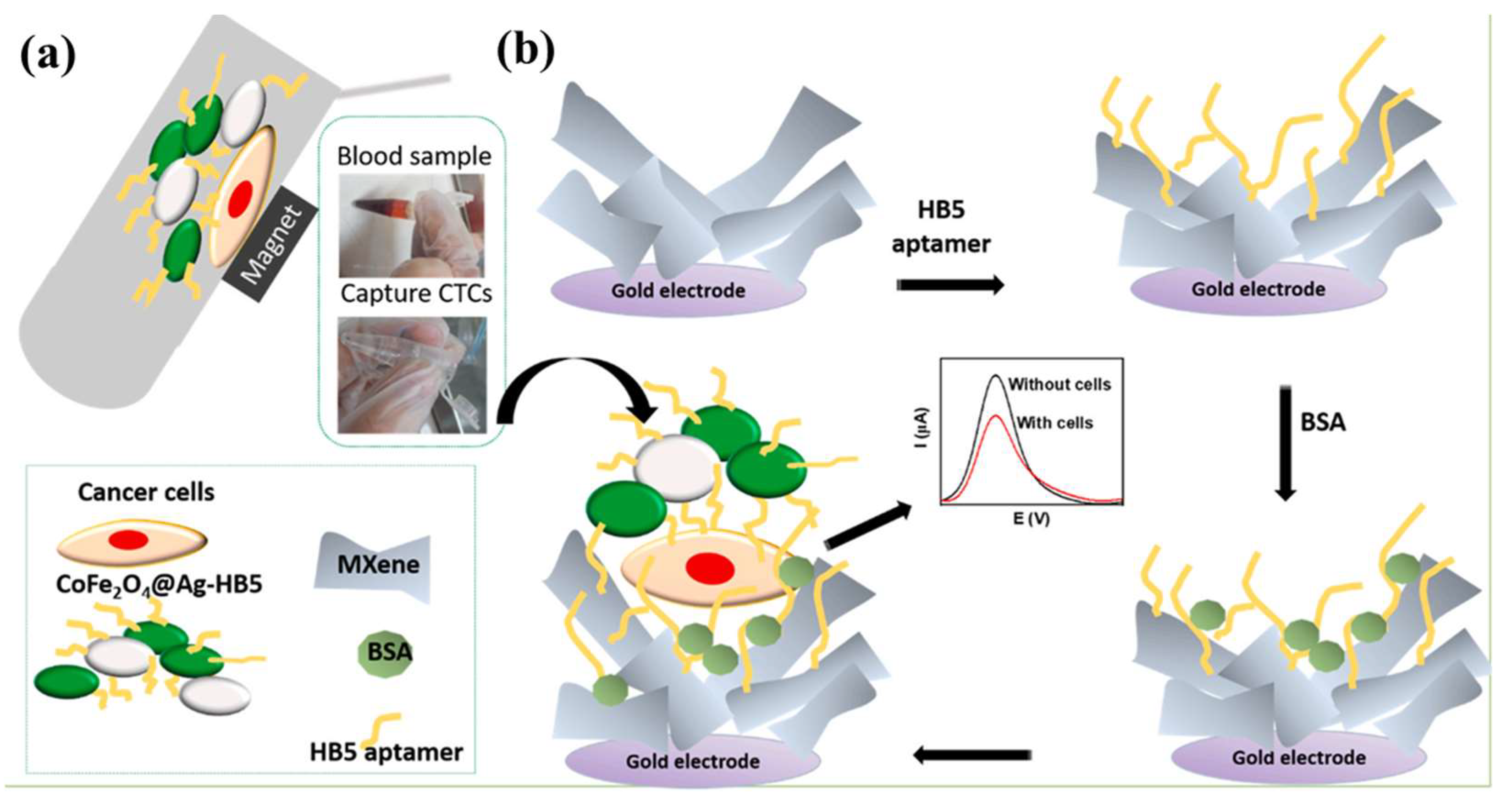
  135. Vajhadin, F.; Mazloum-Ardakani, M.; Shahidi, M.; Moshtaghioun, S.M.; Haghiralsadat, F.; Ebadi, A.; Amini, A. MXene-based cytosensor for the detection of HER2-positive cancer cells using CoFe2O4@Ag magnetic nanohybrids conjugated to the HB5 aptamer. Biosens. Bioelectron. 2022, 195, 113626. [Google Scholar] [CrossRef]
  136. Xu, X.; Chen, H.; Cao, Y.C.; Lin, Y.J.; Liu, J.A. A Novel Fluorescent Nanoparticle for Sensitive Detection of Cry1Ab Protein In Vitro and In Vivo. J. Fluoresc. 2018, 28, 863–869. [Google Scholar] [CrossRef]
  137. Wu, S.; Huang, H.; Shang, M.; Du, C.; Wu, Y.; Song, W. High visible light sensitive MoS2 ultrathin nanosheets for photoelectrochemical biosensing. Biosens. Bioelectron. 2017, 92, 646–653. [Google Scholar] [CrossRef]
  138. Karaman, C.; Bölükbaşı, Ö.S.; Yola, B.B.; Karaman, O.; Atar, N.; Yola, M.L. Electrochemical neuron-specific enolase (NSE) immunosensor based on CoFe2O4@Ag nanocomposite and AuNPs@MoS2/rGO. Anal. Chim. Acta 2022, 1200, 339609. [Google Scholar] [CrossRef]
  139. Mphuthi, N.; Sikhwivhilu, L.; Ray, S.S. Functionalization of 2D MoS2 Nanosheets with Various Metal and Metal Oxide Nanostructures: Their Properties and Application in Electrochemical Sensors. Biosensors 2022, 12, 386. [Google Scholar] [CrossRef]
  140. Li, F.; Feng, J.; Gao, Z.; Shi, L.; Wu, D.; Du, B.; Wei, Q. Facile Synthesis of Cu2O@TiO2-PtCu Nanocomposites as a Signal Amplification Strategy for the Insulin Detection. ACS Appl. Mater. Interfaces 2019, 11, 8945–8953. [Google Scholar] [CrossRef]
  141. Song, Y.; Qiao, J.; Li, W.; Ma, C.; Chen, S.; Li, H.; Hong, C. Bimetallic PtCu nanoparticles supported on molybdenum disulfide-functionalized graphitic carbon nitride for the detection of carcinoembryonic antigen. Mikrochim. Acta 2020, 187, 538. [Google Scholar] [CrossRef]
  142. Hu, K.; Cheng, J.; Wang, K.; Zhao, Y.; Liu, Y.; Yang, H.; Zhang, Z. Sensitive electrochemical immunosensor for CYFRA21-1 detection based on AuNPs@MoS2@Ti3C2Tx composites. Talanta 2022, 238, 122987. [Google Scholar] [CrossRef]
  143. Yu, H.; Chong, Y.; Zhang, P.; Ma, J.; Li, D. A D-shaped fiber SPR sensor with a composite nanostructure of MoS2-graphene for glucose detection. Talanta 2020, 219, 121324. [Google Scholar] [CrossRef]
  144. Beibei, W.; Chao, W.; Yuyang, L.; Xuejing, L.; Dan, W.; Qin, W. Electrochemiluminescence biosensor for cardiac troponin I with signal amplification based on a MoS2@Cu2O-Ag-modified electrode and Ce:ZnO-NGQDs. Analyst 2022, 147, 4768–4776. [Google Scholar]
  145. Posha, B.; Sandhyarani, N. Highly sensitive endotoxin detection using a gold nanoparticle loaded layered molybdenum disulfide-polyacrylic acid nanocomposite. Analyst 2020, 145, 3939–3947. [Google Scholar] [CrossRef] [PubMed]
  146. Surendranath, A.; Mohanan, P.V. Solvothermal exfoliation assisted synthesis of transition metal dichalcogenide based tungsten disulphide quantum dots (WS2 QDs) and cellular QD-bio interaction in LN-229 human glioblastoma cells. Mater. Sci. Eng. B Adv. Funct. Solid-State Mater. 2022, 284, 115907. [Google Scholar] [CrossRef]
  147. Niknam, S.; Dehdast, S.A.; Pourdakan, O.; Shanbani, M.; Koohi, M.K. Tungsten Disulfide Nanomaterials (WS2 NM) Application in Biosensors and Nanomedicine: A review. Nanomed. Res. J. 2022, 7, 214–226. [Google Scholar] [CrossRef]
  148. Chen, Y.H.; Tamming, R.R.; Chen, K.; Zhang, Z.P.; Liu, F.J.; Zhang, Y.F.; Hodgkiss, J.M.; Blaikie, R.J.; Ding, B.Y.; Qiu, M. Bandgap control in two-dimensional semiconductors via coherent doping of plasmonic hot electrons. Nat. Commun. 2021, 12, 4332. [Google Scholar] [CrossRef]
  149. Wang, L.; Kutana, A.; Yakobson, B.I. Many-body and spin-orbit effects on direct-indirect band gap transition of strained monolayer MoS2 and WS2. Ann. Der Phys. 2014, 526, L7–L12. [Google Scholar] [CrossRef]
  150. Yao, Y.; Lin, Z.; Li, Z.; Song, X.; Moon, K.-S.; Wong, C.-P. Large-scale production of two-dimensional nanosheets. J. Mater. Chem. 2012, 22, 13494–13499. [Google Scholar] [CrossRef]
  151. Zhang, J.; Han, D.; Wang, S.; Zhang, X.D.; Yang, R.Q.; Ji, Y.C.; Yu, X. Electrochemical detection of adenine and guanine using a three-dimensional WS2 nanosheet/graphite microfiber hybrid electrode. Electrochem. Commun. 2019, 99, 75–80. [Google Scholar] [CrossRef]
  152. Xi, Q.; Zhou, D.M.; Kan, Y.Y.; Ge, J.; Wu, Z.K.; Yu, R.Q.; Jiang, J.H. Highly Sensitive and Selective Strategy for MicroRNA Detection Based on WS2 Nanosheet Mediated Fluorescence Quenching and Duplex-Specific Nuclease Signal Amplification. Anal. Chem. 2014, 86, 1361–1365. [Google Scholar] [CrossRef]
  153. Banerjee, S.; Lollar, C.T.; Xiao, Z.; Fang, Y.; Zhou, H.-C. Biomedical Integration of Metal–Organic Frameworks. Trends Chem. 2020, 2, 467–479. [Google Scholar] [CrossRef]
  154. Fu, J.H.; Zhong, Z.; Xie, D.; Guo, Y.J.; Kong, D.X.; Zhao, Z.X.; Zhao, Z.X.; Li, M. SERS-Active MIL-100(Fe) Sensory Array for Ultrasensitive and Multiplex Detection of VOCs. Angew. Chem. Int. Ed. 2020, 59, 20489–20498. [Google Scholar] [CrossRef]
  155. Wang, N.; Xie, M.G.; Wang, M.K.; Li, Z.X.; Su, X.G. UiO-66-NH2 MOF-based ratiometric fluorescent probe for the detection of dopamine and reduced glutathione. Talanta 2020, 220, 121352. [Google Scholar] [CrossRef]
  156. Zhou, L.; Yang, L.; Wang, C.; Jia, H.; Xue, J.; Wei, Q.; Ju, H. Copper doped terbium metal organic framework as emitter for sensitive electrochemiluminescence detection of CYFRA 21-1. Talanta 2022, 238, 123047. [Google Scholar] [CrossRef]
  157. Liu, X.; Guo, J.; Li, Y.; Wang, B.; Yang, S.; Chen, W.; Wu, X.; Guo, J.; Ma, X. SERS substrate fabrication for biochemical sensing: Towards point-of-care diagnostics. J. Mater. Chem. B 2021, 9, 8378–8388. [Google Scholar] [CrossRef]
  158. Ali, G.K.; Omer, K.M. Ultrasensitive aptamer-functionalized Cu-MOF fluorescent nanozyme as an optical biosensor for detection of C-reactive protein. Anal. Biochem. 2022, 658, 114928. [Google Scholar] [CrossRef]
  159. Duan, X.; Zhang, N.; Li, Z.; Zhang, L.; Sun, F.; Zhou, Z.; Liu, H.; Guo, Y.; Sun, X.; Jiang, J.; et al. Ultrasensitive electrochemiluminescent aptasensor for trace detection of kanamycin based-on novel semi-sandwich gadolinium phthalocyanine complex and dysprosium metal-organic framework. J. Colloid. Interface Sci. 2023, 632, 171–178. [Google Scholar] [CrossRef]
  160. El-Sheikh, S.M.; Sheta, S.M.; Salem, S.R.; Abd-Elzaher, M.M.; Basaleh, A.S.; Labib, A.A. Prostate-Specific Antigen Monitoring Using Nano Zinc(II) Metal-Organic Framework-Based Optical Biosensor. Biosensors 2022, 12, 931. [Google Scholar] [CrossRef]
  161. Fallah, F.; Shishehbore, M.R.; Sheibani, A. Fabrication of a novel sensor based on Cu quantum dot and SH-SiO2 nanoparticles supported on copper-based metal organic framework (Cu QD-SH-SiO2@Cu-MOF) and its application for the simultaneous determination of norepinephrine, piroxicam and epinephrine. Talanta 2023, 252, 123776. [Google Scholar] [CrossRef]
  162. Fang, J.; Dai, L.; Feng, R.; Wu, D.; Ren, X.; Cao, W.; Ma, H.; Wei, Q. High-Performance Electrochemiluminescence of a Coordination-Driven J-Aggregate K-PTC MOF Regulated by Metal-Phenolic Nanoparticles for Biomarker Analysis. Anal. Chem. 2023, 95, 1287–1293. [Google Scholar] [CrossRef]
  163. Gao, Y.; Li, M.; Zeng, Y.; Liu, X.; Tang, D. Tunable Competitive Absorption-Induced Signal-On Photoelectrochemical Immunoassay for Cardiac Troponin I Based on Z-Scheme Metal-Organic Framework Heterojunctions. Anal. Chem. 2022, 94, 13582–13589. [Google Scholar] [CrossRef]
  164. Luo, L.; Ou, Y.; Yang, Y.; Liu, G.; Liang, Q.; Ai, X.; Yang, S.; Nian, Y.; Su, L.; Wang, J. Rational construction of a robust metal-organic framework nanozyme with dual-metal active sites for colorimetric detection of organophosphorus pesticides. J. Hazard. Mater. 2022, 423, 127253. [Google Scholar] [CrossRef] [PubMed]
Figure 1. A schematic diagram of the bionanonetworks based on organic compounds and inorganic nanoparticles as an SPR signal amplification strategy for BNP detection (Reprinted with permission from Ref. [31]. Copyright 2016, Microchimica Acta).
Metals 13 00792 g001
Figure 2. Schematic diagram of SPR/SERS dual-mode plasmonic biosensor based on CHA-induced AuNP network for the detection of miRNA-652. (a) Preparation of the Probe 1 and Probe 2. (b) SPR/SERS dual-mode sensing strategy based on the CHA-induced AuNP network (Reprinted with permission from Ref. [38]. Copyright 2021, Elsevier).
Metals 13 00792 g002
Figure 3. Schematic diagram for fluorescence quenching in SiO2-Ag colloidal nanoparticles with the addition of biomolecules (Reprinted with permission from Ref. [44]. Copyright 2021, Elsevier).
Metals 13 00792 g003
Figure 4. Illustration of the AgNPs@MIL-101 (Fe)-based SERS biosensor to detect cholesterol (Reprinted with permission from Ref. [46]. Copyright 2022, Elsevier).
Metals 13 00792 g004
Figure 5. Schematic illustration of the (f-MWCNT/AgNP) nanocomposites-modified electrode fabrication process (Reprinted with permission from Ref. [48]. Copyright 2021, Nanocomposites).
Metals 13 00792 g005
Figure 6. Schematic illustration of the AChE-ChO/PtNPs/MOF nano-composite onto the Au electrode and subsequent recognition of Ach (Reprinted with permission from Ref. [53]. Copyright 2019, Elsevier).
Metals 13 00792 g006
Figure 7. Schematic diagram of (a) a three-electrode measuring system and (b) the preparing CS-Py/TiO2 nanocomposite films on FTO (Reprinted with permission from Ref. [78]. Copyright 2021, Biosensors (Basel)).
Metals 13 00792 g007
Figure 8. Schematic diagram for the development of G/TiO2/PSE-modified electrochemical immunosensor for dengue detection (Reprinted with permission from Ref. [86]. Copyright 2021, Elsevier).
Metals 13 00792 g008
Figure 12. Schematic Illustration of the miRNA Assay Based on WS2 Nanosheet Mediated Fluorescence Quenching and Duplex-Specific Nuclease Signal Amplification (Reprinted with permission from Ref. [153]. Copyright 2014, Analytical Chemistry).
Metals 13 00792 g012
Figure 13. Tubular SERS gas sensor. (a) SEM image of the cross-section of the capillary tube loaded with Ag@ZIF-67/4-ATP powder and (b) high magnification SEM image of the particle clusters. (c) Schematic diagram of the sensing device with the glass capillary loaded with Ag@ZIF-67/4-ATP nanoparticles and fixed in a resin channel, which was connected with two tubes at the ends of the capillary. (d,e) Fluorescence microscopy images for the capillary filled with (d) Ag@ZIF-67/4-ATP and (e) Ag/4-ATP nanoparticles upon the absorption of fluorescent gas molecules. (f) Relative fluorescent intensity obtained from the above fluorescence images (d,e) processed by ImageJ to show the difference in absorbed fluorescent gas molecules between the tube sensor filled with Ag@ZIF-67/4-ATP and AgNPs/4-ATP nanoparticles, respectively (Reprinted with permission from Ref. [134]. Copyright 2022, American Chemical Society).
Metals 13 00792 g013
Figure 14. Breath analysis based on SERS sensor. (a) Schematic diagram of SERS sensor-based diagnostic workflow. (b) Typical SERS spectrum of breath sample obtained from (i) a gastric cancer patient and (ii) a healthy volunteer, and the barcodes were converted from the corresponding spectra (Reprinted with permission from Ref. [134]. Copyright 2022, American Chemical Society).
Metals 13 00792 g014
Table 2. Summary of organic materials hybridized with TiO2 for biosensor applications.
Hybrid MaterialDetection MethodAnalyteLODRef.
TiO2-CSDPVSARS-CoV-23.42 ag mL−1[79]
MIPs-TiO2-rGOCV and DPV and EISTZR0.21 μg/L[80]
PANI@TiO2UVXn0.1 µM[81]
TiO2/TiCT-NUFDPVUA
DA
0.2 nM
0.18 nM
[82]
LDH/Au-EVIMC-TiNTs-PANI ITOCV and EISLactate1.65 × 10−7 M[83]
Table 5. Magnetic metal oxide nanoparticles biosensors for biomedical applications.
NanomaterialDetection MethodLODLinear RangeAnalyteRef.
CDs-Fe3O4@PDAfluorescence method7.6 × 104 nM0.5–100 nMmiRNA-167[122]
Fe3O4 nanoringULF-NMR10 ppb-calreticulin[123]
Fe3O4/ITOCV4.3 × 10−14 M10−5–10−14 Mmethotrexate[124]
CH-Fe3O4 NPsCV0.4 ppm4–1200 ppmurea/glucose[125]
Urease-CH-Fe3O4 NPsCA-0.1–80 mMurea[126]
Fe3O4/Au NPsSERS0.10 μM
0.08 μM
1–150 μΜ
1–100 μM
GSH
cholesterol
[127]
Polydopamine@Fe3O4DPASV0.11 nM0.5–400 nMdiclofenac[128]
γ-Fe2O3/CeO2-PDIfluorescence method0.45 μM0.5–5 μMVitamin C[129]
Disclaimer/Publisher’s Note: The statements, opinions and data contained in all publications are solely those of the individual author(s) and contributor(s) and not of MDPI and/or the editor(s). MDPI and/or the editor(s) disclaim responsibility for any injury to people or property resulting from any ideas, methods, instructions or products referred to in the content.

