Article

Evaluation of Mineral Carbonation of Asbestos-Tex and Analysis of Airborne Asbestos Concentrations

1
Department of Architectural Engineering, RCIT, Jeonbuk National University, Jeonju-si 54896, Korea
2
Department of Architectural Engineering, Innovative Research and Education Center for Integrated Bioactive Materials-BK21 FOUR, Jeonbuk National University, Jeonju-si 54896, Korea
*
Author to whom correspondence should be addressed.
Buildings 2022, 12(9), 1372; https://doi.org/10.3390/buildings12091372
Submission received: 26 July 2022 / Revised: 31 August 2022 / Accepted: 31 August 2022 / Published: 2 September 2022
(This article belongs to the Section Building Materials, and Repair & Renovation)

Abstract

:
Asbestos is a human carcinogen that causes diseases, such as lung cancer and malignant mesothelioma. In Korea, approximately 1.23 × 10⁹ kg of asbestos raw materials was imported for about 30 years. More than 80% of this were used as building material, such as interior materials and ceiling materials. Among the manufactured asbestos-containing materials, the closest product to the human body is asbestos-tex, used as ceiling material. In this study, asbestos contained in asbestos-tex was transformed into a shape that is physically safe for the human body through mineral carbonation and the results were verified through the analysis of airborne asbestos concentrations. We found that asbestos-tex powder in a buffer solution at 100 °C and at partial CO2 pressures of greater than 10 MPa transformed its constituent chrysotile asbestos moiety ((Mg3Si2O5(OH)4) into magnesite (MgCO3). Consequently, the needle-shaped asbestos fibers (diameters ≤ 3 µm) were converted to an angular rod-shaped mineral (diameters > 5 µm) that is safe for humans.

1. Introduction

Asbestos is a fibrous silicate mineral with fiber lengths > 5 µm, diameters < 3 µm, and an aspect ratio greater than 3:1 [1]. Any material that contains more than 1 wt% asbestos is referred to as an asbestos-containing material. As asbestos is fire proof and is an excellent insulator, it has been used in roofing, wall insulation, and ceiling construction. However, asbestos is an extremely thin fibrous material that can be inhaled. It can enter the lungs through the bronchi, reach the alveoli, and can remain semi-permanently in the body and cause lung damage. A prolonged exposure to asbestos fibers can cause serious public-health issues, including asbestosis, lung cancer, and mesothelioma, after 15–30 years of incubation [2,3,4]. However, asbestos is not only harmful by its size [5,6].
Subject to guidance from the World Health Organization (WHO), the International Agency for Research on Cancer (IARC) classified asbestos as a human carcinogen in 1977 and initiated full-scale regulation. In South Korea, manufacturers of asbestos-containing materials have been prohibited from synthesizing it since 1999 and its import, transfer, provision, and use were banned in 2006 [7]. Furthermore, the dismantled asbestos-containing material should be treated initially by a high-temperature melting and solidification process (as an intermediate treatment process) and disposed of in landfills approved by the local environmental agency for final treatment, in accordance with the Korean Waste Management Act [8].
In South Korea, the manufacturing, import, transfer, provision, and use of asbestos-containing products were prohibited in 2006. Prior to this date, approximately 1.23 × 10⁹ kg of asbestos raw materials was imported over a period of approximately 30 years. Approximately 82% of the total quantity of imported asbestos in the 1990s was used in the manufacturing of building materials for walls and ceilings. Considerable numbers of asbestos-containing materials are still being used in public and multiuse facilities [7]. Additionally, a recent study estimated the existence of 48 million tons of possible asbestos waste in the Asia-Pacific region between 2013 and 2022 [9].
A number of measures have been proposed for its treatment in recognition of the hazards in asbestos waste. Various solutions have been proposed, including its decomposition with hydrochloric and sulfuric acids [10,11,12], applications of anti-scattering paint [13], complete sealing of asbestos waste using solidifying agents, such as concrete or polymers [14], and the detoxification of asbestos by high-temperature treatments above 1000 °C, the temperature at which the key material of asbestos (chrysotile) undergoes a material change [15,16]. However, practical applications of paints or solidifying agents are limited because of several issues, including the requirement for continuous monitoring owing to asbestos scattering in worn areas, an insufficiency of acceptable landfill spaces, high-energy consumption required to sustain the high-temperature incinerator above 1000 °C, and the emission of harmful gases. Therefore, the development and application of an environmentally friendly and safe treatment method for asbestos waste is necessary to appropriately treat asbestos waste.
In this study, we demonstrate mineral carbonation as a means of treating asbestos waste. This method has an added benefit, pertaining to the consumption of carbon dioxide (CO2), which is a potent atmospheric greenhouse gas. Mineral carbonation is a reaction between the alkaline earth metal components of minerals and carbon dioxide and produces a thermodynamically stable mineral. It is considered as a suitable method for the long-term fixation of carbon dioxide [17]. Since 1999, the year in which the concept of mineral carbonation was introduced, the formation of magnesite was demonstrated by applying high-pressure CO2 to the MgO component of the mineral serpentine, which was heat treated at 630 °C [18]. Similarly, carbonate mineral formation after the acid extraction of Mg ions followed by treatment with CO2 has also been demonstrated [19,20,21]. Recently, fixation of CO2 based on mineral carbonation of industrial wastes containing alkaline earth metals, such as red gypsum and blast furnace slag waste, has been demonstrated [22,23].
Although studies on mineral carbonation have been reported, pyrolysis at high temperatures or strong acids, such as hydrochloric and nitric acids, are still required to extract alkaline earth metals from waste materials. These methods require a large amount of energy to sustain the high-temperature incinerator, generate harmful gases, and use high-risk chemicals and are, therefore, suboptimal.
In this study, we investigated the effects of the partial pressure of CO2 on mineral carbonation based on the application of a range of CO2 partial pressures to asbestos-tex, which is a representative asbestos-containing ceiling material. Thus, the use of high-risk chemicals in the extraction of Mg ions, such as strong acids, is replaced with sodium bicarbonate; the need for a high-temperature melting process is also omitted. Furthermore, the concentration of airborne asbestos was evaluated to confirm the degree of scattering of asbestos waste and the level of detoxification possible by mineral carbonation.

2. Materials and Methods

2.1. Materials

Asbestos-containing ceiling materials (asbestos-tex) typically contain 3–8% chrysotile. The asbestos-tex used in this study was obtained from a multiuse facility built in the 1990s (which is currently still in use). The tex used was asbestos-tex obtained from Byucksan Co., ltd., Jung-gu, Seoul, Korea. Asbestos-tex was crushed and filtered through a 150 µm sieve and the chemical compositions are summarized in Table 1.

2.2. Mineral Carbonation

2.2.1. Asbestos-Tex Slurry

A buffer solution was prepared by mixing 1000 mL of distilled water, 58.43 g of NaCl (1 M), and 53.77 g of NaHCO3 (0.64 M) in a 1000 mL flask, stirring with a magnetic stirrer [24]. Subsequently, 1000 mL of the buffer solution and 100 g of asbestos-tex powder were mixed using a magnetic stirrer to prepare an asbestos-tex slurry. NaCl in the buffer solution was responsible for the ion exchange on the particle surface during the carbonation reaction and it promoted the dissolution of Mg [25]. NaHCO3 was used to adjust the pH of the slurry to weakly alkaline values to facilitate carbonate precipitation of CO2 [26].

2.2.2. Carbonation Experiments

Experiments were conducted using a medium-temperature high-pressure autoclave (UTO Engineering, Korea) that was able to sustain a temperature range of 0–200 °C, a pressure range of 0–15 MPa, and 0–200 revolutions per minute (rpm). The synthesized asbestos-tex slurry was placed in the reaction vessel and sealed. The reaction vessel was heated to 100 °C and high-purity CO2 (99.9%) was directly added to the slurry at final pressures of 3, 5, 10, or 12 MPa. Constant temperature and pressure were maintained by cooling to balance the reaction heat and to supplement the consumed CO2 (Figure 1). In addition, continuous mechanical stirring at 150 rpm within the reaction vessel was used to catalyze the reaction by dispersing asbestos-tex particles throughout the slurry.

2.2.3. Characterization

After the CO2 injection and pressurized heat treatment, the sample was dried along with the raw material in an oven at 90 °C for 24 h. The dried samples were qualitatively analyzed by X-ray diffraction (XRD) and scanning electron microscopy (SEM) to confirm the microstructure of the crystal phase in the mixture. X-ray diffraction was performed with the use of a tube current of 30 mA, generator voltage of 40 kV, scanning angle range (2θ) from 5° to 80° at intervals of 0.02° with CuKα on a D/max-III-A type, Rigacu Co., Akishima-shi, Tokyo Japan. SEM was conducted using a JEOL JSM-5900 at magnifications of 500× and 2000× and an acceleration voltage of 15 kV.

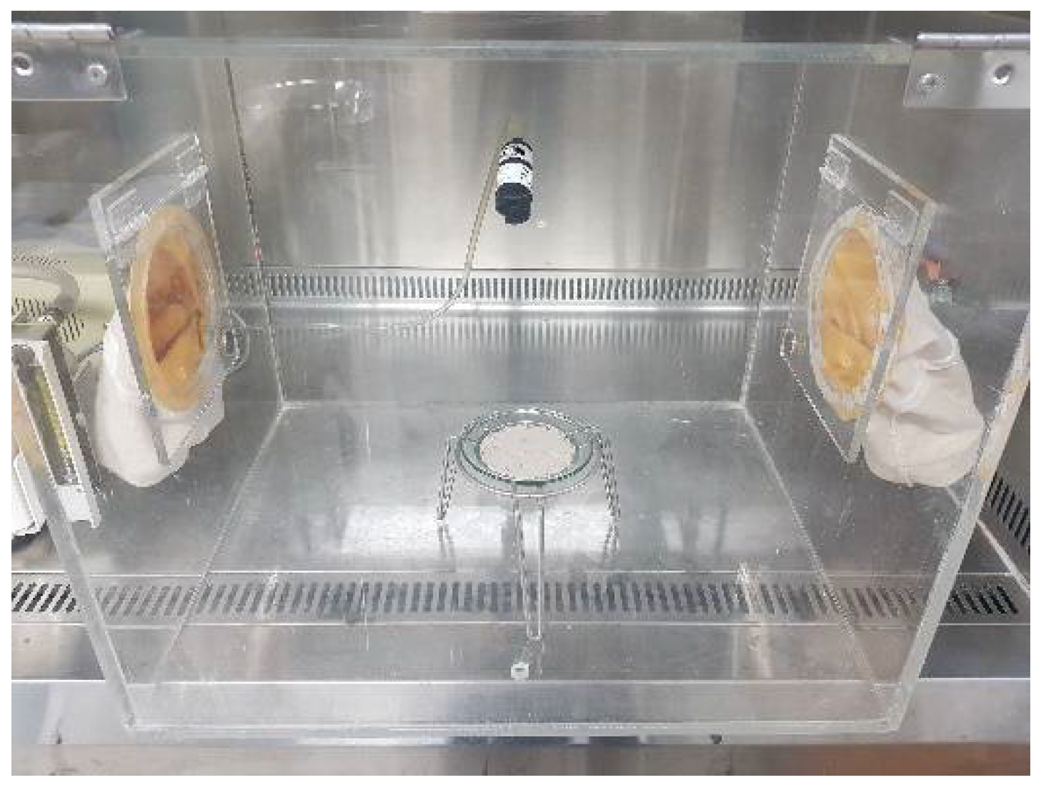
2.3. Airborne Asbestos Concentration

The analysis of airborne asbestos concentrations was performed to confirm the presence of fibrous materials in the samples after mineral carbonation. An isolated and sealed (with parafilm) acryl box with dimensions of 400 mm × 500 mm × 400 mm (length × width × height) was constructed with a diameter hole of 1 cm for air sampling (Figure 2). A mini fan was used to configure the same conditions as the wind speed occurring in an available room. Considering that the maximum wind speed of a general indoor air conditioner is 3.5 m/s, a fan with a wind speed of 3.2 m/s was used as a replacement for air circulation facilities such as air conditioners and fans used indoors. The NIOSH (National Institute for Occupational Safety and Health) 7400 [27] method of the NIOSH was used for sampling and measurements. Additionally, a kit was used which comprised a membrane cellulose ester (MCE) filter (25 mm, SKC) with a pore size of 0.8 µm and an extension cowl of 50 mm. The kit was attached at a distance of 30 cm from the bottom at an angle of 45° and the sample was situated at 15 cm from the bottom. On each watch glass of diameter 12 cm, 5 g each was evenly distributed with a diameter of 10 cm, and air was collected at a flow rate of 10 L/min for 120 min, to sample 1200 L in total.
Following sample collection, the filter was cut into quarters and the sample was then made transparent using an acetone vaporizer. It was observed at a magnification of 400× with a phase contrast microscope (Nikon Eclipse 80i, City, Japan) equipped with a Walton Beckett graticule. Fibers within fibrous ducts were counted among the particulate matters in a single field of the Walton Beckett graticule when their lengths exceeded 5 μm and the length-to-diameter ratio was larger than 3:1. This was repeated for 100 fields. The number of fibers per unit area of the filter was calculated with the use of Equation (1) and the airborne fiber concentration was calculated with the use of Equation (2).
E = ( F n f ) ( F n f ) A f  
where E is the number of fibers per unit area (f/mm2), F is the number of fibers in the sample, n f is the total field count observed in the sample, B is the number of fibers in a blank sample, n b is the total field count observed in a blank sample, and A f is the graticule field area (0.00785 mm2),
C = E × A c V × 10 3 ,  
where C is the airborne asbestos and fiber concentration (f/cc), E the number of fibers per unit area (f/mm2), A c is the effective area of the filter (approximately 385 mm2), and V is the volume of the collected air (L).

3. Results

3.1. Mineral Carbonation

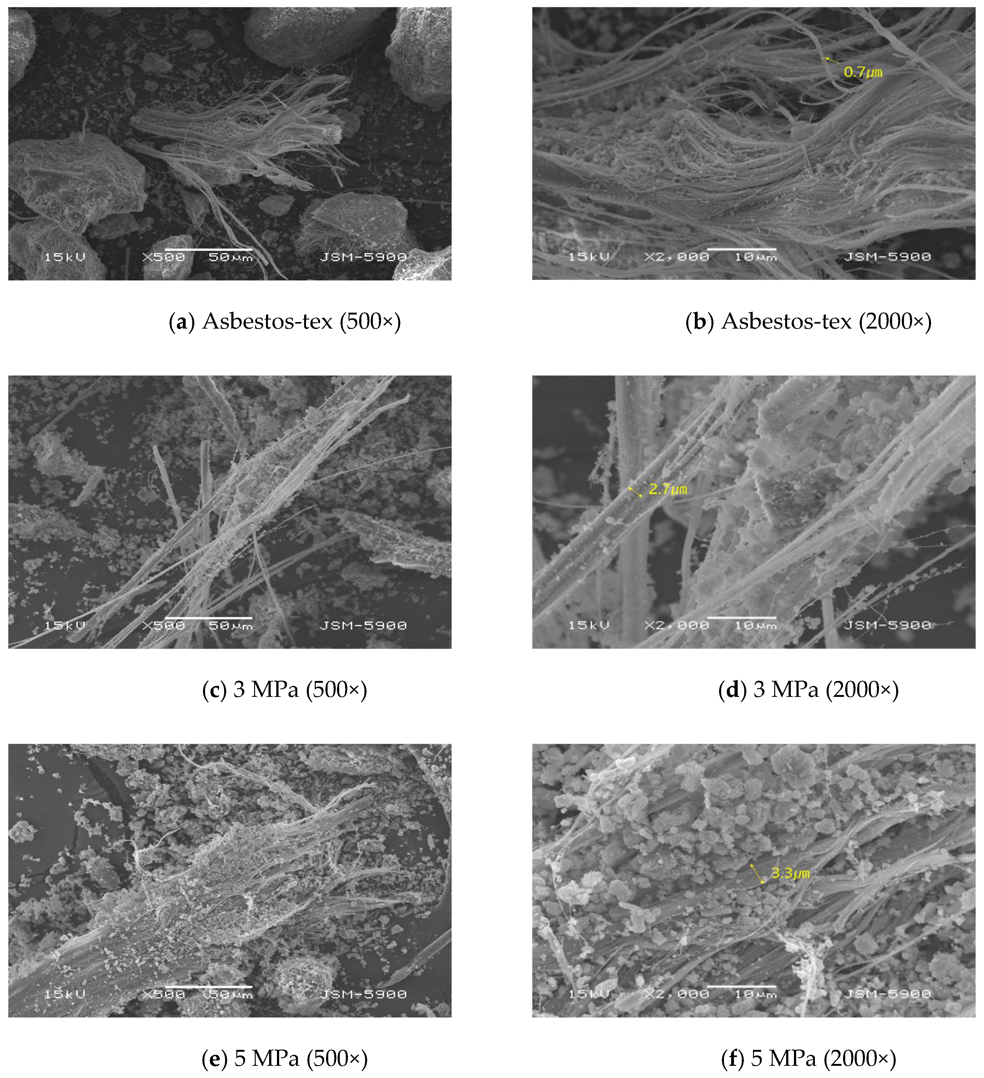
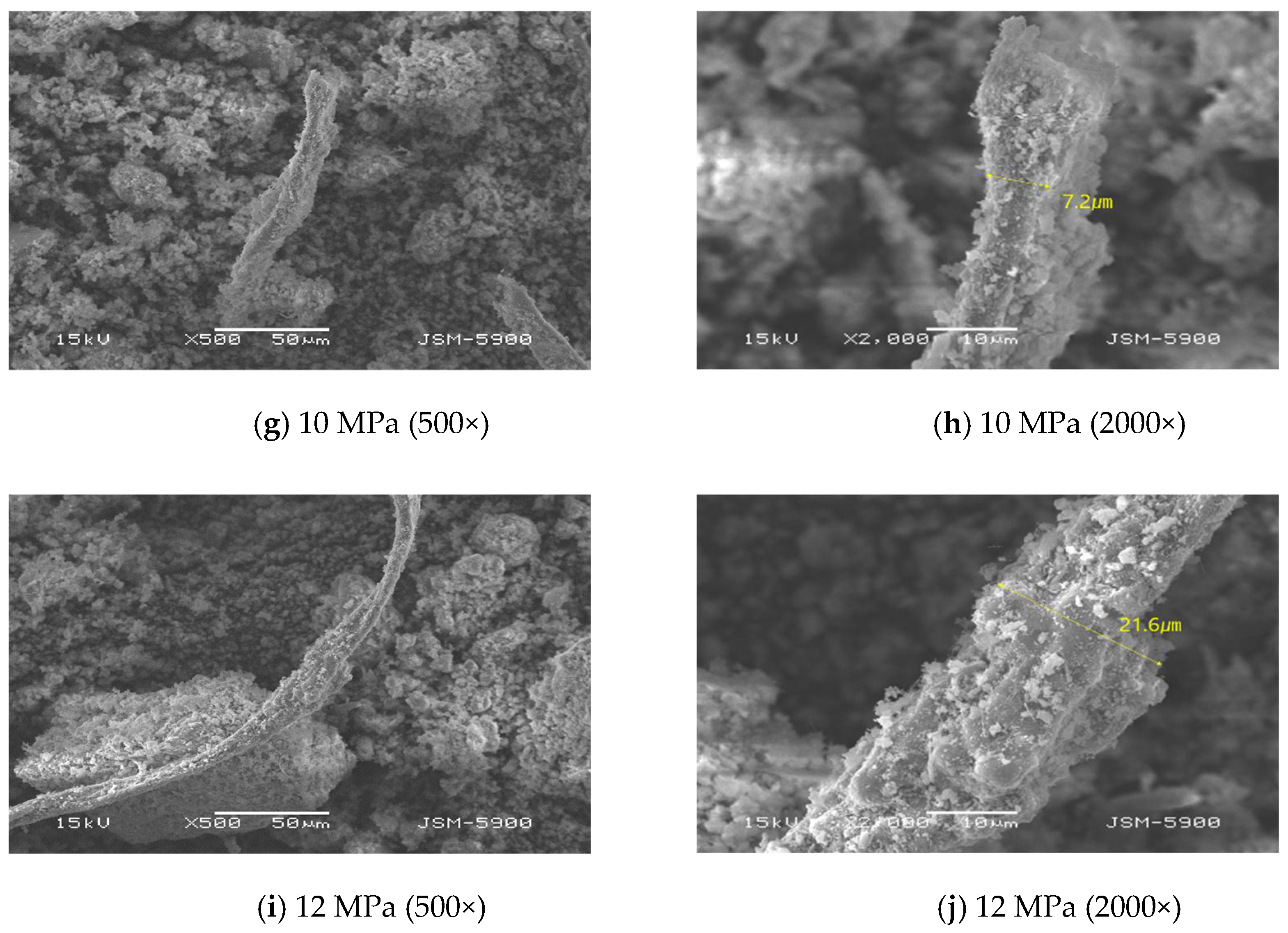
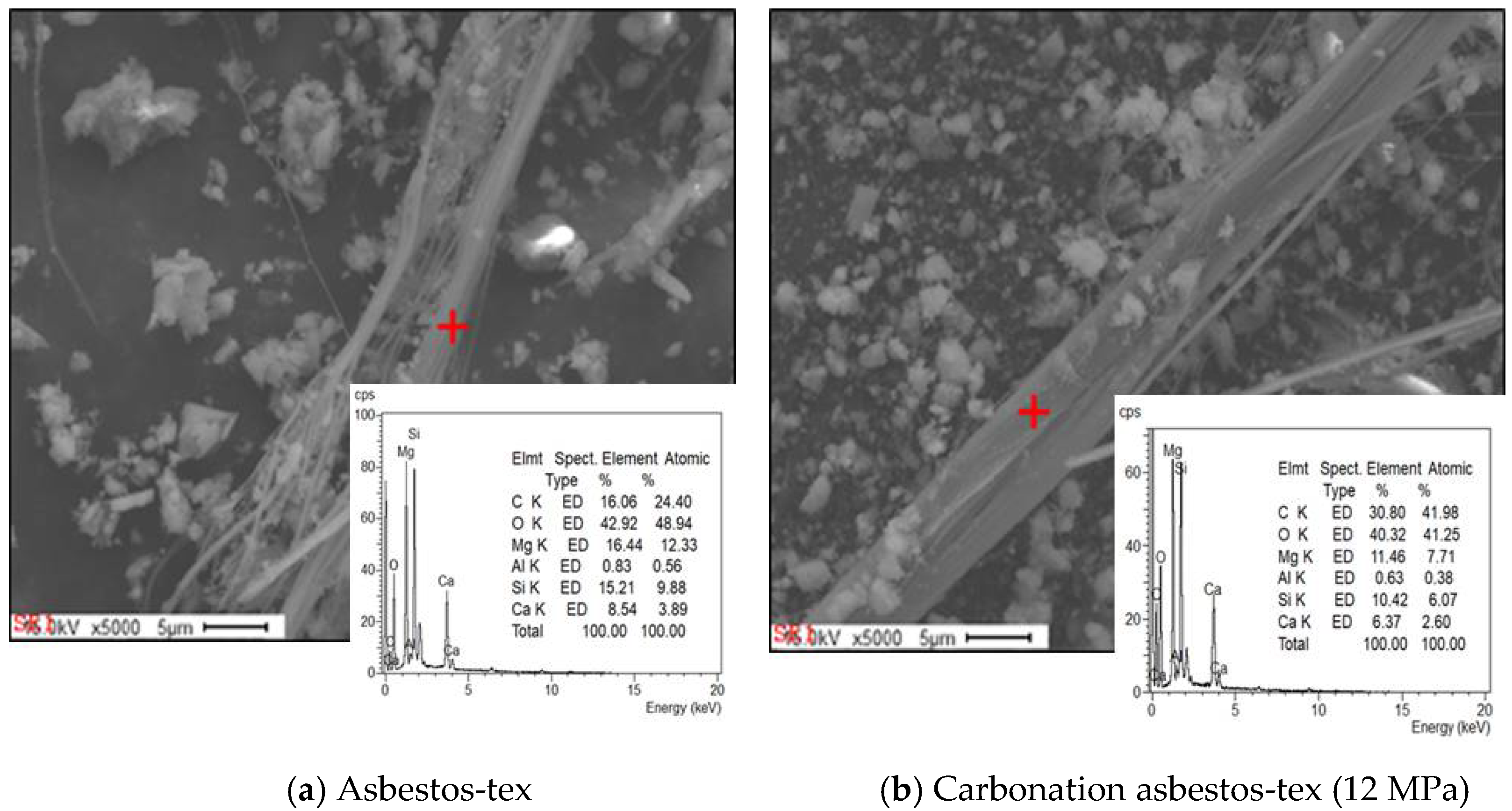
3.1.1. Microstructure

SEM analysis was conducted to assess the physical characteristics of the asbestos minerals in the samples and to confirm the presence of the asbestos minerals after mineral carbonation.
Figure 3 depicts the SEM images of all the studied samples. The raw material and sample, which reacted at 3 MPa (Figure 3a–d), comprised fiber bundles, in which thread-like fibers were aggregated. The diameter of the fibers was very fine (minimum value of 0.02 µm), while the diameters of the fiber bundles comprising aggregated fibers varied in diameter (maximum value of 5 µm with sharp ends). These particles had diameters < 3 µm. This range is within the diameter range of harmful substances that cause respiratory diseases following their penetrations in deep regions within the lung [28,29]. Similarly, in the sample exposed to 5 MPa with a high-partial pressure of CO2, an asbestos-type material with a similar diameter and length as those of the sample exposed to 3 MPa and a high-partial pressure of CO2 was observed, along with many angular particles around the original fiber material (Figure 3e,f).
Samples that underwent mineral carbonation at 10 MPa and 12 MPa displayed elongated rod-shaped minerals with sharp ends. The aspect ratio exceeded 3:1, which represents a long rod shape. However, they differed from the asbestos minerals because their diameters exceeded 5 µm and had angular ends (Figure 3g–j). These materials were larger than 3 µm in diameter. These dimensions ensure that they are harmless and are blocked by nasal hairs, the mucous membranes within the nose, or by the bronchial cilia after inhalation [30].

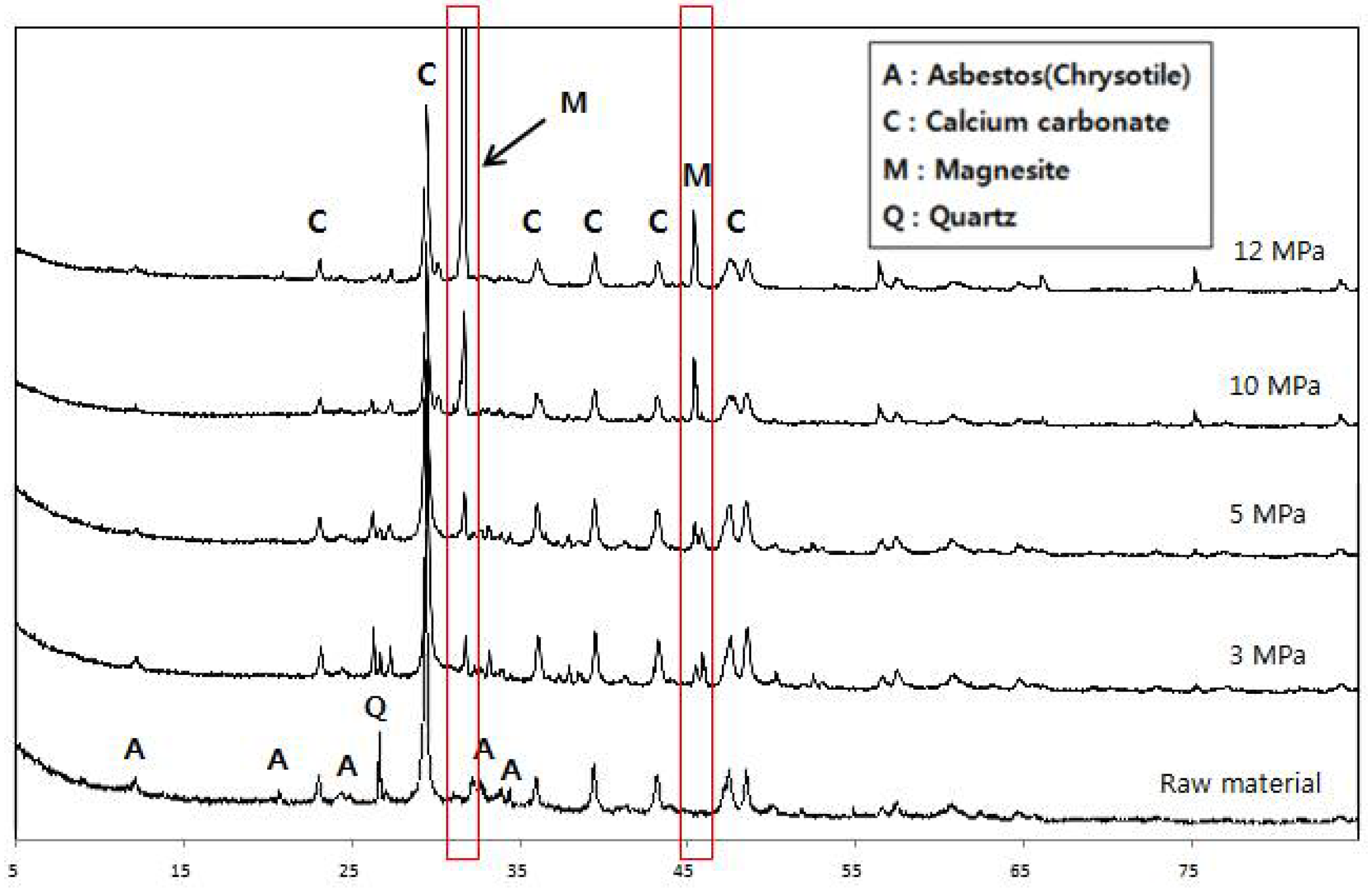
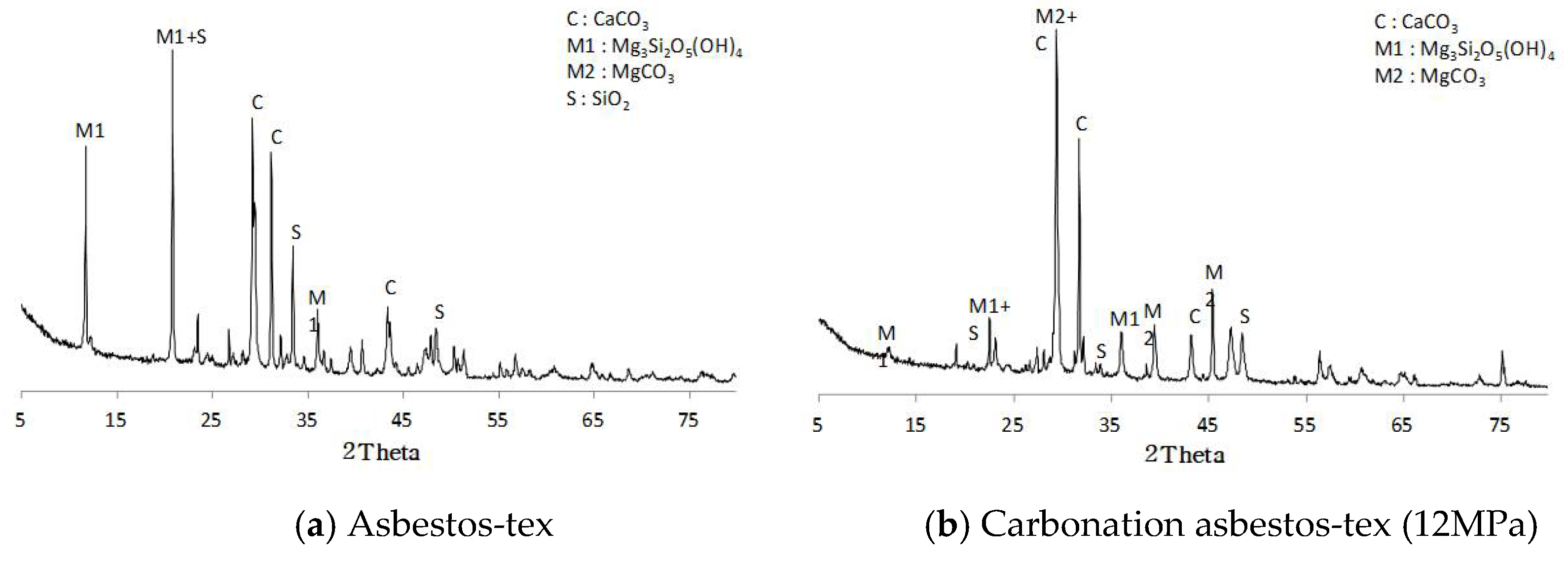
3.1.2. X-ray Diffraction Analysis

The phase transition of minerals was observed by comparing the constituent minerals through an XRD analysis of the asbestos-tex raw material and samples after mineral carbonation. The asbestos-tex raw material was composed mainly of calcite (CaCO3), serpentine-based chrysotile asbestos (Mg3Si2O5(OH)4), and quartz (SiO2). At 3 MPa and 5 MPa, a steady decrease was observed for peaks around the scattering angles of 12°, 24°, and 35°, which correspond to the main peaks of chrysotile. Peaks at 32° and 46°, which correspond to magnesite (MgCO3), were also observed [31]. These peaks were not observed in the raw materials. This is because the magnesium ions of asbestos combined with CO2 during the mineral carbonation reaction and formed magnesite crystals. The sample exposed to 10 MPa demonstrated small peaks owing to the phase transition of most of the asbestos and, at 12 MPa, efficient asbestos reduction. In contrast, the main peaks of magnesite became sharper as the pressure increased.
2Mg3Si2O5(OH)4 = 3Mg2SiO4 + SiO2 + 4H2O
Mg2SiO4(s) + 4H+(aq) = 2Mg2+(aq) + H4SiO4(aq)
CO2(g) + H2O(l) = H2CO3(aq)
H2CO3(aq) = H+(aq) + HCO(aq)
HCO(aq) = H+(aq) + CO32− (aq)
The crystal structure of chrysotile was transformed into forsterite (Mg2SiO4) because of the vaporization of H+ and OH in chrysotile (Equation (3)) during pressurized heat treatment in a high-temperature, high-pressure autoclave. Furthermore, the bicarbonate in the buffer solution promoted the mineral carbonation reaction following the increases in the concentration of H+ and HCO3, which acted as constituents of carbonation, according to Equations (4)–(7) at conditions of high CO2 partial pressures [26]. Additionally, Si was temporarily dissolved in aqueous H4SiO4 and was degraded by amorphous silica; this resulted in the removal of the Si-rich layer [25,26]. The chloride and sodium ions of sodium chloride promoted ion exchange at the particle surface and dissolution of magnesium during mineral carbonation [17,25] to induce the formation of magnesite. The overall equation for the mineral carbonation of asbestos is shown in Equation (8).
Mg2SiO4(s) + 2CO2(a) + 2H2O(l) = 2MgCO3(s) + H4SiO4(aq),
The decrease in the quartz peak as the mineral carbonation progressed (as shown in Figure 4) was attributed to the dissolution of SiO2, which remained in liquid form, as H4SiO4. Consequently, only the peaks of precipitates, calcite, and magnesite were observed in samples exposed to pressures >10 MPa.
As a result of XRD analysis (Figure 5), asbestos-tex (raw materials) was mainly composed of calcite (CaCO3), serpentine asbestos, Chrysotile Asbestos (Mg3Si2O5(OH)4), and quartz (Quartz, SiO2). SEM-EDS analysis (Figure 6) also showed a similar pattern to XRD; considering that the general chemical composition of white asbestos is Mg 26.3%, Si 20.3%, H 1.45%, and O 51.96%, most of the asbestos analyzed in tex is white asbestos and some of it is calcite. On the other hand, in the case of the mineral carbonated sample (12 MPa), magnesite, which was not found in the raw material, was observed and formed by combining the Mg ion of white asbestos with CO2 by the mineral carbonation reaction. Most of the white asbestos was decomposed during the pressure heat-treatment process, so the peak was low and it was confirmed that the main peak of magnesite increased sharply as the pressure increased. In addition, changes in chemical composition were also observed in the EDS results and it was found that the carbon level increased about twice that of the raw material.

3.2. Airborne Asbestos Concentration Analysis

Table 2 illustrates airborne asbestos concentrations of the samples, which reacted (mineral carbonation) with raw asbestos-tex materials in a sealed and enclosed space. Irrespective of the mineral carbonation reaction, the airborne asbestos concentrations did not exceed the limit of 0.01 f/cc regulated by the Korean Indoor Air Quality Control Act. This was because no flow of wind was present in the enclosed space. However, a numerical comparison analysis was conducted to evaluate the safety of asbestos. The number of airborne asbestos-type fibers in the raw material was 0.00046 f/cc and this decreased slightly for samples exposed to either 3 MPa or 5 MPa; a decrease of 15% was observed at 5 MPa and the asbestos concentration was 0.00039 f/cc.
For samples that underwent the mineral carbonation reaction at pressures higher than 10 MPa, the number of airborne asbestos fibers per unit area was 0.000065 f/cc. This is equivalent to detecting only 0.5% fibers in 100 observation field counts, thereby verifying the significant decrease in the presence of asbestos fibers following mineral carbonation.

4. Conclusions

In this study, asbestos-tex, a well-known and extensively used asbestos-containing material, was powdered and phase transferred to a carbonate mineral. This resulted in detoxification. The only requirements to accomplish this were the use of sodium bicarbonate, a sodium chloride buffer solution, and a high CO2 partial pressure; high-temperature melting or chemical pretreatment with strong acids was not required.
  • SEM analysis revealed that the asbestos-tex raw material was in the form of needle-like fibrous minerals. At 3 MPa, the mineral form was not significantly different from the raw material. However, at 5 MPa, carbonate particles were generated. At 10 MPa or above, it was transformed into thick rod-like minerals following mineral carbonation.
  • Asbestos-tex is a mixture of calcium carbonate, quartz, and chrysotile. At the CO2 partial pressures of 3 and 5 MPa, only some parts of the chrysotile were transformed into magnesite. At CO2 partial pressures of 10 MPa or above, most of the chrysotile was transformed into magnesite; at 12 MPa, a large growth of magnesite crystals was observed.
  • The analysis of airborne asbestos concentration did not show any significant difference between the carbonated samples (at 3 or 5 MPa) and the raw material. However, at 10 MPa or above, it was significantly reduced to one-seventh of that of the raw material. This confirmed that the asbestos-type fibrous material was transformed into a mineral through the carbonation process.
  • Mineral carbonation of asbestos-tex was conducted at a constant temperature of 100 °C and a range of CO2 partial pressures. Only a fraction of the chrysotile of asbestos-tex was transformed into magnesite at low-partial pressures. However, at CO2 partial pressures in excess of 10 MPa, the detoxification of asbestos-tex was achieved up to a level that made it more significant than 3 µm.

Author Contributions

Y.L.; methodology, data curation H.J.; formal analysis, review and editing S.S. All authors have read and agreed to the published version of the manuscript.

Funding

This research received no external funding.

Acknowledgments

This study supported by a grant (21CTAP-C163524–01) from infrastructure and transportation technology promotion research program funded by the Ministry of Land, Infrastructure and Transport of the Korean government.

Conflicts of Interest

The authors declare no conflict of interest.

References

  1. WHO/EURO Technical Committee for Monitoring and Evaluating MMMF. The WHO/EURO man-made mineral fiber reference scheme. Scand. J. Work Environ. Health 1985, 11, 123–129. [Google Scholar] [CrossRef]
  2. Becklake, M.R.; Gibbs, G.W.; Arhirii, M.; Hurwitz, S. Exposure to asbestos and respiratory abnormality: The influence of fibre type and nature of exposure. In Biological Effects of Mineral Fibers; Wagner, J.C., Ed.; International Agency for Resarch on Cancer Scientific Publication: Genova, Switzerland, 1980; Volume 30, pp. 763–768. [Google Scholar]
  3. Stayner, L.; Welch, L.S.; Lemen, R. The worldwide pandemic of asbestos related diseases. Annu. Rev. Public Health 2013, 34, 205–216. [Google Scholar] [CrossRef] [PubMed]
  4. Tossavainen, A. Global use of asbestos and the incidence of mesothelioma. Int. J. Occup. Environ. Health 2004, 10, 22–25. [Google Scholar] [CrossRef]
  5. David, J.B.; Celeste, M.B.; David, P.C.; Cardozo-Pelaez, F.; Pfau, J.C. Internalization of Libby Amphibole Asbestos and Induction of Oxidative Stress in Murine Macrophages. Toxicol. Sci. 2007, 19, 277–288. [Google Scholar]
  6. Vijayalakshmi, P.; Sigmund, A.W.; Navdeep, S.C.; David, W.K. Mitochondrial-derived free radicals mediate asbestos-induced alveolar epithelial cell apoptosis. Am. J. Physiol. Lung Cell Mol. Physiol. 2004, l286, 1220–1227. [Google Scholar]
  7. Ministry of Environment. Asbestos Management Overview. Korea Ministry of Environment, 2009; p. 456. Available online: http://me.go.kr/home/web/main.do (accessed on 5 January 2021).
  8. Kim, T.H.; Song, T.H.; Shin, H.G.; Jang, K.P. A Study on the Recycling of Detoxified Waste Asbestos. J. Rec. Const. Resour. 2020, 8, 161–166. [Google Scholar] [CrossRef]
  9. Li, J.; Dong, Q.; Yu, K.; Liu, L. Asbestos and asbestos waste management in the Asian-Pacific region: Trends, challenges and solutions. J. Clean Prod. 2014, 81, 218–226. [Google Scholar] [CrossRef]
  10. Yoo, K.K.; Kim, B.S.; Kim, M.S.; Lee, J.C.; Jeong, J. Dissolution of magnesium from serpentine mineral in sulfuric acid solution. Mater. Trans. 2009, 50, 1225–1230. [Google Scholar] [CrossRef]
  11. Fedoročková, A.; Hreus, M.; Paschman, P.; Sučik, G. Dissolution of magnesium from calcined serpentinite in hydrochloric acid. Miner. Eng. 2012, 32, 1–4. [Google Scholar] [CrossRef]
  12. Rozalen, M.; Huertas, F.J. Comparative effect of chrysotile leaching in nitric, sulfuric and oxalic acids at room temperature. Chem. Geol. 2013, 352, 134–142. [Google Scholar] [CrossRef]
  13. Chung, Y.H.; Han, J.H.; Sung, J.H.; Song, K.S.; Lim, K.T.; Yu, I.J. Investigation of Asbestos and MMMF Containing Construction Material in Korean Buildings. J. Odor. Indoor Environ. 2005, 1, 156–165. [Google Scholar]
  14. Shafer, R.; Throwe, S.; Salgado, O.; Garlow, C.; Hoerath, E.; Asbestos/NESHAP Adequately Wet Guidance’. North Carolina: Alliance Technologies Corporation. 1990. Available online: https://s23893.pcdn.co/wp-content/uploads/2013/09/Pamphlet-–-AsbestosNESHAP-Adequately-Wet-Guidance-December-1990.pdf (accessed on 13 September 2021).
  15. Gualtieri, A.F.; Tartaglia, A. Thermal decomposition of asbestos and recycling in traditional ceramics. J. Eur. Ceram. Soc. 1999, 20, 1409–1418. [Google Scholar] [CrossRef]
  16. Zaremba, T.; Peszko, M. Investigation of the thermal modification of asbestos wastes for potential use in ceramic formulation. J. Therm. Anal. Calorim. 2008, 92, 873–877. [Google Scholar] [CrossRef]
  17. O’Connor, W.K.; Dahlin, D.C.; Turner, P.C.; Walters, R.P. Carbon Dioxide Sequestration by Ex-Situ Mineral Carbonation; Albany Research Center: Albany, Oregon, 1999. Available online: https://www.osti.gov/servlets/purl/r875354 (accessed on 5 October 2015).
  18. Schulze, R.K.; Hill, M.A.; Field, R.D.; Papin, P.A.; Hanrahan, R.J.; Byler, D.D. Characterization of carbonated serpentine using XPS and TEM. Energy Conv. Manag. 2004, 45, 3169–3179. [Google Scholar] [CrossRef]
  19. Fouda, M.F.R.; Amin, R.E.; Abd-Elzaher, M.K. Extraction of magnesia from Egyptian serpentine ore via reaction with different acids. I. Reaction with sulfuric acid, Bull. Chem. Soc. Jpn. 1996, 69, 1907–1912. [Google Scholar] [CrossRef]
  20. Teir, S.; Revitzer, H.; Eloneva, S.; Fogelholm, C.J.; Zevenhoven, R. Dissolution of natural serpentinite in mineral and organic acids. Int. J. Miner. Process. 2007, 83, 36–46. [Google Scholar] [CrossRef]
  21. Azdarpour, A.; Asadullah, M.; Junin, R.; Mohammadian, E.; Hamidi, H.; Daud, A.R.M.; Manan, M. Extraction of calcium from red gypsum for calcium carbonate production. Fuel Process. Technol. 2015, 130, 12–19. [Google Scholar] [CrossRef]
  22. Rahmani, O. CO2 sequestration by indirect mineral carbonation of industrial waste red gypsum. J. CO2 Util. 2018, 27, 374–380. [Google Scholar] [CrossRef]
  23. Gao, J.; Li, C.; Liu, W.; Hu, J.; Wang, L.; Liu, Q.; Liang, B.; Yue, H.; Zhang, G.; Luo, D.; et al. Process simulation and energy integration in the mineral carbonation of blast furnace slag. Chin. J. Chem. Eng. 2019, 27, 157–167. [Google Scholar] [CrossRef]
  24. Gerdermann, S.J.; O’Connor, W.K.; Dahlin, D.C.; Penner, L.R.; Rush, H. Ex situ aqueous mineral carbonation. Environ. Sci. Technol. 2007, 41, 2587–2593. [Google Scholar] [CrossRef]
  25. Wang, F.; Dreisinger, D.; Jarvis, M.; Hitchins, T. Kinetics and mechanism of mineral carbonation of olivine for CO2 sequestration. Miner. Eng. 2019, 131, 185–197. [Google Scholar] [CrossRef]
  26. Huijgen, W.J.J.; Witkamp, G.; Comans, R.N.J. Mechanisms of aqueous wollastonite carbonation as a possible CO2 sequestration process. Chem. Eng. Sci. 2006, 61, 4242–4251. [Google Scholar] [CrossRef]
  27. National Institute of Occupational Safety and Health (NIOSH). Asbestos and Other Fibers by PCM 7400′. In NIOSH Manual of Analytical Methods, 4th ed.; DHHS (NIOSH) Publication: Cincinnati, OH, USA, 1994; pp. 133–147. [Google Scholar]
  28. Commins, B.T. The Significance of Asbestos and Other Mineral Fibres in Environmental Ambient Air; Commins Associates: Berkshire, UK, 1985. [Google Scholar]
  29. Rom, W.N.; Markowitz, S.B. Environmental and Occupational Medicine, 4th ed.; Lippincott-Raven Publishers: Philadelphia, PA, USA, 2007. [Google Scholar]
  30. National Research Council. Committee on Nonoccupational Health Risks of Asbestiform Fibers. In Asbestiform Fibers: Nonoccupational Health Risks; National Academy Press: Washington, DC, USA, 1984. [Google Scholar] [CrossRef]
  31. Jo, H.J.; Jang, Y.N.; Jo, J.H. A low temperature detoxification method for treatment of Chrysotile-containing waste roofing slate. Minerals 2017, 7, 144. [Google Scholar] [CrossRef] [Green Version]
Figure 1. Schematic of the experimental system for mineral carbonation in a stirred reactor.
Buildings 12 01372 g001
Figure 2. Internal space used in the airborne asbestos concentration experiment.
Buildings 12 01372 g002
Figure 3. Scanning electron microscopy (SEM) analysis of asbestos-tex samples after mineral carbonation for a range of pressures.
Buildings 12 01372 g003aBuildings 12 01372 g003b
Figure 4. X-ray diffraction patterns of asbestos-tex and other samples after mineral carbonation at various pressures.
Buildings 12 01372 g004
Figure 5. X-ray diffraction patterns of asbestos-tex after carbonation.
Buildings 12 01372 g005
Figure 6. SEM-EDS (energy dispersive X-ray spectroscopy) of asbestos-tex after carbonation.
Buildings 12 01372 g006
Table 1. Chemical specifications of asbestos-tex (wt%).
TypeOxide Composition (%)
CaOSiO2MgOFe2O3Al2O3SO3K2OOthers
Asbestos-Tex59.420.66.864.793.883.210.650.61
Table 2. Airborne asbestos concentrations of Asbestos-tex and mineral carbonated samples.
Exposure PressuresObserved Field CountNumber of Fibers
(1 f)
Number of Fibers
(1/2 f)
Number of FibersNumber of Fibers per Unit Area (f/mm2)Asbestos Concentration
(f/cc)
Atmospheric pressure100313.54.4590.00046
3 MPa313.54.4590.00046
5 MPa2233.8210.00039
10 MPa010.50.6370.000065
12 MPa010.50.6370.000065
Publisher’s Note: MDPI stays neutral with regard to jurisdictional claims in published maps and institutional affiliations.

