Article

Correlation between Pathogenic Determinants Associated with Clinically Isolated Non-Typhoidal Salmonella

by
Boimpoundi Eunice Flavie Ouali
1,†,
Tsyr-Huei Chiou
1,†,
Jenn-Wei Chen
2,3,†,
I-Chu Lin
1,
Ching-Chuan Liu
3,4,
Yu-Chung Chiang
5,6,*,
Tzong-Shiann Ho
3,4,* and
Hao-Ven Wang
1,7,8,*
1
Department of Life Sciences, National Cheng Kung University, Tainan 701, Taiwan
2
Department of Microbiology and Immunology, College of Medicine, National Cheng Kung University, Tainan 701, Taiwan
3
Center of Infectious Disease and Signaling Research, National Cheng Kung University, Tainan 701, Taiwan
4
Department of Pediatrics, National Cheng Kung University Hospital, College of Medicine, National Cheng Kung University, Tainan 701, Taiwan
5
Department of Biological Sciences, National Sun Yat-sen University, Kaohsiung 80424, Taiwan
6
Department of Biomedical Science and Environment Biology, Kaohsiung Medical University, Kaohsiung 80708, Taiwan
7
Center for Bioscience and Biotechnology, National Cheng Kung University, Tainan 701, Taiwan
8
Marine Biology and Cetacean Research Center, National Cheng Kung University, Tainan 701, Taiwan
*
Authors to whom correspondence should be addressed.
These authors contributed equally to this work.
Pathogens 2021, 10(1), 74; https://doi.org/10.3390/pathogens10010074
Submission received: 14 December 2020 / Revised: 1 January 2021 / Accepted: 12 January 2021 / Published: 15 January 2021

Abstract

:
Non-typhoidal and Typhoidal Salmonella are bacterial pathogens source of worldwide and major disease burden. Virulent determinants of specific serovars belonging to non-typhoidal Salmonella have been extensively studied in different models, yet the pathogenesis of this group of bacteria and the development of clinical symptoms globally remains underexplored. Herein, we implemented microbiological and molecular procedures to investigate isolate virulence traits and molecular diversity, likely in association with disease severity. Our results show that selected clinical isolates from a tertiary referring hospital, depending on the richness of the environment and isolate serotypes, exhibited different, and sometimes controversial, virulence properties. The tested strains were susceptible to Ceftriaxone (90%) with decreasing reactivity to Trimethoprim–Sulfamethoxazole (72%), Chloramphenicol (64%), Ampicillin (48%), Gentamicin (44%), and Ciprofloxacin (2%). Disc susceptibility results partially correlated with minimum inhibitory concentration (MIC); however, special attention must be given to antimicrobial treatment, as a rise in multi-resistant isolates to Trimethoprim–Sulfamethoxazole (2/38 µg/mL), Minocycline (8 µg/mL) and Ampicillin (16 µg/mL) has been noticed, with two isolates resistant to Ceftazidime (16 µg/mL). By comparison to previous molecular epidemiology studies, the variation in the gene profiles of endemic pathogens supports the need for continuous and up-to-date microbiological and molecular reports.

1. Introduction

Salmonella is a food-borne pathogen notorious for causing various symptomatic events in humans, from self-limiting pathologies to more complicated forms of infection. The infectious pathogen origin of enteric fever or acute gastroenteritis includes the non-typhoidal strains, the typhoidal strains, and the invasive non-typhoidal strains recently found in Sub-Saharan Africa [1,2,3]. Non-typhoidal strains responsible for food poisoning are widespread globally. The World Health Organization estimates infections due to non-typhoidal Salmonella enterica to bear the highest burden of foodborne diseases, with 180 million diarrheal cases, and Africa and South East Asia were identified as the largest exposed regions [4,5]. Diseases and outbreaks are mostly attributed to contaminated vegetables, meats, drinks, and animal exposure [6,7,8,9]. In Taiwan, non-typhoidal Salmonella is still a prevalent pathogen [10] which causes the highest annual incidence of bacteremia among the population in the South [11].
In recent years, salmonellosis management has improved considerably due to multidrug resistance trace-back methods, real-time surveillance, and the monitoring of outbreaks [10,12]. However, despite this progress, the prevalence of salmonellosis remains globally consistent and has even increased in some areas [5]. The expansion of industrialization, the evolution of microbial host adaptation, and the emergence of antibiotic-resistant clones largely contribute to the persistence of this disease [13].
The immune condition of the human host, in addition to bacterial serotype and virulence, plays a clinically important and concomitant role in the severity of the diseases caused by Salmonella [14]. The pathogen utilizes a large variety of virulence factors, including plasmid virulence genes, cell surface structure, flagella proteins, and pathogenicity islands (SPIs) to trigger disease by invading the host and escaping host defenses [15,16]. Salmonella is recognized to bind to host cell targets, to be engulfed, and to migrate through the host blood or lymphatic circulatory systems, resulting in bacteremia, septicemia, more complicated forms of the disease [17,18,19] and long-lasting carriage [20]. To invade a host cell, Salmonella injects invasion signaling molecules through the type III secretion system encoded by the SPI-1 which, in turn, activate the host cell signaling pathways that lead to the internalization of the pathogen and, subsequently, the reconstruction of the cytoskeleton actin protein [19,21]. The SPIs carry the genes responsible for invasion, survival, and extra-intestinal spreading [21]. Hence, bacteria can be restricted to the gastrointestinal tract or can be expanded to other organs through the bloodstream.
Serotyping, genotyping, and pulsed-field gel electrophoresis have been extensively used for disease surveillance in Taiwan; however, microbiological examination of the bacterial physiology, coupled with molecular epidemiology studies, is neglected. Herein, we seek to examine the correlation between virulence determinants in association with Salmonella pathogenicity by investigating the distribution of virulence traits among Salmonella clinical isolates and by comprehending their interconnection.

2. Results

2.1. Overview of Salmonella Isolates Adherence and Invasiveness to Epithelial Cells

To determine the ability of the isolates to attach to and pervade epithelial cells, FHs 74 Int cells were infected with the bacterial inoculum. While no statistical differences were noticed for isolates invading the cells, most of the blood and stool isolates adhered onto and invaded the epithelial cells (Figure 1). The blood isolate B2 and the stool isolates S4 and S5 exhibited the highest adhesion rates, whereas B2, followed by S4, had the greatest invasion rates among the isolates. B2 showed a significant increase in adhesion rate compared to B1. The adhesion and invasion rates of B2, respectively, approximate 20% and 12%, whereas S4 and S5 had each an adhesion rate of about 15%.

2.2. Environmental Conditions Affecting Bacterial Growth

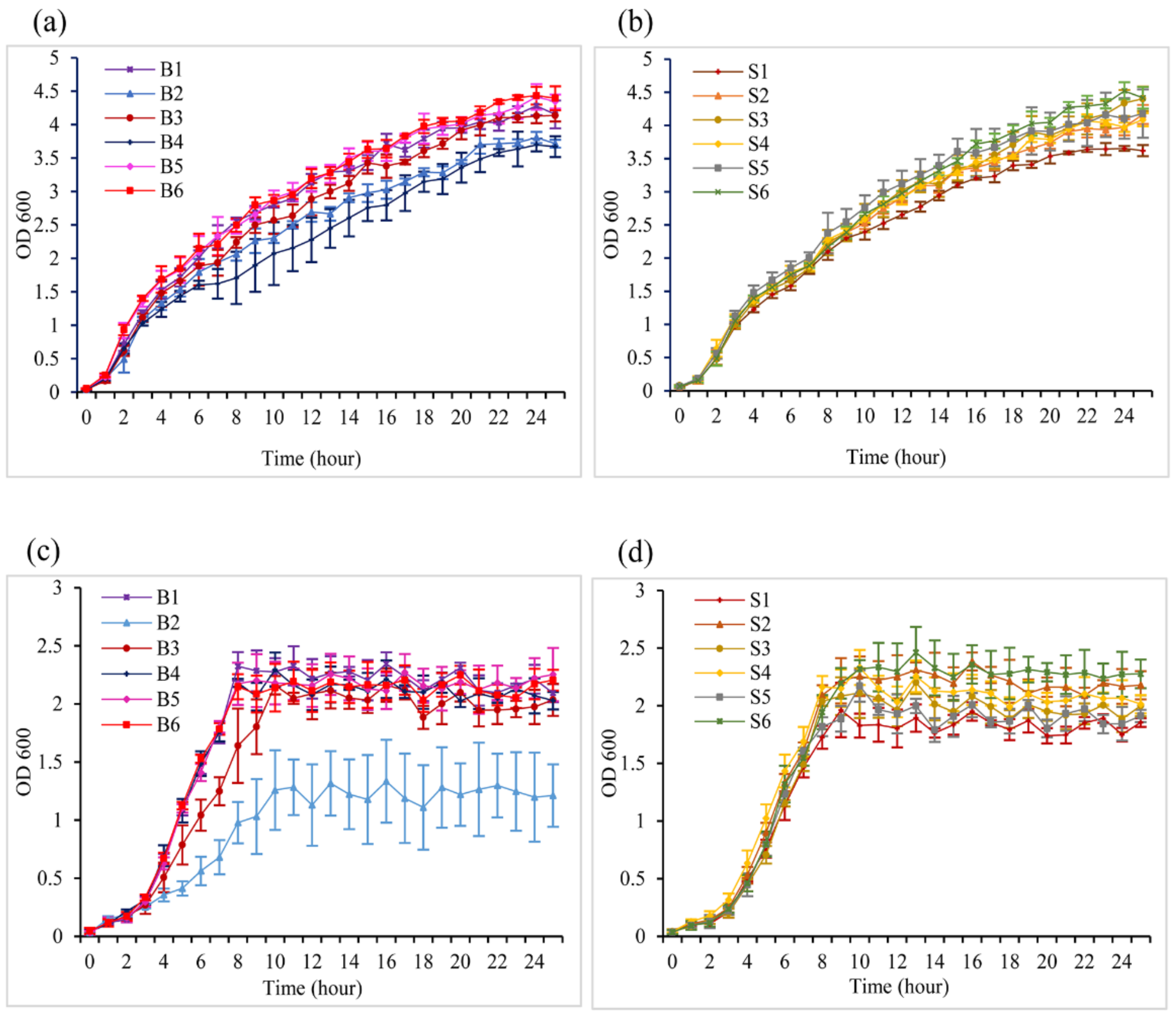
To analyze the bacterial growth status and its possible relation with infection, Salmonella isolates were grown in a lysogeny broth and minimal medium, with the absorbance measured at 600 nm. The blood (Figure 2a) and stool isolates (Figure 2b) showed a high potency to multiply in the LB medium with a continuous exponential phase. On the other hand, in the M9 culture media, the growth of the blood (Figure 2c) and stool isolates (Figure 2d) decreased relatively, with a noticeable short log phase and bacteria reaching the stationary phase after 9 h of incubation. A drastic reduction in B2 isolate multiplication was noted in the depleted M9 media.

2.3. Motility of the Bacterial Isolates

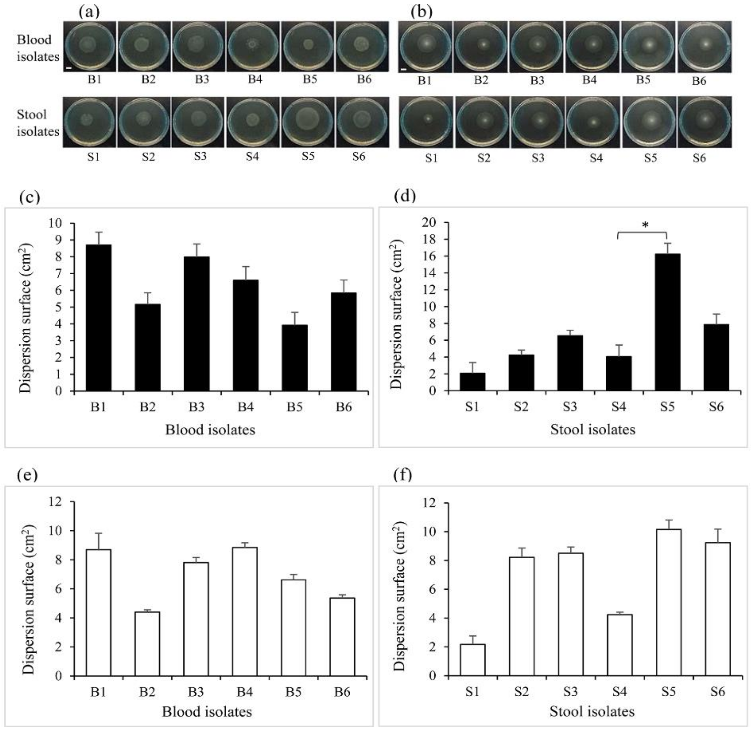
Bacterial motility was measured to assess the ability of the isolates to spontaneously and actively move and to differentially swarm and swim on a semisolid surface. The 12 clinical isolates were all capable of swarming (Figure 3a) and swimming (Figure 3b) on 0.5% and 0.3% LB-agar, respectively. Partly based on the colony pattern formation described by Kearns [22], swarmer isolates exhibited dendritic, bull’s-eye, or a thick circular colony pattern, whereas swimmer isolates essentially presented a uniformly round, featureless colony. The dispersion of the blood isolates on swarm plates was not statistically different (Figure 3c), whilst stool isolate S5 had the significantly highest swarming dispersion area by comparison to S4 (Figure 3d). No significant differences were supported for the swimming dispersion surface of blood (Figure 3e) and stool (Figure 3f) isolates. However, we noticed that the stool S1 had a lesser trend to swim and swarm on agar plates.

2.4. The Effects of the Intrinsic Nature of the Bacteria and Culture Medium on Biofilm Formation

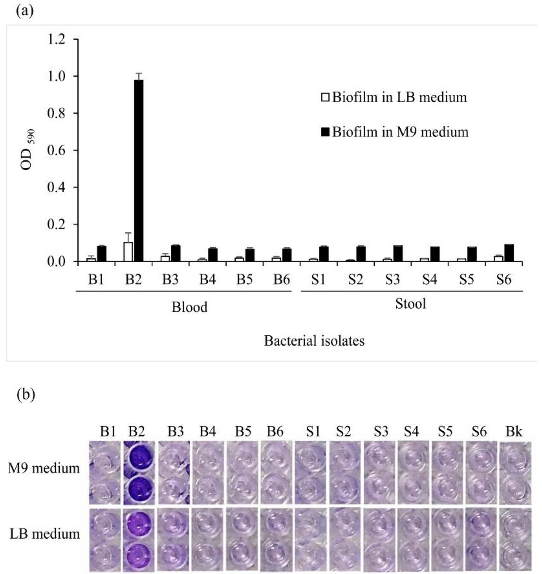
Most of the 12 bacterial isolates were not able to organize into a biofilm. The blood isolates B2 solely exhibited a substantial ability to form a relatively weak cell aggregate when cultured in the LB medium. In M9 medium, isolates B1, B3, S3, and S6 were identified as weak biofilm producers, whereas the B2 isolate was recognized as a strong biofilm producer. The absorbance of B2 cultured in M9 was equal to 0.97 against 0.1 in LB (Figure 4a) and resulted in a more pronounced purple color in the corresponding wells (Figure 4b). However, the differences in biofilm formation levels among the isolates were not statistically supported.

2.5. Salmonella Isolates Genotyping by Multiplex PCR

Three independent multiplex PCR assays were performed for each group of genes for the purpose of virulence gene profiling. A comparison to the 100 bp Plus DNA ladder indicated that the observed bands of InvA, SpvC, and FliC (Supplementary Figure S1A) with HilA, FimH, and SopB (Supplementary Figure S1B) were consistent throughout in terms of the estimated length of each amplicon. The targeted genes InvA, FimH, and HilA were all found in the 12 clinical isolates tested. Out of the bacteria, 50% had both SpvC and SopB but lacked FliC; 33.33% had SopB and FliC but lacked SpvC; FliC, SpvC and SopB were all absent in the products generated by the isolate S5, while all the amplicon bands were found in the electrophoresed product issued from isolate S1 (Table 1).
Products from multiplex PCR were run on agarose gels. The presence of the specific gene was attested by the visualization of a band at their corresponding size.

2.6. Comparative Virulence Gene Expression and Serotyping

To quantify the expression of virulence genes, quantitative PCR was performed using total RNA extracted from the 50 bacteria isolates. All the isolates expressed InvA, FimH, and SopB mRNA, with values between 0.67 and 4.96, 0.94 and 5.62, 0.26 and 1.16 arbitrary units, respectively (a.u.) (Figure 5). Overall, 30% of the isolates expressed FliC and 34% expressed SpvC at relatively low values. Out of the 50 isolates, only one blood isolate did not express HilA. Respective positive and negative controls with S. Typhimurium and E. coli were used for comparison purposes and results’ validation. Serotyping of the 50 isolates based on the O-antigen showed that the most prevalent serogroups were B and D1, each with 30% (15/50), followed by the serogroup C2 (20%, 10/50), and the serogroups C1 and E, which identically counted for 10% of the isolates (5/50).

2.7. Salmonella Isolates In Vitro Susceptibility to Antibiotics

About 90% of the 50 isolates showed sensitivity to Ceftriaxone. Decreased sensitivity was noticed with Trimethoprim–Sulfamethoxazole, Chloramphenicol, Ampicillin, Gentamicin, and Ciprofloxacin (Table 2). Among the isolates, 50% and 36% were greatly resistant to Ampicillin and Chloramphenicol, respectively, whereas Ciprofloxacin and Gentamicin had intermediate effect on, respectively, 76% and 48% of the bacteria tested.
All the isolates were sensitive to Cefepime at the minimum inhibitory concentration (MIC) breakpoint value of ≤1 and ≤0.25 µg/mL for Carbapenems (Ertapenem, Imipenem, and Meropenem). Although Ampicillin, Trimethoprim-Sulfamethoxazole, and Chloramphenicol were not considered to be good empirical regimens for Salmonella infections due to the high frequency of resistance [23], we found that most of the stool isolates were still susceptible to Trimethoprim–Sulfamethoxazole.

3. Discussion

The microbiological and molecular characterization of virulence determinants associated with human pathogens is primordial for effective disease control and management. A series of tests were performed here on Salmonella clinical isolates to examine their virulence factor profiles and the correlation between them and the bacterial pathogenicity. Initially, microbiological analysis was conducted on 12 Salmonella clinical isolates which included adherence and invasion assay, growth, motility, biofilm formation, and genotyping assays. For consideration of a more representative sample size and for wider bacterial screening, the number of isolates was eventually increased to a total of 50. The 50 isolates were then considered for the quantification of virulence gene expressions and antimicrobial testing. The 12 clinical isolates were able to attach and invade small intestine cells regardless of their isolation origins. This suggests that the isolation sites within the host do not necessarily define Salmonella virulence. In an early study, Suez and colleagues indicated that invasive non-typhoidal Salmonella generally does not present a high cell invasion competency in vitro [24]. In parallel, very few studies have been conducted using FHs-74 Int as a model for pathogenic Salmonella infection. FHs 74 Int, infected by a mammalian intestinal protozoan parasite, revealed that the cell is more susceptible to pathogen invasion and better mimics an in vivo infection compared to Caco-2 epithelial cells [25]. Although not discussed in the present paper, this was also confirmed with data in our preliminary studies with Caco-2 cells. Thus, our study proposes the relevant use of FHs 74 Int cells for Salmonella clinical isolate infection investigations.
The blood and stool isolates exhibited similar growth rates which, seemingly, were affected by environmental nutrient availability. However, it was interesting to find that fast-growing isolates such as B5 and B6 did not present a high potency to adhere/invade epithelial cells, suggesting no direct correlation between the isolate growth rate and their degree of invasiveness. Concurrently, it is known that bacteria that multiply may also undergo space colonization for nutrient uptake and escaping macrophages and drugs within the host [26,27]. Thereby, the fast-growing isolates might exploit this ability to trigger other responses, which do not include host cell invasion. The tested blood and stool isolates showed an ability to move on a semi-solid surface, and various Salmonella strains were also found to be motile [28,29]. Bacteria helical fimbriae and flagellum are known to drive swimming and swarming motility, respectively [30]. Nonetheless, the high swarmer isolates S5, as well as some blood and stool isolates, were shown to not express FliC, suggesting that other genes such as FljB might take part in the formation of flagella in the absence of FliC in those specific isolates [31]. This hypothesis has, however, not been verified in the present work, but constitutes a topic for further studies. Regarding the motile isolates which do not invade the cells, we assumed that flagella recruitment might not direct invasiveness, but may contribute to it, since the adherent strain S5 was highly motile. Our results suggested that the isolates are highly competitive in terms of nutrient availability in specific microbiota. While Liu et al. suggested intact motility to be an invasion-related factor for the specific case of S. Typhi [32], we observed that B2 and S4, with high invasion rates, did not exhibit a particularly high motility potency. This implies either an alteration in the intrinsic motility of the isolates or a species-dependent characteristic.
Salmonella flagella and motility play a concomitant role in biofilm initiation by generating the starting signal on a suitable surface [33]. Our data showed that the sole isolate identified as a strong biofilm producer was also the most invasive. This finding implies a correlation between bacterial invasiveness and biofilm formation potency, as stated earlier [34]. However, selection pressures acting on biofilm including host adaptation may inhibit invasive serovars’ ability to form a biofilm for the pathogen to improve its survival [34,35,36,37]. This may explain why the isolates S4 and S5, which lost the biofilm formation potency, were still invasive. The biofilm formed by the strain B2 was also influenced by nutrient availability, since salt variation in the environment affected Salmonella motility and persistence within the host [33,38]. The lysogenic medium LB provides all the nutrients required for the isolates to grow, whereas the minimal medium triggered stress and survival responses, resulting in biofilm formation. Biofilm is also known as an important virulence factor used by bacteria to escape antibiotic treatment [33,39]. We then hypothesized that the blood isolate B2 might display distinct features in reaction to antibiotic treatments. The MIC results showed that the invasive and biofilm-forming isolate was, in fact, resistant to a wide range of antibiotics. To prevent overgeneralization, all the isolates were submitted to the exact same conditions, and their phenotypes were compared between them. Thus, reference strain controls were not used in the present case for the microbiological investigations.
The ability of Salmonella to grow, to be motile, and to form a biofilm was directed by the presence of virulence genes. Genes encoding for effector proteins and transcription regulation were targeted to depict their contribution to Salmonella pathogenicity. Effector proteins, including SopB, FimH, InvA, and FliC are involved in epithelial cell adhesion, invasion, and colonization [21,26,40,41], while transcriptional regulator HilA, and SpvC are shown to positively regulate virulence-associated genes and invasion genes [40,42]. From an early epidemiological study, most Salmonella clinical isolates from hospitals in Taiwan were shown to harbor the SpvC virulence plasmid [43]. While this was partly the case in our investigation, the clinical implication has yet not been elucidated. We speculated that the lack of SpvC in B2, S4, and S5 might contribute to increased adherence to FHs 74 Int cells. Moreover, bacteria with no SpvC gene exhibited either an ability to form a biofilm, to grow quickly, or to invade epithelial cells, conditions that lead to symptomatic manifestations in a human host. Nevertheless, these outcomes, drawn from the study of 12 isolates, may not be sustained when considering all 50 isolates, which show a more diverse virulence gene expression profile. Most of the isolates from serogroup B, C1, C2, and E do not express SpvC mRNA whereas most isolates from serogroup D1 lack FliC mRNA (Supplementary Table S2). A more recent study stated that the Salmonella serogroup C2 was more involved in host bacteremia [44].
The emergence of multidrug-resistant Salmonella strains has led investigators to make attempts to find an appropriate, effective antibiotic treatment against invasive serotypes involved in salmonellosis [45,46]. Our disc diffusion results indicated that Ceftriaxone is an effective antibiotic against the isolate sample, and it is widely suggested for the treatment of Salmonella infections [46]. However, we found one stool and one blood isolate resistant to Ceftriaxone from the MIC result, as increasing cases have also been noticed [47,48,49]. The clinical implications of this result need further study. A correlation between bacterial growth rates and antimicrobial treatment in Salmonella infections has recently been found [50]. While studies depicted the efficacy of some widely used drugs in the treatment of Salmonella infections [51,52], some show an increase in resistance for the same antimicrobial [45,53], leading to consideration of the nature of the clinical isolates strains prior to medication. Nevertheless, in order not to waste time and dispense immediate and adequate treatment upon medical examination, we propose a restriction on antibiotic usage based on the antimicrobial profiles of the endemic prevalent pathogen. The antimicrobial susceptibility profiles and the distribution of virulence genes such as SpvC greatly differed from previous molecular epidemiology studies also carried out in Taiwan [43,48,54]. Further investigations are thus needed to attest and comprehend the correlation between SpvC and Salmonella virulence.

4. Materials and Methods

4.1. Bacterial Samples and Cell Cultures

A total of 50 Salmonella isolates were used in this study. The pathogens were obtained from hospitalized children at National Cheng Kung University Hospital in Tainan, Taiwan. The identified culture sites were categorized as stool and blood. A total of 25 of the samples were recovered from patients’ blood and 25 from patients’ stool. Before considering this bacterial sample size, we initially focused on 12 isolates, six isolated from patients’ blood and six from stool samples, for microbiological investigations (Table 3). These 12 isolates were eventually included in the larger sample of 50 Salmonella isolates. Data on patient’s age, Salmonella serogroup and culture site were obtained from medical records. All Salmonella isolates were cultured and identified according to standard microbiological procedures in the microbiology laboratory of the Department of Pathology [55]. The bacteria were stored at −80 °C in Luria Bertani (LB broth-Miller, AthenaES, Baltimore, MD, USA) media, supplemented with 50% glycerol (Sigma-Aldrich, Darmstadt, Germany). This study was approved by the Institutional Review Board (IRB) of National Cheng Kung University Hospital (NCKUH-IRB approval number: A-ER-103-113, 2018/1/17). Our data were fully de-identified and anonymized to protect the participants’ privacy.
Human fetal small intestine FHs 74 Int (ATCC®CCL-241TM, Manassas, VA, USA) cells were grown in a complete growth medium of Hybri-Care Medium ATCC 46-X (ATCC, Manassas, VA, USA) supplemented with 30 ng/mL epidermal growth factor (EGF, Invitrogen, Carlsbad, CA, USA), and 10% fetal bovine serum (Gibco by Life Technologies, Grand Island, NE, USA), and were maintained at 37 °C in a 5% CO2 and humidified atmosphere.

4.2. Bacterial Adhesion and Invasion Assay

The bacterial adhesion and invasion assays were based on a modified method described by Gagnon et al. [56]. An overnight bacterial culture was 1:100 sub-cultured in fresh LB broth for 2 h at 37 °C under 200 rpm shaking. Following the absorbance measurement at 600 nm, the bacteria were collected using centrifugation at 6000× g for 10 min and washed twice with sterile phosphate-buffered saline (PBS, Protech, Taipei, Taiwan). The Salmonella isolates inoculum was prepared in complete growth media with a Multiplicity of Infection (M.O.I) equal to 50. Beforehand, FHs 74 Int cells at the late exponential stage were used to seed a 24-well polystyrene plate, followed by an incubation at 37°C for 48 h in a 5% CO2 atmosphere, until the cells reached approximately an 80% confluence. After infection, the plates were incubated for 2 h at 37 °C in 5% CO2 and washed with PBS to remove non-adherent bacteria.
For the bacterial adhesion test, 200 µL of 0.1% Triton X-100 (AppliChem, Darmstadt, Germany) was added to each well, and the plate was incubated for 10 min at 37 °C to allow cell lysis. Bacterial suspensions were collected and serial 10-fold dilution with 0.85% Saline Solution was achieved. Aliquots of 100 µL bacterial suspension were spread on LB-agar (Hispanagar, Burgos, Spain) plate. The plates were incubated overnight at 37 °C, and the bacterial colonies were counted.
The process followed for the bacterial invasion test was similar to that of bacterial adhesion assay. Subsequent to cell infection, 200 µL of complete growth media containing 150 µg/mL of Gentamicin (Gibco by Life Technologies, Grand Island, NE, USA) was added to each well in order to eliminate the bacteria outside the cells, and the plate was incubated for a further 1 h at 37 °C. Cell lysis, bacterial dilution, and plating were performed as previously described. The Salmonella adhesion and invasion rate were evaluated as the percentage ratio of the number of adhered-to or invaded bacteria relative to the initial number of bacteria in the inoculum.

4.3. Bacterial Growth Curve

One colony of an overnight Salmonella culture streaked onto a LB-agar plate was suspended into 2 mL LB broth and incubated for 15 h at 37 °C under 200 rpm shaking. The bacterial culture was 1:100 diluted into a fresh LB broth or an M9 minimal media and incubated from 0 to 25 h. M9 media contained, at the final concentration, 72 mM Na2HPO4-2H2O, 22 mM KH2PO4, 8.56 mM NaCl, 18.7 mM NH4Cl, 2 mM MgSO4, 0.4% glucose, and 0.1mM CaCl2. The Optical Density (OD) was measured hourly using a spectrophotometer at a wavelength of 600 nm. A 1:5 dilution of bacteria culture was performed prior to the measurement to keep the values within the accuracy range.

4.4. Bacterial Motility

The swarming and swimming motility of Salmonella were investigated by pipetting 2 µL of an overnight bacterial culture onto the center of a swarming or swimming plate. The swarm plates were composed of LB and 0.5% agar powder supplemented with 0.5% glucose (D-(+)-Glucose, anhydrous, VWR International, Pennsylvania, USA), and the swim plates were made of LB and 0.3% agar. The plates were prepared a day prior to the experiment. After inoculation, the plates were incubated for 6 h at 37 °C with the lid facing up. Photographs of the plates were taken and processed by using ImageJ software (National Institutes of Health, Bethesda, Maryland, USA) for evaluation of the swarming and swimming area. Isolates that spread on the agar plate away from the initial spot of inoculation were considered motile, whereas non-motile isolates were identified as almost static to where the inoculum was pipetted.

4.5. Salmonella Isolates Biofilm Formation

A crystal violet colorimetric assay was used for the evaluation of Salmonella biofilm formation based on a conjoined modified method used by Kalai Chelvam et al. [28] and Peng [57]. An overnight bacterial culture was 1:100 diluted into fresh LB and M9 media. Then, 100 µL of the diluted suspension was pipetted into a 96-well polystyrene microtiter plate (JetBiofil, Guangzhou, China) and incubated for 48 h at 37 °C. Non-adherent cells were washed out three times with distilled water, and the plate was dried at room temperature. Staining was achieved by adding 100 µL of 0.4% crystal violet (v/v) into each well, followed by incubation in the dark for 20 min. Afterward, the dye was discarded by successive washes with distilled water. Adherent cells were solubilized into 100 µL anhydrous ethanol (Nihon Shiyaku Reagent, Kyoto, Japan), and absorbance was measured at 590 nm. Incubated with the crystal violet dye, adherent cells display a purple color, whose solute optical intensity is proportional to the biofilm biomass produced. The assay was carried out in triplicate with wells containing only media and no bacteria as blank. For data interpretation, three standard deviations above the mean of OD of the negative control (blank) were defined as the cut-off OD, as suggested by Díez-García et al. [58]. Thereby, the isolates were classified into non-biofilm, weak biofilm, moderate biofilm, or strong biofilm producers.

4.6. Virulence Genes Genotyping

Multiplex PCR method was performed on the 12 clinical isolates including positive and negative controls. The targeted genes and their corresponding primers are listed in Table 4.
Initially, monoplex PCR was performed, and the products were sequenced to verify primer specificity. Based on the size of the amplicons, the multiplex PCR was optimized by establishing two reaction groups. The first group simultaneously amplified FliC, SpvC, and InvA genes, and the second group FimH, SopB, and HilA genes.
An overnight bacterial cultured on an LB-agar plate was used for each trial. The crude bacterial extract was prepared by suspending three bacterial colonies into 60 µL of lysis buffer (Tris-EDTA, pH 8, 0.1% Triton X-100). The mixture was then boiled at 98 °C for 5 min and spun down at 13,000 g for 10 min. The supernatant was collected and used for further amplification steps.
To obtain a final volume of 50 µL, the multiplex PCR reaction mixture contained 2 µL of the crude bacterial extract, 1 µL of 10 µM forward and reverse primers for each group of genes, 25 µL of One Taq®2X MM w/Standard Buffer (New England Biolabs, Massachusetts, USA) and 17 µL autoclaved deionized water.
The reaction was set in a 2720 Thermal Cycler (Applied Biosystems, California, USA) under the following conditions: initial denaturation at 95 °C for 3 min followed by 30 cycles of second denaturation at 94 °C for 30 s, annealing at 58 °C for 30 s, elongation at 72 °C for 2 min, and final extension at 72 °C for 10 min with a hold at 4 °C. The PCR products were electrophoresed on 3% Agarose gel (Invitrogen, Carlsbad, CA, USA ) pre-stained with Health View TM nucleic Acid Stain (Genomics Bioscience and Technology, Taipei, Taiwan). The band pattern was revealed under UV light using the Syngene U: Genius imaging system.

4.7. Quantitative-PCR (qPCR) Analysis

In order to provide wider insight into the genetic profile of Salmonella isolates, relative quantification of targeted virulence genes was implemented by using the qPCR method with SYBR green reagent on the 50 isolates. An overnight bacterial culture was 1:100 sub-cultured in 5 mL LB broth to an OD between 0.4 and 0.8. Bacteria were collected by centrifugation at 6000× g for 10 min and washed twice with PBS. Total RNA was extracted using RNeasy Mini Kit (Qiagen, Hilden, Germany), following the manufacturer’s instructions. RNA concentration was determined using NanoDrop 2000 spectrophotometer (Thermofisher, Massachusetts, USA ) and RNA integrity was evaluated on agarose gel electrophoresis. Complementary DNA (cDNA) was synthesized using iScript cDNA Synthesis Kit (BioRad, California, USA), as described by the manufacturer, and samples were conserved at −20 °C prior to downstream experiments.
The primers used for this step were designed and listed in Table 5. The primers’ annealing temperature was later optimized by gradient PCR. The qPCR test tubes (Applied Biosystems, Massachusetts, USA) contained 10 µL of duplicate sample mix constituted of 5 µL of Power SYBR Green PCR Master Mix (Applied Biosystems), 0.5 µL of Forward and Reverse primer relative to the final concentration of 500 nM, 1 µL of synthesized cDNA, and 3 µL of PCR grade water. No template control tubes were included. StepOnePlus Real-Time PCR System (Applied Biosystems, Massachusetts, USA) was used to perform qPCR runs at the following conditions: 95 °C for 10 min, 40 cycles of 95 °C for 15 s, and 58 °C for 1 min. Amplification specificity was assessed by melting curve. The Ct values that were obtained were normalized against the RNA polymerase sigma factor RpoD gene, and the expression of mRNA was calculated using the 2−∆Ct, a variation of the Livak method [59,60].

4.8. Antibiotic Susceptibility Test and MIC

The Salmonella isolates antibiotic susceptibility test was conducted based on the Kirby–Bauer method [61]. The bacterial suspension of the 50 isolates, adjusted to a 0.5 McFarland standard, was spread over Mueller–Hinton Agar (Hi-media, Mumbai, India) using sterile swabs. The experiment was performed using commercial antibiotic discs (Hi-Media, India) of chloramphenicol (C, 30 µg), ciprofloxacin (CIP, 5 µg), ampicillin (AMP, 10 µg), gentamicin (GEN, 10 µg), ceftriaxone (CTR, 30 µg) and trimethoprim-sulfamethoxazole (COT, 1.25/23.7 µg). The antibiotic discs were individually placed on top of the inoculated agar plate and incubated for 18 h at 37°C. Zone diameter sizes were measured, and the results were interpreted according to the Clinical Laboratory Standards Institute (CLSI) and European Committee on Antimicrobial Susceptibility Testing (EUCAST) guidelines, provided by the company along with the antibiotics.
For serotyping and determination of the MIC, all 50 isolates were sent to Ruei Fu Shih Medical Lab at Taichung (Taiwan). By using a standard procedure on the BD Phoenix™ Automated Microbiology System, the MIC of 17 antimicrobial agents including amikacin (AK), ampicillin (AMP), ampicillin–sulbactam (A/S), cefazolin (CZ), cefepime (CPM), cefmetazole (CMZ), cefotaxime (CTX), ceftazidime (CAZ), ceftriaxone (CTR), ertapenem (ETP), gentamicin (GEN), imipenem (ETP), meropenem (MRP), minocycline (MI), piperacillin–tazobactam (PIT), tigecycline (TGC), and trimethoprim–sulfamethoxazole (COT) was resolved. The MIC values were interpreted according to antimicrobial breakpoints in Supplementary Table S3.

4.9. Statistical Analysis

The data were analyzed using SPSS statistics 17.0 software (IBM Corporation, New York, USA). One-way or two-way analysis of variance (ANOVA) pairwise comparison with Bonferroni corrections was made to evaluate significant differences. The results were significantly different when the p value was less than or equal to 0.05.

5. Conclusions

Our data ultimately show that selected clinical isolates have various virulence factor profiles, which may or not be correlated. The pathogenic determinants contributing to the clinical symptoms and the persistence of the pathogens within the host might be complex and underestimated. Thus, these factors could not be entirely covered by the present study. Almost all 50 isolates expressed InvA, FimH, SopB, and HilA mRNA, but the expression of FliC and SpvC was highly heterogeneous. Notwithstanding some limitations in this study, we believe that the presence of SpvC might paradoxically attenuates the virulence of the Salmonella isolates. For future consideration, the generation and testing of isogenic mutants and their complemented variants could help to clear up this hypothesis.

Supplementary Materials

The following are available online at https://www.mdpi.com/2076-0817/10/1/74/s1, Supplementary Figure S1: Agarose gels displaying (a) FliC, SpvC, InvA and (b) SopB, FimH, HilA amplicons produced by the multiplex PCR, Supplementary Table S1: Antimicrobial susceptibility summary from MIC test, Supplementary Table S2: Salmonella isolates virulence gene and mRNA expression levels, Supplementary Table S3: MIC breakpoint ranges.

Author Contributions

Conceptualization, B.E.F.O., T.-S.H.; H.-V.W.; methodology, B.E.F.O. and T.-H.C.; validation, B.E.F.O. and H.-V.W.; formal analysis, B.E.F.O. and T.-H.C.; investigation, B.E.F.O.; resources, Y.-C.C. and H.-V.W.; data curation, B.E.F.O.; writing—original draft preparation, B.E.F.O.; writing—review and editing, B.E.F.O., J.-W.C., I-C.L., C.-C.L., Y.-C.C., T.-S.H.; H.-V.W.; supervision, T.-S.H. and H.-V.W.; funding acquisition, H.-V.W. All authors have read and agreed to the published version of the manuscript.

Funding

This research was funded by grants from Taiwan Centers for Disease Control [MOHW103-CDC-C-114-000802, MOHW104-CDC-C-114-113701, MOHW105-CDC-C-114-123302].

Institutional Review Board Statement

The study was conducted according to the guidelines of the Declaration of Helsinki, and approved by the Institutional Review Board of National Cheng Kung University Hospital (A-ER-103-113, 2018/1/17).

Informed Consent Statement

Informed consent was obtained from all subjects involved in the study.

Data Availability Statement

Data is contained within the article or supplementary material.

Acknowledgments

The authors would like to thank Tzu-Yu Liu and Yi-Ru Gao for their technical assistance.

Conflicts of Interest

The authors declare no conflict of interest. The funders had no role in the design of the study; in the collection, analyses, or interpretation of data; in the writing of the manuscript, or in the decision to publish the results.

References

  1. Harth, R. In Africa, a Deadly Salmonella Strain Takes Hold. 2015. Available online: https://biodesign.asu.edu/news/africa-deadly-salmonella-strain-takes-hold (accessed on 22 May 2019).
  2. Yang, J.; Barrila, J.; Roland, K.L.; Kilbourne, J.; Ott, C.M.; Forsyth, R.J.; Nickerson, C.A. Characterization of the Invasive, Multidrug Resistant Non-typhoidal Salmonella Strain D23580 in a Murine Model of Infection. PLoS Negl. Trop. Dis. 2015, 9, e0003839. [Google Scholar] [CrossRef] [PubMed] [Green Version]
  3. Kagirita, A.A.; Baguma, A.; Owalla, T.J.; Bazira, J.; Majalija, S. Molecular Characterization of Salmonella from Human and Animal Origins in Uganda. Int. J. Bact. 2017, 4604789. [Google Scholar] [CrossRef] [Green Version]
  4. Kirk, M.D.; Pires, S.M.; Black, R.E.; Caipo, M.; Crump, J.A.; Devleesschauwer, B.; Döpfer, D.; Fazil, A.; Fischer-Walker, C.L.; Hald, T.; et al. World Health Organization Estimates of the Global and Regional Disease Burden of 22 Foodborne Bacterial, Protozoal, and Viral Diseases, 2010: A Data Synthesis. PLoS Med. 2015, 12, e1001921. [Google Scholar] [CrossRef] [Green Version]
  5. Besser, J.M. Salmonella epidemiology: A whirlwind of change. Food Microbiol. 2018, 71, 55–59. [Google Scholar] [CrossRef] [PubMed]
  6. Pires, S.M.; Vieira, A.R.; Hald, T.; Cole, D. Source Attribution of Human Salmonellosis: An Overview of Methods and Estimates. Foodborne Pathog. Dis. 2014, 11, 667–676. [Google Scholar] [CrossRef] [PubMed] [Green Version]
  7. Imanishi, M.; Anderson, T.C.; Routh, J.; Brown, C.; Conidi, G.; Glenn, L.; Reddy, V.; Waechter, H.; Malavet, M.; Nyaku, M.; et al. Salmonellosis and meat purchased at live-bird and animal-slaughter markets, United States, 2007–2012. Emerg. Infect. Dis. 2014, 20, 167–169. [Google Scholar] [CrossRef] [PubMed] [Green Version]
  8. De Knegt, L.V.; Pires, S.M.; Hald, T. Attributing foodborne salmonellosis in humans to animal reservoirs in the European Union using a multi-country stochastic model. Epidemiol. Infect. 2015, 143, 1175–1186. [Google Scholar] [CrossRef] [PubMed] [Green Version]
  9. Rezende, A.C.; Crucello, J.; Moreira, R.C.; Silva, B.S.; Sant’Ana, A.S. Incidence and growth of Salmonella enterica on the peel and pulp of avocado (Persea americana) and custard apple (Annona squamosa). Int. J. Food Microbiol. 2016, 235, 10–16. [Google Scholar] [CrossRef] [PubMed]
  10. Chiou, C.S.; Liao, Y.S.; Liao, C.H.; Tsao, C.S.; Kuo, J.C. Salmonellosis Surveillance and Epidemiological Trend in Taiwan. Taiwan Epidemiol. Bullet. 2015, 31. [Google Scholar] [CrossRef]
  11. Wu, C.J.; Chen, P.L.; Tang, H.J.; Chen, H.M.; Tseng, F.C.; Shih, H.I.; Hung, Y.P.; Chung, C.H.; Ko, W.C. Incidence of Aeromonas bacteremia in Southern Taiwan: Vibrio and Salmonella bacteremia as comparators. J. Microbiol. Immunol. Infect. 2014, 47, 145–148. [Google Scholar] [CrossRef] [Green Version]
  12. Kuo, H.C.; Lauderdale, T.L.; Lo, D.Y.; Chen, C.L.; Chen, P.C.; Liang, S.Y.; Kuo, J.C.; Liao, Y.S.; Liao, C.H.; Tsao, C.S.; et al. An Association of Genotypes and Antimicrobial Resistance Patterns among Salmonella Isolates from Pigs and Humans in Taiwan. PLoS ONE 2014, 9, e95772. [Google Scholar] [CrossRef] [PubMed] [Green Version]
  13. Nair, D.V.T.; Venkitanarayanan, K.; Johny, A.K. Antibiotic-Resistant Salmonella in the Food Supply and the Potential Role of Antibiotic Alternatives for Control. Foods 2018, 7, 167. [Google Scholar] [CrossRef] [Green Version]
  14. Hook, E.W. Salmonellosis: Certain factors influencing the interaction of Salmonella and the human host. Bull. N. Y. Acad. Med. 1961, 37, 499–512. [Google Scholar]
  15. Peterson, J.W. Bacterial Pathogenesis. In Medical Microbiology, 4th ed.; Baron, S., Ed.; University of Texas Medical Branch at Galveston: Galveston, TX, USA, 1996. [Google Scholar]
  16. Andino, A.; Hanning, I. Salmonella enterica: Survival, Colonization, and Virulence Differences among Serovars. Sci. World J. 2015, 2015, 16. [Google Scholar] [CrossRef] [PubMed] [Green Version]
  17. Tapia, M.D.; Tennant, S.M.; Bornstein, K.; Onwuchekwa, U.; Tamboura, B.; Maiga, A.; Sylla, M.; Sissoko, S.; Kourouma, N.; Toure, A.; et al. Invasive Nontyphoidal Salmonella Infections Among Children in Mali, 2002–2014: Microbiological and Epidemiologic Features Guide Vaccine Development. Clin. Infect. Dis. 2015, 61, S332–S338. [Google Scholar] [CrossRef] [PubMed] [Green Version]
  18. Hakim, S.; Davila, F.; Amin, M.; Hader, I.; Cappell, M.S. Infectious Aortitis: A Life-Threatening Endovascular Complication of Nontyphoidal Salmonella Bacteremia. Case. Rep. Med. 2018, 2018. [Google Scholar] [CrossRef] [PubMed] [Green Version]
  19. Hume, P.J.; Singh, V.; Davidson, A.C.; Koronakis, V. Swiss Army Pathogen: The Salmonella Entry Toolkit. Front. Cell Infect. Microbiol. 2017, 7, 348. [Google Scholar] [CrossRef]
  20. Gunn, J.S.; Marshall, J.M.; Baker, S.; Dongol, S.; Charles, R.C.; Ryan, E.T. Salmonella chronic carriage: Epidemiology, diagnosis and gallbladder persistence. Trends Microbiol. 2014, 22, 648–655. [Google Scholar] [CrossRef] [Green Version]
  21. Rhen, M.; Maskell, D.; Mastroeni, P.; Threlfall, J. Salmonella: Molecular Biology and Pathogenesis, 1st ed.; Horizon Bioscience: Wymondham, Norfolk, UK, 2007. [Google Scholar]
  22. Kearns, D.B. A field guide to bacterial swarming motility. Nat. Rev. Microbiol. 2010, 8, 634–644. [Google Scholar] [CrossRef] [Green Version]
  23. Crump, J.A.; Mintz, E.D. Global trends in typhoid and paratyphoid. Fever. Clin. Infect. Dis. 2010, 50, 241–246. [Google Scholar] [CrossRef]
  24. Suez, J.; Porwollik, S.; Dagan, A.; Marzel, A.; Schorr, Y.I.; Desai, P.T.; Agmon, V.; McClelland, M.; Rahav, G.; Gal-Mor, O. Virulence Gene Profiling and Pathogenicity Characterization of Non-Typhoidal Salmonella Accounted for Invasive Disease in Humans. PLoS ONE 2013, 8, e58449. [Google Scholar] [CrossRef] [PubMed] [Green Version]
  25. Varughese, E.A.; Bennett-Stamper, C.L.; Wymer, L.J.; Yadav, J.S. A new in vitro model using small intestinal epithelial cells to enhance infection of Cryptosporidium parvum. J. Microbiol. Methods 2014, 106, 47–54. [Google Scholar] [CrossRef] [PubMed]
  26. Deditius, J.A.; Felgner, S.; Spöring, I.; Kühne, C.; Frahm, M.; Rohde, M.; Weiß, S.; Erhardt, M. Characterization of Novel Factors Involved in Swimming and Swarming Motility in Salmonella enterica Serovar Typhimurium. PLoS ONE 2015, 10, e0135351. [Google Scholar] [CrossRef] [PubMed] [Green Version]
  27. Harshey, R.M.; Partridge, J.D. Shelter in a swarm. J. Mol. Biol. 2015, 427, 3683–3694. [Google Scholar] [CrossRef] [Green Version]
  28. Chelvam, K.K.; Chai, L.C.; Thong, K.L. Variations in motility and biofilm formation of Salmonella enterica serovar Typhi. Gut Pathog. 2014, 6. [Google Scholar] [CrossRef] [Green Version]
  29. Park, S.Y.; Pontes, M.H.; Groisman, E.A. Flagella-independent surface motility in Salmonella enterica serovar Typhimurium. Proc. Natl. Acad. Sci. USA 2015, 112, 1850–1855. [Google Scholar] [CrossRef] [Green Version]
  30. Partridge, J.D.; Harshey, R.M. Swarming: Flexible roaming plans. J. Bacteriol. 2013, 195, 909–918. [Google Scholar] [CrossRef] [Green Version]
  31. Choi, E.; Han, Y.; Cho, Y.J.; Nam, D.; Lee, E.J. A trans-acting leader RNA from a Salmonella virulence gene. PNAS 2017, 114, 10232–10237. [Google Scholar] [CrossRef] [Green Version]
  32. Liu, S.L.; Ezaki, T.; Miura, H.; Matsui, K.; Yabuuchi, E. Intact Motility as a Salmonella typhi Invasion-Related Factor. Infect. Immun. 1988, 56, 1967–1973. [Google Scholar] [CrossRef] [Green Version]
  33. Chakroun, I.; Mahdhi, A.; Morcillo, P.; Cordero, H.; Cuesta, A.; Bakhrouf, A.; Mahdouani, K.; Esteban, M.Á. Motility, biofilm formation, apoptotic effect and virulence gene expression of atypical Salmonella Typhimurium outside and inside Caco-2 cells. Microb. Pathog. 2018, 114, 153–162. [Google Scholar] [CrossRef]
  34. MacKenzie, K.D.; Palmer, M.B.; Köster, W.L.; White, A.P. Examining the Link between Biofilm Formation and the Ability of Pathogenic Salmonella Strains to Colonize Multiple Host Species. Front. Vet. Sci. 2017, 4. [Google Scholar] [CrossRef] [PubMed] [Green Version]
  35. MacKenzie, K.D.; Wang, Y.; Musicha, P.; Hansen, E.G.; Palmer, M.B.; Herman, D.J.; Feasey, N.A.; White, A.P. Parallel evolution leading to impaired biofilm formation in invasive Salmonella strains. PLoS Genet. 2019, 15, e1008233. [Google Scholar] [CrossRef] [PubMed]
  36. Ramachandran, G.; Aheto, K.; Shirtliff, M.E.; Tennant, S.M. Poor biofilm-forming ability and long-term survival of invasive Salmonella Typhimurium ST313. Pathog. Dis. 2016, 74. [Google Scholar] [CrossRef] [Green Version]
  37. Paudyal, N.; Pan, H.; Elbediwi, M.; Zhou, X.; Peng, X.; Li, X.; Fang, W.; Yue, M. Characterization of Salmonella Dublin isolated from bovine and human hosts. BMC Microbiol. 2019, 19, 226. [Google Scholar] [CrossRef]
  38. Speranza, B.; Corbo, M.R.; Sinigaglia, M. Effects of nutritional and environmental conditions on Salmonella sp. biofilm formation. J. Food Sci. 2010, 76. [Google Scholar] [CrossRef]
  39. González, J.F.; Alberts, H.; Lee, J.; Doolittle, L.; Gunn, J.S. Biofilm Formation Protects Salmonella from the Antibiotic Ciprofloxacin In Vitro and In Vivo in the Mouse Model of chronic Carriage. Sci Rep. 2018, 8. [Google Scholar] [CrossRef] [Green Version]
  40. Boddicker, J.D.; Knosp, B.M.; Jones, B.D. Transcription of the Salmonella Invasion Gene Activator, hilA, Requires HilD Activation in the Absence of Negative Regulators. J. Bacteriol. 2003, 185, 525–533. [Google Scholar] [CrossRef] [Green Version]
  41. Kisiela, D.; Laskowska, A.; Sapeta, A.; Kuczkowski, M.; Wieliczko, A.; Ugorski, M. Functional characterization of the FimH adhesin from Salmonella enterica serovar Enteritidis. Microbiology 2006, 152, 1337–1346. [Google Scholar] [CrossRef] [Green Version]
  42. Guiney, D.G.; Fierer, J. The Role of the spv Genes in Salmonella Pathogenesis. Front. Microbiol. 2011, 2, 129. [Google Scholar] [CrossRef] [Green Version]
  43. Su, L.H.; Chiu, C.H.; Wu, T.L.; Chu, C.; Chia, J.H.; Kuo, A.J.; Lee, C.C.; Sun, C.F.; Ou, J.T. Molecular Epidemiology of Salmonella entericaSerovar Enteritidis Isolated in Taiwan. Microbiol. Immunol. 2002, 46, 833–840. [Google Scholar] [CrossRef]
  44. Tseng, C.F.; Chiu, N.C.; Huang, C.Y.; Huang, D.T.N.; Chang, L.; Kung, Y.H.; Huang, F.Y.; Chi, H. The epidemiology of non-typhoidal Salmonella gastroenteritis and Campylobacter gastroenteritis in pediatric inpatients in northern Taiwan. J. Microbiol. Immunol. Infect. 2019, 52, 449–455. [Google Scholar] [CrossRef] [PubMed]
  45. Eng, S.K.; Pusparajah, P.; Ab Mutalib, N.S.; Ser, H.L.; Chan, K.G.; Lee, L.H. Salmonella: A review on pathogenesis, epidemiology and antibiotic resistance. Front. Life Sci. 2015, 8, 284–293. [Google Scholar] [CrossRef] [Green Version]
  46. Christenson, J.C. Salmonella infections. Pediatr. Rev. 2013, 34, 375–383. [Google Scholar] [CrossRef] [PubMed] [Green Version]
  47. Li, W.C.; Huang, F.Y.; Liu, C.P.; Weng, L.C.; Wang, N.Y.; Chiu, N.C.; Chiang, C.S. Ceftriaxone Resistance of Nontyphoidal Salmonella enterica Isolates in Northern Taiwan Attributable to Production of CTX-M-14 and CMY-2 β-Lactamases. J. Clin. Microbiol. 2005, 43, 3237–3243. [Google Scholar] [CrossRef] [Green Version]
  48. Lo, H.Y.; Lai, F.P.; Yang, Y.J. Changes in epidemiology and antimicrobial susceptibility of nontyphoid Salmonella in children in southern Taiwan, 1997–2016. J. Microbiol. Immunol. Infect. 2018, 54, 585–591. [Google Scholar] [CrossRef]
  49. Su, L.H.; Teng, W.S.; Chen, C.L.; Lee, H.Y.; Li, H.C.; Wu, T.L.; Chiu, C.H. Increasing Ceftriaxone Resistance in Salmonellae, Taiwan. Emerg. Infect. Dis. 2011, 17, 1086–1090. [Google Scholar] [CrossRef]
  50. Rossi, O.; Dybowski, R.; Maskell, D.J.; Grant, A.J.; Restif, O.; Mastroeni, P. Within-host spatiotemporal dynamics of systemic Salmonella infection during and after antimicrobial treatment. J. Antimicrob. Chemother. 2017, 72, 3390–3397. [Google Scholar] [CrossRef]
  51. Khanal, P.R.; Satyal, D.; Bhetwal, A.; Maharjan, A.; Shakya, S.; Tandukar, S.; Parajuli, N.P. Renaissance of Conventional First-Line Antibiotics in Salmonella enterica Clinical Isolates: Assessment of MICs for Therapeutic Antimicrobials in Enteric Fever Cases from Nepal. Biomed. Res. Int. 2017, 2017. [Google Scholar] [CrossRef] [Green Version]
  52. Ed-Dra, A.; Karraouan, B.; Allaoui, A.E.; Khayatti, M.; Ossmani, H.E.; Filali, F.R.; ElMdaghri, N.; Bouchrif, B. Isolation, antimicrobial resistance and genetic diversity of Salmonella Infantis isolates from foods and human samples in Morocco. J. Glob. Antimicrob. Resist. 2018, 14, 297–301. [Google Scholar] [CrossRef]
  53. Sjölund-Karlsson, M.; Joyce, K.; Blickenstaff, K.; Ball, T.; Haro, J.; Medalla, F.M.; Fedorka-Cray, P.; Zhao, S.; Crump, J.A.; Whichard, J.M. Antimicrobial Susceptibility to Azithromycin among Salmonella enterica Isolates from the United States. Antimicrob. Agents Chemother. 2011, 55, 3985–3989. [Google Scholar] [CrossRef] [Green Version]
  54. Chiu, C.H.; Su, L.H.; Hung, C.C.; Chen, K.L.; Chu, C. Prevalence and Antimicrobial Susceptibility of Serogroup D Nontyphoidal Salmonella in a University Hospital in Taiwan. J. Clin. Microbiol. 2004, 42, 415–417. [Google Scholar] [CrossRef] [PubMed] [Green Version]
  55. Murray, P.R. (Ed.) Enterobacteriaceae: Introduction and identification. In Manual of Clinical Microbiology, 6th ed.; ASM Press: Washington, DC, USA, 1995; pp. 438–449. [Google Scholar]
  56. Gagnon, M.; Zihler, B.A.; Chervet, N.; Chassard, C.; Lacroix, C. Comparison of the Caco-2, HT-29 and the mucus-secreting HT29-MTX intestinal cell models to investigate Salmonella adhesion and invasion. J. Microbiol. Methods 2013, 94, 274–279. [Google Scholar] [CrossRef] [PubMed]
  57. Peng, D. Biofilm Formation of Salmonella. In Microbial Biofilms—Importance and Applications; Dhanasekaran, D., Thajuddin, N., Eds.; IntechOpen: London, UK, 2016. [Google Scholar] [CrossRef] [Green Version]
  58. Díez-García, M.; Capita, R.; Alonso-Calleja, C. Influence of serotype on the growth kinetics and the ability to form biofilms of Salmonella isolates from poultry. Food Microbiol. 2012, 31, 173–180. [Google Scholar] [CrossRef]
  59. Livak, K.J.; Schmittgen, T.D. Analysis of Relative Gene Expression Data Using Real-Time Quantitative PCR and the 2(-Delta Delta C(T)) Method. Methods 2001, 25, 402–408. [Google Scholar] [CrossRef] [PubMed]
  60. Magalhães, A.P.; França, A.; Pereira, M.O.; Cerca, N. RNA-based qPCR as a tool to quantify and to characterize dual-species biofilms. Sci. Rep. 2019, 9. [Google Scholar] [CrossRef] [Green Version]
  61. Hudzicki, J. Kirby-Bauer Disk Diffusion Susceptibility Test Protocol in A.S.M. 2009. Available online: https://asm.org/getattachment/2594ce26-bd44-47f6-8287-0657aa9185ad/Kirby-Bauer-Disk-Diffusion-Susceptibility-Test-Protocol-pdf.pdf (accessed on 13 September 2018).
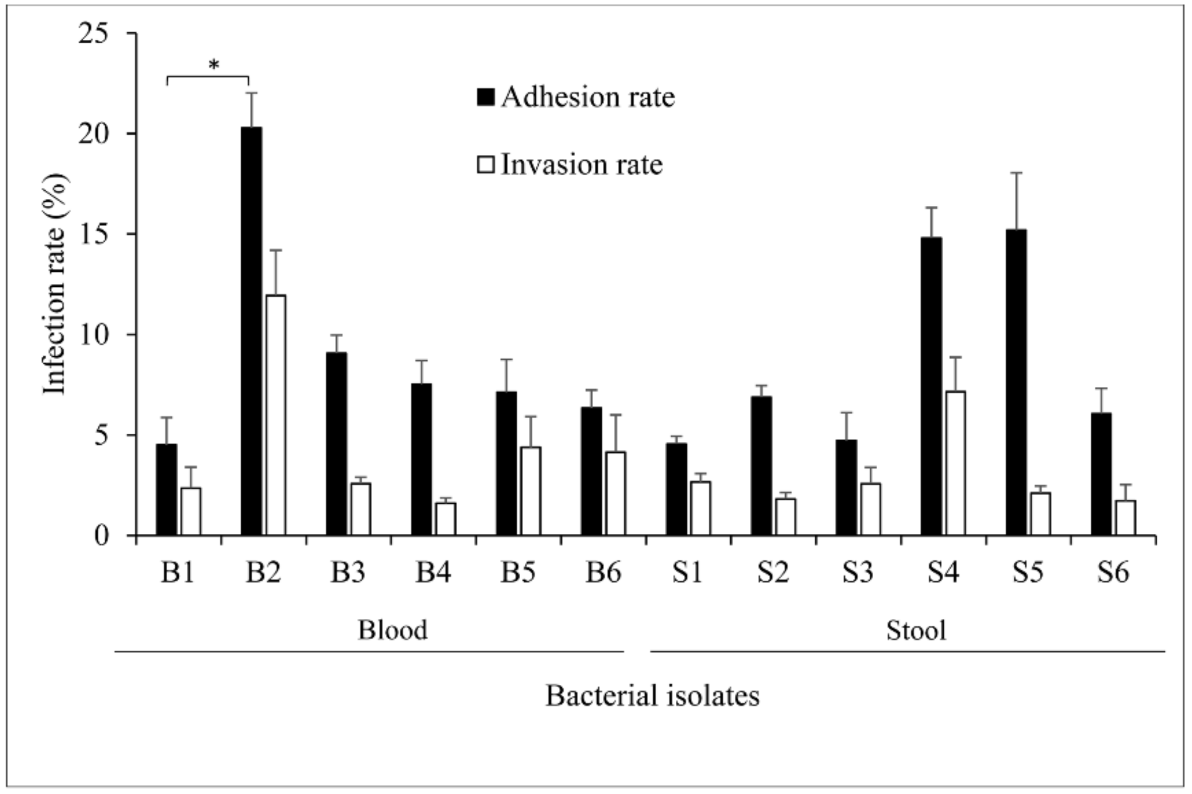
Figure 1. Infection rates of different Salmonella isolates on FHs 74 Int cells. Bacteria inoculum suspended in complete Hybri-Care Medium was directly added to the cultured cells in a 24-well plate to a multiplicity of infection (M.O.I_ of 50:1 and incubated for 2 h at standard conditions. Bacterial colony forming unit (CFU) count was achieved after cell wash and lysis. The adhesion rate was calculated as the percentage ratio of bacteria attaching the cell membrane and probably penetrating the cells to the initial concentration of bacteria in the inoculum. The invasion rate solely considered the engulfed bacteria since Gentamicin treatment discarded the external bacteria. Values were expressed as the mean of four independent repeats with standard error of the mean (±SEM). A general linear model including repeated measures was used for statistical analysis. (*) p = 0.007.
Pathogens 10 00074 g001
Figure 2. Growth curves of different Salmonella isolates. (a) Blood isolates in LB medium. (b) Stool isolates in LB. (c) Blood isolates in M9 medium. (d) Stool isolates in M9 medium. The optical density (OD) of cultured bacteria was read at 600 nm. The values represented the mean of three independent measurements with the standard error of the mean (±SEM).
Pathogens 10 00074 g002
Figure 3. The motility patterns of the Salmonella isolates. Salmonella isolates (a) swarming and (b) swimming pattern revealed on agar plates. The pictures were taken 6 h after plate inoculation. Swarming dispersion surface of (c) blood and (d) stool isolates on 0.5% agar supplemented with 0.5% glucose. Swimming dispersion surface of (e) blood and (f) stool isolates on 0.3% agar plates. Comparative dispersion was represented as the mean of three repeats with the standard error of the mean (±SEM). One-way analysis of variance was used for statistical analysis. (*) p = 0.03. The scale bar corresponds to 1 cm.
Pathogens 10 00074 g003
Figure 4. Biofilm formed by Salmonella isolates. (a) Comparative optical density (OD) of bacterial forming biofilm cultured in LB and M9 medium. Bacteria adhering to the well of a 96-well plate after an incubation of 48 h at 37 °C were stained with crystal violet and dissolved into anhydrous ethanol. The OD was eventually measured at 590 nm with consideration of a negative control referred to as blank. By using the blank cut-off OD (ODc), isolates have been identified as non-biofilm, weak, or strong biofilm-forming when OD ≤ ODc, ODc < OD ≤ (2 × ODc), and (4 × ODc) < OD, respectively. Two-way analysis of variance method was used for statistical analysis. (b) Visual representation of biofilm formed by Salmonella isolates stained and dissolved in an ethanolic solvent. Data presented as the mean ± SEM of three measurements performed in three independent experiments. Bk: blank.
Pathogens 10 00074 g004
Figure 5. Quantitative mRNA expression of virulence genes in Salmonella isolates. Total RNA from bacteria cultured in LB was extracted. The cDNA was synthesized and used for qPCR. The sample Ct values were first normalized against the endogenous RpoD gene control, then the level of gene expression was calculated by using the formula 2 −∆Ct. Data were presented as mean ±SEM of three independent experimental repeats. Salmonella Typhimurium LT2 (ST, positive control), and E. coli (EC, negative control). Ctrl: control.
Pathogens 10 00074 g005
Table 1. Distribution of virulence genes among the bacterial isolates.
Bacterial IsolatesFliCSpvCInvAFimHSopBHilA
B1-+++++
B2+-++++
B3-+++++
B4-+++++
B5+-++++
B6+-++++
S1++++++
S2-+++++
S3-+++++
S4+-++++
S5--++-+
S6-+++++
+: Presence of gene, -: Absence of gene.
Table 2. Salmonella isolates and antibiotic susceptibility profiles.
AntimicrobialsSusceptibility Characteristics (n = 50)
Sensitive
N (%)
Intermediate
N (%)
Resistant
N (%)
Chloramphenicol32 (64%)0 (0%)18 (36%)
Ciprofloxacin2 (4%)38 (76%)10 (20%)
Ampicillin24 (48%)1 (2%)25 (50%)
Gentamicin22 (44%)24 (48%)4 (8%)
Ceftriaxone45 (90%)4 (8%)1 (2%)
Trimethoprim-Sulfamethoxazole36 (72%)0 (0%)14 (28%)
N: number; %: percentage.
Table 3. Salmonella enterica used for microbiology study.
Bacterial Identification NameBacterial Collection NumberIsolation SourceSerotyping Group
B11517BloodD1
B21596BloodB
B31462BloodD1
B41793BloodD1
B51829BloodB
B61833BloodB
S11792StoolB
S21785StoolD1
S31794StoolD1
S41795StoolB
S51798StoolB
S61762StoolD1
Table 4. Targeted Salmonella virulence genes and their primers.
Target GeneOligo SequenceAmplicon Size (bp)NCBI Reference Sequence
FliCF: AACGCAGTAAAGAGAGGACG162NC 003197.2
R: ACACCGTAAACAACCTGACT
SpvCF: TCACGTAAAGCCTGTCTCTG214NC 003277.2
R: CAGACCAGGAAAATTCGCAG
FimHF: CGGAAAAATCAGGTTGGGTC360NC 003197.2
R: CGGTAGAGGTCGTCACATAG
SopBF: CTTTTGCAGGTAAGCCATCC235NC 003197.2
R: CACTCGCTGCATAACCTCTA
InvAF: TCCACGAATATGCTCCACAA381NC 003197.2
R: CCAACAATCCATCAGCAAGG
HilAF: GCTTTAGGATTACTGGGGCT407NC003197.2
R: AGTCCTGTTATTTCCTGCGT
F: Forward; R: Reverse.
Table 5. Primers implemented for qPCR.
Target GeneOligo Sequence
FliCF: TCTGTACCGCACCCAGGTCA
R: CGGCTGCTACAACCACCGAA
SpvCF: CACTCTGGCGCATCCCTGAA
R: GCTGCTTATGATGGGGCGGA
FimHF: TGACGATCCCTCGCCAGACA
R: ACGACCTGTCCGGCATTCAC
SopBF: GTATTGCGCCAGCCATTCGC
R: GCCGAGGCGCTACATCAGTT
InvAF: CGACTTCCGCGACACGTTCT
R: TAGCCTGGCGGTGGGTTTTG
HilAF: TCGTAGTGGTGTCTCCGCCA
R: CTCAGGCCAAAGGGCGCATA
RpoDF: GTTGACCCGGGAAGGCGAAA
R: GGGTATTCGGCAACGGAGCA
F: Forward; R: Reverse.
Publisher’s Note: MDPI stays neutral with regard to jurisdictional claims in published maps and institutional affiliations.