Article

Rapid Detection of Equine Piroplasms Using Multiplex PCR and First Genetic Characterization of Theileria haneyi in Egypt

1
Parasitology and Animal Diseases Department, Veterinary Research Institute, National Research Centre, Dokki, Giza 12622, Egypt
2
Tick and Tick-Borne Diseases Research Unit, Veterinary Research Institute, National Research Centre, Dokki, Giza 12622, Egypt
3
Parasitology Department, Faculty of Veterinary Medicine, Cairo University, Giza 12622, Egypt
4
Department of Veterinary Microbiology and Pathology, College of Veterinary Medicine, Washington State University, Pullman, WA 99164, USA
5
Department of Veterinary Tropical Diseases, Faculty of Veterinary Science, University of Pretoria, Private Bag X04, Onderstepoort 0110, South Africa
6
Department of Parasitology, Faculty of Veterinary Medicine, University of Firat, Elazig 23119, Turkey
*
Authors to whom correspondence should be addressed.
Pathogens 2021, 10(11), 1414; https://doi.org/10.3390/pathogens10111414
Submission received: 26 September 2021 / Revised: 27 October 2021 / Accepted: 28 October 2021 / Published: 31 October 2021

Abstract

:
Equine Piroplasmosis (EP) is an infectious disease caused by the hemoprotozoan parasites Theileria equi, Babesia caballi, and the recently identified species T. haneyi. Hereby, we used a multiplex PCR (mPCR) targeting the 18S rRNA gene of T. equi and B. caballi for the simultaneous detection of EP in Egyptian equids and examined the presence of T. haneyi infections in Egypt. Blood samples from 155 equids (79 horses and 76 donkeys) collected from different governorates of Egypt were examined by mPCR and PCR targeting T. hayeni. The mPCR method revealed a prevalence of T. equi of 20.3% in horses and of 13.1% in donkeys and a prevalence of B. caballi of 1.2% in horses. B. caballi was not detected in donkeys in the current study. The mPCR method also detected coinfections with both species (2.5% and 1.3% in horses and donkeys, respectively). Additionally, we report the presence of T. haneyi in Egypt for the first time in 53.1% of the horse and 38.1% of the donkey tested samples. Coinfection with T. haneyi and T. equi was found in 13.5% of the samples, while infection with the three EP species was found in 1.9% of the samples.

1. Introduction

In rural areas of many developing countries, including Egypt, there is a huge reliance on working equids, which include horses, donkeys, mules, and ponies. These animals play important roles in sustaining the livelihoods of millions of people by providing support in industries that include agriculture, construction, tourism, mining, and public transport [1,2]. The health and welfare of domesticated equids are often overlooked in rural areas. Although equids can be affected by a myriad of diseases that include amongst others, African Horse Sickness, Epizootic Lymphangitis (EZL), Tetanus, Rabies, Trypanosomiasis, and Piroplasmosis, there is a general lack of knowledge regarding the identification, management, and prevention of infectious diseases [3].
Equine piroplasmosis [EP] is one of the infectious tick-borne diseases (TBDs) of equids, caused by the hemoprotozoan parasites T. equi, B. caballi [4], and the newly identified species T. haneyi [5]. There are over 30 species of ticks that have been described as vectors of T. equi, B. caballi, or both, including the genera Hyalomma, Rhipicephalus, Dermacentor, Amblyomma, and Haemaphysalis [6]. In Egypt, three species of ticks have been detected in equids, i.e., Hyalomma dromedarii, Hyalomma excavatum, and Rhipicephalus annulatus [7]. However, investigations on vector competence for T. haneyi has not been reported [8]. Infection with T. equi and B. caballi can cause severe economic losses in the equine industry due to the cost of treatment, especially for acutely infected horses, which, in the absence of appropriate treatment, can die [2]. It was found that T. haneyi rarely causes clinical signs in field horses [9], even in splenectomized horses experimentally infected with T. haneyi using the intravenous (IV) route [5,10]. Horses infected with T. equi remain persistently infected, while those affected by B. caballi are infected for an extended period [11].
Phylogenetic analysis of published T. equi and B. caballi 18S rRNA (SSU rRNA) gene sequences have led to the identification of five T. equi genotypes (A, B, C, D, and E) and two B. caballi genotypes (A, B) globally. The genotype B of B. caballi was reclassified into two subgenotypes called genotype B1 and genotype B2 [12,13], but genotype B2 was renamed as genotype C [14,15,16]. Theileria haneyi was first detected in a horse at the U.S.– Mexico border, near Eagle Pass, Texas [5,10]. Phylogenetic evidence places this species into a clade distinct from that of T. equi [5], and this species also lacks the equi merozoite antigen 1 (ema-1) gene that occurs in T. equi, explaining the failure of a diagnostic cELISA test based on recombinant T. equi ema-1 antigen in detecting T. haneyi [9].
The control and treatment of EP in non-endemic countries depend mainly on serological and molecular techniques [17]. In Egypt, the national tick control program recommends the use of acaricides like doramectin to reduce tick exposure [18,19]. The diagnosis of EP based solely on clinical signs is not specific, and differentiation between the EP causative agents is not possible based on clinical signs alone [20]. Microscopical examination (ME) of blood films has limited utility due to its low sensitivity, particularly in carrier animals with low parasitemia [21]. In addition, these diagnostic tools cannot identify and genetically characterize species of Babesia and Theileria infecting equids. In Egypt, the diagnosis of EP is based on ME and/or small-scale surveys using conventional PCR (cPCR) for the diagnosis of each species separately [22,23,24,25]. Moreover, serological diagnosis (IFA and ELISA techniques) is used mainly in the case of chronically infected animals [20,23,24]. The effective treatment of EP, therefore, depends on the ability to differentiate between T. equi and B. caballi [26]. Thus, accurate and sensitive diagnostic methods that can differentiate between T. equi, B. caballi, and T. haneyi in animals that have mixed infections are required as a step toward implementing adequate control measures. To overcome the diagnostic drawbacks faced in Egypt, more sensitive and specific DNA amplification methods like PCR followed by sequencing of the amplicons could be used, especially in the prepatent phase infection of piroplasms [27].
Although uniplex (u) PCR assays are effective in the detection of single-species infection, they are time-consuming and expensive when applied on many samples that may have mixed infections [28]. The reverse line blot (RLB) assay has overcome this problem to a large extent by allowing the simultaneous detection of multiple parasite species in a single sample [29], but RLB requires expertise and specialized equipment, and the protocol is very labor-intensive [28]. Multiplex PCR (mPCR) is a single, lower cost, and technically less challenging approach that is able to amplify two or more target loci from one or more organisms using a mixture of specific primer pairs in a single reaction. Thus, mPCR could be a favorable tool for the diagnostic and epidemiological evaluation of TBD in endemic regions [28,30]. Therefore, the current work aimed to study the prevalence of EP using mPCR to detect T. equi and B. caballi simultaneously, targeting the 18S rRNA of both species. In addition, the current research addressed, for the first time, the detection of T. haneyi in Egypt through the examination of blood samples by conventional PCR (cPCR) followed by amplicon sequence comparison with South African and American T. haneyi isolates.

2. Results

2.1. Molecular Detection of Equine Piroplasmosis

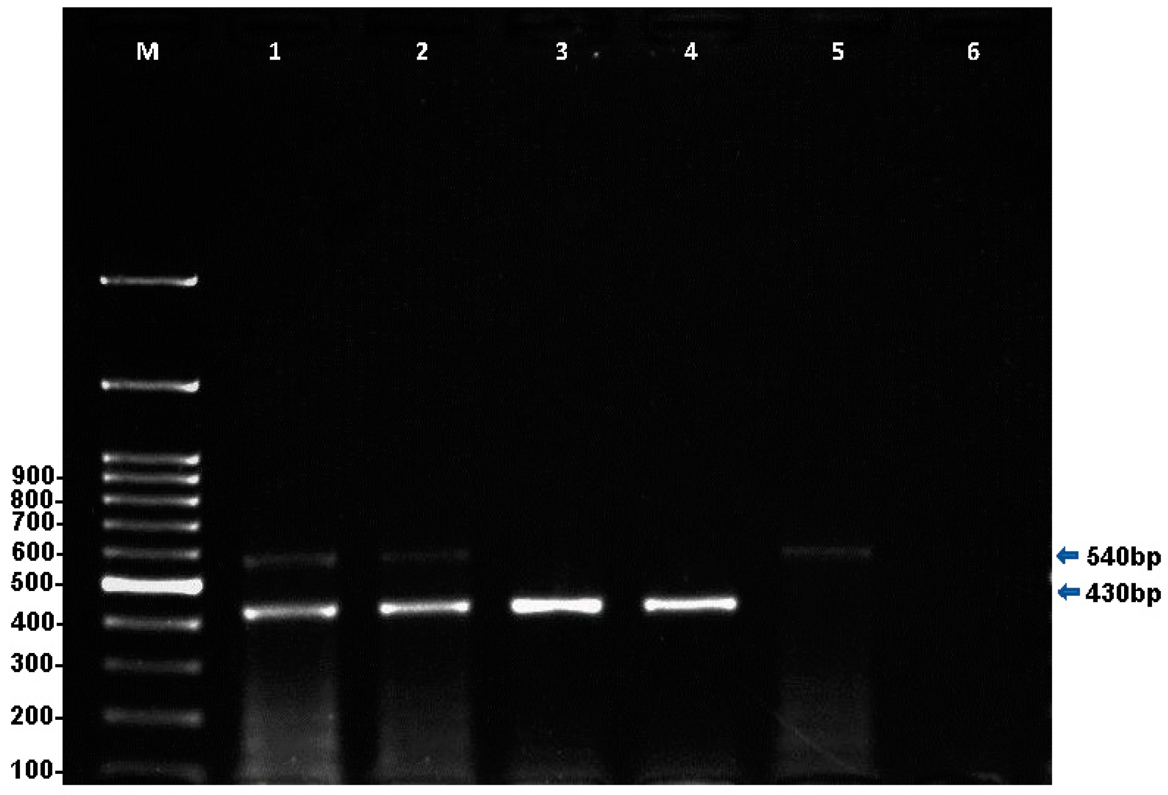
2.1.1. Multiplex PCR for the Simultaneous Detection of T. equi and B. caballi

Multiplex PCR detected single T. equi infections in 26 (16.7%) (95% CI, 10.1–22.5%) equids, 16 (20.3%) (95% CI, 11.1–29.1%) horses, and 10 (13.1%) (95% CI, 5.5–20.6%) donkeys at the expected amplicon size of 430 bp (Figure 1). Similarly, a single infection with B. caballi was detected in one horse (1.2%) (95% CI, −0.1–3.6%) with an expected amplicon size of 540 bp (Figure 1). Co-infections with both parasites were found in two horses (2.5%) (95% CI, 0–5.9%) and one donkey (1.3%) (95% CI, 0.1–3.8%), with an overall prevalence of 1.9% (95% CI, 0–4.0) (three equines) (Table 1). Statistically, there was no significant difference in EP infection among horses and donkeys on the basis of mPCR data (p > 0.05); however, the difference between single T. equi and B. caballi infections was statistically significant (p < 0.05) as T. equi infection was more prevalent.

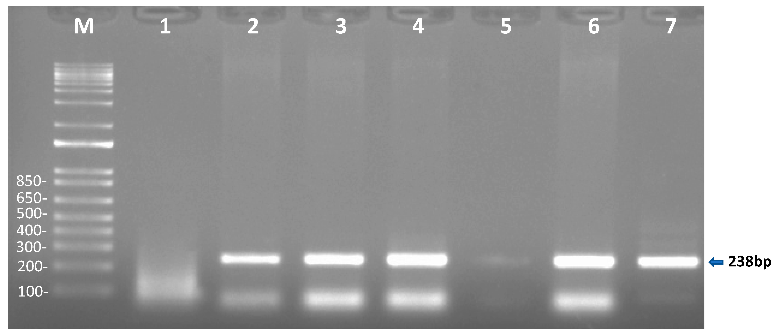
2.1.2. Conventional PCR Analysis for the Detection of T. haneyi in Egyptian Equids

Theileria haneyi was detected in 71 (45.8%) (95% CI, 37.4–53.6%) equids; 42 (53.1%) (95% CI, 40.4–62.1%) of these samples were derived from horses, and 29 (38.1%) (95% CI, 27.2–49.0%) from donkeys (Table 2). The positive samples gave the expected amplicon size of 238 bp (Figure 2). Statistically, there were no significant differences in infection with T. haneyi between horses and donkeys on the basis of the cPCR data (p > 0.05).

2.1.3. Coinfections with T. haneyi (cPCR), T. equi, and B. caballi (mPCR)

The analysis of the mPCR and cPCR results of the 155 samples tested indicated that 3 horses and 18 donkeys were co-infected with both T. haneyi and T. equi. Additionally, co-infections with all three parasites (T. equi, T. haneyi, and B. caballi) were observed in two horses (95% CI, 0–5.9%) and one donkey (Table 2). Co-infections with T. haneyi and B. caballi were not observed.

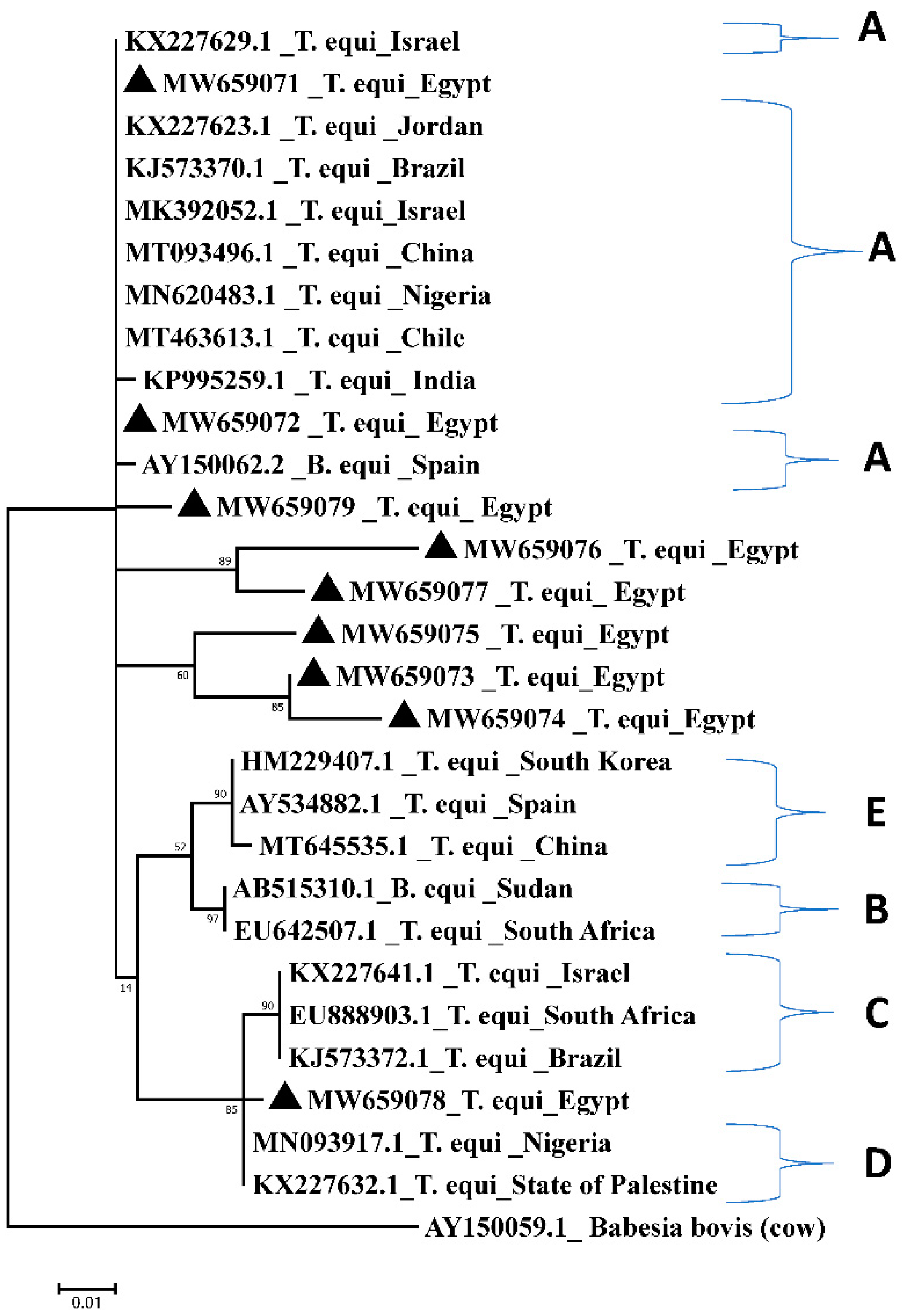
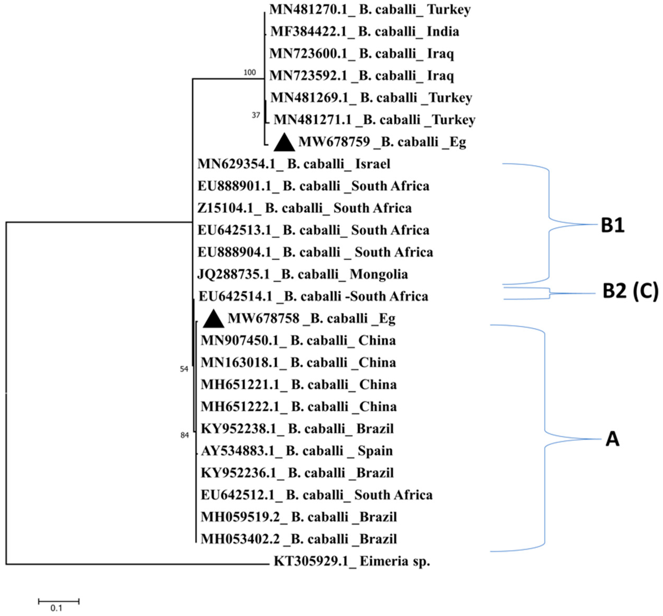
2.2. Comparative Analysis and Sequence Conservation of the 18S rRNA Amplicons among Different Isolates

The 360-bp fragment of the T. equi 18S rRNA gene was amplified and sequenced from nine selected positive samples. The identity percent among the different Egyptian amplicons from T. equi and B. caballi is shown in supplementary Tables S1 and S2. Blast analysis indicated that the amplicon derived from the Egyptian isolates showed between 95.7 and 99% identity to previously published T. equi 18S rRNA gene sequences. In addition, the amplified B. caballi amplicon (540 bp) from two selected positive sample was sequenced. Blast analysis indicated that the B. caballi Egyptian isolate showed an identity percent ranging from 98.1 to 99.3% to published B. caballi isolates.
Comparative analysis showed that one T. equi Egyptian amplicon derived from one horse with accession number MW659071.1 and two amplicons from donkeys with accession numbers MW659072.1 and MW659079.1 clustered with sequences from Chile (MT463613.1) [31], Israel (MK932052.1) [13], China (MT093496.1) [31], Jordan (KX227623.1) [32], and Nigeria (MN620483.1) [33], whereas only one Egyptian amplicon derived from one donkey (MW659078.1) clustered with sequences from the State of Palestine (KX227632.1) [32] and Nigeria (MN093917.1) [34]. In addition, three sequences derived from horses (MW659073.1, MW659074.1, and MW659075.1) and two from donkeys (MW659076.1 and MW659077.1) clustered together in a separate group from the other sequences obtained in the current study (Figure 3).
Similarly, the B. caballi Egyptian isolates showed 98.1–99.3% sequence identity with B. caballi sequences from China, Brazil, South Africa, Israel, Iraq, Turkey, and India. Comparative analysis of the B. caballi isolate (MW678758.1) from horses clustered with sequences from China (MN907451.1), Brazil (KY952238.1) [35], and South Africa (EU642512.1) [12], while a B. caballi isolate (MW678759.1) isolated from donkeys clustered in a separate clade with sequences from Iraq (MN723592.1), Turkey (MN481269.1), and India (MF384422.1) (Figure 4).

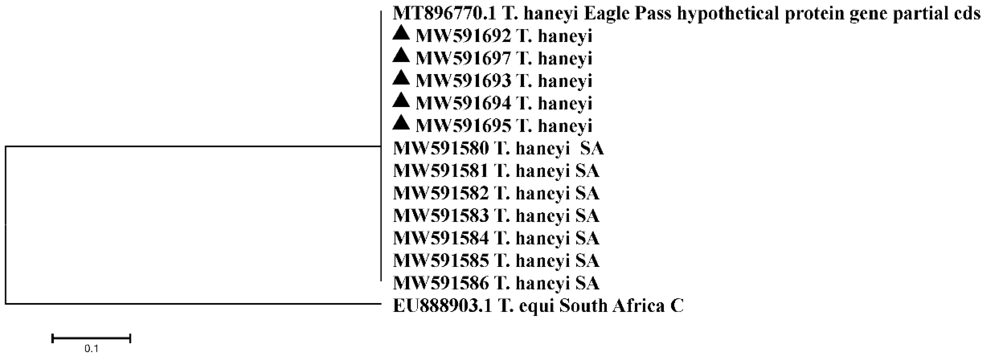
2.3. Sequencing Analysis of a T. haneyi Hypothetical-Protein-Coding Gene

BLASTn analysis of the five T. haneyi Egyptian samples sequenced in this study showed 100% sequence identity to published T. haneyi sequences from South African isolates (MW591580-MW591586) [36] and to the published sequences of T. haneyi Eagle Pass strain gene for a hypothetical protein (MT896770.1) (Figure S1). The comparative analysis, based on amplicons derived from infected Egyptian horses (n = 2) (MW591694.1, MW591695.1) and donkeys (n = 3) (MW591692.1, MW591693.1, MW591697.1), indicated that the Egyptian T. haneyi sequences all clustered together with the reference T. haneyi sequence and with sequences from South African isolates; T. equi genotype C (18S r RNA) was selected as an outgroup (Figure 5).

3. Discussion

Piroplasms are Apicomplexa tick-borne parasites distributed worldwide which are responsible for piroplasmosis (theileriosis and babesiosis) in vertebrates. The aim of the present study was to use molecular methods for the detection of the prevalence of EP in Egypt caused by T. equi and B. caballi. We also aimed at detecting the occurrence of T. haneyi in equids in Egypt, which was unknown. Importantly, the DNA sequence data generated in this study also allowed for some genetic characterization of T. equi, B. caballi, and T. haneyi Egyptian strains currently circulating in this country.
The prevalence of T. equi was higher than that of B. caballi, and this is consistent with previous reports [37,38]. This phenomenon may be due to the increased susceptibility of B. caballi to treatment compared to T. equi. In addition, the horse immune system may be more efficient in eliminating B. caballi-infected erythrocytes than T. equi-infected ones, the latter parasites having a long persistence [9,39].
The result of this study also indicate that the prevalence of coinfections with both parasites (T. equi and B. caballi) in equids was 1.9%, which is lower than that detected in Mongolia (7.7%) [40] and Iraq (5.15%) [41] using mPCR and in Cuba (20%) [21] and Nigeria (2.7%) using nested PCR [9].
The observed difference in the prevalence of EP compared to other countries may be due to the type of equids (race or working) examined, hygienic measures, differences in environmental conditions—which can have a significant impact on tick activity—tick control strategies, number of samples analyzed, and type of PCR used for molecular diagnosis [24].
Blast analysis of the amplified fragments from T. equi and B. caballi showed sequence identities between 96 and 99% to published sequences. While lower sequence similarities may indicate distinct parasite species, it is important to note that the analysis was based on small fragments of the 18S rRNA gene. However, initial epidemiological studies on South African T. equi and B. caballi 18S rRNA gene sequences reported identities between 96.1 and 99.9% to the previously published T. equi sequence from South Africa (accession number: Z15105) and between 96.9 and 99.9% to a published B. caballi sequence from South Africa (accession number: Z15104). Phylogenetic analysis of the South African sequences and subsequently of sequences from other parts of the world led to the identification of distinct parasite genotypes, which may even represent distinct parasite species [36]. Therefore, the sequences obtained in this study could represent Egyptian isolates that belong to theses distinct parasite genotypes. However, amplification and sequencing of the complete 18S rRNA gene would be necessary to confirm these identities.
Theileria haneyi was defined as a new species infective to equids [5] and has since been reported to occur in several countries in North and South America, Africa, and Asia [5,9,36,42]. In the current study, T. haneyi was identified in both horses and donkeys in Egypt, and the sequence of the hypothetical-protein-coding gene was identical to the published T. haneyi Eagle Pass reference sequence and to sequences from South African isolates, confirming the presence of T. haneyi in Egypt, as reported here for the first time.
The results of the current study are in agreement with Sears et al., [10] who reported that coinfection of T. haneyi and T. equi could be induced experimentally in horses, which can explain the presence of the three parasites in naturally infected animals in our study. That means there was no cross immunity induced by T. haneyi and other two equine piroplasm (T. equi and B. caballi) and the infection with these two parasites does not protect equines from the infection with T. haneyi and vice versa.
The prevalence of T. haneyi either as single or as a mixed infection with T. equi and B. caballi was higher than that recorded for imported Argentine horses in Nigeria (2.7% and 0.6%, respectively) [9], and this observation may be explained by the factors mentioned earlier that include environmental conditions, husbandry, and tick vectors. Differences in sampling size and time of sample collection could also be contributing factors.
The application of new technologies with higher sensitivities and specificities could better facilitate the diagnosis of EP in Egypt. A multiplex EP real-time PCR assay targeting the 18S rRNA gene was developed for the simultaneous, quantitative detection of T. equi and B. caballi in field animals. Quantitative molecular genotyping assays for T. equi were also developed and enable the rapid detection of distinct T. equi parasite genotypes. Future studies in Egypt should focus on further characterizing the T. equi and B. caballi genotypes that may be circulating within the different governorates, with a view to determining risk factors in disease control. It has been noted that T. haneyi species classification was based on differences in the equi merozoite antigen (EMA) multigene family, and the identification of T. haneyi in South African horses infected with T. equi genotype C indicated that T. haneyi may be a subgroup of T. equi Genotype C [5,36]. The identification of T. haneyi in Egyptian equids is not surprising but warrants further investigation.

4. Materials and Methods

4.1. Collection of Field Samples

Blood samples were collected from 155 apparently healthy equids (79 horses and 76 donkeys) from different governorates in Egypt (Cairo 30°2′0″ N, 31°14′0″ E, Giza 29°59′13.2″ N, 31°12′42.48″ E, Monufia 30°31′12″ N, 30°59′24″ E, Faiyum 29°18′30.14″ N, 30°50′38.78″ E, Beni Suef 29°4′0″ N, 31°5′0″ E, Ismailia 30°35′0″ N, 32°16′0″ E, and Alexandria 31°10′0″ N, 29°53′0″ E) (Figure 6).
The equid samples were collected from the following places: the Police Academy and Elzahraa-Stud in Cairo and the zoological garden Abattoir in Giza, National Research Centre veterinary caravans to Almonofia, Al fayoum, Beni Suef, Ismailia, and Alexandria governorates, Egypt. The blood samples were collected on EDTA-containing vials and transferred to the laboratory in ice boxes. Blood spots were prepared by applying 100 µL of blood on Whatman WB120410 FTA Elute Micro Card (GE Healthcare and Cytiva, North Bend, OH, USA). Ethical clearance for sample collection from equids was obtained through the Institutional Animal Care and Use Committee (IACUC) (Vet CU28/04/2021/297 and 28/04/2021).

4.2. DNA Extraction

Genomic DNA was extracted from FTA Elute Micro Card [43,44], following the manufacturer’s instructions.
Positive control DNA samples extracted from T. equi and B. caballi in vitro cultures were provided by the OIE equine piroplasmosis reference lab located in Pullman, WA, USA.

4.3. Molecular Detection of Equine Piroplasmosis by Three PCR Approaches

4.3.1. Multiplex PCR (mPCR) for the Detection of T. equi and B. caballi

Field samples were tested for the presence of equine piroplasmosis using a published conventional mPCR assay designed for the simultaneous detection of T. equi and B. caballi infections [40]. The 18S rRNA gene was used, targeting the 943–1300-bp region for T. equi and the 562–1141-bp region for B. caballi [38,40]. Briefly, the universal forward primer Bec-UF2 and species-specific reverse primers (Cab-R, B. caballi; Equi-R, T. equi) were combined in reactions containing 3 µL of DNA sample, 12.5 µL of Sigma 2× JumpStart™ REDTaq® ReadyMix™ (Foster City, California, USA), 5 μM of each primer, and 7.5 µL of nuclease-free water in a 25 µL total volume. Primers sequences are shown in Table 3. The amplification conditions were according to Abedi et al. [38], with minor modifications, which included an initial denaturation for 5 min at 94 °C, followed by 35 cycles each of 94 °C for 1 min as a denaturation period, an annealing period of 54 °C for 1 min, and an extension period at 72 °C for 1 min, with the addition of a final extension period of 7 min at 72 °C. The DNA extracted from T. equi and B. caballi in vitro cultures was used as a positive control, and the negative control was a no-template control (NTC). All amplicons were visualized by 2% agarose gel electrophoresis (Invitrogen, Waltham, USA).

4.3.2. Uniplex PCR (uPCR) for Confirmation of the mPCR Results for the Detection of T. equi and B. caballi

Samples that tested positive for piroplasmosis using the mPCR assay were confirmed by performing uPCR assays. For the amplification of T. equi parasite DNA, the primers TBM and Equi-R were used, while the amplification of B. caballi was done using the primers Bec-UF2 and Cab-R (Table 3). The reactions were set up as previously described, and PCR amplification conditions were the same as those reported for the mPCR assay.

4.3.3. Detection of T. haneyi

For the detection of T. haneyi, instead of performing a nested PCR as done by Knowles et al. [5], a gradient annealing temperature in PCR using the internal nested primers described in Table 3 was used. The best annealing temperature was 56 °C, which was chosen to complete the amplification process. Amplicons were visualized by 1.5% agarose gel electrophoresis.

4.4. Sequencing and Sequence Analysis

Samples (T. equi n = 9; B. caballi n = 2 and T. haneyi n = 5) that gave strong positive amplification reactions were selected for further sequencing and comparative analyses. Briefly, amplicons were purified using the GeneDirex PCR clean-up and Gel Extraction kit (Taiwan) according to the manufacturer’s instructions and sent for bi-directional sanger sequencing to Macrogen ( Seoul, South Korea ) using ABI3730XL DNA Sanger sequencer (ThermoFisher) (Waltham, MA, United States) All sequence data were edited using MEGA 7 software (https://www.megasoftware.net/download_form accessed on 2 January 2021). Query coverage and the percent of identity among the compared sequences were calculated by non-redundant National Centre for Biotechnology Information (NCBI) and Clustal Omega (https://blast.ncbi.nlm.nih.gov/Blast.cgi accessed on 2 January 2021) and (https://www.ebi.ac.uk/Tools/msa/clustalo/ accessed on 1 March 2021). In the present study, samples were aligned with the reference sequences for 18S rRNA representing T. equi (Z15105.1) [45] and for a gene coding a hypothetical protein of unknown function but specific for T. haneyi genome (MT896770.1 T. haneyi Eagle Pass strain) [5], available in the NCBI database. In addition, B. caballi gene sequence was kindly provided by Lowell S. Kappmeyer [Animal Diseases Research Unit, USDA-ARS, Pullman, WA 99164-6630, US]. Moreover, the T. equi and B. caballi sequences of the present study were compared with different 18S rRNA reference sequences collected from distinct geographical areas worldwide and available in GenBank (Tables S3 and S4) [46,47,48,49,50,51,52,53,54]. T. haneyi sequences were compared with the sequence of a hypothetical-protein-coding gene of T. haneyi Eagle Pass strain present in GenBank and with six T. haneyi South African (SA) isolate sequences [36]. All sequence data were edited using MEGA 7 software. Query cover and identity percentage among the compared sequences were calculated by NCBI and Clustal Omega (https://blast.ncbi.nlm.nih.gov/Blast.cgi accessed on 16 March 2021) and (https://www.ebi.ac.uk/Tools/msa/clustalo/ accessed on 23 February 2021). The resulted sequences data were submitted to GenBank to get accession numbers for T. equi, B. caballi, and T. haneyi Egyptian isolates.

4.5. Comparative Analysis

To assess the genetic diversity of hemoparasites within the study samples, species-specific dendrograms were constructed using a phylogenetic tree prediction generated by MEGA 7 (https://www.megasoftware.net/download_form accessed on 3 April 2021). This dendrogram was constructed using the Maximum Likelihood method based on the Kimura 2-parameter mode [55]. Egyptian T. equi and B. caballi isolates and the 18S rRNA gene of T. equi and B. caballi of different reference sequences in GenBank were used for comparative analyses, which were classified into genotypes A, B, C, D, and E for T. equi and genotypes A, B1, and B2 (C). The 18S rRNA gene sequences of B. bovis (AY150059.1) [56] were included in the dendrogram as outgroups for the T. equi dendrogram, while Eimeria sp. cytochrome oxidase subunit I (COI) gene (KT305929.1) [52] was used as the outgroup for the B. caballi dendrogram. Hypothetical-protein-coding gene of unknown function of T. haneyi Egyptian isolates, South African isolate (SA) [36], and T. haneyi Eagle Pass strain reference sequence [5] were used in T. haneyi’s dendrogram construction. Theileria equi genotype C South Africa (EU888903.1) [12] was used as the outgroup.

4.6. Statistical Analysis

The chi-square (χ2) test was applied at a probability of p < 0.05 to compare infection rates between equids determined by mPCR and cPCR. Significant associations were identified when a p value of less than 0.05 was observed [57].

5. Conclusions

The mPCR technique is a rapid diagnostic method for the simultaneous detection of both T. equi and B. caballi, especially in mixed-infected cases. This study represents a first report on the presence of T. haneyi in Egyptian equids and, specifically, in donkeys. Further investigations are required to determine the T. equi and B. caballi genotypes in Egypt and to study the impact of the presence of T. haneyi either as a single or as a co-infecting agent with other EP in disease control and how that can be involved in pathogen evolution.

Supplementary Materials

The following are available online at https://www.mdpi.com/article/10.3390/pathogens10111414/s1, Table S1: Identity percent between T. equi Egyptian isolates analyzed in the present study. Table S2: Identity percent between B. caballi Egyptian isolates analyzed in the present study. Table S3: T. equi accession numbers of different 18s gene isolates used in dendrogram construction and their references. Table S4: B. caballi accession numbers of different 18s gene isolates used in dendrogram construction and their references. Figure S1: Alignment of the DNA sequences of five T. haneyi Egyptian (Eg) isolates (GenBank accession no. MW591692:MW591695 and MW591697) and six T. haneyi South African (SA) isolates of a hypothetical-protein-coding gene (GenBank accession number MW591580: MW591586) [BioEdit software].

Author Contributions

Conceptualization, A.M.N., H.F.A., M.S.M. and B.S.M.E.; methodology, B.S.M.E., H.F.A. and M.S.M.; software, B.S.M.E., H.F.A. and R.V.B.; validation, A.M.N., H.F.A., M.S.M., and B.S.M.E.; formal analysis, B.S.M.E., H.F.A. and M.S.M.; investigation, M.S.M., H.F.A., O.A.M., S.O., and B.S.M.E.; resources, H.F.A., M.S.M., O.M.K. and A.M.N.; data curation, M.S.M. and H.F.A.; writing—original draft preparation, B.S.M.E., H.F.A., M.S.M. and O.M.K.; writing—review and editing, A.M.N., B.S.M.E., O.A.M., M.S.M., H.F.A., O.M.K. and S.O.; and visualization, H.F.A., R.V.B., M.S.M., A.M.N., O.A.M. and B.S.M.E.; supervision, O.A.M., A.M.N., H.F.A., M.S.M. and O.M.K. All authors have read and agreed to the published version of the manuscript.

Funding

This research was funded by B.S.M.E, National research center, (NRC) fund.

Institutional Review Board Statement

The study was approved by the Institutional Animal Care and Use Committee (IACUC) (Vet CU28/04/2021/297 and 28/04/2021).

Informed Consent Statement

Not applicable.

Data Availability Statement

Not applicable.

Acknowledgments

We are so thankful to Carlos Suarez, United States Department of Agriculture, for his great help and support in this study. The thanks also go to Sobhy abdel-Shafy, Parasitology and Animal Diseases Department, National Research Center, Dokki, Giza, Egypt, for kindly providing the caravan equine samples and for his help in the statistical analysis of the data. We are also thankful to Giza Zoo team, Giza, Egypt, for their help in blood sample collection.

Conflicts of Interest

The authors declare no conflict of interest.

References

  1. Valette, D. Invisible Workers. The Economic Contributions of Working Donkeys, Horses and Mules to Livelihoods; The Brooke: Louisville, KY, USA, 2015; pp. 1–23. Available online: https://www.thebrooke.org/sites/default/files/Advocacy-and-policy/Invisible-workers-report.pdf (accessed on 20 May 2021).
  2. Mahmoud, M.S.; Kandil, O.M.; Abu Ezz, N.T.E.; Hendawy, S.H.M.; Elsawy, B.S.M.; Knowles, D.P.; Bastos, R.G.; Kappmeyer, L.S.; Laughery, J.M.; Alzan, H.F.; et al. Identification and Antigenicity of the Babesia caballi Spherical Body Protein 4 (SBP4). Parasit. Vectors 2020, 13, 369. [Google Scholar] [CrossRef]
  3. Church, S. BEASTS of Burden Targeting Disease in Africa’s Working Donkeys and Horses. Available online: https://thehorse.com/features/beasts-of-burden-africas-working-horses-and-donkeys/ (accessed on 17 May 2021).
  4. Romero-Salas, D.; Solis-Cortés, M.; Zazueta-Islas, H.M.; Flores-Vásquez, F.; Cruz-Romero, A.; Aguilar-Domínguez, M.; Salguero-Romero, J.L.; de León, A.P.; Fernández-Figueroa, E.A.; Lammoglia-Villagómez, M.Á.; et al. Molecular Detection of Theileria equi in Horses from Veracruz, Mexico. Ticks Tick-Borne Dis. 2021, 12, 101671. [Google Scholar] [CrossRef] [PubMed]
  5. Knowles, D.P.; Kappmeyer, L.S.; Haney, D.; Herndon, D.R.; Fry, L.M.; Munro, J.B.; Sears, K.; Ueti, M.W.; Wise, L.N.; Silva, M.; et al. Discovery of a novel species, Theileria haneyi n. sp. infective to equids, highlights exceptional genomic diversity within the genus Theileria: Implications for apicomplexan parasite surveillance. Int. J. Parasitol. 2018, 48, 679–690. [Google Scholar] [CrossRef]
  6. Scoles, G.A.; Ueti, M.W. Vector Ecology of Equine Piroplasmosis. Annu. Rev. Entomol. 2015, 60, 561–580. [Google Scholar] [CrossRef]
  7. Okely, M.; Anan, R.; Gadallah, S.; Samy, A. Hard ticks (Acari: Ixodidae) infesting domestic animals in Egypt: Diagnostic characters and a Taxonomic key to the collected species. Med. Vet. Entomol. 2021, 10, 111. [Google Scholar]
  8. Idoko, I.S.; Edeh, R.E.; Adamu, A.M.; Machunga-mambula, S.; Okubanjo, O.O.; Balogun, E.O.; Adamu, S.; Johnson, W.; Kappmeyer, L.; Mousel, M.; et al. Molecular and Serological Detection of Piroplasms in Horses from Nigeria. Pathogen 2021, 10, 508. [Google Scholar] [CrossRef] [PubMed]
  9. Mshelia, P.W.; Kappmeyer, L.; Johnson, W.C.; Kudi, C.A.; Oluyinka, O.O.; Balogun, E.O.; Richard, E.E.; Onoja, E.; Sears, K.P.; Ueti, M.W. Molecular detection of Theileria species and Babesia caballi from horses in Nigeria. Parasitol. Res. 2020, 119, 2955–2963. [Google Scholar] [CrossRef]
  10. 1Sears, K.P.; Kappmeyer, L.S.; Wise, L.N.; Silva, M.; Ueti, M.W.; White, S.; Reif, K.E.; Knowles, D.P. Infection dynamics of Theileria equi and Theileria haneyi, a newly discovered apicomplexan of the horse. Vet. Parasitol. 2019, 271, 68–75. [Google Scholar]
  11. Ueti, M.W.; Palmer, G.H.; Scoles, G.A.; Kappmeyer, L.S.; Knowles, D.P. Persistently infected horses are reservoirs for intrastadial tick-borne transmission of the apicomplexan parasite Babesia equi. Infect. Immun. 2008, 76, 3525–3529. [Google Scholar] [CrossRef] [Green Version]
  12. Bhoora, R.; Franssen, L.; Oosthuizen, M.C.; Guthrie, A.J.; Zweygarth, E.; Penzhorn, B.L.; Jongejan, F.; Collins, N.E. Sequence Heterogeneity in the 18S rRNA Gene within Theileria equi and Babesia caballi from horses in South Africa. Vet. Parasitol. 2009, 159, 112–120. [Google Scholar] [CrossRef] [PubMed] [Green Version]
  13. Tirosh-Levy, S.; Steinman, A.; Levy, H.; Katz, Y.; Shtilman, M.; Gottlieb, Y. Parasite load and genotype are associated with clinical outcome of piroplasm—Infected equines in Israel. Parasit. Vectors 2020, 13, 267. [Google Scholar] [CrossRef]
  14. Qablan, M.A.; Oborník, M.; Petrželková, K.J.; Sloboda, M.; Shudiefat, M.F.; Hořín, P.; Lukeš, J.; Modrý, D. Infections by Babesia caballi and Theileria equi in Jordanian equids: Epidemiology and genetic diversity. Parasitology 2013, 140, 1096–1103. [Google Scholar] [CrossRef]
  15. Camino, E.; Cruz-lopez, F.; Juan, L.D.; Dominguez, L.; Shiels, B.; Coultous, R.M. Phylogenetic analysis and geographical distribution of Theileria equi and Babesia caballi sequences from horses residing in Spain. Ticks Tick-Borne Dis. 2020, 11, 101521. [Google Scholar] [CrossRef]
  16. Wang, J.; Liu, J.; Yang, J.; Wang, X.; Li, Z.; Jianlin, X.; Li, X.; Xiang, Q.; Li, Y.; Liu, Z.; et al. The First molecular detection and genetic diversity of Babesia caballi and Theileria equi in horses of Gansu Province, China. Ticks Tick-Borne Dis. 2019, 10, 528–532. [Google Scholar] [CrossRef]
  17. Ueti, M.W.; Tan, Y.; Broschat, S.L.; Ortiz, E.J.C.; Camacho-Nuez, M.; Mosqueda, J.J.; Scoles, G.A.; Grimes, M.; Brayton, K.A.; Palmerc, G.H. Expansion of variant diversity associated with a high prevalence of pathogen strain superinfection under conditions of natural transmission. Infect. Immun. 2012, 80, 2354–2360. [Google Scholar] [CrossRef] [PubMed] [Green Version]
  18. Tirosh-Levy, S.; Gottlieb, Y.; Fry, L.M.; Knowles, D.P.; Steinman, A. Twenty years of equine piroplasmosis research: Global distribution, molecular diagnosis, and Phylogeny. Pathogens 2020, 9, 926. [Google Scholar] [CrossRef]
  19. Salib, F.A.; Youssef, R.R.; Rizk, L.G.; Said, S.F. Epidemiology, diagnosis and therapy of Theileria equi infection in Giza, Egypt. Vet. World 2013, 6, 76–82. [Google Scholar] [CrossRef]
  20. Selim, A.; Khater, H. Seroprevalence and risk factors associated with equine piroplasmosis in North Egypt. Comp. Immunol. Microbiol. Infect. Dis. 2020, 73, 101549. [Google Scholar] [CrossRef] [PubMed]
  21. Díaz-Sánchez, A.A.; Pires, M.S.; Estrada, C.Y.; Cañizares, E.V.; Del Castillo Domínguez, S.L.; Cabezas-Cruz, A.; Rivero, E.L.; da Fonseca, A.H.; Massard, C.L.; Corona-González, B. First molecular evidence of Babesia caballi and Theileria equi Infections in horses in Cuba. Parasitol. Res. 2018, 117, 3109–3118. [Google Scholar] [CrossRef] [PubMed]
  22. Ibrahim, A.K.; Gamil, I.S.; Abd-El baky, A.A.; Hussein, M.M.; Tohamy, A.A. Comparative molecular and conventional detection methods of Babesia equi (B. equi) in Egyptian equine. Glob. Vet. 2011, 7, 201–210. [Google Scholar]
  23. Mahmoud, M.S.; Abu El-Ezz, N.T.; Abdel-Shafy, S.; Nassar, S.A.; Namaky, A.H.E.; Khalil, W.K.B.; Knowles, D.; Kappmeyer, L.; Silva, M.G.; Suarez, C.E. Assessment of Theileria equi and Babesia caballi infections in equine populations in Egypt by molecular, serological and hematological approaches. Parasit. Vectors 2016, 9, 260. [Google Scholar] [CrossRef] [PubMed] [Green Version]
  24. Mahdy, O.A.; Nassar, A.S.; Mohamed, B.S.; Mahmoud, M.S. Comparative diagnosis utilizing molecular and serological techniques of Theileria equi in distinct equine poupulation in Egypt. Int. J. ChemTech Res. 2016, 9, 185–197. [Google Scholar]
  25. El-seify, M.A.; City, K.E.; Helmy, N.; Mahmoud, A.; Soliman, M. Use molecular techniques as an alternative tool for diagnosis and characterization of Theileria equi. Iraqi J. Vet. Sci. 2018, 32, 5–11. [Google Scholar]
  26. Rothschild, C.M. Equine Piroplasmosis. J. Equine Vet. Sci. 2013, 33, 497–508. [Google Scholar] [CrossRef]
  27. El-Ashker, M.; Hotzel, H.; Gwida, M.; El-Beskawy, M.; Silaghi, C.; Tomaso, H. Molecular biological identification of Babesia, Theileria, and Anaplasma species in cattle in Egypt using PCR Assays, Gene sequence analysis and a novel DNA microarray. Vet. Parasitol. 2015, 207, 329–334. [Google Scholar] [CrossRef] [PubMed] [Green Version]
  28. Bilgiç, H.B.; Karagenç, T.; Simuunza, M.; Shiels, B.; Tait, A.; Eren, H.; Weir, W. Development of a multiplex PCR assay for simultaneous detection of Theileria annulata, Babesia bovis and Anaplasma marginale in cattle. Exp. Parasitol. 2013, 133, 222–229. [Google Scholar] [CrossRef] [Green Version]
  29. Georges, K.; Loria, G.R.; Riili, S.; Greco, A.; Caracappa, S.; Jongejan, F.; Sparagano, O. Detection of haemoparasites in cattle by reverse line blot hybridisation with a note on the distribution of ticks in Sicily. Vet. Parasitol. 2001, 99, 273–286. [Google Scholar] [CrossRef]
  30. Markoulatos, P.; Siafakas, N.; Moncany, M. Multiplex polymerase chain reaction: A practical approach. J. Clin. Lab. Anal. 2002, 16, 47–51. [Google Scholar] [CrossRef] [PubMed]
  31. Zhao, S.; Wang, H.; Zhang, S.; Xie, S.; Li, H.; Zhang, X.; Jia, L. First report of genetic diversity and risk Factor analysis of equine piroplasm Infection in equids in Jilin, China. Parasit. Vectors 2020, 13, 459. [Google Scholar] [CrossRef]
  32. Ketter-ratzon, D.; Tirosh-levy, S.; Nachum-biala, Y.; Saar, T.; Qura’n, L.; Zivotofsky, D.; Abdeen, Z.; Baneth, G.; Steinman, A. Characterization of Theileria equi genotypes in horses in Israel, the Palestinian Authority and Jordan. Ticks Tick-Borne Dis. 2017, 8, 499–505. [Google Scholar] [CrossRef]
  33. Onyiche, T.E.; Taioe, M.O.; Ogo, N.I.; Sivakumar, T.; Biu, A.A.; Mbaya, A.W.; Xuan, X.; Yokoyama, N.; Thekisoe, O. Molecular evidence of Babesia caballi and Theileria equi in equines and ticks in Nigeria: Prevalence and risk factors analysis. Parasitology 2020, 147, 1238–1248. [Google Scholar] [CrossRef] [PubMed]
  34. Idoko, S.I.; Tirosh-Levy, S.; Leszkowicz Mazuz, M.; Mohammed Adam, B.; Sikiti Garba, B.; Wesley Nafarnda, D.; Steinman, A. Genetic characterization of piroplasms in donkeys and horses from Nigeria. Animals 2020, 10, 324. [Google Scholar] [CrossRef] [Green Version]
  35. Costa, S.; Braga, D.O.; Costa, F.N.; Regina, D.; Gomes, M.; Xavier, D.R.; André, M.R.; Gonçalves, L.R.; Freschi, C.R.; Machado, R.Z. Genetic diversity of piroplasmids species in equids from Island of São Luís, Northeastern Brazil. Rev. Bras. Parasitol. Vet. 2017, 26, 331–339. [Google Scholar]
  36. Bhoora, R.V.; Collins, N.E.; Schnittger, L.; Troskie, C.; Marumo, R.; Labuschagne, K.; Smith, R.M.; Dalton, D.L.; Mbizeni, S. Molecular genotyping and epidemiology of equine piroplasmids in South Africa. Ticks Tick-Borne Dis. 2020, 11, 101358. [Google Scholar] [CrossRef]
  37. Wise, L.N.; Kappmeyer, L.S.; Mealey, R.H.; Knowles, D.P. Review of equine piroplasmosis. J. Vet. Intern. Med. 2013, 27, 1334–1346. [Google Scholar] [CrossRef]
  38. Abedi, V.; Razmi, G.; Seifi, H.; Naghibi, A. Molecular and serological detection of Theileria equi and Babesia caballi Infection in horses and Ixodid ticks in Iran. Ticks Tick-Borne Dis. 2014, 5, 239–244. [Google Scholar] [CrossRef]
  39. Brüning, A. Equine piroplasmosis an update on diagnosis, treatment and prevention. Br. Vet. J. 1996, 152, 139–151. [Google Scholar] [CrossRef]
  40. Alhassan, A.; Pumidonming, W.; Okamura, M.; Hirata, H.; Battsetseg, B.; Fujisaki, K.; Yokoyama, N.; Igarashi, I. Development of a single-round and multiplex PCR method for the simultaneous detection of Babesia caballi and Babesia equi in horse blood. Vet. Parasitol. 2005, 129, 43–49. [Google Scholar] [CrossRef] [PubMed]
  41. Aziz, K.J.; Mohammed, Z.A.; Naqid, I.A. Molecular identification and phylogenetic analysis of Theileria equi and Babesia caballi infections in equids from Erbil Province, North of Iraq. Adv. Anim. Vet. Sci. 2019, 7, 1060–1066. [Google Scholar] [CrossRef]
  42. Bishop, R.P.; Kappmeyer, L.S.; Onzere, C.K.; Odongo, D.O.; Githaka, N.; Sears, K.P.; Knowles, D.P.; Fry, L.M. Equid Infective Theileria Cluster in Distinct 18S RRNA Gene Clades Comprising Multiple Taxa with Unusually Broad Mammalian Host Ranges. Parasit. Vectors 2020, 13, 261. [Google Scholar] [CrossRef]
  43. Sumbria, D.; Singla, L.D.; Sharma, A.; Kumar, S.; Bal, M.S. Comparative sensitivity and specificity of PCR assays for the detection of Theileria equi coupled with three DNA template extraction methods. J. Equine Vet. Sci. 2016, 38, 87–93. [Google Scholar] [CrossRef]
  44. Sharma, S. A Comparative study between blood Smear, whole Blood PCR and FTA card PCR for diagnosis of Theileria Annulata and Theileria Orientalis in Cattle. J. Anim. Res. 2019, 9, 585–588. [Google Scholar] [CrossRef]
  45. Allsopp, M.T.E.P.; Cavalier-Smith, T.; Waal, D.T.D.; Allsopp, B.A. Phylogeny and evolution of the piroplasms. Parasitology 1994, 108, 147–152. [Google Scholar] [CrossRef]
  46. Criado-Fornelio, A.; Martinez-Marcos, A.; Buling-Saraña, A.; Barba-Carretero, J.C. Molecular studies on Babesia, Theileria and Hepatozoon in Southern Europe: Part I. Epizootiological Aspects. Vet. Parasitol. 2003, 113, 189–201. [Google Scholar] [CrossRef]
  47. Kumar, S.; Sudan, V.; Shanker, D.; Devi, A. Babesia (Theileria) equi genotype A among Indian equine population. Vet. Parasitol. Reg. Stud. Rep. 2020, 19, 100367. [Google Scholar] [CrossRef] [PubMed]
  48. Peckle, M.; Pires, M.S.; Silva, C.B.D.; Costa, R.L.D.; Vitari, G.L.V.; Senra, M.V.X.; Dias, R.J.P.; Santos, H.A.; Massard, C.L. Molecular characterization of Theileria equi in horses from the State of Rio de Janeiro, Brazil. Ticks Tick-Borne Dis. 2018, 9, 349–353. [Google Scholar] [CrossRef]
  49. Salim, B.; Bakheit, M.A.; Kamau, J.; Nakamura, I.; Sugimoto, C. Nucleotide sequence heterogeneity in the small subunit ribosomal RNA gene within Theileria equi from horses in Sudan. Parasitol. Res. 2009, 106, 493–498. [Google Scholar] [CrossRef]
  50. Seo, M.-G.; Yun, S.-H.; Choi, S.-K.; Cho, G.-J.; Park, Y.-S.; Cho, K.-H.; Kwon, O.-D.; Kwak, D. Molecular and hylogenetic analysis of equine piroplasms in the republic of Korea. Res. Vet. Sci. 2013, 94, 579–583. [Google Scholar] [CrossRef]
  51. Nagore, D.; García-Sanmartín, J.; García-Pérez, A.L.; Juste, R.A.; Hurtado, A. Detection and identification of equine Theileria and Babesia species by reverse line blotting: Epidemiological survey and phylogenetic analysis. Vet. Parasitol. 2004, 123, 41–54. [Google Scholar] [CrossRef] [PubMed]
  52. Al-Habsi, K.; Yang, R.; Ryan, U.; Miller, D.W.; Jacobson, C. Morphological and molecular characterization of three Eimeria species from Captured Rangeland goats in Western Australia. Vet. Parasitol. Reg. Stud. Rep. 2017, 9, 75–83. [Google Scholar] [CrossRef] [PubMed] [Green Version]
  53. Valente, J.D.M.; Mongruel, A.C.B.; Machado, C.A.L.; Chiyo, L.; Leandro, A.S.; Britto, A.S.; Martins, T.F.; Barros-Filho, I.R.; Biondo, A.W.; Perotta, J.H.; et al. Tick-borne pathogens in carthorses from Foz do Iguaçu City, Paraná State, Southern Brazil: A Tri-border area of Brazil, Paraguay and Argentina. Vet. Parasitol. 2019, 273, 71–79. [Google Scholar] [CrossRef] [PubMed]
  54. Narankhajid, M.; Yeruult, C.; Gurbadam, A.; Battsetseg, J.; Aberle, S.W.; Bayartogtokh, B.; Joachim, A.; Duscher, G.G. Some aspects on tick species in Mongolia and their potential role in the transmission of equine piroplasms, Anaplasma phagocytophilum and Borrelia burgdorferi L. Parasitol. Res. 2018, 117, 3557–3566. [Google Scholar] [CrossRef] [PubMed] [Green Version]
  55. Kumar, S.; Stecher, G.; Tamura, K. Molecular evolutionary genetics analysis version 7.0 for bigger datasets. Mol. Biol. Evol. 2016, 33, 1870–1874. [Google Scholar] [CrossRef] [PubMed] [Green Version]
  56. Criado-fornelio, A.; Martinez-marcos, A.; Buling-Saraña, A.; Barba-Carretero, J.C. Molecular studies on Babesia, Theileria and Hepatozoon in Southern Europe Part II. Phylogenetic Analysis and evolutionary history. Vet. Parasitol. 2003, 114, 173–194. [Google Scholar] [CrossRef]
  57. Snedecor, G.W.; Cochran, W.G. Statistical Methods, 8th ed.; Iowa State University Press: Ames, IA, USA, 1989; pp. 217–235. [Google Scholar]
Figure 1. Multiplex PCR for the simultaneous detection of T. equi and B. caballi using a 2% agarose gel stained with SYBR safe; M: DNA ladder, lane 1: T. equi (430 bp) and B. caballi (540 bp) positive control DNA, lane 2: mixed infection with T. equi and B. caballi, lanes 3 and 4: sample infected with T. equi, lane 5: sample infected with B. caballi, and lane 6: negative control.
Pathogens 10 01414 g001
Figure 2. Conventional PCR of T. haneyi using a 1.5% agarose gel stained with SYBR safe; M: Ladder, lane 1: negative control, and lanes 2–7: T. haneyi positive amplicon.
Pathogens 10 01414 g002
Figure 3. Comparative analysis by the Maximum Likelihood method of T. equi 18S rRNA gene. Egyptian isolates are labelled with a triangle. A, B, C, D, and E mean different genotypes. Babesia bovis AY150059 gene sequence was used as an outgroup.
Pathogens 10 01414 g003
Figure 4. Comparative analysis by the Maximum Likelihood method of B. caballi 18S rRNA gene. Egyptian equine B. caballi isolates are labelled with a triangle. A, B1, and B2 (C) mean different B. caballi genotypes. Eimeria sp. KT305929 gene was used as an outgroup.
Pathogens 10 01414 g004
Figure 5. Comparative analysis by the Maximum Likelihood method of T. haneyi gene coding for a hypothetical protein. Egyptian samples are labelled with a black triangle. SA: South Africa T. haneyi isolates. Theileria equi genotype C of South Africa was used as the outgroup.
Pathogens 10 01414 g005
Figure 6. Geographic locations of the sampling sites in Egypt. 1: Alexandria, 2: Monufia, 3: Ismailia, 4: Giza, 5: Faiyum, 6: Beni Suef, and 7: Cairo.
Pathogens 10 01414 g006
Table 1. Results of mPCR for the detection of T. equi and B. caballi.
Tested AnimalNo.Positive mPCR
EPSingle InfectionCo-Infection (T. equi and B. caballi)
T. equiB. caballi
No. (%, 95% CI)No. (%, 95% CI)No. (%, 95% CI)No. (%, 95% CI)
Horses7919 (24.1%, 14.7–33.5%)16 (20.3%, 11.1–29.1%)1(1.2%,0.1–3.6)2 (2.5%, 0–5.9%)
Donkeys7611 (14.4%, 6.5–22.2%)10 (13.1%, 5.5–20.6%)01 (1.3%, 0.1–3.8%)
Total equine15530 (19.3%, 13.1–25.5%)26 (16.7%, 10.1–22.5%)1(0.6%, 0–1.8%)3 (1.9%, 0–4.0)
Table 2. Results for T. haneyi by cPCR and coinfection with T. equi and B. caballi in horses and donkeys determined by using mPCR.
AnimalNo. of Tested AnimalsPositive
T. haneyiT. haneyi and T. equiT. haneyi, T. equi and B. caballi
No. (%, 95% CI)No. (%, 95% CI)No. (%, 95% CI)
Horses7942 (53.1%, 40.4–62.1%)3 (4.5%, 0–9.0%)2 (2.5%, 0–5.9%)
Donkeys7629 (38.1%, 27.2–49.0%)18 (26.8%, 16.1–36.7%)1 (1.3%, 0.1–3.8%)
Total equine15571 (45.8%, 37.3–53.6%)21(13.5%, 8.8–18.8%)3 (1.9%, 0–4.0%)
Table 3. Oligonucleotide primers used in molecular diagnosis.
ParasitePrimer NameGene NamePCR TypeAmplicon SizePrimer ForwardPrimer ReverseReference
B. caballiB. caballi (diagnosis and sequencing)18S rRNAmPCR540 bpBec-UF2
5-TCG AAG ACG ATC AGA TAC CGT CG-3
Cab-R
5-CTCGTTCATGATTTAGAATTG CT-3
[38,40]
T. equiT. equi 1 (diagnosis)18S rRNAmPCR430 bpEqui-R 5-TGCCTTAAACTTCCTTGCGAT-3
T. equi 2
(sequencing)
18SrRNAuPCR360 bpTBM
5′-CTTCAGCACCTTGAGAGAAATC-3′
Equi-R
5′-TGCCTTAAACTTCCTTGCGAT-3
[14]
T. haneyiTh int. (diagnosis and sequencing)hypothetical protein gene of unknown functioncPCR238 bpThan_intfor
5′-GACAACAGAGAGGTGATT-3
Than_intrev
5′-CGTTGAATGTAATGGGAAC-3
[5]
Publisher’s Note: MDPI stays neutral with regard to jurisdictional claims in published maps and institutional affiliations.

