Article

Molecular and Serological Detection of Piroplasms in Horses from Nigeria

1
Department of Veterinary Pathology, Faculty of Veterinary Medicine, University of Abuja, Gwagwalada 902101, Nigeria
2
Department of Veterinary Medicine, Surgery and Radiology, Faculty of Veterinary Medicine, University of Jos, Jos 930222, Nigeria
3
Department of Veterinary Public Health and Preventive Medicine, Faculty of Veterinary Medicine, University of Abuja, Gwagwalada 902101, Nigeria
4
Department of Microbiology, Faculty of Sciences, University of Abuja, Gwagwalada 902101, Nigeria
5
Department of Veterinary Parasitology and Entomology, Faculty of Veterinary Medicine, Ahmadu Bello University, Zaria 810107, Nigeria
6
Department of Biochemistry, Faculty of Life Sciences, Ahmadu Bello University, Zaria 810107, Nigeria
7
Department of Veterinary Pathology, Faculty of Veterinary Medicine, Ahmadu Bello University, Zaria 810107, Nigeria
8
Animal Disease Research Unit, USDA-ARS, Pullman, WA 99164-6630, USA
9
Department of Veterinary Microbiology & Pathology, College of Veterinary Medicine, Washington State University, Pullman, WA 99164-7040, USA
*
Author to whom correspondence should be addressed.
Pathogens 2021, 10(5), 508; https://doi.org/10.3390/pathogens10050508
Submission received: 22 March 2021 / Revised: 20 April 2021 / Accepted: 21 April 2021 / Published: 23 April 2021

Abstract

:
Equine piroplasmosis, an economically important disease of equids caused by the hemoprotozoan parasites Theileria equi, T. haneyi, and Babesia caballi, has a worldwide distribution. These parasites are transmitted by ixodid ticks. To improve the detection of horses in Nigeria exposed to piroplasm parasites, 72 horses with variable clinical signs of piroplasmosis were sampled from Northwest and Northcentral Nigeria and tested by nPCR and cELISA. Blood and serum samples were collected from each horse via jugular venesection. Individually, nPCR or cELISA failed to identify all horses exposed to piroplasms. A combination of species-specific nPCR and the OIE-approved T. equi and B. caballi cELISAs enhanced the detection of horses exposed to parasites. The results also demonstrated horses showing abnormal hematology were positive for only T. equi, except for one sample that was coinfected with T. equi and T. haneyi. We also identified ticks collected from some of the horses, with Rhipicephalus evertsi evertsi being the most prevalent. This study shows that a larger proportion of horses in the sample set were exposed to T. equi than B. caballi or T. haneyi. Additionally, ticks that have been previously reported as potential vectors for these parasites were found to have infested sampled horses. Further studies are needed to investigate which tick species are competent vectors for Theileria spp. and Babesia caballi in Nigeria.

1. Introduction

Equine piroplasmosis (EP) is an economically important disease of equids caused by the hemoprotozoan parasites Theileria equi and Babesia caballi [1], and the recently defined T. haneyi [2]. In Nigeria, economic benefits arise from the use of horses in ceremonies (durbars and parades), recreation, agriculture, food, and as companion animals [3]. Those economic benefits are challenged by numerous problems, including disease, poor management practices, malnutrition, and unrestricted cross-border movement of horses [4]. All these factors contribute to the maintenance and spread of EP parasites but may be mitigated by a better understanding of parasite distribution within regions of this large, ecologically, and culturally diverse country.
Additionally, EP parasites are directly associated with the presence and distribution of their vectors [5]. These parasites are transmitted naturally by ixodid ticks, including Rhipicephalus [6,7], Dermacentor [8], Amblyomma [5,9], Haemaphysalis [10], and Hyalomma [11]. The transmission Theileria spp by ticks may be transstadial or intrastadial [10,12]. Transstadial transmission occurs when a tick stage acquires the pathogen from an infected horse, and the next life cycle stage within the same tick generation transmits the pathogen, whereas intrastadial transmission is when the same tick stage that acquired the parasite transmits the pathogen [7,12]. In contrast, the transovarial transmission of B. caballi occurs when the parasite is transmitted by the next tick generation [8]. In utero transmission rarely occurs [13,14,15], and iatrogenic transmission via the use of blood-contaminated equipment or needles and transfusion of infected blood is well documented [9,16,17]. After transmission, horses suffer acute infection showing high fever, anemia, jaundice, and lethargy [17]. Theileria equi is the most virulent among the horse piroplasms; horses become persistently infected with piroplasms for life and are reservoirs for pathogen transmission [12]. Drug treatment has been shown to eliminate parasites efficiently [18,19] and reduce transmission risk. However, antibodies remained detectable for months after parasite elimination [19].
Equine piroplasmosis is endemic and has long been documented in Nigeria. The epidemiological data for EP in Nigeria, as reviewed by Onyiche et al. (2020), were based on low sensitivity assays, such as microscopic examination of a stained blood smear or a single assay, including PCR or competitive ELISA (cELISA) [20]. In the case of PCR, detection of the parasite can precede the development of a detectible antibody response. In the case of cELISA, after drug therapy, horses remain serologically positive for months [19]. Therefore, there is a need for combined high sensitivity molecular and serological studies that enhance the detection of animals exposed to the parasites that cause EP in Nigeria. Only in the last year has the use of molecular tools been reported in Nigeria [4,21,22]. Here, we report using the World Organization for Animal Health (OIE) approved cELISA serological tests [23] combined with molecular detection for T. equi, T. haneyi, and B. caballi to increase our ability to detect horses exposed to pathogens that cause EP.
Further, the association of EP parasites with competent tick vectors in specific districts will contribute to the understanding of risk levels for uninfected horses. Targeting symptomatic animals, while not helpful in describing prevalence, will help make associations with tick presence and disease symptoms. In this study, blood samples from symptomatic horses from Northwest and Northcentral Nigeria were evaluated for exposure to EP parasites by combining molecular and serological assays, and they were associated with tick species present on individual horses.

2. Results

Before sample collection, the 72 horses examined in this study showed variable clinical signs. However, microscopic examination of Giemsa-stained thin blood smear did not detect any infected erythrocytes. Red blood cell (RBC) count, packed cell volume (PCV), and hemoglobin showed 41 horses were below the normal range for at least one out of the three parameters (Table 1, Table S1).
Evaluation of 72 horses by nPCR or cELISA demonstrated 88.88% (64/72) of horses had been exposed to EP parasites (Table 1 and Table S1). Eighteen horses were both T. equi nPCR and cELISA positive. Forty-three horses were T. equi cELISA positive and nPCR negative. Two horses were nPCR positive and cELISA negative (Table 2). In addition, one T. equi positive horse sample was also positive for B. caballi (z-1) (Table S2). Two horse samples positive for T. equi were also positive for T. haneyi (z-42 and z-46) (Table S1). One horse sample was only T. haneyi nPCR positive (z-54) (Table S1). Nineteen horses had RBC count, PCV, and hemoglobin below the normal ranges. Of the 19, 18 samples were positive for T. equi, and one horse sample, z-42, was positive for T. equi and T. haneyi. Eleven horses had two of three parameters below the normal ranges. Nine samples were positive for only T. equi, sample z-1 was positive for T. equi and B. caballi, and z-26 was negative for all tests. Eleven horses had two of three parameters below the normal ranges, of which nine samples were positive for T. equi and two samples were negative for all tests (Table S1).
In this study, we identified ixodid tick species feeding on horses in Nigeria. Of 444 collected ticks, 51.1% and 48.9% were identified as male and female ticks, respectively (Table 3 and Table S2). A few of the tick species identified in this study have been implicated in previous studies for transmitting pathogens that cause equine piroplasmosis, including Rhipicephalus evertsi evertsi, R. sanguineus, Hyalomma dromedarii, and H. truncatum [4].
Binomial categorization of nPCR for T. equi showed that horses from Zaria were significantly more infected (p < 0.007) compared to horses from Igabi or Keffi. We did not find any association between T. equi infection, as detected by nPCR, and horse sex (p > 0.84). Competitive ELISA results highlighted that mares and horses from Zaria were significantly more positive for T. equi (p < 0.001). Female horses were significantly more likely to be detected as infected with T. equi than males (p < 0.02) when both testing methods were analyzed together. Overall, horses from Zaria were significantly more infected (p < 0.001) than horses from other locations sampled. No differences between horse sex or location were detected for T. haneyi (sex: p > 0.18; location: p > 0.70) or B. caballi (sex: p =1; location: p > 0.79) parasites. We did not demonstrate significant differences between the two detection methods employed in this study (i.e., nPCR and cELISA) for the tick species identified on horses for the parasite species tested (p > 0.45). No ticks were found on horses that tested positive for T. haneyi. Moreover, we did not detect any significant differences in parasite prevalence in the three tested locations (p > 0.34) when tick species were included in the model. Sex could not be tested as there were only males in Zaria, where most of the horses sampled had ticks. The number of ticks per horse was higher in Zaria for all testing methods (p < 0.02) and strongly associated with T. equi nPCR (p < 0.001) in positive animals having more ticks. Competitive ELISA of T. equi was not associated with tick number (p > 0.46). Sex was not significant for the number of ticks for any testing method (p > 0.77).

3. Discussion

Equine piroplasmosis is spread worldwide, including Africa, the Americas, Asia, Australia, and Europe. However, a few countries are considered to have equine piroplasmosis-free status [17]. Nevertheless, the movement of live horses between countries could widely disseminate new parasite variants [26]. Understanding the infection status of horses may assist in preventing parasites from spreading into different geographical regions. This study’s focus was to determine the exposure status of horses showing clinical signs of EP in Northwest and Northcentral Nigeria by molecular and serological assays. The results demonstrated that most horses showing clinical signs suggestive of piroplasmosis were exposed to T. equi. The result also showed a predominant infestation of R. evertsi evertsi, a vector implicated for transmission of parasites that cause EP [5]. However, this study was limited in concluding the effect of horse breed and sex due to the unbalanced representation of breed and sex in the three counties. Future work should consider an experimental design with increased sample size and a more balanced representation of horse breed and sex across geographical locations being sampled.
Although almost half of the horses sampled in this study presented variable clinical signs consistent with piroplasmosis, parasitemia was below the detection limit by micro-scopic examination of Giemsa-stained blood film. Motloang et al. (2008) did not detect B. caballi or T. equi in the blood smears they investigated from EP suspected horses but recorded high seroprevalence of the parasites using immune fluorescence antibody tests [27]. Moloi et al. (2010) did not detect any parasites using thin blood smears from 543 horses screened for EP [28]. The mere absence of positive results by microscopic examination does not indicate that horses are not infected with EP parasites. It suggests difficulty detecting B. caballi and T. equi at the onset of acute disease and in carrier animals with low parasitemia [29,30]. During acute parasitemia, nPCR detects infection before an antibody response develops, yielding a negative cELISA result. In chronic infection, the low parasitemia stimulates antibody production for life. This again justifies the use of combined molecular and serological assays to overcome the constraint of single assays.
The current study documented evidence of anti-T. equi antibodies to the epitope defined by the mAb 36/133.97 [31] in horse samples from Nigeria. In this study, T. equi detection was highest using cELISA rather than species-specific nPCR. This finding corroborates that of Ibrahim et al. (2011) in horse samples from Egypt. The prevalence of T. equi infection using cELISA was higher, followed by PCR, and then microscope examination of Giemsa-stained blood film [32]. Possible explanations for the lower nPCR detection are a low number of parasites in the peripheral blood of carrier state animals or horses having received treatment to eliminate or reduce parasitemia. Both scenarios may result in antibody detection with nPCR being negative as previously described [1].
Individually, nPCR or cELISA failed to identify all horses infected with piroplasms and emphasized the need for combined assays to detect horses exposed to EP parasites. We demonstrated that ~85% of samples were positive for T. equi (21 samples nPCR positive and 61 cELISA positive), which was higher than that for B. caballi (1 sample nPCR positive). These findings agree with previous reports [33,34]. A possible explanation for the low detection of B. caballi in this study may be associated with genetic diversity between the rap-1 genes of the Nigerian B. caballi isolate and the isolate used for the currently available RAP-1-cELISA and rap-1-PCR. Recent epidemiological evidence reported discordance in the sequence conservation between B. caballi isolates from Egypt [35], Israel [36], South Africa [37], and Spain [38] and the epitope used as the antibody recognition site in the OIE-approved B. caballi RAP-1-cELISA [23,39].
A few horses in this study were positive for T. haneyi by nPCR. Unfortunately, there are no commercial cELISA or IFAT available to detect antibodies to T. haneyi. Recently, a T. haneyi ELISA has been developed and should be included in future epidemiological studies [40]. Based on our findings using available nPCR and cELISA in this study, we expect serological assays to reveal a different outcome regarding horses exposed to T. haneyi in Nigeria. Considering that some horses may be exposed to different variants, in this study, we may have missed horses that were exposed to T. equi-like variant strains [26]. Therefore, additional research concerning different variants, if not species of parasites, that infect horses is needed. Recently, a Theileria species 18S rDNA variant was elevated as a distinct species [2]. Recent investigations using phylogenetic analysis of rDNA showed Theileria piroplasm variants from horses in Nigeria, including T. haneyi [21,41].
In this study, we also identified ticks that are potential vectors for EP parasites feeding on horses showing signs of disease. Previous studies demonstrated that R. evertsi evertsi, R. sanguineus, and H. dromedarii are vectors for T. equi and B. caballi [4]. Hyalomma truncatum is a vector of B. caballi [42]. However, there are no studies concerning vector competency for T. haneyi. Therefore, transmission studies to confirm the tick competence of T. equi, B. caballi, and T. haneyi in Nigeria will be critical for the development of control strategies to prevent the spread of these parasites in Nigerian horse populations.

4. Materials and Methods

4.1. Study Population

A purposive sampling technique using 72 horses of West African Barb or Argentine Polo Pony breeds and sexes (Figure 1) showing varying clinical signs suggestive of piroplasmosis was utilized for this study. Clinical signs included congested to icteric ocular and vaginal mucous membranes, pyrexia, edema around the fetlock, weight loss, anorexia, and unilateral and bilateral epiphora. Blood and serum samples were collected from horses in Zaria and Igabi, the Local Government Areas of Kaduna State, and the Keffi Local Government Area of Nasarawa State (Figure 1).
The consent of the client and the attending veterinarian was sought before sample collection. Three milliliters of whole blood were dispensed into vacutainers containing ethylenediaminetetraacetic acid (EDTA), out of which 200 µL was spotted on Whatman FTATM classic cards (Whatman® International Ltd., Hercules, CA, USA) and allowed to dry at room temperature. Complete blood counts were performed for all 72 horses by the College of Veterinary Medicine, University of Abuja, Nigeria, using an automated hematology analyzer (Pioway Medical Lab Equipment Co., Ltd., Nanjing, China). A few drops of EDTA blood from the respective horses were used to perform Giemsa-stained thin blood smear on glass slides and 60 fields viewed using light microscopy at 100x oil immersion magnifications. Another 5 mL of whole blood was dispensed into plain vacutainers and allowed to stand at room temperature for 1 h, after which sera were harvested and transferred into 2 mL Eppendorf tubes and stored at −4 °C until used.

4.2. Polymerase Chain Reaction (PCR)

One-millimeter disposable biopsy punches with a plunger (Miltex, Inc. Princeton, NJ, USA) were used to punch out discs from each card into labeled PCR amplification tubes. Discs were washed three times with 200 µL of FTA card purification reagent added into each PCR tube and incubated at room temperature for 5 min. Two hundred microliters of Dulbecco’s phosphate buffered saline (DPBS) (Life Technologies Corporation, Grand Island, NY, USA) were added to each PCR tube and incubated at room temperature for 5 min. The DPBS was removed, and the step repeated one more time. The lid of each PCR tube was opened, and the disc was allowed to dry for 1 h at room temperature.
Modified nPCRs for T. equi, B. caballi, and T. haneyi were performed as previously described [2,8]. The primer sets are presented in Table 4. The first-round PCR reaction was performed in 25 µL reaction volumes with 12.5 µL of PCR master mix (Roche, Indianapolis, IN, USA), 7.5 µL of external primer mix, and 5 µL of water containing the sample disc. Amplification was conducted using a Bio-Rad 1000TM Thermal cycler (Bio-Rad Laboratories, Inc., Summit, MS, USA); cycling conditions were 95 °C for 5 min, followed by 30 cycles of 95 °C for 20 s, 60 °C for 20 s, and 72 °C for 20 s, and then a final extension of 72 °C for 5 min, and held at 4 °C. Second-round reactions contained 1 µL of the first-reaction product, 12.5 µL of PCR master mix (Roche, Indianapolis, IN, USA), and 11.5 µL of internal primer mix. The cycling conditions were 95 °C for 5 min, followed by 30 cycles of 95 °C for 5 s, 60 °C for 5 s, and 72 °C for 5 s, a final 72 °C for 5 min extension, and held at 4 °C. The PCR products were separated on 2% agarose gel (SeaKem LE, Lonza, Rockland, ME, USA) in 1× TAE (0.04 M Tris, 0.004 M acetate, 0.001 EDTA, pH 8.3). Agarose gels were electrophoresed for 1 h at 70 volts and stained with SYBR Safe DNA stain according to manufacturer directions (ThermoFisher Scientific, Waltham, MA, USA).

4.3. Detection of T. equi- and B. caballi-Specific Antibodies

Serological testing was carried out using a competitive Enzyme-linked Immunosorbent Assay (cELISA) kit (VMRD, Pullman, WA, USA) as directed by the manufacturer, with ≥40% inhibition as the definition of a positive reaction, per the manufacturer protocol. All the samples were run in duplicates. There is no commercially available ELISA test to detect T. haneyi-specific antibodies.

4.4. Morphological Identification of Ticks

Ticks collected from the horses were transferred into Petri dishes, mounted on slides, and examined using a dissecting microscope at a magnification of 40× and sorted by genus, species, and sex. Ticks were morphologically identified using features described by Walker et al. (2014) [43].

4.5. Statistical Analysis

Data were analyzed with a general linear model in SAS 9.4 with fixed effects of county and sex for response variables of the testing method. Sex was categorized as male and female to reduce confounding within counties. The breed of horse was not included in the analysis as West African Barb horses were confounded in one county. To test if tick species impacted detection of parasites, data were analyzed with a general linear model with fixed effects of tick species and county. Sex was not included in this model as Zaria had the most identified ticks and only male horses. The number of ticks on horses was tested with a general linear model with fixed effects of sex, county, and parasite testing method. All p-values < 0.05 were considered significant.

5. Conclusions

A combination of molecular and serologic tests improved our ability to detect horses exposed to EP pathogens in Nigeria. Both West African Barb horses and the Argentine Polo Pony breeds had high exposure rates, which may be associated with poor management practices among the locals. Hematology and microscopic examination proved to be unreliable in determining piroplasm exposure. Therefore, to prevent spreading parasites that cause EP in Nigeria, continuous surveillance using a combination of molecular and serologic assays is critically important.

Supplementary Materials

The following are available online at https://www.mdpi.com/article/10.3390/pathogens10050508/s1: Table S1: Hematology, molecular, and serological data of horse samples collected in Nigeria; Table S2: Ticks collected from horses in Nigeria.

Author Contributions

Conceptualization, I.S.I., W.J., L.K., M.W.U.; methodology, I.S.I., W.J., L.K., M.M. and M.W.U.; formal analysis, I.S.I., W.J., L.K., M.M., and M.W.U.; investigation, I.S.I., W.J., L.K.; resources, M.W.U.; data curation, I.S.I., W.J., L.K.; writing—original draft preparation, I.S.I., W.J., L.K., M.M., M.W.U.; writing—review and editing, R.E.E., A.M.A., S.M.-M., O.O.O., E.O.B., S.A.; visualization, I.S.I., W.J., L.K., M.W.U.; supervision, M.W.U.; project administration, M.W.U.; funding acquisition, M.W.U. All authors have read and agreed to the published version of the manuscript.

Funding

This work was supported by the United States Department of Agriculture-Agricultural Research Service project # 2090-32000-039-00D.

Institutional Review Board Statement

Ethical clearance for the humane handling of horses was obtained from the University of Abuja Ethical Committee under the assigned number UAE-CAU/2017/0041.

Informed Consent Statement

Informed consent was obtained from all subjects in the study.

Data Availability Statement

Data are contained within the article and Supplementary Materials. Additional raw data are available upon request to the corresponding author.

Acknowledgments

The authors are grateful to the field veterinarian and horse owners. They are also grateful to the former Vice Chancellor of the University of Abuja, Michael Umale Adikwu and Chudy Nduaka, the President/Founder of the Africa Education Initiative, Lionville, USA for supporting this study.

Conflicts of Interest

The authors declare no conflict of interest. The funders had no role in the design of the study; in the collection, analyses, or interpretation of data; in the writing of the manuscript; or in the decision to publish the results.

References

  1. Wise, L.N.; Kappmeyer, L.S.; Silva, M.G.; White, S.N.; Grause, J.F.; Knowles, D.P. Verification of post-chemotherapeutic clearance of Theileria equi through concordance of nested PCR and immunoblot. Ticks Tick Borne Dis. 2018, 9, 135–140. [Google Scholar] [CrossRef] [PubMed]
  2. Knowles, D.P.; Kappmeyer, L.S.; Haney, D.; Herndon, D.R.; Fry, L.M.; Munro, J.B.; Sears, K.; Ueti, M.W.; Wise, L.N.; Silva, M.; et al. Discovery of a novel species, Theileria haneyi n. sp., infective to equids, highlights exceptional genomic diversity within the genus Theileria: Implications for apicomplexan parasite surveillance. Int. J. Parasitol. 2018, 48, 679–690. [Google Scholar] [CrossRef] [PubMed]
  3. Ehizibolo, D.O.; Kamani, J.; Ehizibolo, P.O.; Egwu, K.O.; Dogo, G.I.; Salami-Shinaba, J.O. Prevalence and Significance of Parasites of Horses in Some States of Northern Nigeria. J. Equine Sci. 2012, 23, 1–4. [Google Scholar] [CrossRef] [PubMed] [Green Version]
  4. Onyiche, T.E.; Taioe, M.O.; Ogo, N.I.; Sivakumar, T.; Biu, A.A.; Mbaya, A.W.; Xuan, X.; Yokoyama, N.; Thekisoe, O. Molecular evidence of Babesia caballi and Theileria equi in equines and ticks in Nigeria: Prevalence and risk factors analysis. Parasitology 2020, 147, 1238–1248. [Google Scholar] [CrossRef] [PubMed]
  5. Scoles, G.A.; Ueti, M.W. Vector Ecology of Equine Piroplasmosis. Annu. Rev. Èntomol. 2015, 60, 561–580. [Google Scholar] [CrossRef] [PubMed]
  6. Ueti, M.W.; Palmer, G.H.; Kappmeyer, L.S.; Scoles, G.A.; Knowles, D.P. Expression of Equi Merozoite Antigen 2 during Development of Babesia equi in the Midgut and Salivary Gland of the Vector Tick Boophilus microplus. J. Clin. Microbiol. 2003, 41, 5803–5809. [Google Scholar] [CrossRef] [PubMed] [Green Version]
  7. Ueti, M.W.; Palmer, G.H.; Kappmeyer, L.S.; Statdfield, M.; Scoles, G.A.; Knowles, D.P. Ability of the Vector Tick Boophilus microplus to Acquire and Transmit Babesia equi following Feeding on Chronically Infected Horses with Low-Level Parasitemia. J. Clin. Microbiol. 2005, 43, 3755–3759. [Google Scholar] [CrossRef] [PubMed] [Green Version]
  8. Schwint, O.N.; Knowles, D.P.; Ueti, M.W.; Kappmeyer, L.S.; Scoles, G.A. Transmission of Babesia caballi by Dermacentor nitens (Acari: Ixodidae) is restricted to one generation in the absence of alimentary reinfection on a susceptible equine host. J. Med. Entomol. 2008, 45, 1152–1155. [Google Scholar] [CrossRef] [PubMed] [Green Version]
  9. Scoles, G.A.; Hutcheson, H.J.; Schlater, J.L.; Hennager, S.G.; Pelzel, A.M.; Knowles, D.P. Equine Piroplasmosis Associated with Amblyomma cajennense Ticks, Texas, USA. Emerg. Infect. Dis. 2011, 17, 1903–1905. [Google Scholar] [CrossRef] [PubMed]
  10. Ikadai, H.; Matsuu, A.; Sasaki, M.; Ishida, H.; Fujisaki, K.; Oyamada, T.; Igarashi, I. Molecular evidence of Babesia equi transmission in Haemaphysalis longicornis. Am. J. Trop. Med. Hyg. 2007, 76, 694–697. [Google Scholar] [CrossRef]
  11. Zapf, F.; Schein, E. The development of Babesia (Theileria) equi (Laveran, 1901) in the gut and the haemolymph of the vector ticks, Hyalomma species. Parasitol. Res. 1994, 80, 297–302. [Google Scholar] [CrossRef] [PubMed]
  12. Ueti, M.W.; Palmer, G.H.; Scoles, G.A.; Kappmeyer, L.S.; Knowles, D.P. Persistently Infected Horses Are Reservoirs for Intrastadial Tick-Borne Transmission of the Apicomplexan Parasite Babesia equi. Infect. Immun. 2008, 76, 3525–3529. [Google Scholar] [CrossRef] [PubMed] [Green Version]
  13. Chhabra, S.; Ranjan, R.; Uppal, S.K.; Singla, L.D. Transplacental transmission of Babesia equi (Theileria equi) from carrier mares to foals. J. Parasit. Dis. 2011, 36, 31–33. [Google Scholar] [CrossRef] [PubMed] [Green Version]
  14. Georges, K.C.; Ezeokoli, C.D.; Sparagano, O.; Pargass, I.; Campbell, M.; D’Abadie, R.; Yabsley, M.J. A case of transplacental transmission of Theileria equi in a foal in Trinidad. Vet. Parasitol. 2011, 175, 363–366. [Google Scholar] [CrossRef]
  15. Tirosh-Levy, S.; Gottlieb, Y.; Mimoun, L.; Mazuz, M.L.; Steinman, A. Transplacental Transmission of Theileria equi Is Not a Common Cause of Abortions and Infection of Foals in Israel. Animals 2020, 10, 341. [Google Scholar] [CrossRef] [Green Version]
  16. Short, M.A.; Clark, C.K.; Harvey, J.W.; Wenzlow, N.; Hawkins, I.K.; Allred, D.R.; Knowles, D.P.; Corn, J.L.; Grause, J.F.; Hennager, S.G.; et al. Outbreak of equine piroplasmosis in Florida. J. Am. Vet. Med. Assoc. 2012, 240, 588–595. [Google Scholar] [CrossRef]
  17. Wise, L.; Kappmeyer, L.; Mealey, R.; Knowles, D. Review of Equine Piroplasmosis. J. Vet. Intern. Med. 2013, 27, 1334–1346. [Google Scholar] [CrossRef]
  18. Grause, J.F.; Ueti, M.W.; Nelson, J.T.; Knowles, D.P.; Kappmeyer, L.S.; Bunn, T.O. Efficacy of imidocarb dipropionate in eliminating Theileria equi from experimentally infected horses. Vet. J. 2013, 196, 541–546. [Google Scholar] [CrossRef]
  19. Ueti, M.W.; Mealey, R.H.; Kappmeyer, L.S.; White, S.N.; Kumpula-McWhirter, N.; Pelzel, A.M.; Grause, J.F.; Bunn, T.O.; Schwartz, A.; Traub-Dargatz, J.L.; et al. Re-Emergence of the Apicomplexan Theileria equi in the United States: Elimination of Persistent Infection and Transmission Risk. PLoS ONE 2012, 7, e44713. [Google Scholar] [CrossRef] [Green Version]
  20. Onyiche, T.E.; Suganuma, K.; Igarashi, I.; Yokoyama, N.; Xuan, X.; Thekisoe, O. A Review on Equine Piroplasmosis: Epidemiology, Vector Ecology, Risk Factors, Host Immunity, Diagnosis and Control. Int. J. Environ. Res. Public Health 2019, 16, 1736. [Google Scholar] [CrossRef] [Green Version]
  21. Mshelia, P.W.; Kappmeyer, L.; Johnson, W.C.; Kudi, C.A.; Oluyinka, O.O.; Balogun, E.O.; Richard, E.E.; Onoja, E.; Sears, K.P.; Ueti, M.W. Molecular detection of Theileria species and Babesia caballi from horses in Nigeria. Parasitol. Res. 2020, 119, 2955–2963. [Google Scholar] [CrossRef] [PubMed]
  22. Mshelia, W.P.; Sambo, K.W.; Adamu, S.; Edeh, E.R.; Onoja, I.I. Persistence of equine piroplasmosis in horses in Nigeria. J. Equine Vet. Sci. 2016, 39, S98–S107. Available online: https://core.ac.uk/download/pdf/82099107.pdf (accessed on 7 March 2021). [CrossRef] [Green Version]
  23. World Organization for Animal Health (OIE). Laboratory Criteria: OIE Manual of Standards for Diagnostic Tests and Vaccines for Terrestrial Animals; Chapter 2.5.8; World Organization for Animal Health (OIE): Paris, France, 2005. [Google Scholar]
  24. Harvey, J.W. Normal hematologic values. In Equine Clinical Neonatology; Koterba, A.M., Drummond, W.H., Kosch, P.C., Eds.; Lea and Febiger: Philadelphia, PA, USA, 1990; pp. 561–570. [Google Scholar]
  25. Harvey, J.W.; Asquith, R.L.; McNulty, P.K.; Kivipelto, J.; Bauer, J.E. Haematology of foals up to one year old. Equine Vet. J. 1984, 16, 347–353. [Google Scholar] [CrossRef] [PubMed]
  26. Bishop, R.P.; Kappmeyer, L.S.; Onzere, C.K.; Odongo, D.O.; Githaka, N.; Sears, K.P.; Knowles, D.P.; Fry, L.M. Equid infective Theileria cluster in distinct 18S rRNA gene clades comprising multiple taxa with unusually broad mammalian host ranges. Parasites Vectors 2020, 13, 1–7. [Google Scholar] [CrossRef] [PubMed]
  27. Motloang, M.Y.; Thekisoe, O.M.; Alhassan, A.; Bakheit, M.; Motheo, M.P.; Masangane, F.E.; Thibedi, M.L.; Inoue, N.; Igarashi, I.; Sugimoto, C.; et al. Prevalence of Theileria equi and Babesia caballi infections in horses belonging to resource-poor farmers in the north-eastern Free State Province, South Africa. Onderstepoort J. Vet. Res. 2008, 75, 141–146. [Google Scholar] [CrossRef]
  28. Moloi, T.P. Serology, Molecular Epidemiology and Strain Diversity of Equine Piroplasms in South Africa. Master’s Thesis, University of the Free State Qwaqwa Campus, Phuthaditjhaba, South Africa, February 2010. [Google Scholar]
  29. Nagore, D.; García-Sanmartín, J.; García-Pérez, A.L.; Juste, R.A.; Hurtado, A. Detection and identification of equine Theileria and Babesia species by reverse line blotting: Epidemiological survey and phylogenetic analysis. Vet. Parasitol. 2004, 123, 41–54. [Google Scholar] [CrossRef]
  30. Qablan, M.A.; Oborník, M.; Petrželková, K.J.; Sloboda, M.; Shudiefat, M.F.; Hořín, P.; Lukeš, J.; Modrý, D. Infections by Babesia caballi and Theileria equi in Jordanian equids: Epidemiology and genetic diversity. Parasitology 2013, 140, 1096–1103. [Google Scholar] [CrossRef] [PubMed]
  31. Knowles, D.P.; E Perryman, L.; Goff, W.L.; Miller, C.D.; Harrington, R.D.; Gorham, J.R. A monoclonal antibody defines a geographically conserved surface protein epitope of Babesia equi merozoites. Infect. Immun. 1991, 59, 2412–2417. [Google Scholar] [CrossRef] [PubMed] [Green Version]
  32. Ibrahim, A.K.; Gamil, I.S.; Abd-El Baky, A.A.; Hussein, M.M.; Tohamy, A.A. Comparative molecular and conventional detection methods of Babesia equi (B. Equi) in Egyptian Equine. Glob. Vet. 2011, 7, 201–210. [Google Scholar]
  33. Kamyingkird, K.; Yangtara, S.; Desquesnes, M.; Cao, S.; Adjou Moumouni, P.K.; Jittapalapong, S.; Nimsupan, B.; Terkawi, M.A.; Masatani, T.; Nishikawa, Y.; et al. Seroprevalence of Babesia caballi and Theileria equi in horses and mules from Northern Thailand. J. Protozool. Res. 2014, 24, 11–17. [Google Scholar]
  34. Turaki, U.A.; Kumsha, H.A.; Biu, A.A.; Bokko, P.B. Prevalence of Piroplasmosis amongst local horses in Northeastern Nigeria. IOSR J. Agric. Vet. Sci. 2014, 7, 4–7. [Google Scholar]
  35. Mahmoud, M.S.; Abu El-Ezz, N.T.; Abdel-Shafy, S.; Nassar, S.A.; El Namaky, A.H.; Khalil, W.K.B.; Knowles, D.; Kappmeyer, L.; Silva, M.G.; Suarez, C.E. Assessment of Theileria equi and Babesia caballi infections in equine populations in Egypt by molecular, serological and hematological approaches. Parasites Vectors 2016, 9, 260. [Google Scholar] [CrossRef] [PubMed] [Green Version]
  36. Rapoport, A.; Aharonson-Raz, K.; Berlin, D.; Tal, S.; Gottlieb, Y.; Klement, E.; Steinman, A. Molecular characterization of the Babesia caballi rap-1 gene and epidemiological survey in horses in Israel. Infect. Genet. Evol. 2014, 23, 115–120. [Google Scholar] [CrossRef] [PubMed]
  37. Bhoora, R.; Quan, M.; Zweygarth, E.; Guthrie, A.J.; Prinsloo, S.A.; Collins, N.E. Sequence heterogeneity in the gene encoding the rhoptry-associated protein-1 (RAP-1) of Babesia caballi isolates from South Africa. Vet. Parasitol. 2010, 169, 279–288. [Google Scholar] [CrossRef] [PubMed]
  38. Cortés, M.G.M.; Fernández-García, J.L.; Martínez-Estéllez, M. Ángel H. Seroprevalence of Theileria equi and Babesia caballi in horses in Spain. Parasite 2017, 24, 14. [Google Scholar] [CrossRef] [Green Version]
  39. Kappmeyer, L.S.; Perryman, L.E.; Hines, S.A.; Baszler, T.V.; Katz, J.B.; Hennager, S.G.; Knowles, D.P. Detection of Equine Antibodies to Babesia caballi by Recombinant B. caballi Rhoptry-Associated Protein 1 in a Competitive-Inhibition Enzyme-Linked Immunosorbent Assay. J. Clin. Microbiol. 1999, 37, 2285–2290. [Google Scholar] [CrossRef] [Green Version]
  40. Bastos, R.; Sears, K.; Dinkel, K.; Kappmeyer, L.; Ueti, M.; Knowles, D.; Fry, L. Development of an Indirect ELISA to Detect Equine Antibodies to Theileria haneyi. Pathogens 2021, 10, 270. [Google Scholar] [CrossRef] [PubMed]
  41. Idoko, I.S.; Tirosh-Levy, S.; Mazuz, M.L.; Adam, B.M.; Garba, B.S.; Nafarnda, D.W.; Steinman, A. Genetic Characterization of Piroplasms in Donkeys and Horses from Nigeria. Animals 2020, 10, 324. [Google Scholar] [CrossRef] [Green Version]
  42. de Waal, D.T. The transovarial transmission of Babesia caballi by Hyalomma truncatum. Onderstepoort J. Vet. Res. 1990, 57, 99–100. [Google Scholar]
  43. Walker, A.R.; Bouattour, A.; Camicas, J.-L.; Estrada-Pena, A.; Horak, I.; Latif, A.A.; Pegram, R.G.; Preston, P.M. Ticks of Domestic Animals in Africa: A Guide to Identification of Species; Bioscience Reports: Edinburgh, UK, 2014. [Google Scholar]
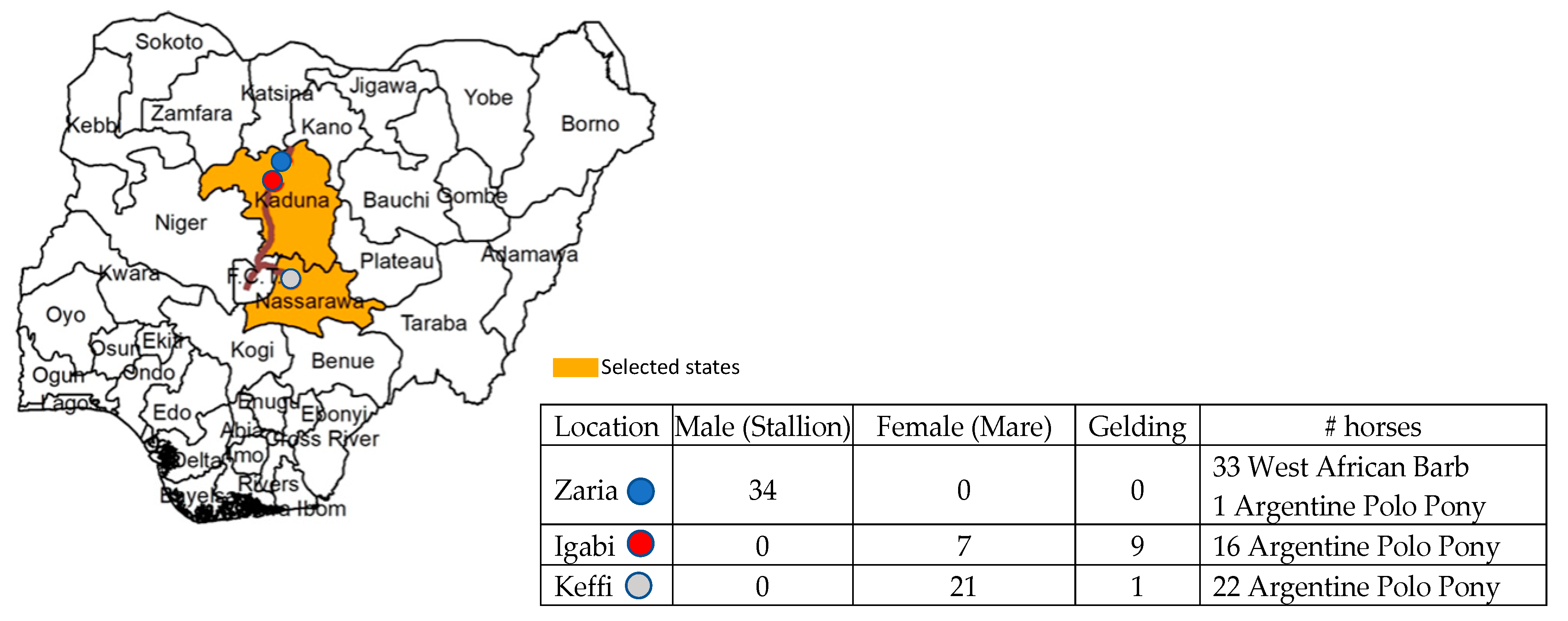
Figure 1. Site of sampling in Nigeria and category of horses utilized in this study.
Pathogens 10 00508 g001
Table 1. Summary of hematology, molecular, and serological data of horse samples collected in Nigeria.
Piroplasm
T. equiB. caballiT. haneyi
HematologyHorses testednPCRcELISAnPCRcELISAnPCRcELISA
Abnormal411537101NA
Normal31524002NA
Total722061103NA
RBC: red blood cells (normal range: 6.6–106/µL); PCV: packed cell volume (normal range: 31%–50%); Hb: hemoglobin (normal range: 11.4–17.3 g/dL) [24,25]; Abnormal: samples below normal range for at least one parameter; nPCR: nested PCR; cELISA: competitive ELISA; NA: not commercially available.
Table 2. Comparison of molecular and serologic assays to detect T. equi and B. caballi in horses.
Piroplasm
nPCR/cELISAT. equiB. caballi
+/−21
−/+430
+/+180
Total631
nPCR: nested PCR; cELISA: competitive ELISA.
Table 3. Distribution of various tick species infesting Nigerian horses.
Tick SpeciesMaleFemalesTotal
Rhipicephalus evertsi evertsi164 (50.8%)159 (49.2%)323 (69.6%)
Rhipicephalus sanguineus4 (40.0%)6 (60.0%)10 (2.2%)
Amblyomma variegatum37 (55.2%)30 (44.8%)67 (14.4%)
Hyalomma dromedarii22 (68.8%)10 (31.2%)32 (6.9%)
Hyalomma impeltatum04 (100%)4 (0.9%)
Hyalomma truncatum01 (100%)1 (0.2%)
Boophilus decoloratus07 (100%)7 (1.5%)
Total227 (51.1%)217 (48.9%)444 (100%)
Table 4. Species-specific nested polymerase chain reaction.
PrimerSequence (5’-3’)Expected Product Size Target Gene
T. equi external-forward GAGGAGGAGAAACCCAAG549 bpema1
T. equi external-reverseGCCATCGCCCTTGTAGAG
T. equi internal-forward TCAAGGACAACAAGCCATAC229 bp
T. equi internal-reverseTTGCCTGGAGCCTTGAAG
B. caballi external-forwardGATTACTTGTCGGCTGTGTCT374 bprap1
B. caballi external-reverse CGCAAAGTTCTCAATGTCAG
B. caballi internal-forwardGCTAAGTACCAACCGCTGA221 bp
B. caballi internal-reverse CGCAAAGTTCTCAATGTCAG
T. haneyi external-forward CCATACAACCCACTAGAG381 bphypothetical single copy gene
T. haneyi external-reverseCTGTCATTTGGGTTTGATAG
T. haneyi internal-forward GACAACAGAGAGGTGATT238 bp
T. haneyi internal-reverse CGTTGAATGTAATGGGAAC
Publisher’s Note: MDPI stays neutral with regard to jurisdictional claims in published maps and institutional affiliations.