Article

Regulation of Expression of the TIR-Containing Protein C Gene of the Uropathogenic Escherichia coli Strain CFT073

by
Julia Ittensohn
1,2,†,
Jacqueline Hemberger
1,2,†,
Hannah Griffiths
1,2,
Maren Keller
1,2,
Simone Albrecht
1,2 and
Thomas Miethke
1,2,*
1
Medical Faculty of Mannheim, Institute of Medical Microbiology and Hygiene, University of Heidelberg, Theodor-Kutzer-Ufer 1-3, 68167 Mannheim, Germany
2
Medical Faculty of Mannheim, Mannheim Institute for Innate Immunoscience (MI3), University of Heidelberg, Ludolf-Krehl-Str. 13-17, 68167 Mannheim, Germany
*
Author to whom correspondence should be addressed.
These authors contributed equally.
Pathogens 2021, 10(5), 549; https://doi.org/10.3390/pathogens10050549
Submission received: 27 November 2020 / Revised: 23 April 2021 / Accepted: 27 April 2021 / Published: 1 May 2021

Abstract

:
The uropathogenic Escherichia coli strain CFT073 causes kidney abscesses in mice Toll/interleukin-1 receptor domain-containing protein C (TcpC) dependently and the corresponding gene is present in around 40% of E. coli isolates of pyelonephritis patients. It impairs the Toll-like receptor (TLR) signaling chain and the NACHT leucin-rich repeat PYD protein 3 inflammasome (NLRP3) by binding to TLR4 and myeloid differentiation factor 88 as well as to NLRP3 and caspase-1, respectively. Overexpression of the tcpC gene stopped replication of CFT073. Overexpression of several tcpC-truncation constructs revealed a transmembrane region, while its TIR domain induced filamentous bacteria. Based on these observations, we hypothesized that tcpC expression is presumably tightly controlled. We tested two putative promoters designated P1 and P2 located at 5′ of the gene c2397 and 5′ of the tcpC gene (c2398), respectively, which may form an operon. High pH and increasing glucose concentrations stimulated a P2 reporter construct that was considerably stronger than a P1 reporter construct, while increasing FeSO4 concentrations suppressed their activity. Human urine activated P2, demonstrating that tcpC might be induced in the urinary tract of infected patients. We conclude that P2, consisting of a 240 bp region 5′ of the tcpC gene, represents the major regulator of tcpC expression.

Graphical Abstract

1. Introduction

Urinary tract infections are frequent, often recurrent and most often caused by uropathogenic Escherichia coli (UPEC). Expanding antibiotic resistance hampers successful treatment of urinary tract infections with UPECs. Therefore, exploration of the host-pathogen interaction is crucial for the elucidation of new treatment mechanisms and approaches. We detected a new class of virulence factors in the uropathogenic E. coli strain CFT073 and in Brucella spp., which impair Toll-like receptor (TLR) signaling [1]. By binding to myeloid differentiation factor 88 (MyD88) and TLR4 they impair the secretion of pro-inflammatory cytokines such as TNF-α and IL-6 [1,2]. Other groups detected structurally similar virulence factors with almost identical mechanisms in human pathogens such as Salmonella enterica subsp. enterica Serovars Enteritidis, Dublin, Gallinarum, Yersinia spp., Brucella spp., Staphylococous aureus MSSA476, Enterococcus faecalis, but also in non-pathogens such as Paracoccus denitrificans [3,4,5,6,7,8,9,10,11]. At least some of these TLR-inhibiting virulence factors significantly increase disease severity in different murine infection models [1,3,4,6,12].
We also described that the Toll/interleukin-1-receptor domain (TIR)-containing protein C (TcpC) from CFT073 impairs the NACHT leucin-rich repeat PYD protein 3 (NLRP3) inflammasome by binding to NLRP3 and caspase-1 [13]. This event prevents the prion-like condensation of apoptosis-associated speck-like protein (ASC), but also of caspase-1 to form intracellular specks, the hallmark of NLRP3 inflammasome activation. Consequently, the maturation and secretion of IL-1β is prevented [13].
Recently, TcpC from E. coli Nissle was reported to strengthen epithelial barrier function by stimulating PKCζ and ERK1/2 signaling in HT-29/B6 cells demonstrating another influence of TcpC on host cells [14]. Moreover, outer membrane vesicles and soluble factors, such as TcpC, released by E. coli Nissle as well as ECOR63 enhanced barrier function in intestinal epithelial cells [15].
The tcpC gene is only found in the phylogenetic group B2 of extra-intestinal E. coli strains (ExPECs) and is chromosomally located in the serU island, which co-segregates with the high pathogenicity island (HPI) of ExPECs [16]. The latter finding indicated that the serU-island and the HPI were transferred en bloc between ExPECs [16]. Around 40% of UPECs isolated from patients suffering from pyelonephritis carry the tcpC gene, while this gene is only present in 21% of UPECs responsible for cystitis, in 16% during asymptomatic bacteriuria and in only 8% of commensal E. coli strains in stool, respectively [1]. Thus, the frequency of tcpC+ UPECs correlates with disease severity in humans [1,16].
Structurally, TcpC consists of a Toll/Interleukin-1 receptor (TIR) domain, which is located in the C-terminal half of the molecule (bp 508–924) and interacts with the TLR-signaling cascade and the NLRP3 inflammasome [1,13]. We crystallized the homologous domain of TcpB from Brucella spp., which shares a 55% amino acid sequence identity to the TIR domain of TcpC, and demonstrated that the tertiary structure of the bacterial TIR domain has an arrangement of five central β-sheets and five surrounding α-helices which characterizes eukaryotic TIR domains [17]. TcpC but not TcpB is predicted to also contain a N-terminal transmembrane domain [1].
Although we published earlier that co-culture of RAW264.7 cells with CFT073 induced the production of TcpC, its mechanism of induction is still unknown and a detailed analysis is missing [1]. This is an important issue, since on the one hand the secreted TcpC could act in infected humans as an immunosuppressant during urosepsis with UPECs such as CFT073. Indeed, systemic application of TcpC was used to successfully treat mice suffering from autoimmune arthritis, which indicates the immunosuppressive potential of the molecule [18]. On the other hand, the potential consequences of TcpC expression on CFT073 itself are unknown as well. We thus determined the effect of TcpC overexpression on the multiplication of CFT073. Moreover, we explored the localization of different truncated constructs of TcpC in CFT073 and their influence on the morphology of the bacterium upon overexpression.
Based on the arrangement of genes surrounding the tcpC gene on the chromosome and their direction of gene transcription, the tcpC gene may form an operon with the 5′-located gene c2397 within the serU island, which encodes a hypothetical protein. Thus, promoter regions, which control transcription of tcpC, may be located at 5′ of the gene c2397 and/or c2398. We explored their functional relevance in the present study.

2. Results

2.1. Overexpression of TcpC Stops Growth and Provokes Filamentation of the Uropathogenic E. coli Strain CFT073

To study the effects of TcpC expression on the UPEC strain CFT073, we transformed the bacterium with two different non-leaky plasmids, which allowed the anhydrotetracycline (ATc)-inducible expression of TcpC fused either to an enhanced yellow fluorescent protein (eYFP) or to a Strep-tag. ATc did not influence the growth of untransformed CFT073 significantly (Figure 1A). However, ATc induction of two empty vector controls demonstrated that the induction of the plasmids at the beginning of the culture period impaired the replication of CFT073 (Figure 1B,D). We did not observe growth inhibition when both plasmids were induced at an OD600 of 0.5 (Figure 1B,D). Growth of CFT073 was impaired when the tcpC gene was induced at the beginning of the culture and it stopped when the gene was induced after the culture reached an OD600 density of 0.5 (Figure 1C). In the latter case, growth of CFT073 significantly differed from the growth observed upon induction of the control plasmid pASK-IBA3 (Figure 1B,C). We were worried that the addition of eYFP might yield a toxic protein being responsible for the growth retardation (Figure 1C), however, the addition of the much smaller Strep-tag to TcpC again prevented growth of the bacterium, indicating that TcpC itself but not eYFP is responsible for growth retardation (Figure 1E). As discussed above, growth of CFT073 upon induction of the plasmid pStrep-tag tcpC or the corresponding control plasmid differed significantly if they were induced at an OD600 of 0.5 (Figure 1D,E). Since TcpC was reported to cause NAD+ loss in E. coli, which in turn impairs its growth, we generated an E244A-mutant of TcpC, which lacks the ability to cleave NAD+ [19]. However, E244A-mutated TcpC impaired the growth of CFT073 as efficiently as wild-type TcpC (Figure 1F).
We subsequently explored whether morphologic changes imposed on the bacterium accompany the negative influence of TcpC on bacterial growth and if so, which part of the molecule might be responsible for these changes. We constructed a series of truncated constructs of tcpC fused to eYFP. At four hours post induction of the different constructs major morphologic changes were imposed on CFT073. Thus, all plasmids containing the TIR domain (bp 508–924), i.e., p(tcpC 508–924), p(tcpC 127–924) and p(tcpC 1–924), caused strong filamentation of the bacteria (Figure 2, Supplementary Materials Figure S1). We observed this to a much lower degree or not at all with plasmids p(tcpC 1–150), p(tcpC 127–507) and p(tcpC 1–507), expressing different N-terminal parts of TcpC. We also realized that the different constructs varied in their cellular distribution. Microscopy of the CFT073 transformants with higher resolution demonstrated that TcpC 1–150 was mainly located at the cell wall. This finding confirmed the predicted transmembrane domain in this part of the molecule (Figure 3A,B). The culture of CFT073 p(tcpC 1–150):eYFP:AMP in LB medium apparently increased the cell wall localization of TcpC 1–150 (Figure 3B). It appeared that TcpC 127–507 was mainly localized at the cell poles (Figure 3C) while TcpC 1–507 and TcpC 508–924 were homogeneously expressed (Figure 3D,E). Finally, TcpC 127–924 and full-length TcpC 1–924 again showed the polarized expression pattern observed with TcpC 127–507 (Figure 3F,G). Filamented bacteria could no longer be observed 24 h post induction (Figures S2 and S3). The principal expression pattern of the TcpC constructs within CFT073 observed at four hours post induction remained at 24 h post induction (Figure 4) with the exception of TcpC 127–507, which lost its pure polarized expression pattern, and TcpC 508–924, which now was expressed in specks (Figure 4D). In summary, overexpression of TcpC caused growth retardation and presumably impairment of cell division as indicated by filamented bacteria.

2.2. Endogenous Promoters Controlling the tcpC Gene

Based on our observation that TcpC exerts fundamental changes on CFT073, we concluded that the bacterium tightly controls the expression of TcpC. We, therefore, explored endogenous promoters putatively involved in the regulation of tcpC expression. We assumed that an untranslated region 5′ of the gene c2397, encoding for a hypothetical protein, might function as the promoter since this gene may form an operon with the tcpC gene (c2398) (Figure 5A). In addition, we also tested an untranslated region 5′ of the tcpC gene (c2398) as a putative promoter. We thus cloned a 645 bp DNA fragment starting at position 2200564 and ending at position 2201209 and a 240 bp element starting at position 2202535 and ending at position 2202775 of the chromosome of CFT073, respectively. We designated the 645 bp element as promoter 1 (P1) and the 240 bp element as promoter 2 (P2) (Figure 5A). We fused both putative promoter regions with the green fluorescent protein mut2 (gfpmut2) to generate plasmid-based reporter constructs. As shown in Figure 5B–D, we transformed CFT073 with three different plasmids containing either P1 or P2 or P1 plus P2 in each case fused to gfpmut2. In addition, we also replaced tcpC by gfpmut2 on the chromosome of CFT073 generating the tcpC-deficient strain CFT073 tcpC::gfpmut2 (Figure 5E). We cultured the four different CFT073 strains in glucose-containing M9-minimal medium and analyzed the expression of the reporter constructs and the size of the bacteria by flow cytometry. The activity of P1 was hardly detectable in comparison to CFT073 lacking a reporter construct (Figure 5B,F). In contrast, P2 was active and the presence of P1 did not diminish the activity of P2 (Figure 5C,D). The chromosomal reporter construct showed a clearly detectable activity although weaker than P2 as expected (Figure 5E). Glucose-containing M9-minimal medium did not influence the forward scatter behavior of the bacteria (Figure 5), indicating that the filamentation seen with induced expression results from overexpression of the TIR domain of TcpC.
To provide further evidence for the existence of the P1 and P2 promoters, we analyzed RNA transcripts encompassing either c2397 and c2398 or 2398 alone. We used CFT073 or CFT073 transformed with the plasmid pTcpC containing a DNA fragment starting at 535 bp 5′ of the start codon of c2397 and ending at the stop codon of c2398. We used the latter strain to increase the sensitivity of the reverse transcription PCR. The strains were cultured in glucose-containing M9-minimal medium, pH8, which stimulates promoter activity (see below). While the long transcript was not detectable, we could demonstrate the transcript of the c2398 gene in case of the plasmid strain CFT073 pTcpC (Figure 6A). We used a new set of primers to amplify a shorter fragment of c2398 RNA post reverse transcription and could demonstrate a c2398 transcript in CFT073 (Figure 6B). We then performed a 5′RACE PCR to determine the transcription start of the 2398 transcript. We detected a terminal deoxynucleotidyl transferase (TdT)-dependent transcript (Figure 6C, compare lanes 5 and 6), which we could re-amplify with a second forward but the same reverse primer (Figure 6D). We sequenced this PCR product and found that transcription of c2398 starts 44 bp 5′ of a possible start codon of the c2398 RNA (Figure 6E). Interestingly, the c2398 RNA demonstrated in Figure 6A was obtained with the forward primer c2398fw binding 5′ of the transcription start demonstrated in Figure 6E with 16 of 22 bases. This may be compatible with the existence of a second promoter 5′ of the c2397 gene, although we could not demonstrate a long transcript presumably due to sensitivity issues.
We then explored which factors might induce tcpC promoters and analyzed the four different CFT073 strains in glucose- or tryptone-containing M9-minimal medium at different pH, since pH values may vary from 5.2 to 7.4 in the urinary tract. The experiment revealed that high pH strongly induced P2 but only weakly P1 if the strains were incubated in glucose- but not in tryptone-containing M9-minimal media (Figure 7A,B, Figure S4). P1 and P2 combined induced the reporter as strongly as P2 alone, demonstrating that P1 did not attenuate P2 (Figure 7C). High pH also induced the expression of gfpmut2 in CFT073 tcpC::gfpmut2, again if cultured in glucose-containing M9-minimal medium (Figure 7D, Figure S4).

2.3. Glucose Induces the tcpC Promoter

Given the fact that increasing pH induced P1 and P2 only in the glucose-containing medium, we wondered whether glucose itself would induce tcpC promoters. Since P2 was more sensitive to pH changes, we tested this promoter first for its sensitivity against a range of glucose concentrations. A glucose concentration of 3 mmol/L induced P2 significantly and higher glucose doses increased its activity further in a dose-dependent manner (Figure 8A). Again, P1 reacted much weaker (Figure 8B) and the activity of P1 plus P2 was as strong as P2 alone (Figure 8C). The chromosomal reporter strain CFT073 tcpC::gfpmut2 also increased the expression of its reporter glucose dependently and significantly (Figure 8D).

2.4. FeSO4 Dampens tcpC Promoter Activity

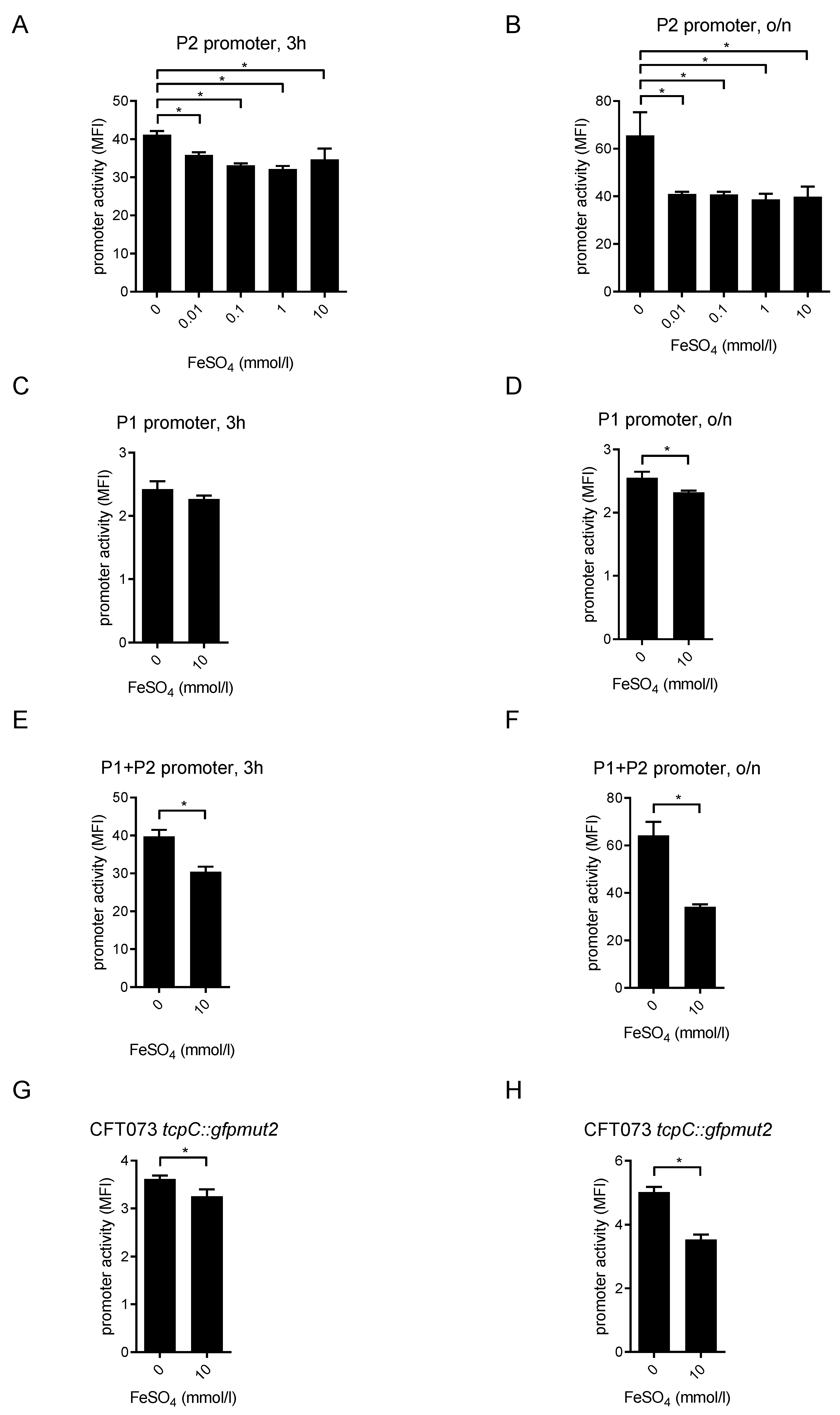
Iron is essential for many bacterial pathogens, including UPECs, to maintain their own growth. To secure iron supply, UPECs including CFT073 possess siderophores, which bind Fe3+ and the generated complexes are subsequently transferred across the bacterial cell wall [20]. Since iron levels in urine are low [21], we wondered whether reduced iron levels would facilitate the expression of the tcpC gene. The expression of virulence factors such as TcpC might be an advantageous adaptation under these growth-limiting conditions. Incubation of the P2-reporter CFT073 strain with increasing FeSO4 concentrations demonstrated that the activity of P2 was reduced already 3 h post addition (Figure 9A). This effect was even more pronounced after overnight incubation with FeSO4 (Figure 9B). The activity of P1 was only weakly reduced by FeSO4, and the differences were only significant after an overnight incubation with FeSO4 (Figure 9C,D). In contrast, the combined activity of P1 and P2 was already reduced after 3 h incubation with FeSO4 and this was more pronounced after incubation overnight (Figure 9E,F). FeSO4 also reduced the activity of the chromosomal reporter strain CFT073 tcpC::gfpmut2 significantly after incubation of 3 h and overnight (Figure 9G,H). To further analyze the influence of FeSO4 on the P2 promoter, we used the iron chelator 2,2’-bipyridine, which is also known to deplete intracellular iron from E. coli [22]. We found that incubation of the P2-reporter strain with the iron chelator significantly increased the activity of the P2 promoter (Figure 10). Co-administration of FeSO4 neutralized this effect (Figure 10). Taken together, FeSO4 clearly dampened the activity of the tcpC promoter and influenced plasmid or chromosomal reporter constructs similarly. Since increasing pH and glucose concentrations stimulated while FeSO4 impaired plasmid and chromosomal reporter constructs in a similar manner, we conclude that the regulation of the tcpC gene on the chromosome of CFT073 follows the rules observed with the plasmid reporter constructs.

2.5. Human Urine Induces the tcpC Promoter

The bacterial burden of CFT073 in the urine and kidneys of infected mice was by several orders of magnitude higher compared to its tcpC-deficient mutant CFT073 tcpC::KAN, indicating that the tcpC promoter is active in urine during an infection [1,23]. To verify this assumption, we incubated our collection of reporter strains in urine of healthy human donors. Only pure urine induced the promoter P2 after an incubation period of 4 h and 24 h (Figure 11A,B). P1 was almost not induced while the combination P1 and P2 was activated as strongly as P2 alone (Figure 11C). Urine induced the chromosomal reporter strain CFT073 tcpC::gfpmut2 to some extent, however the difference to the negative control strain CFT073 pAMP was not significant (Figure 11D). Due to the better signal to noise ratio of the plasmid reporter constructs and thus their higher sensitivity, we think that the latter constructs clearly demonstrate that P2 senses urine and induces the expression of the tcpC gene in urine.

3. Discussion

Our results demonstrate that overexpression of TcpC influences the UPEC strain CFT073 fundamentally in that growth is impaired and filamentation is induced. Thus, aside from TcpC’s influence on the innate immune system, the protein also provokes drastic changes on CFT073. Therefore, we explored the regulation of the tcpC gene. We found that a 240 bp region 5′ of the tcpC gene (c2398) served as the crucial promoter element, which we designated P2, while a second, functionally minor regulatory region, designated P1, is present on a 645 bp region 5′ of the gene c2397. We could map the site of transcription start of the c2398 mRNA to position 2202778 of the CFT073 genome, or 44 bp 5′ of a possible start codon. P2, and to a lower extent, P1, were induced by high pH, glucose concentrations and urine, while increasing concentrations of FeSO4 lowered promoter activity.
We also used the promoter prediction tool iPro70-FMWin, which predicts σ70 promoters with comparably high accuracy [24]. We focused on σ70 promoters since these are the most common promoters in E. coli [25]. IPro70-FMWin predicts that P1 and P2 contain a σ70 promoter with a probability score of 0.98 and 0.52, respectively. Both putative σ70 promoters are at a distance of around 480 and 150 bp to the translation start codon of c2397 and c2398, respectively. Most 5′ untranslated regions (UTR) in E. coli vary between 20 to 40 nucleotides in length, while some have longer 5′ UTRs between 100 and 290 nucleotides [25]. Thus, the length of 5′ UTR of the predicted σ70 promoter in P1 is unusual and our own results demonstrate a 5′UTR of only 44 bp in case of the P2 promoter.
The TIR domains of bacterial TIR containing proteins including TcpC and TcpB were recently shown to act as NAD+ hydrolases [19,26]. Overexpression of the TIR domain of TcpC reduced the intracellular bacterial concentration of NAD+ [19]. Since NAD+-dependent DNA ligases are present in all bacteria and are essential for growth, overexpression of TcpC may stop replication of CFT073 by a lack of NAD+-dependent DNA ligases [27]. However, our findings using mutant E244A TcpC do not support this conclusion since the mutant impaired the growth of CFT073-like wild-type TcpC.
So far, only overexpression of the TIR domain of TcpC induced bacterial filamentation, while tcpC promoter induction with glucose or high pH did not influence bacterial morphology. Filamentation of UPECs such as the UTI89 strain was described as a bacterial defense mechanism during urinary tract infection in mice and is presumably relevant for the entry and exit of bladder epithelial cells [28,29]. Interestingly, filamentation was not induced in mice lacking TLR4, indicating that innate immune responses trigger this morphologic change of the invading bacterium [28,30]. Functionally, filamented bacteria evaded phagocytosis by polymorphonuclear neutrophils [30]. Filamentation of UPECs was also observed in vitro upon infection of human bladder epithelial cells and in the urine of females with urinary tract infections, indicating that this bacterial escape mechanism is also operative in infected humans [31,32]. In the UTI89 strain the bacterial cell wall inhibitor SulA is active during filamentation and a sulA-deficient UTI89 mutant was attenuated to sustain a bladder infection in wild-type mice [28]. It remains to be seen whether other conditions such as an infection of host cells increases tcpC promoter activity and induces filamentation. Obviously, there are tcpC-independent pathways to induce filamentation since UTI89 does not contain a tcpC homolog; nevertheless, it is capable of adopting this morphologic change [30,33].
P2 was clearly more relevant than P1 for the induction of tcpC expression at all conditions tested. Thus, P2 was more sensitive to pH changes, increasing glucose or decreasing FeSO4 concentrations. It was also more active than P1 in human urine. Although P1 was only weakly induced, the changes were nevertheless significant. P1 never impaired the activity of P2 but instead the combination of both promoters was in almost all cases, except in the presence of urine, as active as the individual ones. Unfortunately, we could not demonstrate a polycistronic transcript encoding the mRNA of c2397 and c2398, but we also cannot rule out this possibility. In any case, we could not verify the relevance of P1 for the transcription of the tcpC (c2398) gene and we think that the mapped P2 promoter region is mainly responsible for tcpC transcription.
The fact that P2 was active in human urine is important since it demonstrates that the virulence factor TcpC may be expressed during a urinary tract infection of humans. This assumption is supported by the observation that the frequency of UPECs harboring the tcpC gene was particularly high in patients with pyelonephritis [1,16]. Thus, tcpC expression might be relevant in particular during an upper urinary tract infection. Moreover, CFT073 in comparison to its tcpC-deficient mutant multiplied to a significantly higher bacterial burden in murine urine and kidneys and only the CFT073 wild-type strain damaged kidneys during an urinary tract infection of mice [1]. However, other groups failed to detect expression of tcpC in E. coli strains isolated from the urinary tract of patients. Accordingly, analysis of eight different E. coli isolates by CFT073-specific microarrays and another five E. coli isolates by RNA-seq from patients with bacteriuria or uncomplicated urinary tract infection, respectively, revealed the induction of iron acquisition and peptide transport systems but not of tcpC [34,35]. However, in patients with cystitis the frequency of isolates harboring the tcpC gene is only around 21% [1] and due to the limited number of isolates analyzed the collection did not include a tcpC+ isolate.
Our results also indicate which urine properties might influence the expression of TcpC. On the one hand, urine contains only low amounts of iron and therefore the negative influence exerted by FeSO4 is not present in the urinary tract [21]. On the other hand, the pH of urine is around six, which supports the expression of TcpC. The pH of the kidneys equals seven and therefore expression of TcpC may be further facilitated. However, glucose levels of urine are below 1 mmol/l and our results demonstrate that high pH values only induce the expression of tcpC in the presence of glucose. Therefore, we assume that additional urine components influence the expression of tcpC gene during an infection.
UPEC strains like CFT073 are not only known to cause pyelonephritis—and the strain was originally isolated from a patient suffering from this infection—[36] but also urosepsis, the most devastating complication of a urinary tract infection. Blood, in contrast to urine, contains glucose levels required for tcpC expression and a pH of seven, which is also compatible with tcpC expression. Thus, we speculate that TcpC is also produced during urosepsis.
We published earlier that TcpC may contain a transmembrane domain in its N-terminal as predicted in silico [1]. We now confirm the prediction, as the truncated construct tcpC (1–150) was expressed at the bacterial cell wall. We also reported earlier that TcpC was secreted and this process was inhibited by the efflux pump inhibitor phenylalanine-arginine-β-naphtylamide (PAβN) [1]. Possibly, this N-terminal part of TcpC is crucial for its secretion. It is at present unclear why this expression pattern is not seen with the full-length construct. However, since TcpC is rapidly cleaved at least after transfection in eukaryotic cells and since the eYFP label is added at the C-terminus of TcpC, we may fail to observe this because the fluorescent label may be cleaved off inside CFT073 [13].
In summary, we demonstrate here that overexpression of tcpC impaired growth of the UPEC strain CFT073 and provoked its filamentation. We defined the location of the tcpC promoter 5′ of tcpC and c2397 and we explored its regulation: pH, glucose and urine switched the promoter on while FeSO4 switched it off.

4. Materials and Methods

4.1. Bacterial Strains

The UPEC strain CFT073 was purchased from ATCC (Manassas, VA, USA) [36]. The tcpC-deficient reporter strain CFT073 tcpC::gfpmut2 was constructed using the λ-red system [37]. Briefly, we amplified the 5′UTR of tcpC fused to gfpmut2 from pPc2398:gfpmut2:KAN containing this region and using primers SN10 (fw, 5′-TACTATCTCGAGGCAGGAGTCTATGGTAACG-3′) and SN19 (rev, 5′-GAAGCAGCTCCAGCCTACACTTATTTGTACAATTCATCC-3′). The latter contains the P1 site of pKD13, encoding the kanamycin resistance gene. The downstream region of tcpC was amplified from the CFT073 genome with primers SN22 (fw, 5′-ATAGTAGAGACCGGAAGACACGGATTCCATG-3′) having an overhang with the P4 site of pKD13 and SN23 (rev, 5′-TGCCCATTAACATCACCATC-3′). Subsequently, the kanamycin cassette was amplified from pKD13 using the above fragments and outer primers SN10 and SN23. The obtained donor DNA was transformed into recipient strain CFT073 expressing the λ-red system from helper plasmid pKD46 by electroporation. Expression of lambda red recombinase was induced by treatment with 100 mM arabinose at 30 °C. Transformants were recovered in S.O.C medium (Invitrogen Corp., Paisley, UK). After selection on kanamycin (50 mg/mL), the resistance cassette was flipped out using the curable temperature-sensitive plasmid pCP20 encoding FLP recombinase.

4.2. Plasmids

All plasmids generated for this study are listed in Table 1. We generated the plasmid pTcpC, which contains a DNA fragment starting 535 bp 5′ of the start codon of c2397 and ending at the stop codon of c2398. We cloned full-length tcpC into the non-leaky bacterial expression vector pASK-IBA5plus (IBA GmbH, Göttingen) containing a N-terminal Strep-tag and tcpC:eYFP into pASK-IBA3plus (IBA GmbH, Göttingen) containing a C-terminal Strep-tag. We also cloned full-length E244A-mutated tcpC into pASK-IBA5plus. We constructed three different gfpmut2-reporter constructs to define the endogenous tcpC promoter, five different tcpC-truncated constructs and one full-length tcpC construct, which were IPTG-inducible and transformed into CFT073. Sequences of primers used are listed in Table 2, the primer combinations to amplify P1, P2, P1 plus P2, full-length tcpC and truncated constructs of tcpC in Table 3. Restriction enzymes used to clone amplicons into the appropriate plasmid are also described in Table 2 and Table 3. In total, 100 μL of electro-competent CFT073 and 0.1–0.3 μL of a plasmid-miniprep were mixed and the plasmids electroporated (5 ms, 1700 V, Multiporator, Eppendorf, Germany) into CFT073. Subsequently, 100 μL LB medium was added, and the bacterial suspension was cultured on LB agar plates supplemented with kanamycin or ampicillin. We picked single colonies after overnight incubation. The correct composition of all plasmids was verified by sequencing (GATC, Konstanz, Germany).

4.3. Reagents and Culture Media

Isopropyl-β-D-thiogalactopyranosid (IPTG) came from Applichem GmbH (Darmstadt, Germany), anhydrotetracycline (ATc) from Toku-e (Gentaur GmbH, Aachen, Germany), Luria-Bertani broth (LB) and LB agar plates were purchased from Roth (Karlsruhe, Germany). Components for the M9-minimal medium (di-sodium-hydrogenphosphate (33.9 g/L), potassium-dihydrogenphosphate (15 g/L), sodium chloride (2.5 g/L), ammonium-chloride (5 g/L)) came from Merck (Darmstadt, Germany). This medium was either supplemented with glucose (0.4%), thiamin (10 µg/mL, Roth) and nicotic acid (0.0025%, Roth) or tryptone (0.2%, BD Chemicals, Greenwood Village, CO, USA). 2,2′-bipyridine, 5 g, was bought from Roth. Human urine was donated by healthy individuals. The urine was filtered (0.2 µm pore size) and used on the day of donation.

4.4. Culture of Bacteria

The tcpC-deficient reporter strain CFT073 tcpC::gfpmut2 and plasmid-transformed CFT073 strains were cultured overnight in M9-minimal medium containing glucose or in case of the tcpC promoter stimulation with urine in LB medium in the presence of ampicillin (100 μg/mL) or kanamycin (25 μg/mL); wild-type CFT073 was cultured in the absence of antibiotics. After three wash steps, bacterial densities were adjusted to an OD600 of 0.5, diluted and further cultured in LB or M9-minimal medium supplemented with either glucose, thiamin and nicotic acid or tryptone as indicated in the figure legends.

4.5. Growth Determination

Bacterial growth was determined by measuring the optical density using an OD600 DiluPhotometer (IMPLEN, Munich, Germany).

4.6. Reporter Assays

CFT073 strains transformed with the different truncated or full-length tcpC reporter plasmids were induced with IPTG (20 μmol/L) and incubated for 4 or 24 h. At each time point, bacteria were fixed with PFA/PBS (1%, 30 min, room temperature). After several wash steps with PBS, bacterial pellets were immobilized on agarose pads (1%) and analyzed with a Leica Leitz DMR fluorescence microscope (Leica, Wetzlar, Germany) equipped with an Orca flash 4.0 LT camera (Hamamatsu Photonics Deutschland GmbH, Herrsching am Ammersee, Germany) using an excitation wavelength of 524 nm.
We used flow cytometry (FACScan, BD Biosciences, Germany) to explore the fluorescence activity of CFT073 strains transformed with the three different tcpC-promoter reporter plasmids and the tcpC-deficient CFT073 tcpC::gfpmut2 strain. We analyzed the data with FlowJo (Ashland, OR, USA).

4.7. Reverse Transcription PCR and 5′ RACE PCR

To detect a polycistronic mRNA of the hypothetical gene c2397 and c2398 (tcpC) or just the mRNA of c2398, RNA was extracted from CFT073 and CFT073 p(TcpC). Bacterial cultures of CFT073 and CFT073 + p(TcpC) were grown overnight in glucose-M9-minimal medium pH8 to induce the promoter. The medium for CFT073 p(TcpC) contained chloramphenicol (34 μg/mL). At mid-logarithmic phase, around 2.5 mL of the culture (corresponding to approximately 1.5 × 10⁹ bacteria) were harvested, washed three times with Dulbecco’s Phosphate-Buffered Solution (DPBS) and resuspended in 100 µL DPBS plus 250 µL DNA/RNA Shield (Zymo Research, Freiburg, Germany). After incubating the samples for five minutes and centrifugation (10 min, 5000 g), the supernatant was removed and 200 µL TE-buffer (10 mM Tris·Cl, 1 mM EDTA, pH8) containing 1 mg/mL lysozyme was added. The samples were vortexed and incubated for five minutes under shaking and then mixed with 700 μL RLT-buffer (RNeasy Mini Kit (Qiagen, Hilden, Germany)) plus 7 µL β-mercaptoethanol. After vortexing and centrifuging (2 min, 16000 g) the samples, the supernatant was mixed with 500 µL ethanol (100%). The RNA was then extracted with the RNeasy Mini Kit (Qiagen). During the procedure, two on-column DNase digests were conducted (RNase-Free DNase Set (Qiagen)) and the purified RNA was eluted in 50 µL RNAse-free H2O. Then, another DNase digest was performed in solution according to the kit manual to get rid of residual DNA traces. The DNase was removed with a second column purification (RNeasy Mini Kit) and the pure RNA was eluted in 50 µL RNAse free H2O. RNA quality was examined with gel electrophoresis of 10 µL sample on a 1% agarose-TAE agarose gel and ethidium bromide staining (60 min, 7 V/cm).
We also took 20 µL aliquots of the overnight cultures, incubated them at 95 °C for 10 min and stored them at −20 °C as PCR positive controls.
The RNA was transcribed to cDNA with the RevertAid First Strand cDNA Synthesis Kit (Thermo Scientific, Karlsruhe, Germany) and the Primer c2398rev (Table 2, Figure 12) that binds at the end of c2398. For each of the two samples, two reactions with and without reverse transcriptase (+/−RT) were set up to test for DNA contamination in the PCR. GAPDH-RNA served as a positive control.
In total, 2 µL of the +/− RT-cDNA and boiled bacteria samples (positive control) were then amplified using two different primer sets spanning either the entire operon (c2397fw—c2398rev, Table 2, Figure 12) or just c2398 (c2398fw—c2398rev, Table 2, Figure 12). Then 10 µL of PCR product was analyzed on an 1% agarose gel and ethidium bromide staining (60 min, 7 V/cm).
The 5′ RACE PCR was performed using the kit “5′ RACE System for Rapid Amplification of cDNA Ends, Version 2.0” (Invitrogen, ThermoFisher, Waltham, MA, USA). In brief, the extracted RNA was reverse transcribed into cDNA with SuperScript II Reverse Transcriptase and the primer GSP1 (Table 2, Figure 12), binding 336 bp downstream of the putative tcpC start codon. The RNA was degraded with RNase Mix and the cDNA purified with a S.N.A.P column to remove dNTPs, GSP1 and proteins. In the next step, a poly-C-tail was added to the 5′-end of the cDNA with the enzyme terminal deoxynucleotidyl transferase (TdT). A negative control without TdT was included. The tailed product was then amplified in a PCR using the poly-G and deoxyinosine-containing primer AAP (Table 2) that binds to the poly-C tail and the nested primer GSP2 (Table 2, Figure 12) that binds the cDNA strand. Additionally, a second positive control PCR was performed using GSP3 (Table 3, Figure 12) that binds 34 bp downstream of the putative start codon and GSP2 (Figure 12). Aliquots of the PCR products were analyzed on an agarose gel and the rest of the RACE-PCR sample was purified with the QIAquick PCR Purification Kit (Qiagen). To generate enough specific product, the product was re-amplified in a second PCR with the AUAP primer (Table 2) which was homologous to the 5′-part of the AAP-primer and GSP2 (Figure 12). We purified the PCR product via gel electrophoresis and extracted the amplicon using the kit NucleoSpin Gel and PCR Clean-up (Machery-Nagel GmbH & Co. KG, Düren, Germany). The purified product was then analyzed by Sanger-sequencing (LGC Genomics GmbH, Germany) with the primer GSP2 (Figure 12).

4.8. Statistics

We used Prism 6.07 (GraphPad software, San Diego, CA, USA) to analyze experimental results for statistically significant differences. One-way ANOVA and Tukey’s test as post-hoc tests served to compare the experimental groups. We considered p values of less than 0.05 to be significant.

Supplementary Materials

The following are available online at https://www.mdpi.com/article/10.3390/pathogens10050549/s1, Figure S1. The TIR-domain of TcpC induces filamented bacteria. Microphotographs represent the corresponding light microscopy images of Figure 2. Figure S2. The filamentation of CFT07 induced by the TIR-domain of TcpC is no longer detectable 24 hours post induction.Bacteria were transformed as described in Figure 2 but analyzed 24 hours post induction with IPTG by fluorescence-microscopy. Note that CFT073 transformed with p(tcpC 508–924):eYFP:AMP (E) with p(tcpC 127–924):eYFP:AMP (F) and p(tcpC 1–924):eYFP:AMP (G), respectively, is no longer filamented. We repeated the experiment once with identical results. Figure S3. The filamentation of CFT07 induced by the TIR-domain of TcpC is no longer detectable 24 hours post induction. Microphotographs represent the corresponding light microscopy images of Figure S2. Figure S4. pH7 induces the promoter P1 and the chromosomal reporter construct significantly. We tested CFT073 transformed with the plasmids pPc2397:gfpmut2:KAN containing the promoter P1 (A), the chromosomal reporter strain CFT073 tcpC::gfpmut2 (B) and CFT073 (C). Bacteria were incubated overnight in M9-minimal medium containing glucose at a pH of 7. We determined expression of GFPmut2 by flow cytometry and determined the mean fluorescence intensity (MFI). The values indicated by the bars represent three independent experiments (D). * p < 0.05, ANOVA posthoc Tukey.

Author Contributions

J.I., J.H., H.G., M.K.: acquisition, analysis and interpretation of data; S.A., T.M.: conception and design of the study; T.M.: writing of the manuscript. All authors have read and agreed to the published version of the manuscript.

Funding

This work was supported by the German Research Foundation, grant reference number MI 471/9-1.

Institutional Review Board Statement

Not applicable.

Informed Consent Statement

Not applicable.

Data Availability Statement

Data available on request from the authors.

Acknowledgments

We thank Silke Neumann-Pfeifer for the generation of plasmids and expert support.

Conflicts of Interest

Authors have no conflict of interest to declare.

References

  1. Cirl, C.; Wieser, A.; Yadav, M.; Duerr, S.; Schubert, S.; Fischer, H.; Stappert, D.; Wantia, N.; Rodriguez, N.; Wagner, H.; et al. Subversion of Toll-like receptor signaling by a unique family of bacterial Toll/interleukin-1 receptor domain-containing proteins. Nat. Med. 2008, 14, 399–406. [Google Scholar] [CrossRef]
  2. Snyder, G.A.; Cirl, C.; Jiang, J.; Chen, K.; Waldhuber, A.; Smith, P.; Römmler, F.; Snyder, N.; Fresquez, T.; Durr, S.; et al. Molecular mechanisms for the subversion of MyD88 signaling by TcpC from virulent uropathogenic Escherichia coli. Proc. Natl. Acad. Sci. USA 2013, 110, 6985–6990. [Google Scholar] [CrossRef] [Green Version]
  3. Newman, R.M.; Salunkhe, P.; Godzik, A.; Reed, J.C. Identification and characterization of a novel bacterial virulence factor that shares homology with mammalian Toll/interleukin-1 receptor family proteins. Infect. Immun. 2006, 74, 594–601. [Google Scholar] [CrossRef] [Green Version]
  4. Norenberg, D.; Wieser, A.; Magistro, G.; Hoffmann, C.; Meyer, C.; Messerer, M.; Schubert, S. Molecular analysis of a novel Toll/interleukin-1 receptor (TIR)-domain containing virulence protein of Y. pseudotuberculosis among Far East scarlet-like fever serotype I strains. Int. J. Med. Microbiol. 2013, 303, 583–594. [Google Scholar] [CrossRef] [PubMed]
  5. Salcedo, S.P.; Marchesini, M.I.; Lelouard, H.; Fugier, E.; Jolly, G.; Balor, S.; Muller, A.; Lapaque, N.; Demaria, O.; Alexopoulou, L.; et al. Brucella control of dendritic cell maturation is dependent on the TIR-containing protein Btp1. PLoS Pathog. 2008, 4, e21. [Google Scholar] [CrossRef] [Green Version]
  6. Askarian, F.; van Sorge, N.M.; Sangvik, M.; Beasley, F.C.; Henriksen, J.R.; Sollid, J.U.; van Strijp, J.A.; Nizet, V.; Johannessen, M. A Staphylococcus aureus TIR domain protein virulence factor blocks TLR2-mediated NF-kappaB signaling. J. Innate Immun. 2014, 6, 485–498. [Google Scholar] [CrossRef] [PubMed]
  7. Kraemer, T.D.; Haro, O.D.Q.; Domann, E.; Chakraborty, T.; Tchatalbachev, S. The TIR domain containing locus of Enterococcus faecalis is predominant among urinary tract infection isolates and downregulates host inflammatory response. Int. J. Microbiol. 2014, 2014, 918143. [Google Scholar] [CrossRef] [PubMed] [Green Version]
  8. Zou, J.; Baghdayan, A.S.; Payne, S.J.; Shankar, N. A TIR domain protein from E. faecalis attenuates MyD88-mediated signaling and NF-kappaB activation. PLoS ONE 2014, 9, e112010. [Google Scholar] [CrossRef] [PubMed]
  9. Low, L.Y.; Mukasa, T.; Reed, J.C.; Pascual, J. Characterization of a TIR-like protein from Paracoccus denitrificans. Biochem. Biophys. Res. Commun. 2007, 356, 481–486. [Google Scholar] [CrossRef] [Green Version]
  10. Chan, S.L.; Low, L.Y.; Hsu, S.; Li, S.; Liu, T.; Santelli, E.; le Negrate, G.; Reed, J.C.; Woods, V.L., Jr.; Pascual, J. Molecular mimicry in innate immunity: Crystal structure of a bacterial TIR domain. J. Biol. Chem. 2009, 284, 21386–21392. [Google Scholar] [CrossRef] [Green Version]
  11. Waldhuber, A.; Snyder, G.A.; Rommler, F.; Cirl, C.; Muller, T.; Xiao, T.S.; Svanborg, C.; Miethke, T. A comparative analysis of the mechanism of toll-like receptor-disruption by tir-containing protein C from uropathogenic Escherichia coli. Pathogens 2016, 5, 25. [Google Scholar] [CrossRef] [PubMed] [Green Version]
  12. Salcedo, S.P.; Marchesini, M.I.; Degos, C.; Terwagne, M.; von Bargen, K.; Lepidi, H.; Herrmann, C.K.; Lacerda, T.L.S.; Imbert, P.R.; Pierre, P.; et al. BtpB, a novel Brucella TIR-containing effector protein with immune modulatory functions. Front. Cell. Infect. Microbiol. 2013, 3, 28. [Google Scholar] [CrossRef] [PubMed] [Green Version]
  13. Waldhuber, A.; Puthia, M.; Wieser, A.; Cirl, C.; Durr, S.; Neumann-Pfeifer, S.; Albrecht, S.; Rommler, F.; Muller, T.; Zheng, Y.; et al. Uropathogenic Escherichia coli strain CFT073 disrupts NLRP3 inflammasome activation. J. Clin. Investig. 2016, 126, 2425–2436. [Google Scholar] [CrossRef] [PubMed] [Green Version]
  14. Hering, N.A.; Richter, J.F.; Fromm, A.; Wieser, A.; Hartmann, S.; Gunzel, D.; Bucker, R.; Fromm, M.; Schulzke, J.D.; Troeger, H. TcpC protein from E. coli Nissle improves epithelial barrier function involving PKCzeta and ERK1/2 signaling in HT-29/B6 cells. Mucosal Immunol. 2014, 7, 369–378. [Google Scholar] [CrossRef] [Green Version]
  15. Alvarez, C.S.; Badia, J.; Bosch, M.; Gimenez, R.; Baldoma, L. Outer membrane vesicles and soluble factors released by probiotic Escherichia coli nissle 1917 and commensal ECOR63 enhance barrier function by regulating expression of tight junction proteins in intestinal epithelial cells. Front. Microbiol. 2016, 7, 1981. [Google Scholar] [CrossRef] [Green Version]
  16. Schubert, S.; Norenberg, D.; Clermont, O.; Magistro, G.; Wieser, A.; Romann, E.; Hoffmann, C.; Weinert, K.; Denamur, E. Prevalence and phylogenetic history of the TcpC virulence determinant in Escherichia coli. Int. J. Med. Microbiol. 2010, 300, 429–434. [Google Scholar] [CrossRef] [PubMed]
  17. Snyder, G.A.; Deredge, D.; Waldhuber, A.; Fresquez, T.; Wilkins, D.Z.; Smith, P.T.; Durr, S.; Cirl, C.; Jiang, J.; Jennings, W.; et al. Crystal structures of the Toll/Interleukin-1 receptor (TIR) domains from the Brucella protein TcpB and host adaptor TIRAP reveal mechanisms of molecular mimicry. J. Biol. Chem. 2014, 289, 669–679. [Google Scholar] [CrossRef] [Green Version]
  18. Pasi, S.; Kant, R.; Surolia, A. Toll/interleukin-1 receptor domain derived from TcpC (TIR-TcpC) ameliorates experimental autoimmune arthritis by down-modulating Th17 cell response. J. Biol. Chem. 2016, 291, 12358–12369. [Google Scholar] [CrossRef] [Green Version]
  19. Essuman, K.; Summers, D.W.; Sasaki, Y.; Mao, X.; Yim, A.K.Y.; di Antonio, A.; Milbrandt, J. TIR domain proteins are an ancient family of NAD(+)-consuming enzymes. Curr. Biol. 2018, 28, 421–430. [Google Scholar] [CrossRef] [Green Version]
  20. Welch, R.A.; Burland, V.; Plunkett, G., 3rd; Redford, P.; Roesch, P.; Rasko, D.; Buckles, E.L.; Liou, S.R.; Boutin, A.; Hackett, J.; et al. Extensive mosaic structure revealed by the complete genome sequence of uropathogenic Escherichia coli. Proc. Natl. Acad. Sci. USA 2002, 99, 17020–17024. [Google Scholar] [CrossRef] [Green Version]
  21. Mann, R.; Mediati, D.G.; Duggin, I.G.; Harry, E.J.; Bottomley, A.L. Metabolic adaptations of uropathogenic E. coli in the urinary tract. Front. Cell. Infect. Microbiol. 2017, 7, 241. [Google Scholar] [CrossRef] [PubMed]
  22. Fontenot, C.R.; Tasnim, H.; Valdes, K.A.; Popescu, C.V.; Ding, H. Ferric uptake regulator (Fur) reversibly binds a [2Fe-2S] cluster to sense intracellular iron homeostasis in Escherichia coli. J. Biol. Chem. 2020, 295, 15454–15463. [Google Scholar] [CrossRef]
  23. Yadav, M.; Zhang, J.; Fischer, H.; Huang, W.; Lutay, N.; Cirl, C.; Lum, J.; Miethke, T.; Svanborg, C. Inhibition of TIR domain signaling by TcpC: MyD88-dependent and independent effects on Escherichia coli virulence. PLoS Pathog. 2010, 6, e1001120. [Google Scholar] [CrossRef] [PubMed]
  24. Cassiano, M.H.A.; Silva-Rocha, R. Benchmarking bacterial promoter prediction tools: Potentialities and limitations. mSystems 2020, 5. [Google Scholar] [CrossRef] [PubMed]
  25. Mendoza-Vargas, A.; Olvera, L.; Olvera, M.; Grande, R.; Vega-Alvarado, L.; Taboada, B.; Jimenez-Jacinto, V.; Salgado, H.; Juarez, K.; Contreras-Moreira, B.; et al. Genome-wide identification of transcription start sites, promoters and transcription factor binding sites in E. coli. PLoS ONE 2009, 4, e7526. [Google Scholar] [CrossRef] [Green Version]
  26. Coronas-Serna, J.M.; Louche, A.; Rodriguez-Escudero, M.; Roussin, M.; Imbert, P.R.C.; Rodriguez-Escudero, I.; Terradot, L.; Molina, M.; Gorvel, J.P.; Cid, V.J.; et al. The TIR-domain containing effectors BtpA and BtpB from Brucella abortus impact NAD metabolism. PLoS Pathog. 2020, 16, e1007979. [Google Scholar] [CrossRef] [PubMed] [Green Version]
  27. Sriskanda, V.; Shuman, S. Conserved residues in domain Ia are required for the reaction of Escherichia coli DNA ligase with NAD+. J. Biol. Chem. 2002, 277, 9695–9700. [Google Scholar] [CrossRef] [Green Version]
  28. Justice, S.S.; Hunstad, D.A.; Seed, P.C.; Hultgren, S.J. Filamentation by Escherichia coli subverts innate defenses during urinary tract infection. Proc. Natl. Acad. Sci. USA 2006, 103, 19884–19889. [Google Scholar] [CrossRef] [Green Version]
  29. Justice, S.S.; Hunstad, D.A.; Cegelski, L.; Hultgren, S.J. Morphological plasticity as a bacterial survival strategy. Nat. Rev. Microbiol. 2008, 6, 162–168. [Google Scholar] [CrossRef]
  30. Justice, S.S.; Hung, C.; Theriot, J.A.; Fletcher, D.A.; Anderson, G.G.; Footer, M.J.; Hultgren, S.J. Differentiation and developmental pathways of uropathogenic Escherichia coli in urinary tract pathogenesis. Proc. Natl. Acad. Sci. USA 2004, 101, 1333–1338. [Google Scholar] [CrossRef] [Green Version]
  31. Andersen, T.E.; Khandige, S.; Madelung, M.; Brewer, J.; Kolmos, H.J.; Moller-Jensen, J. Escherichia coli uropathogenesis in vitro: Invasion, cellular escape, and secondary infection analyzed in a human bladder cell infection model. Infect. Immun. 2012, 80, 1858–1867. [Google Scholar] [CrossRef] [Green Version]
  32. Rosen, D.A.; Hooton, T.M.; Stamm, W.E.; Humphrey, P.A.; Hultgren, S.J. Detection of intracellular bacterial communities in human urinary tract infection. PLoS Med. 2007, 4, e329. [Google Scholar] [CrossRef]
  33. Meyer, C.; Hoffmann, C.; Haas, R.; Schubert, S. The role of the galU gene of uropathogenic Escherichia coli in modulating macrophage TNF-alpha response. Int. J. Med. Microbiol. 2015, 305, 893–901. [Google Scholar] [CrossRef]
  34. Hagan, E.C.; Lloyd, A.L.; Rasko, D.A.; Faerber, G.J.; Mobley, H.L. Escherichia coli global gene expression in urine from women with urinary tract infection. PLoS Pathog. 2010, 6, e1001187. [Google Scholar] [CrossRef] [Green Version]
  35. Subashchandrabose, S.; Hazen, T.H.; Brumbaugh, A.R.; Himpsl, S.D.; Smith, S.N.; Ernst, R.D.; Rasko, D.A.; Mobley, H.L. Host-specific induction of Escherichia coli fitness genes during human urinary tract infection. Proc. Natl. Acad. Sci. USA 2014, 111, 18327–18332. [Google Scholar] [CrossRef] [PubMed] [Green Version]
  36. Mobley, H.L.; Green, D.M.; Trifillis, A.L.; Johnson, D.E.; Chippendale, G.R.; Lockatell, C.V.; Jones, B.D.; Warren, J.W. Pyelonephritogenic Escherichia coli and killing of cultured human renal proximal tubular epithelial cells: Role of hemolysin in some strains. Infect. Immun. 1990, 58, 1281–1289. [Google Scholar] [CrossRef] [PubMed] [Green Version]
  37. Datsenko, K.A.; Wanner, B.L. One-step inactivation of chromosomal genes in Escherichia coli K-12 using PCR products. Proc. Natl. Acad. Sci. USA 2000, 97, 6640–6645. [Google Scholar] [CrossRef] [Green Version]
  38. Zaslaver, A.; Bren, A.; Ronen, M.; Itzkovitz, S.; Kikoin, I.; Shavit, S.; Liebermeister, W.; Surette, M.G.; Alon, U. A comprehensive library of fluorescent transcriptional reporters for Escherichia coli. Nat. Methods 2006, 3, 623–628. [Google Scholar] [CrossRef] [PubMed]
  39. Amann, E.; Ochs, B.; Abel, K.J. Tightly regulated tac promoter vectors useful for the expression of unfused and fused proteins in Escherichia coli. Gene 1988, 69, 301–315. [Google Scholar] [CrossRef]
  40. Sommer, E.; Koler, M.; Frank, V.; Sourjik, V.; Vaknin, A. The sensory histidine kinases TorS and EvgS tend to form clusters in Escherichia coli cells. PLoS ONE 2013, 8, e77708. [Google Scholar] [CrossRef] [PubMed]
Figure 1. Overexpression of tcpC impairs growth of the uropathogenic E. coli strain CFT073. We used untransformed CFT073 as ATc control (A), CFT073 transformed with pASK-IBA3plus (B) as empty vector control or with tcpC fused to eYFP (C). We further transformed CFT073with pASK-IBA5plus (D) as empty vector control or with tcpC or E244A-mutated tcpC fused to a Strep-tag (E,F). The plasmids contained an ATc-inducible promoter. The plasmids were not induced (dots), induced with ATc (0.2 µg/mL) at the initiation of the culture (triangles) or induced at an OD600 of 0.5 (diamonds). Bacteria were cultivated in LB medium for the indicated periods. The graphs represent three independent experiments. * p < 0.05 not induced vs. induced at OD600 = 0.5 and not induced vs. induced at t = 0; # p < 0.05 not induced vs. induced at t = 0; § p < 0.05 CFT073 pASK-IBA5 induced at t = 0 vs. CFT073 pStrep-tag tcpC-E244A induced at t = 0; $ p < 0.05 CFT073 pTcpC-eYFP induced at OD600 = 0.5 vs. CFT073 pASK-IBA3 induced at OD600 = 0.5; % p < 0.05 CFT073 pStrep-tag tcpC induced at OD600 = 0.5 vs. CFT073 pASK-IBA5 induced at OD600 = 0.5; CFT073 pStrep-tag tcpC-E244A induced at OD600 = 0.5 vs. CFT073 pASK-IBA5 induced at OD600 = 0.5; ANOVA post-hoc Tukey’s test.
Pathogens 10 00549 g001
Figure 2. The TIR domain of TcpC induces filamented bacteria. CFT073 transformed with pAMP, which contained no eYFP, served as the negative control (A). We transformed CFT073 with the IPTG-inducible eYFP-reporter plasmids p(tcpC 1–150):eYFP:AMP (B), p(tcpC 127–507):eYFP:AMP (C), p(tcpC 1–507):eYFP:AMP (D), p(tcpC 508–924):eYFP:AMP (E), p(tcpC 127–924):eYFP:AMP (F) or p(tcpC 1–924):eYFP:AMP (G). We analyzed the expression of the different truncated or full-length constructs of tcpC as well as the morphology of the bacteria by fluorescence microscopy four hours post induction with IPTG. Bacteria were cultured in M9-minimal medium containing glucose, thiamine and nicotinic acid. We repeated the experiment once with identical results.
Pathogens 10 00549 g002
Figure 3. Intracellular distribution of TcpC four hours post induction. High-resolution (magnification ×1000) fluorescence-microscopy of the CFT073 transformants analyzed in Figure 2. Bacteria were transformed with the plasmids as indicated (AG) and cultured in M9-minimal medium containing glucose, thiamine and nicotinic acid (A,CG), except in (B) where LB medium was used. We repeated the experiment once with identical results.
Pathogens 10 00549 g003
Figure 4. Intracellular distribution of TcpC 24 h post induction. High-resolution (magnification ×1000) fluorescence-microscopy of the CFT073 transformants analyzed in Figure S2. The plasmids used are indicated in (AF). We repeated the experiment once with identical results.
Pathogens 10 00549 g004
Figure 5. Genomic location and activity of putative promoter regions controlling the tcpC gene. The scheme depicts the central part of the serU island of CFT073. Top line illustrates the position of this part of the genome within the chromosome of CFT073. Black arrows illustrate gene sizes and the direction of gene transcription. The positions of the putative promotors 1 (P1) and 2 (P2) are also indicated (A). CFT073 was transformed either with the plasmids pPc2397:gfpmut2:KAN, pPc2398:gfpmut2:KAN or p(Pc2397–Pc2398):gfpmut2:KAN containing the promoter P1, or P2 or P1 plus P2, respectively, as indicated (BD). In addition, we analyzed the chromosomal reporter strain CFT073 tcpC::gfpmut2 (E). Untransformed, wild-type CFT073 served as negative control (F). Bacteria were incubated for four hours in M9-minimal medium containing glucose (0.4% = 22.2 mmol/L) and analyzed by flow cytometry directly after the incubation period without fixation. In addition to the depicted experiment, we repeated the experiment twice with identical results.
Pathogens 10 00549 g005
Figure 6. Detection of tcpC transcripts and genomic position of the P2 promoter. We cultured CFT073 or CFT073 transformed with the plasmid pTcpC overnight in M9-minimal medium containing glucose (0.4% = 22.2 mmol/L), pH8 until an OD600 of 0.65 was reached. (A) We prepared RNA and reverse transcribed or not (as indicated) using the primer c2398rev and performed a PCR using the primers c2398fw and c2398rev to amplify the almost complete tcpC (c2398) mRNA. We also used primers c2397fw and c2398rev to detect a long transcript encompassing the genes c2397 and c2398. (B) We cultured CFT073, prepared RNA and reverse transcribed or not as indicated and described in (A). PCR reactions were also performed as described in (A) but in addition we used the primer GSP3 (forward primer) and GSP2 (reverse primer) to amplify a shorter fragment of c2398. Genomic DNA of CFT073 served as the positive PCR control in (A,B). (C) For 5′ RACE PCR we synthesized cDNA using the primer GSP1. We performed a control PCR using the primers GSP3 (forward primer) and GSP2 (reverse primer) to detect tcpC (c2398) mRNA. To detect the start of the c2398 transcript we used the forward primer AAP and the reverse primer GSP2. We only detected a transcript post addition of oligo C by terminal deoxynucleotidyl transferase (TdT) as expected. (D) We re-amplified the 5′ RACE tcpC band depicted in (C) using the forward primer AUAP and GSP2 (rev. primer). (E) Sequencing of the PCR product depicted in (D) revealed that the transcript starts at position 2202778 of the CFT073 genome, or 44 bp 5′ of a possible start codon of c2398. Binding sites of all primers used for reverse transcription PCR and 5′ RACE PCR are depicted in Figure 12.
Pathogens 10 00549 g006
Figure 7. High pH induces the tcpC promoter. CFT073 was transformed either with the plasmids pPc2397:gfpmut2:KAN, pPc2398:gfpmut2:KAN or p(Pc2397–Pc2398):gfpmut2:KAN containing the promoter P1, or P2 or P1 plus P2, respectively (AC). In addition, we analyzed the chromosomal reporter strain CFT073 tcpC::gfpmut2 (D). We incubated bacteria overnight in M9-minimal medium-containing either glucose or tryptone at increasing pH as indicated and measured the expression of GFPmut2 by flow cytometry determining the mean fluorescence intensity (MFI). The values indicated by the bars represent three independent experiments and were normalized to MFI of wild-type CFT073 cultured in M9-minimal medium-containing glucose or tryptone at the pH tested. * p < 0.05, ANOVA post-hoc Tukey’s test.
Pathogens 10 00549 g007
Figure 8. Glucose stimulates activity of the tcpC promoter. We cultured CFT073 carrying the plasmids described in Figure 5 in M9-minimal medium in the absence of glucose for 2 h, then added glucose in concentrations as indicated in the graphs for 3 h and measured the expression of GFPmut2 by flow cytometry. Since P2 was considerably more active than P1 we analyzed the reactivity of this promoter towards titrated amounts of glucose as indicated (A) and tested P1 (B) and P1 + P2 (C) only in the presence of the maximal glucose concentration. (D) demonstrates the chromosomal reporter strain CFT073 tcpC::gfpmut2. * p < 0.05, ANOVA post-hoc Tukey’s test. The values indicated by the bars represent three independent experiments.
Pathogens 10 00549 g008
Figure 9. FeSO4 impairs the tcpC promoter. CFT073 transformed with the plasmids pPc2398:gfpmut2:KAN (A,B), or pPc2397:gfpmut2:KAN (C,D), or p(Pc2397–Pc2398):gfpmut2:KAN (E,F), or the mutant strain CFT073 tcpC::gfpmut2 (G,H) were cultured in M9-minimal medium containing glucose in the presence of FeSO4 as indicated in the graphs. Fluorescence intensity of the reporter constructs was measured by flow cytometry 3 h (A,C,E,G) or overnight (B,D,F,H) post addition of FeSO4. * p < 0.05, ANOVA post-hoc Tukey’s test. In addition to the depicted experiment, we repeated the experiment twice with identical results.
Pathogens 10 00549 g009
Figure 10. The iron chelator 2,2’-bipyridine induces the P2 promoter. CFT073 transformed with the plasmid pPc2398:gfpmut2:KAN were cultured in M9-minimal medium containing glucose in the presence of 2,2’-bipyridine (0.2 mmol/L) or 2,2’-bipyridine (0.2 mmol/L) plus FeSO4 (0.05 mmol/L), as indicated in the graphs. Fluorescence intensity of the reporter construct was measured by flow cytometry after 4 (A) or 24 h (B) of culture. The graphs depict three independent experiments. * p < 0.05, ANOVA post-hoc Tukey’s test.
Pathogens 10 00549 g010
Figure 11. TcpC promoter is active in human urine. CFT073 pPc2398:gfpmut2:KAN was cultured in human urine diluted with M9-minimal medium supplemented with glucose as indicated in the graphs (A,B). Fluorescence intensity of the reporter constructs was measured by flow cytometry after 4 h (A) or 24 h of culture (B). CFT073 p(Pc2397:gfpmut2:KAN), CFT073 pPc2398:gfpmut2:KAN or CFT073 p(Pc2397–Pc2398):gfpmut2:KAN (C), or the mutant strain CFT073 tcpC::gfpmut2 (D) were cultured overnight in urine (100%) and reporter activity was determined by flow cytometry. CFT073 pAMP served as negative control. * p < 0.05, ANOVA post-hoc Tukey’s test. Graphs in (C,D) depict the results of three independent experiments.
Pathogens 10 00549 g011
Figure 12. Binding sites of forward and reverse primers for reverse transcription PCR and 5′ RACE PCR.
Pathogens 10 00549 g012
Table 1. Plasmids generated and transformed into CFT073.
Bacterial HostConstructPlasmid Back BoneResist-AncePlasmid NameFunction
CFT0735′UTR-c2397-c2398pACYC-184cmpTcpC [1]contains a DNA fragment starting at 535 bp 5′ of the start codon of c2397 and ends at the stop codon of c2398.
CFT073tcpC-full-length (bp 1–924)-Strep-tagpASK-IBA5plusamp p(Strep-tag tcpC 1-924):AMPnon-leaky, ATc-inducible expression of Strep tag-TcpC
CFT073tcpC-full-length
(bp 1–924) Strep-tag, E244A
pASK-IBA5plusamp p(Strep-tag E244A tcpC 1-924):AMPnon-leaky, ATc-inducible expression of Strep tag-E244A mutated TcpC
CFT073tcpC-full-length(bp 1–924)-eYFPpASK-IBA3plusampp(tcpC 1-924):eYFP:Strep-tag:AMPnon-leaky, ATc-inducible expression of TcpC-YFP-Strep-tag
CFT073Pc2397:gfpmut2pUA66 [38]kan pPc2397:gfpmut2:KANP1 reporter
CFT073Pc2398:gfpmut2pUA66kanpPc2398:gfpmut2:KANP2 reporter
CFT073Pc2397–Pc2398:gfpmut2pUA66kanp(Pc2397-Pc2398):gfpmut2:KANP1+P2 reporter
CFT073-pTrc99A [39]amppAMPnegative control
CFT073tcpC-fragment
(bp 1–150)-eYFP
pDK112 [40]ampp(tcpC 1–150):
eYFP:AMP
overexpression of YFP-labeled tcpC bp 1–150
CFT073tcpC-fragment
(bp 1–507)-eYFP
pDK112ampp(tcpC 1–507):
eYFP:AMP
overexpression of YFP-labeled tcpC bp 1–507
CFT073tcpC-fragment
(bp 127–507)-eYFP
pDK112ampp(tcpC 127–507):
eYFP:AMP
overexpression of YFP-labeled tcpC bp 127–507
CFT073tcpC-fragment
(bp 508–924)-eYFP
pDK112ampp(tcpC 508–924):
eYFP:AMP
overexpression of YFP-labeled tcpC bp 508–924
CFT073tcpC-fragment
(bp 127–924)-eYFP
pDK112ampp(tcpC 127–924):
eYFP:AMP
overexpression of YFP-labeled tcpC bp 127–924
CFT073tcpC-full-length
(bp 1–924)-eYFP
pDK112ampp(tcpC 1–924):
eYFP:AMP
overexpression of YFP-labeled tcpC bp 1–924
ampicillin, kanamycin.
Table 2. Primer names and sequences used in this study. Primer sequences including restriction sites are indicated by lower case letters.
DesignationSequence
pASK-IBA5 16_tcpC_BsaI_fw5′-ATGGTAggtctcAGCGCCGTGATAGCATATGAAA-ACATAG-3′
pASK-IBA5 16_tcpC_BsaI_rev5′-ATGGTAggtctcATATCATCTTCTCCTGTATGCT-ATTTC-3′
pASK-IBA3_tcpC_BsaI_fw5′-ATGGTAggtctcAAATGGTGATAGCATATGAAAA-CATAG-3′
pASK-IBA3_eYFP_BsaI_rev5′-ATGGTAggtctcAGCGCTCTTGTACAGCTCGTCC-ATGCCG-3′
c2397 5’UTR_XhoI_fw5′-TACTATctcgagCACCTCTTGCTGTTTATACG-3′
c2397 5’UTR_BamHI_rev5′-ATAGTAggatccGCCATTAAAAATATAATCTC-3′
c2398 5’UTR_XhoI_fw5′-TACTATctcgagGCAGGAGTCTATGGTAACG-3′
c2398 5’UTR_BamHI_rev5′-ATAGTAggatccCATATGCTATCACATTTTGAG-3′
tcpC bp 1_NcoI_fw5′-TACTATccatgGTGATAGCATATGAAAACATAG-3′
tcpC bp 150_BamHI_rev5′-ATAGTAggatccCTCTTTGGTTTTTAGGTGCTG-3′
tcpC bp 924_BamHI_rev5′-ATAGTAggatccTCTTCTCCTGTATGCTATTTCA-G-3′
tcpC bp 508_NcoI_fw5′-TACTATccatggACTATGATTTTTTCATATCC-3′
tcpC bp 507_BamHI_rev5′-ATAGTAggatccCGTATTATTGTTATCTTGC-3′
tcpC bp 127_NcoI_fw5′-TACTATccatggGGAAACAGCACCTAAAAACC-3′
c2397fw5‘-ATGGCGATTTTTCATCTG-3‘
c2398fw5‘-GTGATAGCATATGAAAACATAG-3‘
c2398rev5‘-CTTCTCCTGTATGCTATTTC-3‘
GSP15‘-CTTTGCCTCAACCTCCTT-3‘
GSP25‘-CTCCCATCTATAATCGTGGAT-3‘
GSP35‘-GCTTGGTGAATGTTTTGG-3‘
AAP (Abridged Anchor Primer)5‘-GGCCACGCGTCGACTAGTACGGGIIGGGIIGGGIIG-3‘
AUAP (Abridged Universal Amplification Primer)5‘-GGCCACGCGTCGACTAGTAC-3‘
Table 3. Primer combinations used to amplify P1, P2, P1 plus P2, tcpC and different truncated tcpC constructs. Restriction enzymes were used as indicated in primer names to clone amplicons into the appropriate plasmids.
ConstructForward PrimerReverse Primer
Strep tag-tcpC-full-length (bp 1–924)pASK-IBA5 16_tcpC_BsaI_fwpASK-IBA5 16_tcpC_BsaI_rev
tcpC-full-length
(bp 1–924)-eYFP
pASK-IBA3_tcpC_BsaI_fwpASK-IBA3_eYFP_BsaI_rev
Pc2397:gfpmut2c2397 5’UTR_XhoI_fwc2397 5’UTR_BamHI_rev
Pc2398:gfpmut2c2398 5’UTR_XhoI_fwc2398 5’UTR_BamHI_rev
Pc2397–Pc2398:gfpmut2c2397 5’UTR_XhoI_fwc2398 5’UTR_BamHI_rev
tcpC-fragment
(bp 1–150)-eYFP
tcpC bp 1_NcoI_fwtcpC bp 150_BamHI_rev
tcpC-fragment
(bp 1–507)-eYFP
tcpC bp 1_NcoI_fwtcpC bp 507_BamHI_rev
tcpC-fragment
(bp 127–507)-eYFP
tcpC bp 127_NcoI_fwtcpC bp 507_BamHI_rev
tcpC-fragment
(bp 508–924)-eYFP
tcpC bp 508_NcoI_fwtcpC bp 924_BamHI_rev
tcpC-fragment
(bp 127–924)-eYFP
tcpC bp 127_NcoI_fwtcpC bp 924_BamHI_rev
tcpC-full-length
(bp 1–924)-eYFP
tcpC bp 1_NcoI_fwtcpC bp 924_BamHI_rev
Publisher’s Note: MDPI stays neutral with regard to jurisdictional claims in published maps and institutional affiliations.